Article

Artonin E and Structural Analogs from Artocarpus Species Abrogates Estrogen Receptor Signaling in Breast Cancer

1
Pharmacology and Toxicology, Faculty of Veterinary Medicine, Universiti Putra Malaysia, Serdang 43400, Malaysia
2
Department of Pharmacology and Toxicology, University of Uyo, Uyo 520271, Nigeria
3
Department of Veterinary Pathology and Microbiology, Faculty of Veterinary Medicine, University Putra Malaysia, Serdang 43400, Malaysia
4
Department of Pharmacy, Faculty of Medicine, University of Malaya, Kuala Lumpur 50603, Malaysia
5
Department of Veterinary Preclinical Science, Faculty of Veterinary Medicine, Universiti Putra Malaysia, Serdang 43400, Malaysia
6
MAKNA-Cancer Research Laboratory, Institute of Bioscience, Universiti Putra Malaysia, Serdang 43400, Malaysia
7
Department of Agricultural and Food Engineering, University of Uyo, Uyo 520271, Nigeria
8
Laboratory of Vaccine and Immunotherapeutics, Institute of Bioscience, University Putra Malaysia, Serdang 43400, Malaysia
*
Authors to whom correspondence should be addressed.
Molecules 2016, 21(7), 839; https://doi.org/10.3390/molecules21070839
Submission received: 13 April 2016 / Revised: 10 June 2016 / Accepted: 22 June 2016 / Published: 29 June 2016
(This article belongs to the Section Natural Products Chemistry)

Abstract

:
The increasing rate of mortality ensued from breast cancer has encouraged research into safer and efficient therapy. The human Estrogen receptor α has been implicated in the majority of reported breast cancer cases. Molecular docking employing Glide, Schrodinger suite 2015, was used to study the binding affinities of small molecules from the Artocarpus species after their drug-like properties were ascertained. The structure of the ligand-binding domain of human Estrogen receptor α was retrieved from Protein Data Bank while the structures of compounds were collected from PubChem database. The binding interactions of the studied compounds were reported as well as their glide scores. The best glide scored ligand, was Artonin E with a score of −12.72 Kcal when compared to other studied phytomolecules and it evoked growth inhibition of an estrogen receptor positive breast cancer cells in submicromolar concentration (3.8–6.9 µM) in comparison to a reference standard Tamoxifen (18.9–24.1 µM) within the tested time point (24–72 h). The studied ligands, which had good interactions with the target receptor, were also drug-like when compared with 95% of orally available drugs with the exception of Artoelastin, whose predicted physicochemical properties rendered it less drug-like. The in silico physicochemical properties, docking interactions and growth inhibition of the best glide scorer are indications of the anti-breast cancer relevance of the studied molecules.

Graphical Abstract

1. Introduction

The human estrogen receptors belong to the nuclear family of receptors and play a critical role in reproduction and normal physiology [1]. They are reported to have two subtypes, viz., the human estrogen receptor α (hERα) and human estrogen receptor β (hERβ) [2]. In spite of the importance of these receptors in cellular behaviors, their abilities to induce cell proliferation is central to their roles in breast cancer, a disease with untold burden to the world. Among the two subtypes, the hERα status has been reported as the most important predictor of breast cancer prognosis [3]. At the time of breast cancer diagnosis, about 70% of all human breast cancers express hERα [4]. Treatment strategies for estrogen receptor positive cancers involve blocking the action of the receptor; either by inhibiting estrogen production using aromatase inhibitors, or by interfering with the binding of estrogen to its receptor using selective estrogen receptor modulators (SERMs) such as Tamoxifen [5]. Unfortunately, resistance is very common with conventional therapeutic strategies, decreasing the survival rate [6]. The low rate of survival and ensued resistance to current therapy prompted the exploration of small molecules, especially from plant sources, which can inhibit the hERα, being the mostly expressed subtype of breast cancer and hence curb its signaling.
The Moraceae plants have been widely investigated for their rich phytochemicals and one of the main genera in this family is Artocarpus, which is distributed in tropical areas of the globe. Artocarpus species are evergreen Asiatic trees with extruded white latex and fleshy fruit containing lots of seeds [7]. The fruits, roots, bud and leaves of Artocarpus have been widely used as traditional medicine for the treatment of malarial fever, liver cirrhosis, hypertension and diabetes [8,9]. It is exceptionally rich in phenolic secondary metabolites such as flavonoids, chalcones, xanthones and arylbenzofurans [10,11]. Some of these constituents have been reported to possess anti-inflammatory, anti-proliferative [12,13], antimicrobial [12] anti-tubercular [10] and antioxidant properties [14]. However, small molecules from this Artocarpus have not been investigated for their binding affinities to the human estrogen receptor and their drug-likeness has not been reported. This study examined the binding affinities of the following molecules from the Artocarpus genus: Artonin E, Artobiloxanthone, Cycloartocarpesin, Artoelastin, Artonin Y, Artonin U, Artonin L, Artonin T and Artonin S.
Computational modeling is an essential component in modern drug discovery and has proven very useful in the screening and selection of potent inhibitors [15]. It has offered an efficient tool in predicting the possible interactions between the studied ligands and the active site of the target receptors [16], which enhances structure based drug design. Docking studies partly replace the laborious and time consuming in vitro screening and has been extensively employed by pharmaceutical companies in screening for lead compounds during drug discovery [16]. In this context, Malami et al. [17] have recently demonstrated the applicability of molecular docking studies in discovering potential uridine cytdine kinase 2 inhibitors from the rhizomes of Alpinia mutica.
The objective of this study was to utilize molecular docking to explore possible small molecule inhibitors from Artocarpus, examine their binding efficiencies to the ligand-binding domain of the hERα and preliminarily test for the in vitro anti-breast cancer relevance of the best glide scorer among the investigated phytomolecules in halting undue proliferation of an estrogen receptor positive breast cancer cell line. This study will help in the development of new estrogen receptor modulators to prolong the rate of breast cancer survival.

2. Results

2.1. Prediction of Drug-Likeness

Poor physicochemical properties of drugs have often led to the exit of promising drug molecules from clinical trial despite the huge cost and labour involved in the preclinical testings. Most of the reasons for this observation has been attributed to poor physicochemical properties of such compounds [18,19]. Today, with the help of computational techniques, accurate physicochemical properties can be predicted prior to expensive experimental procedures [20]. This computational analysis of drug-likeness is very crucial during drug discovery, as it predicts descriptors of the drug molecule which can be enhanced before taking such compound for further pharmacological analysis [19]. It is obvious that no molecule intended for oral route will produce any potential pharmacological effect except when absorbed via the biological membrane. This process is, however, influenced by certain characteristics of such molecule, which were examined in this study. To evaluate the physicochemical properties of the ligands, certain descriptors, reported to correlate with good oral bioavailabilty [21] were taken into consideration, including the Lipinski’s rule of five, which predicts that poor oral absorption or permeation is more likely to occur when there are more than “5 H-bond donors, 10 H-bond acceptors, molecular weight of more than 500 and the calculated Log P (CLogP) is greater than 5” [18]. The Qikprop module of Schrodinger [22], was used to predict the drug-likeness of the studied compounds. Qikprop compares these predicted descriptors with those of 95% of known orally available drugs (Table 1). The Properties evaluated include: aqueous solubility, molecular weight, octanol/water partition coefficient, estimated number of hydrogen bonds that would be donated and accepted, total solvent accessible surface area, predicted apparent Caco-2 cell permeability in nm/s (a model for gut-blood barrier), predicted brain/blood partition coefficient, number of likely metabolic reactions and predicted human oral absorption on 0% to 100% scale (see Table 1). Comparing the results obtained (Table 1) with those of 95% orally available drugs with respect to the above stated Lipinski rule of five, all the studied ligands had excellent molecular weight, donor and acceptor hydrogen bonds and a better predicted octanol/water partition coefficient (QPlogPo/w), except for Artelastin, Tamoxifen and the native ligand, whose QPlogPo/w, and molecular weight resulted in one violation of the Lipinski’s rule of five. This one violation showed by these compounds is, however, accommodated in qikprop. The ligands also had perfect predicted aqueous solubilities (QPlogS) and human oral availabilities with the exception of the native ligand whose oral availability was moderate (<80%). A previous study by Veber et al. [21], showed that these predicted descriptors correlate well with in vivo bioavailabilty and are critical in developing oral dosage. From the predicted number of metabolic reactions, which indicated the possible number of biotransformation of the compound, Artelastin, unlike other ligands also exceeded the predicted limit. The pemeability of the ligands accessed with in silico Caco-2 model, showed that all the studied compounds were compliant. The Caco-2 model is the most popular and extensively characterized cell-based model employed in pharmaceutical industries and academic research fields in predicting drug permeability [23,24]. The predicted total solvent accessible surface area (SASA) and brain/blood partition coefficient were also within the recommended range for orally available drugs, except the native ligand, which was >1000 (Table 1). Amongst the studied phytomolecules, Artelastin was less drug-like when compared to the rest. The prediction was done with respect to the oral route of drug administration, which is still the most preferred route for new chemical entities (NCEs), in spite the advances in drug delivery methods. This uniqueness is owed to its convenience, low cost and high patient’s compliance. All descriptors analyzed and reported are vital for an orally administered drug to achieve a therapeutic concentration [18].

2.2. Docking Assessment

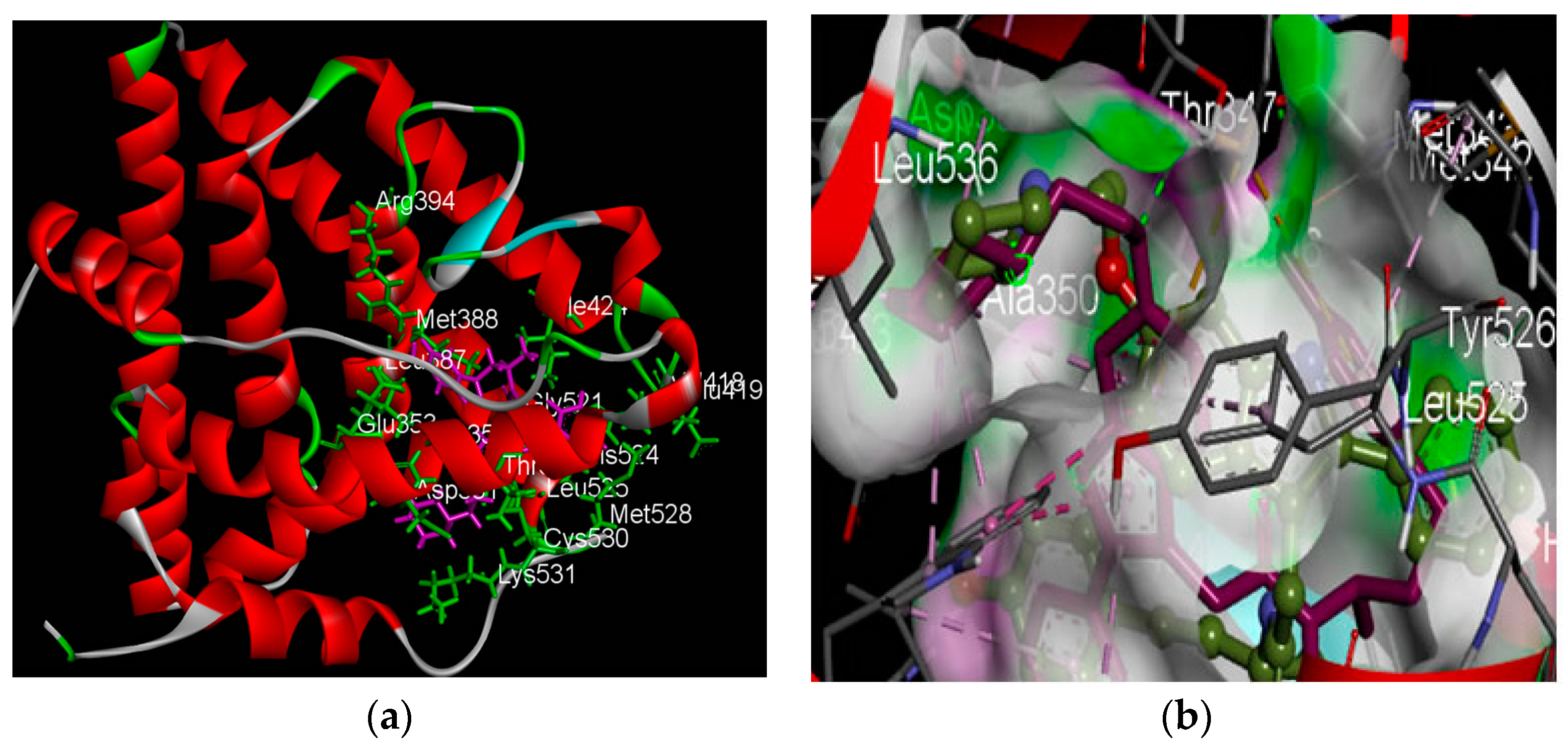
2.2.1. Structure of the Human Estrogen Receptor α, 2IOG

The three-dimensional structure of the hERα was retrieved from the Protein Data Bank with PDB ID: 2IOG determined by X-Ray crystallography at a resolution of 1.60 (Å) and visualized in Discovery studio. The complete X-ray structure of the protein (Figure 1a) is depicted with amino acid residues (viewed within 5 angstrong) shown as green sticks and labeled with their three-letter code. The native ligand, compound 11F, being a co-crystal structure of the target protein, is depicted as purple sticks (see Figure 1a). This particular PDB receptor was chosen based on its crystallographic resolution and species of interest.

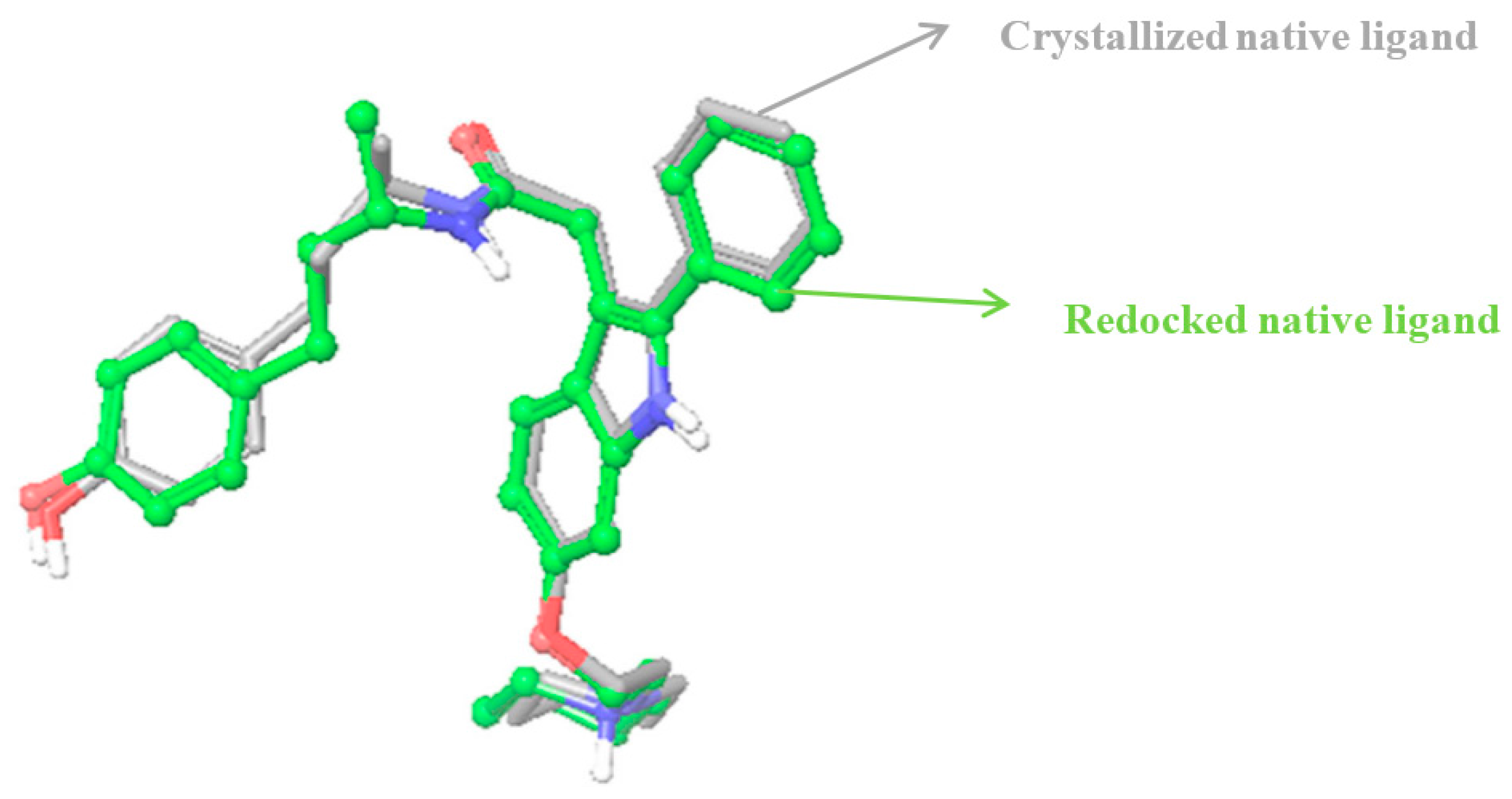
2.2.2. Identification of Estrogen Receptor-Binding Pockets and Validation of Docking Protocol

The human Estrogen receptor catalytic site predictions were carried out using the Cast p program [25]. The server measured analytically the area and volume of each pocket. The best ligand binding site was observed to be at pocket no. 36 of volume 1178.9Å3 and area of 901.1Å2, and consisted of 36 residues ID: Met343, Leu346, Thr347, Leu349, Ala350, Asp351, Glu353, Leu354, Trp383, Leu384, Leu387, Met388, Leu391, Arg394, Phe404, Val418, Glu419, Gly420, Met421, Ileu424, Phe425, Leu428, Gly521, His524, Leu525, Tyr526, Met528, Lys529, Cys530, Lys531, Asn532, Val533, Val534, Pro535, Leu536, and Leu539. Some of these amino acids viewed within 5 angstrong are depicted in Figure 1a after visualizing with discovery studio. This identification is similar to the findings of Suganya et al. [26] and reveals possible residues around the active site with which a potential ligand(s) can bind [27].
To validate the docking protocol, the root mean square deviation between the co-crystallized native ligand and the redocked native ligand should be within 2 angstrong as can be visibly appreciated when the redocked and co-crystal structures are superimposed and the deviation calculated. When comparing the co-crystallized structure of compound 11F, the native ligand to the estrogen receptor, 2IOG, with our redocked compound 11F, the root mean square deviation obtained was 0.7864. Their superimposition was also correctly reproduced (Figure 2) within the binding domain of the target receptor (see Figure 1b). For the purpose of clarity, the receptor was excluded and only the superimposed structures shown in Figure 2.

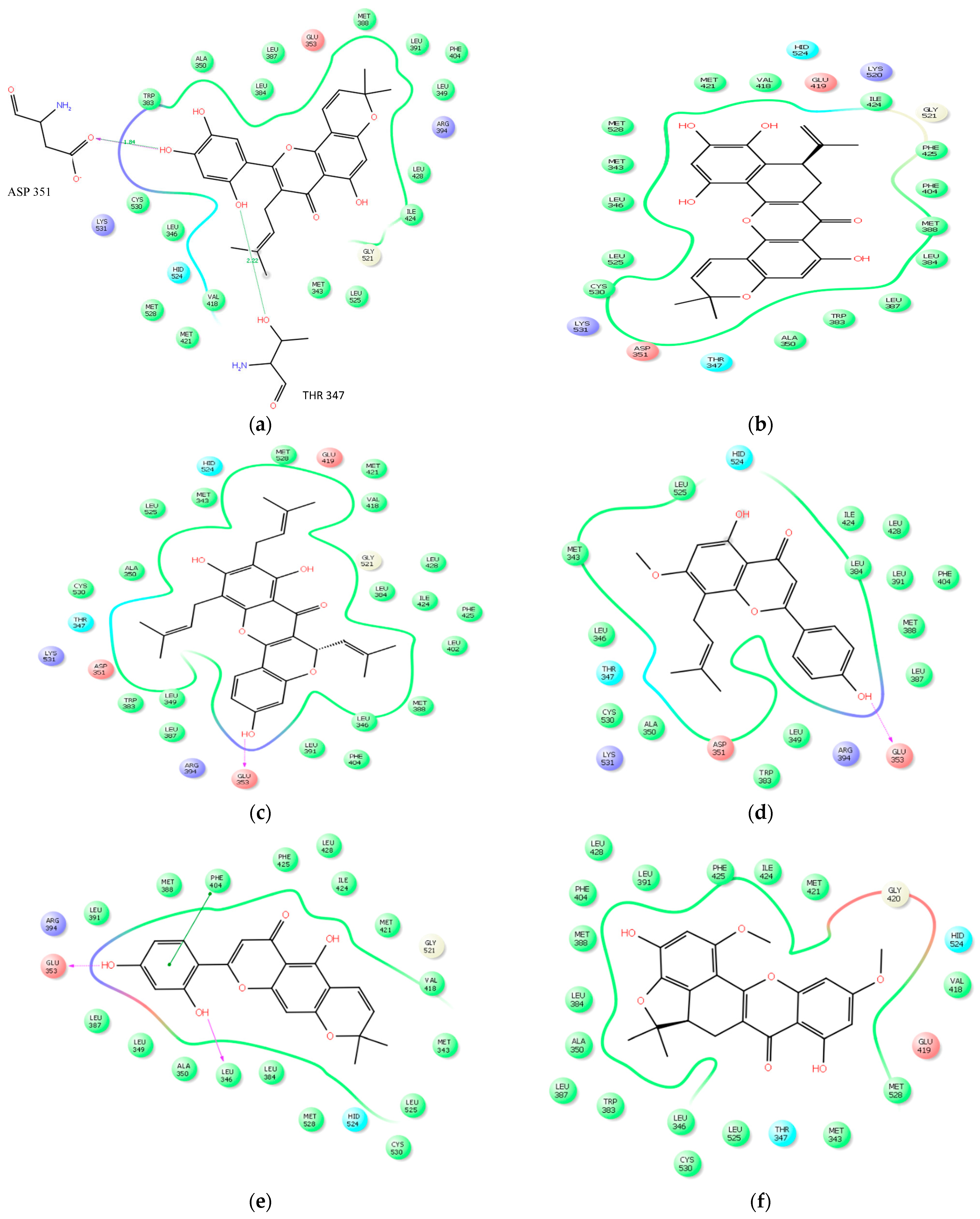
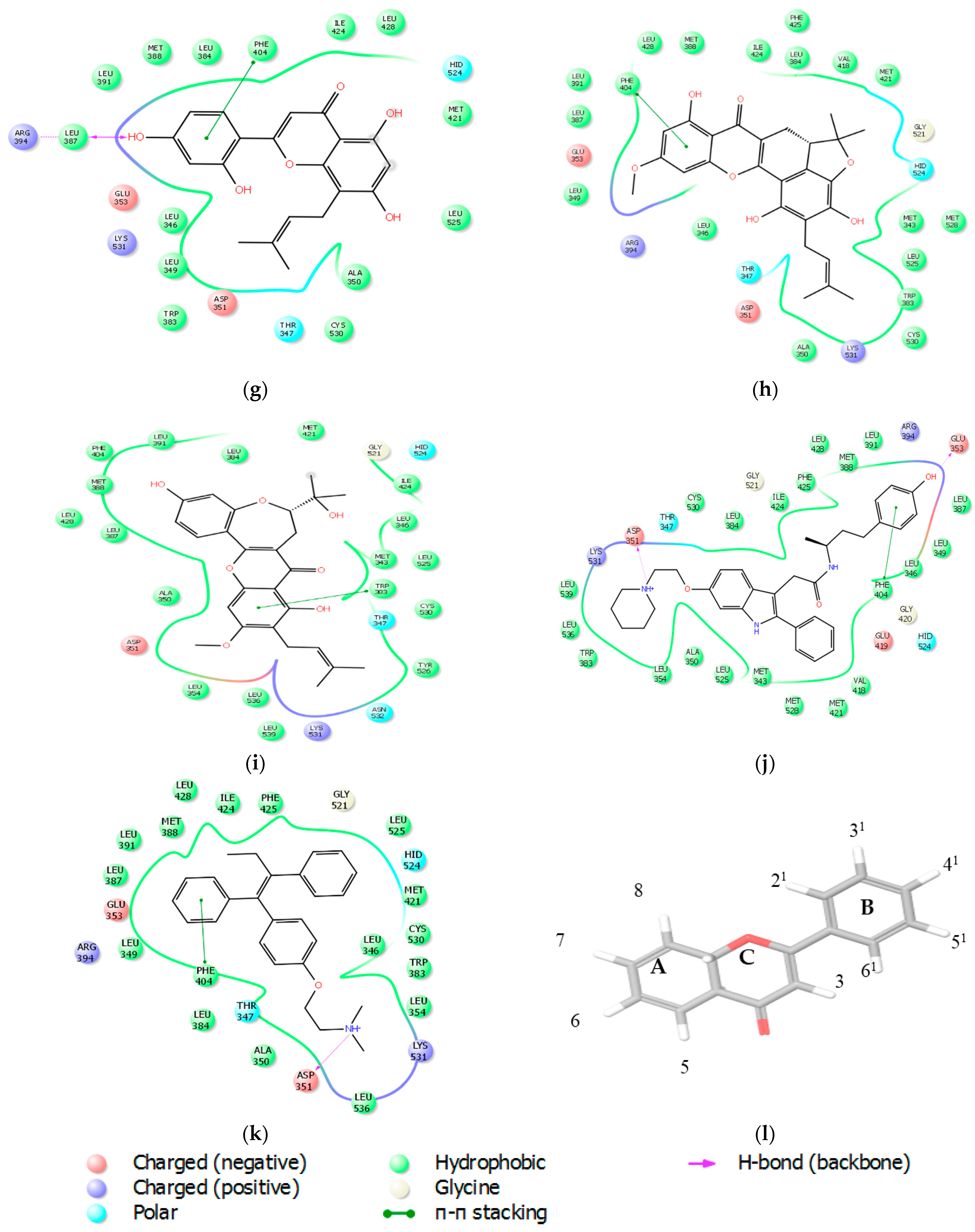
2.2.3. Docking Analysis

The studied molecules were docked alongside with the native ligand and a reference standard, Tamoxifen, which served as controls. The results of the electrostatic interaction between the studied molecules and the target receptor are as depicted in Figure 3a–k. An estimation of the binding affinity, depicted as the glide or docking score was used to access the binding affinities of the studied ligands to the target receptor (see Table 2), increased in electronegativity is a function of the binding affinity [28,29]. Glide has been observed to be more accurate than other docking tools such as GOLD and Surflex methods [28]. Kosh et al. [30], estimated during their in silico studies on phthalates that a Glide score greater than −7 kcal/mol is considered as promising, and that it can go as high as −13 or even more. This study revealed glide scores between −16.81 and −9.10, indicating good affinities to the target receptor (Table 2). All the studied ligands had good Glide scores with the most outstanding score being Artonin E (−12.72) as compared to other molecules from Artocarpus species (see Table 2). The best three glide scorers also showed the best prime ∆G binding energy as depicted in Table 3. The roles of certain crucial amino acids in the ligand-binding domain of the human estrogen receptor α, was also established.
Major non-covalent interactions between the studied ligands and the ligand-binding domain of the hERα was investigated using key amino acids within the receptor. These amino acids have been repeatedly implicated during ligand interaction with the hERα [31] and also play important role in the inhibition of the ligand-binding domain of hERα [26,29,31]. This non-covalent interactions: Van der Waals, columbic interaction, π-π interaction and hydrogen interaction are shown in Table 2 and Figure 3a–l.
Structuarally, all the studied molecules each contain the basic flavone skeleton, i.e., comprising of two benzene rings (A and B as shown in Figure 3l) linked by a three carbon chain that form a closed pyran ring (C). Some of the compounds are prenylated (see Figure 3a,c,d,g–i), while others are not prenylated. Alternatively, the compounds can as well be seen in two forms: those in which the carbon chain forming the C ring is not occupied by any ring structure (Figure 3a,d,e,g or those in which the C ring linking carbon chain is occupied by other ring structures (see Figure 3b,c,f,h,i). From the results as seen in Figure 3a, the 21–OH and 41–OH groups of Artonin E established 1H bond each with negatively charged Aspartate 351, believed to be necessary for antagonism [32], and polar Threonine 347 at distances of 1.84 Ǻ and 2.88 Ǻ respectively (see Figure 3a). The 41–OH groups of Artoelastin, and Artonin U each formed 1H bond with negatively charged glutamate 353 (Figure 3c,d). In Figure 3e, Cycloartocarpesin was observed to form 2H bonds, at 21–OH group with negatively charged Glu 353 and at 41–OH group with hydrophobic Leu 346. The 41–OH group of Artonin Y (Figure 3g) formed 2H bonds with hydrophobic Leu 387 and positively charged Arg 394. Worthy of note is that the hydrogen bond distances, showed in Figure 3a–k, excluded distances beyond 3 Ǻ. However, distances within 4 Ǻ have been reported in Table 4. It is also evident that the benzene rings of cycloartocarpesin (Figure 3e), Artonin Y (Figure 3g), Tamoxifen (Figure 3k) and Artonin T (Figure 3h) each, formed a π π interaction with Phe 404 (see Figure 3e,g,h) respectively, while Artonin S (Figure 3i) formed a π π interaction with Phe 383. Interestingly, all the hydrogen bond interactions appeared to be formed predominantly in the B ring of the flavone skeleton of the ligands (Figure 3a–i). This may be attributed to the vicinal diol groups attached to this ring which enhances their binding to the target receptor. Worthy of note is the observation that the prenylated group together with the 41, 51 vicinal diol of Artonin E appeared to have enhanced the ligand’s binding affinity to the target receptor as shown in its firm hydrogen bond interaction involving the 41, 51 vicinal diols which may be attributed to its high glide score when compared to the other phytomolecules. These vicinal diol group were earlier reported by Reddy et al. [33] to improve the compound’s inhibitory activity towards arachidonate acid.
The clustering of active amino acids around the studied ligands strengthened the ligands anchorage to the target receptor (see Figure 3a–k), the green solid line in each Figure shows hydrophobic interactions with amino acids in the ligand-binding domain of the receptor and the purple arrow indicate the strongest hydrogen bonding interaction of <3 angstrong.
Resistance to current anti-estrogens like Tamoxifen and the recurring cases of breast cancer [34] prompted the attention to search for potential estrogen receptor targeting small molecules from nature. Glide of the Schrodinger suite 2015, was employed to study the binding affinities of some small molecules isolated from the Artocarpus species with the view of finding a potent inhibitor for ligand-binding domain of the human estrogen receptor α. Evidently, the results revealed for the first time, the relative potential of the studied ligands in abrogating estrogen signaling, a unique property in targeting estrogen positive breast cancers.

2.3. Prime Energy Analysis

The Prime molecular mechanics generalized born surface area (MM-GBSA) binding energy was calculated for the studied ligands with the program prime, of the Shrodinger suite [35]. The output properties calculated include: Prime Coulomb energy of the complex ( Δ G b i n d coulomb), Prime Van der Waals energy of the complex ( Δ G b i n d vdW), Prime energy of the complex (Prime MMGBSA complex energy), Prime MMGBSA Ligand Energy, Prime MMGBSA Receptor Energy, Prime MMGBSA Δ G b i n d (kcal/mol) and Prime hydrogen bond ( Δ G b i n d Hbond). The results from the prime energy calculations are as shown in Table 3 with the free energy of binding, Δ G b i n d (kcal/mol) calculated as described in Section 3.4 (Equation (1)).

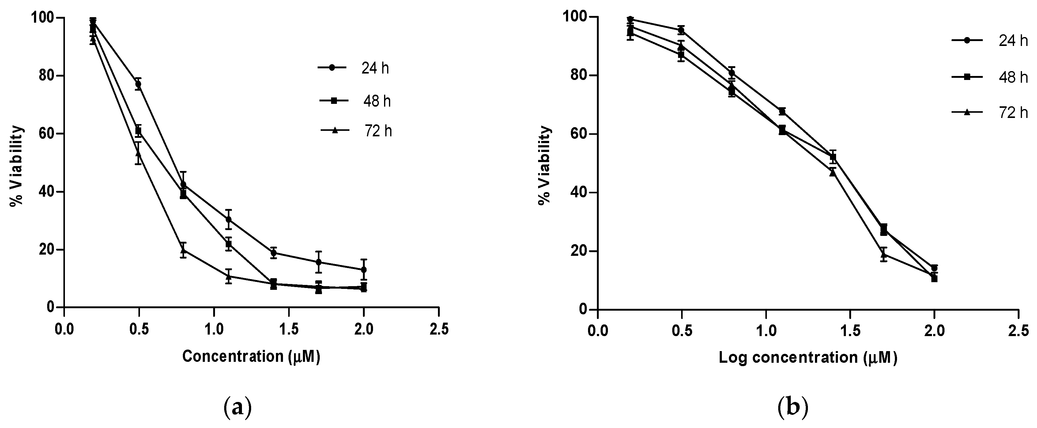
2.4. In Vitro Growth Inhibition Assay

To test the prediction of anticancer relevance of the docking studies, we investigated the growth inhibitory potential of the best glide scorer among the studied phytochemicals, Artonin E and a reference standard, Tamoxifen, in estrogen positive human breast cancer cells, MCF 7 using varying concentrations of the compounds at three different time points. Dose–response curves were plotted for each of the compounds (see Figure 4a,b) and the concentration of tested agents which evoked a 50% growth inhibition of the breast cancer cells were determined, as shown in Table 5 along with their 95% confidence intervals. It was observed that the reference standard, which showed a better docking score of 13.93 kcal, was unable to evoke a stronger in vitro inhibition on the estrogen receptor positive breast cancer cells as compared to Artonin E, whose score was 12.72 kcal. This observation, though consistent with other reported studies [36], was suspected to be due to phenol red, a component of the growth media with which the cells were raised before treatment [37]. The in vitro growth inhibition of the native ligand with a docking score of 16.81 Kcal as previously reported by the crystallographers was 42.7 nM [32] in comparison to Artonin E (docking score of 12.72 kcal and in vitro inhibition of 3.8 µM at 72 h). Generally, the growth inhibition of these compounds was observed to improve upon increasing concentration and time exposure (data available in Supplementary Materials). Comparing the time interval effect, the means of the different growth parameters were statistically significant (p < 0.05) when compared to the values of the 24 h time point as analyzed using Analysis of Variance in GraphPad prism 5.0 (GraphPad Software Inc., La Jolla, CA, USA).

3. Materials and Methods

3.1. Preparation of Ligands

The structures of the ligands were downloaded from the PubChem database and their 3D structures were prepared with Maestro, using ligprep [38], a utility of Schrodinger software suite that combines tools for generating accurate and high quality 3D molecular model from 1D (Smiles) and 2D (SDF) representations. The ligprep applied energy minimization with optimized potentials for liquid simulations-2005 (OPLS_2005) as the applied force field and filtered the ligands before they were used for further computational studies. The output structures were finally written to a file in maestro format.

3.2. Determination of ADMET Properties of the Compounds

Most promising drug candidates often fail during clinical trials owing to poor drug-like characteristics. To nominate potential drug candidates among the studied compounds, certain properties which relate to absorption, distribution, metabolism and excretion were investigated [39]. The QikProp module of Schrodinger Suite was used to predict the following parameters of the studied molecules: absorption, distribution, metabolism, and excretion. In addition to predicting molecular properties, QikProp also provides ranges for comparing a particular molecule’s properties with those of 95% of known orally available drugs. QikProp predicts physically significant descriptors and pharmaceutically relevant properties of organic molecules, either individually or in batches.
In the present study, QikProp was run in a normal processing mode with default options (Qikprop 4.6). The selected properties that are known to influence metabolism, cell permeation and bioavailability are presented in Table 1. These properties were thereafter compared with 95% of known oral drugs and also tested with the Lipinski’s rule of five to be considered as drug-like.

3.3. Molecular Docking Studies

3.3.1. Identification of Binding Pockets and Validation of Docking Protocol

Usually, binding sites and active sites of proteins are often associated with structural pockets and cavities. The castP server was employed for this identification. This program uses the weighted Delaunay triangulation and the alpha complex for shape measurements [40]. It provides identification and measurements of surface accessible pockets as well as interior inaccessible cavities, for proteins and other molecules. It also measures the number of mouth openings, area of the openings, and circumference of mouth lips, in both SA and MS surfaces for each pocket [25] as reported in the results above (see Section 2.2.2).
The predictive ability of the docking protocol was validated by redocking the native ligand, compound 11F to hERα (Protein Data Bank (PDB) ID code 2IOG). To prove the appropriateness of the utilized docking protocol, the native ligand, which was co-crystallized to the target receptor as available in the protein data bank was redocked back to its receptor and such orientation compared by superimposing the co-crystallized ligand to the redocked native ligand and computing the root mean square deviation between them. The root mean square deviation between them should be within 2 angstrong before such docking protocol is validated for the docking studies. Binding sites and ligand poses were correctly identified, as shown in Figure 1b. The redocked native ligand showed similar binding positions and orientations within the binding site and were similar to the co-crystal structures with root mean squares less than 2A (0.7864). The superimposition of the redocked native ligands with its co-crystal structure was also correctly reproduced (Figure 2) within the binding domain of the receptor (see Figure 1b).

3.3.2. Preparation of Protein

The ligand-binding domain of the hERα protein was retrieved from the RCSB protein data bank with the PDB id 2IOG (see X-ray structure in Figure 1a). The resolution, species and bound structure were taken into consideration before choosing the target protein from the database. Protein preparation was processed with the help of the protein preparation wizard from the workflow option of the Schrodinger suite. The force field applied for the preparation of the protein was the optimized potentials for liquid simulations-2005. The water molecules, heteroatoms residues, were deleted while the chain was retained along with H-bond. Hydrogen atoms were added and such bonding network was optimized. Side chains and loops with missing atoms were also built. The complex obtained was finally minimized after the energy gradient converged below 0.05 kcal/mol using the OPLS_2005 force field with Polack-Ribiere Conjugate Gradient (PRCG) algorithm [41]. The receptor-grid was generated with the help of the module glide. Grid generation represents the physical properties like volume of the receptor (specifically the active site) that is needed for carrying out the ligand-docking. The grid boxes were generated by selecting the co-crystallized ligands in the ligand-binding domain and replacing it with the studied ligands during the docking process.

3.3.3. Molecular Docking Studies

Good binders to the target receptor were investigated intensively using Glide extra precision (XP) docking for clear and accurate details along with epik state penalties. The selected entries for the ligands to be docked were being selected as well as the output file of the generated grid. The docking of each potential drug molecule along with the grid generated conformational changes with respect to the active site of the protein, estrogen receptor (PDB ID-2IOG). Following the docking studies, the glide scores or docking scores were displayed and the ligand amongst the phytomolecules, with the least glide score was considered to have the best docked pose or best glide score. The glide scoring system has been well established and has been deemed very accurate in comparison to other docking tools [28]. Glide score approximates the ligand binding free energy.

3.4. Prime Energy Analysis

For the post assessment of docked structures, the molecular mechanics energies combined with the Poisson–Boltzmann or generalized Born and surface area continuum solvation (MM/PBSA and MM/GBSA) methods were used [35]. These are popular approaches to estimate the free energy of the binding of small molecules to biological macromolecules [42]. The calculation uses the OPLS_2005 all-atom force field for protein residues as well as for ligands and cofactors. The input structures for these calculations were taken from a Pose Viewer file Glide output.
The following descriptors were generated by the Prime MM-GBSA approach:
MM-GBSA_∆G_bindLigand binding energy, Δ G b i n d
MM-GBSA_E_complexEnergy of the complex, G c o m p l e x
MM-GBSA_E_proteinEnergy of the receptor without the ligand, G p r o t e i n
MM-GBSA_E_ligandEnergy of the unbound ligand, G l i g a n d
The total free energy of binding was then expressed as:
Δ G bind =   G complex   ( G protein + G ligand )
The other parameters were:
Prime Coulomb energy of the complex Δ G b i n d coulomb
Prime Van der Waals energy of the complex Δ G b i n d vdW
Prime Hydrogen Bond of the Complex Δ G b i n d Hbond
The MM-GBSA scoring along with the experimental binding affinity data of the binding site for studied molecules on 2IOG is presented in Table 3.

3.5. Preparation of Drugs

Artonin E used in these studies was isolated from the stem bark of Artocarpus elasticus [43], while Tamoxifen, the reference standard was purchase from Sigma Aldrich, St. Louis, MO, USA. A stock solution of 100 mM was prepared in DMSO and the final DMSO concentration was 0.01%.

3.6. Cell Culture

The MCF-7 cancer cell line was purchased from the American Type Culture Collection (ATCC, Rockville, MD, USA) and was maintained in RPMI media supplemented with 10% heat-inactivated FBS, 100 U/mL penicillin and 100 µg/mL streptomycin (Sigma). Cells were grown in 25 cm2 tissue culture flasks in a humidified atmosphere containing 5% CO2 at 37 °C. Once the cells reach 80% confluency, 1 mL of trypsin-EDTA solution was added to the flask to detach the monolayer cells. Approximately 0.5 × 10⁶–1 × 10⁶ cells were routinely sub-cultured and maintained appropriately.

3.7. Growth Inhibitory Assay

The growth inhibitory potential of Tamoxifen, a standard estrogen receptor modulator and Artonin E, the best glide scorer among the natural compounds from Artocarpus species was assessed using a cell-based proliferation assay in estrogen positive MCF-7 breast cancer cell line. Briefly, exponentially growing MCF-7 cells was seeded in 96-well micro-plates at a density of 0.5 × 10⁴ cells/well. The Cells were allowed to adhere overnight before being challenged with the compounds. The cells were incubated at various time points with different compound concentrations ranging from 1.56–100 µM. After each treatment time point, 20 µL of MTT (Sigma, St Louis, MO, USA) stock solution (5 mg/mL) was added to each well and incubated for 4 h to allow metabolization of the MTT by cellular mitochondrial dehydrogenases. One hundred microliters of DMSO was thereafter added to each well to solubilize the formazon crystals formed. The absorbance of the converted dye was measured colorimetrically at 570 nm and the assay was carried out in three independent experiments. From the obtained absorbance readings at respective intervals of each tested agent at each concentration, a nonlinear regression was performed using the GraphPad Prism software and the concentration of each agent that evoked a 50% growth inhibition of the estrogen positive breast cancer cells was determined along with their 95% confidence interval (Table 5). A dose–response curve was fitted for each of the compounds with the X-axis being the logarithm of the concentrations used and the Y-axis showing cell viability response calculated using the following formula:
%  of cell viability =   A T A C   × 100
where AT is the absorbance reading of treated samples at each time point, and AC is the absorbance of control samples treated with 0.01% of DMSO equivalent to the amount of DMSO used as a vehicle in the compound-treated wells.

4. Conclusions

Targeting the human estrogen receptor α, a strategy adopted in this study, is a valid approach in discovering new molecules which can halt undue proliferation in estrogen positive breast cancer. The occupation of the binding pocket of hERα by ER targeting small molecules can prevent estrogen from binding to such active site and thus abrogates estrogenic downstream signaling which precedes undue proliferation, as implicated in estrogen receptor positive breast cancer.
The present study used nine ligands from Artocarpus species to study their binding affinities to the ligand-binding domain of the human estrogen receptor α (PDB ID 2IOG) using the Glide module of Schrodinger software after ascertaining their drug-likeness with Qikprop. Artonin E, Artonin U and Cycloartocarpesin were the best amongst the phytomolecules based on their docking scores as well as the Prime MM-GBSA free energy of binding. The in silico study revealed that the studied phytomolecules are potent modulators of estrogen receptor positive breast cancer. The best glide scorer amongst the phytomolecules, Artonin E, was investigated for its time interval growth inhibitory effect on an ER positive breast cancer cell line, MCF-7 along with Tamoxifen, a standard estrogen receptor modulator. In vitro growth inhibition is an essential prerequisite of anticancer drug development, owing to the unique hallmark of cancer cells to resisting cell death and proliferating uncontrollably. Artonin E, with a glide score of 12.72 kcal, showed a better in vitro growth inhibitory effect in submicromolar range as compared to Tamoxifen (glide score of 13.93 kcal), a standard estrogen receptor modulator currently facing much reported resistance. Conclusively, in silico molecular studies have proven very useful in predicting the pharmacokinetic profiles and the binding affinities of suspected new drug candidates before a detailed preclinical and clinical evaluation. This study has revealed potent estrogen receptor modulators with good predicted pharmacokinetic profiles that should be further investigated for their in vitro as well as in vivo activity towards estrogen receptor positive breast cancer. We recommend that the enlisted analogs of Artonin E be screened for their potential anti-breast cancer effects as predicted by our in silico data.

Supplementary Materials

The following are available online at https://www.mdpi.com/1420-3049/21/7/839/s1. In silico physicochemical data, prime energy analysis and in vitro raw data as well as the result analysis and graphs.

Acknowledgments

This study was co-supported by TETFund Nigeria, University Putra Malaysia and Ministry of Science, Technology and Innovation Malaysia (Vote No. 5450742). The authors gratefully acknowledge the costs of publication covered by the Research Management Centre at the University Putra Malaysia.

Author Contributions

Imaobong Etti conceived the research idea, carried out the experiments and analyzed data; Rasedee Abdullah, Najihah Mohd Hashim, Arifah Kadir and Ahmad Bustamam Abdul contributed materials and reagents; Christopher Etti, Ibrahim Malami, Peter Waziri and Chee Wun How participated in the research; and Imaobong Etti and Christopher Etti prepared the manuscript.

Conflicts of Interest

The authors declare no conflict of interest.

References

  1. Hamilton, K.; Arao, Y.; Korach, K. Estrogen hormone physiology: Reproductive findings from estrogen receptor mutant mice. Reprod. Biol. 2014, 14, 3–8. [Google Scholar] [CrossRef] [PubMed]
  2. Nilsson, S.; Makela, S.; Treuter, E.; Tujague, M.; Thomsen, J.; Andersson, G.; Enmark, E.; Pettersson, K.; Warner, M.; Gustafsson, J.A. Mechanism of Estrogen Action. Physiol. Rev. 2001, 81, 1535–1565. [Google Scholar] [PubMed]
  3. Burns, K.; Korach, K. Estrogen receptors and human disease: An update. Arch. Toxicol. 2012, 10, 1491–1504. [Google Scholar] [CrossRef] [PubMed]
  4. Berger, C.E.; Qian, Y.; Liu, G.; Chen, H.; Chen, X. p53, A Target of Estrogen Receptor (ER), Modulates DNA Damage-induced Growth Suppression in ER-positive Breast Cancer Cells. J. Biol. Chem. 2012, 287, 30117–30127. [Google Scholar] [CrossRef] [PubMed]
  5. Belev, B.; Vrbanec, D. Hormonal resistance in breast- and prostate cancer. Period Biol. 2012, 114, 511–517. [Google Scholar]
  6. Andruska, N.D.; Zheng, X.; Yang, X.; Mao, C.; Cherian, M.M.; Mahapatra, L. Estrogen receptor α inhibitor activates the unfolded protein response, blocks protein synthesis, and induces tumor regression. Proc. Natl. Acad. Sci. USA 2015, 112, 4737–4742. [Google Scholar] [CrossRef] [PubMed]
  7. Kochummen, K.M.; Go, R. Tree of Sabah and Sarawak. In Moraceae; Seopadmo, E., Saw, L.G., Eds.; Ampang Press: Kuala Lumpur, Malaysia, 2000; pp. 181–334. [Google Scholar]
  8. Jagtap, U.; Bapat, V.A. Artocarpus: A review of its traditional uses, phytochemistry and pharmacology. J. Ethnopharmacol. 2010, 129, 142–166. [Google Scholar] [CrossRef] [PubMed]
  9. Zhao, T.; Yan, G.-R.; Pan, S.-L.; Wang, H.-Y.; Hou, A.-J. New isoprenylated 2-arylbenzofurans and pancreatic lipase inhibitory constituents from Artocarpus nitidus. Chem. Biodivers. 2009, 6, 2209–2216. [Google Scholar] [CrossRef] [PubMed]
  10. Boonphong, S.; Baramee, A.; Kittakoop, P. Antitubercular and Antiplasmodial Prenylated Flavones from the Roots of Artocarpus altilis. Chiang Mai J. Sci. 2007, 34, 339–344. [Google Scholar]
  11. Jayasinghe, U.L.B.; Samarakoon, T.B.; Kumarihamy, B.M.M. Four new prenylated flavonoids and xanthones from the root bark of Artocarpus nobilis. Fitoterapia 2008, 79, 37–41. [Google Scholar] [CrossRef] [PubMed]
  12. Jamil, S.; King, A.; Jamil, S.; Mariam, S.; Lathiff, A.; Abdullah, S.A. Antimicrobial Flavonoids from Artocarpus anisophyllus Miq and Artocarpus lowii King. J. Teknol. 2014, 71. [Google Scholar] [CrossRef]
  13. Wongpankam, E.; Chunhacha, P.; Pongrakhananon, V. Artonin E Mediates MCL1 Down-regulation and Sensitizes Lung Cancer Cells to Anoikis. Anticancer Res. 2012, 32, 5343–5351. [Google Scholar] [PubMed]
  14. Ramli, F.; Rahmani, M.; Sukari, A.; Hashim, N.M. New Flavonoids Derivatives from Artocarpus Elasticus with Antioxidant, Antimicrobial and Cyotoxic Activities. Open Conf. Proc. J. 2013, 4, 2013. [Google Scholar] [CrossRef]
  15. López-Vallejo, F.; Caulfield, T.; Martínez-Mayorga, K.; Giulianotti, M.A.; Nefzi, A.; Houghten, R.A.; Medina-Franco, J.L. Integrating virtual screening and combinatorial chemistry for accelerated drug discovery. Comb. Chem. High Throughput Screen. 2011, 6, 475–487. [Google Scholar] [CrossRef]
  16. Jacobs, M. In silico tools to aid risk assessment of endocrine disrupting chemicals. Toxicology 2004, 205, 43–53. [Google Scholar] [CrossRef] [PubMed]
  17. Malami, I.; Abdul, A.; Abdullah, R.; Bt Kassim, N.; Waziri, P.; Christopher Etti, I. In silico Discovery of Potential Uridine-Cytidine Kinase 2 Inhibitors from the Rhizome of Alpinia mutica. Molecules 2016, 21, 417. [Google Scholar] [CrossRef] [PubMed]
  18. Lipinski, C.A.; Lombardo, F.; Dominy, B.W.F.P. Experimental and computational approaches to estimate solubility and permeability in drug discovery and development settings. Adv. Drug Deliv. Rev. 2001, 46, 3–26. [Google Scholar] [CrossRef]
  19. Jorgensen, W.L.; Duffy, E.M. Prediction of drug solubility from structure. Adv. Drug Deliv. Rev. 2002, 54, 355–366. [Google Scholar] [CrossRef]
  20. Delaney, J.S. Predicting aqueous solubility from structure. Drug Discov. Today 2005, 4, 289–295. [Google Scholar] [CrossRef]
  21. Veber, D.F.; Johnson, S.R.; Cheng, H.Y.; Smith, B.R.; Ward, K.W.; Kopple, K.D. Molecular Properties That Influence the Oral Bioavailability of Drug Candidates. J. Med. Chem. 2002, 12, 2615–2623. [Google Scholar] [CrossRef]
  22. QikProp, Version 4.6; Schrödinger, LLC: New York, NY, USA, 2015.
  23. Usansky, H.H.; Sinko, P.J. Estimating Human Drug Oral Absorption Kinetics from Caco-2 Permeability Using an Absorption-Disposition Model: Model Development and Evaluation and Derivation of Analytical Solutions for ka and Fa. J. Pharmacol. Exp. Ther. 2005, 314, 391–399. [Google Scholar] [CrossRef] [PubMed]
  24. Zhang, D.; Luo, G.; Ding, X.; Lu, C. Preclinical experimental models of drug metabolism and disposition in drug discovery and development. Acta Pharm. Sin. B Elsevier 2012, 2, 549–561. [Google Scholar] [CrossRef]
  25. Dundas, J.; Ouyang, Z.; Tseng, J.; Binkowski, A.; Turpaz, Y.; Liang, J. CASTp: Computed atlas of surface topography of proteins with structural and topographical residues. Nucleic Acids Res. 2006, 34, W116–W118. [Google Scholar] [CrossRef] [PubMed]
  26. Suganya, J.; Radha, M.; Naorem, D.L. In silico Docking Studies of Selected Flavonoids—Natural Healing Agents against Breast Cancer. Asian Pac. J. Cancer Prev. 2014, 15, 8155–8159. [Google Scholar] [CrossRef] [PubMed]
  27. Henrich, S.; Salo-ahen, O.M.H.; Huang, B.; Rippmann, F.F.; Cruciani, G.; Wade, R.C. Computational approaches to identifying and characterizing protein binding sites for ligand design. J. Mol. Recognit. 2010, 23, 209–219. [Google Scholar] [CrossRef] [PubMed]
  28. Friesner, R.A.; Murphy, R.B.; Repasky, M.P.; Frye, L.L.; Greenwood, J.R.; Halgren, T.A.; Sanschagrin, P.C.; Mainz, D.T. Extra precision glide: Docking and scoring incorporating a model of hydrophobic enclosure for protein-ligand complexes. J. Med. Chem. 2006, 49, 6177–6196. [Google Scholar] [CrossRef] [PubMed]
  29. Celik, L.; Lund, J.; Schiøtt, B. Exploring Interactions of Endocrine-Disrupting Compounds with Different Conformations of the Human Estrogen Receptor α Ligand Binding Domain: A Molecular Docking Study. Chem. Res. Toxicol. 2008, 21, 2195–2206. [Google Scholar] [CrossRef] [PubMed]
  30. Josh, M.K.S.; Pradeep, S.; Adarsh, V.K.; Amma, K.S.V.; Devi, R.S.; Balachandran, S.; Sreejith, M.N.; Jaleel, U.C.A.; Benjamin, S. In silico evidences for the binding of phthalates onto human estrogen receptor α, β subtypes and human estrogen-related receptor γ. Mol. Simul. 2014, 40, 408–417. [Google Scholar] [CrossRef]
  31. Desai, N.; Mahto, M.K.; Alekhya, B.; Naveen, C.R.; Bhaskar, P.M. Comparative docking studies of estrogen receptor inhibitors and their binding interaction analysis. Int. J. Pharm. Sci. Rev. Res. 2012, 16, 91–95. [Google Scholar]
  32. Dykstra, K.D.; Guo, L.; Birzin, E.T.; Chan, W.; Yang, Y.T.; Hayes, E.C.; DaSilva, C.A.; Pai, L.Y.; Mosley, R.T.; Kraker, B.; et al. Estrogen receptor ligands. Part 16: 2-Aryl indoles as highly subtype selective ligands for ER? Bioorg. Med. Chem. Lett. 2007, 17, 2322–2328. [Google Scholar] [CrossRef] [PubMed]
  33. Reddy, G.R.; Ueda, N.; Hada, T.; Sackeyfio, A.C.; Yamamoto, S.; Hano, Y.; Aida, M.; Nomura, T. A prenylflavone, artonin E, as arachidonate 5-lipoxygenase inhibitor. Biochem. Pharmacol. 1991, 41, 115–118. [Google Scholar] [CrossRef]
  34. Jiang, Q.; Zheng, S.; Wang, G. Development of new estrogen receptor-targeting therapeutic agents for tamoxifen-resistant breast cancer. Future Med. Chem. 2013, 9. [Google Scholar] [CrossRef]
  35. Prime, Version 4.2; Schrodinger, LLC: New York, NY, USA, 2015.
  36. Abu, N.; Akhtar, M.N.; Ho, W.Y.; Yeap, S.K.; Alitheen, N.B. 3-Bromo-1-hydroxy-9,10-anthraquinone (BHAQ) inhibits growth and migration of the human breast cancer cell lines MCF-7 and MDA-MB231. Molecules 2013, 18, 10367–10377. [Google Scholar] [CrossRef] [PubMed]
  37. Berthois, Y.; Katzenellenbogen, J.A.; Katzenellenbogen, B.S. Phenol red in tissue culture media is a weak estrogen: Implications concerning the study of estrogen-responsive cells in culture. Proc. Natl. Acad. Sci. USA 1986, 83, 2496–2500. [Google Scholar] [CrossRef] [PubMed]
  38. LigPrep, Version 3.6; Schrödinger, LLC: New York, NY, USA, 2015.
  39. Ekins, S.; Waller, C.; Swaan, P.; Cruciai, G.; Wrigton, S.; Wikel, J. Progress in predicting human ADME parameters in silico. J. Pharmacol. Toxicol. Methods 2000, 44, 251–272. [Google Scholar] [CrossRef]
  40. Binkowski, T.A.; Naghibzadeh, S.; Liang, J. Computed atlas of surface topography of proteins. Nucleic Acids Nucleic Acid Res. 2003, 13, 3352–3355. [Google Scholar] [CrossRef]
  41. Kaminski, G.A.; Friesner, R.A.; Tirado-Rives, J. Evaluation and reparametrization of the OPLS-AA force field for proteins via comparison with accurate quantum chemical calculations on peptides. J. Phys. Chem. 2001, 28, 6474–6487. [Google Scholar] [CrossRef]
  42. Genheden, S.; Ryde, U. The MM/PBSA and MM/GBSA methods to estimate ligand-binding affinities. Expert Opin. Drug Discov. 2015, 10, 449–461. [Google Scholar] [CrossRef] [PubMed]
  43. Rahman, M.A.; Ramli, F.; Karimian, H.; Dehghan, F.; Nordin, N.; Mohd Ali, H.; Mohan, S.; Mohd Hashim, N. Artonin E Induces Apoptosis via Mitochondrial Dysregulation in SKOV-3 Ovarian Cancer Cells. PLoS ONE 2016, 11, e0151466. [Google Scholar] [CrossRef] [PubMed]
  • Sample Availability: Samples of Artonin E are available from the authors.
Figure 1. X-ray Structure of PDB ID 2IOG: (a) Target receptor, 2IOG with co-crystallized native ligand shown in purple sticks and amino acid residues shown as green sticks with their three-letter code and name; and (b) redooked native ligand superimposed with the crystallized native ligand within the binding pocket of 2IOG showing hydrogen bonding in white surface cartoon.
Molecules 21 00839 g001
Figure 2. Docking control. Top ranked ligand pose for the native ligand, compound 11F 3-(4-hydroxyphenyl)-1-methylpropyl]-2-[2-phenyl-6-(2-piperidin-1-ylethoxy)-1h-indol-3-yl]acetamide-LBD hERα superimposed with co-crystallized native ligand.
Molecules 21 00839 g002
Figure 3. Molecular interactions of studied ligands with crucial amino acids at the ligand-binding domain of hERα: (a) Artonin E; (b) Artobiloxanthone; (c) Artelastin; (d) Artonin U; (e) Cycloartocarpesin; (f) Artonin L; (g) Artonin Y; (h) Artonin T; (i) Artonin S; (j) Native ligand, 11F; (k) Tamoxifen; and (l) A flavone skeleton.
Molecules 21 00839 g003aMolecules 21 00839 g003b
Figure 4. Dose–response curves of time-interval effect for: (a) Artonin E and (b)Tamoxifen on MCF 7.
Molecules 21 00839 g004
Table 1. Prediction of drug-likeness and pharmacokinetic profile of studied molecules.
LigandsMWSASADonor HBAccepted HBQPlogPo/wQPlogSQPPCacoQPlogBB# Metab% Human-Oral AbsorptionLipinskis Rule of Five
Artonin E436.5734.135.33.9−6.4124.6−1.97880
Artobiloxanthone434.4689.935.33.6−5.8156.3−1.67870
Cycloartocarpesin352.3608.424.52.9−5.1193.3−1.43850
Artoelastin488.6821.324.56.2−8.1704.4−1.2121001
Artonin Y354.4580.334.52.4−4.075.4−1.87750
Artonin U352.4618.913.83.9−5.4380.3−1.26960
Artonin L396.4635.015.33.5−5.3461.1−1.06950
Artonin T450.5718.925.34.5−6.3451.0−1.291000
Artonin S452.5728.025.34.6−6.4333.7−1.481000
Tamoxifen371.5 730.602.66.6−5.92203.20.431001
Native Ligand547.9 1021.8410.14.3−4.920.7−0.6462.91
Range for 95% known drugs: Molecular weight (MW) = 130.0–725.0; Total solvent accessible surface area (SASA) = 300.0–1000.0; Donor HB = 0.0–6.0; Accept HB = 2.0–20.0; Predicted octanol/water partition coefficient (QPlogPo/w) = −2.0–6.5; Predicted aqueous solubility (QPlogS) = −6.5–0.5; Predicted apparent Caco-2 cell permeability (QPPCaco) ≤25 poor, >500 great; Predicted brain/blood partition coefficient (QPlogBB) = −3.0–1.2; Number of likely metablioc reactions (# metab) = 1–8; % Human oral absorption ≥80% →High, <25% →Poor; Number of violations of Lipinskis Rule of Five; mol MW < 500, QPlogPo/w < 5, donor HB ≤ 5, accpt HB ≤ 10. Compounds that satisfy these rules are considered drug-like.
Table 2. Glide scores of studied ligands with their electrostatic interactions (kcal/mol) with critical amino acid at the ligand-binding domain of hERα.
LigandsGlide ScoreTHR 347ASP 351GLU 353ARG 394
VDWColoumbVDWColoumbVDWColoumbVDWColoumb
Native ligand−16.81−2.46−3.23−1.99−50.430.63−23.30−0.247.52
Tamoxifen−13.93−2.35−2.36−1.47−40.68−0.85−17.79−0.2613.57
Artonin E−12.72−2.35−1.76−1.03−12.11−1.42−2.87−0.601.21
Cycloartocarpesin−11.72−0.41−0.23−0.10−1.091.19−13.830.15−1.67
Artonin U−11.03−3.210.27−0.74−1.51−0.11−11.60−0.35−1.43
Artoelastin−10.90−3.35−0.11−1.16−1.32−1.05−10.05−0.45−1.58
Artonin L−10.70−1.110.36−0.15−0.34−0.24−1.45−0.060.79
Artobiloxanthone−10.50−1.21−1.260.69−1.20−0.222.33−0.04−1.63
Artonin Y−10.50−2.89−0.77−0.57−1.93−1.50−1.21−0.390.66
Artonin T−9.10−1.47−0.26−1.19−1.45−0.79−1.17−0.00−0.01
Artonin S−9.10−2.80−0.61−2.991.04−0.37−3.18−0.092.49
THR—Threonine, ASP—Aspartic acid, GLU—Glutamic acid, ARG—Arginine.
Table 3. Output properties from a Prime MM-GBSA calculation.
IUPAC Names Δ G b i n d (kcal/mol) Δ G b i n d Coulomb Δ G b i n d vdWPrime MMGBSA Complex EnergyPrime MMGBSA Ligand EnergyPrime MMGBSA Receptor Energy Δ G b i n d H Bond
Artonin E 5-hydroxy-8,8-dimethyl-3-(3-methylbut-2-enyl)-2-(2,4,5-trihydroxyphenyl)pyrano[2,3-h]chromen-4-one−47.68−22.59−33.53−9989.98−119.08−9823.22−1.76
Cycloartocarpesin 8-(2,4-dihydroxyphenyl)-5-hydroxy-2,2-dimethylpyrano[3,2-g]chromen-6-one−51.28−18.48−38.52−10,022.60−148.09−9823.22−2.13
Artonin U 5-Hydroxy-2-(4-hydroxyphenyl)-7-methoxy-8-(3-methyl-2-buten-1-yl)-4H-chromen-4-one−60.35−16.22−52.39−9987.84−104.26−9823.22−1.99
Artoelastin 3,8,10-trihydroxy-9,11-bis(3-methylbut-2-enyl)-6-(2-methylprop-1-enyl)-6H-chromeno[4,3-b]chromen-7-one−35.29−13.48−30.50−10,017.30−158.81−9823.22−1.65
Artonin L 3,8-Dihydroxy-1,10-dimethoxy-5,5-dimethyl-5a,6-dihydro-5H,7H-[1]benzofuro[3,4-bc]xanthen-7-one−32.69−5.91−26.01−9949.88−93.968−9823.22−0.05
Artobiloxanthone 6,10,11,13-Tetrahydroxy-9-isopropenyl-3,3-dimethyl-8,9-dihydro-3H,7H-benzo[c]pyrano[3,2-h]xanthen-7-one−11.32−5.45−16.28−9930.81−96.27−9823.22−0.15
Artonin Y 2-(2,4-Dihydroxyphenyl)-5,7-dihydroxy-8-(3-methyl-2-buten-1-yl)-4H-chromen-4-one−44.70−9.09−47.89−10,023.60−155.67−9823.22−1.20
Artonin T 1,3,8-Trihydroxy-10-methoxy-5,5-dimethyl-2-(3-methyl-2-buten-1-yl)-5a,6-dihydro-5H,7H-[1]benzofuro[3,4-bc]xanthen-7-one−27.36−3.95−46.21−9953.46−102.87−9823.22−0.24
ARTONIN S 3,9-dihydroxy-6-(2-hydroxypropan-2-yl)-11-methoxy-10-(3-methylbut-2-enyl)-6,7-dihydrochromeno[3,2-d][1]benzoxepin-8-one−25.703.67−28.01−9957.42−108.50−9823.22−0.33
See structure of ligands in Figure 3a–i.
Table 4. Hydrogen bonding interactions between studied ligands and the critical amino acids at the ligand-binding domain of hERα.
LigandsNo of BondsResiduesDistance
Artonin E4THR 347, ASP 351, LYS 531, CYS 5302.22, 1.84, 3.04, 3.22
Cycloartocarpesin3ARG 394, GLU 353, LEU 3461.94, 3.18, 2.70
Artonin U3GLU 353, ARG 394 GLY 5211.81, 3.41, 3.44
Artoelastin3GLU 353, ARG 394, GLY 5211.94, 2.31, 3.41
Artonin L2CYS 530, MET 528,3.37, 2.08
Artobiloxanthone2LEU 525, MET 3433.37, 3.29
Artonin Y2LEU387, ARG3942.02, 2.25
Artonin T1ARG 3943.50
Artonin S1THR 3472.23
Native ligand3GLU 353, LYS 351,ARG 3941.80, 2.08, 2.11
Tamoxifen2LYS 351, CYS 5301.92, 3.51
THR—Threonine; ASP—Aspartic acid; LYS—Lysine; CYS—Cysteine; ARG—Arginine; GLU—Glutamic acid; LEU—Leucine; MET—Methionine.
Table 5. IC50 Values of Artonin E and Tamoxifen on MCF 7 at Different time interval.
Compounds24 h48 h72 h
IC50 (µM)95% Confidence IntervalIC50 (µM)95% Confidence IntervalIC50 (µM)95% Confidence Interval
Artonin E6.95.5–8.65.14.5–5.83.83.4–4.1
Tamoxifen24.122.6–25.720.618.6–22.918.917.5–20.4

