Article

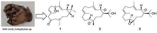
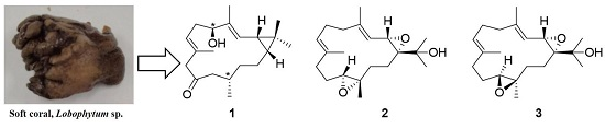
New Casbane and Cembrane Diterpenoids from an Okinawan Soft Coral, Lobophytum sp.

1
Department of Chemistry, Biology, and Marine Science, University of the Ryukyus, 1 Senbaru, Nishihara-cho, Okinawa 903-2013, Japan
2
Department of Bioresources Engineering, Okinawa National College of Technology, 905 Henoko, Nago-shi, Okinawa 905-2192, Japan
*
Authors to whom correspondence should be addressed.
Molecules 2016, 21(5), 679; https://doi.org/10.3390/molecules21050679
Submission received: 16 April 2016 / Revised: 18 May 2016 / Accepted: 18 May 2016 / Published: 23 May 2016
(This article belongs to the Section Natural Products Chemistry)

Abstract

:
A new rare casbane-type diterpenoid 1 and two new cembrane diterpenoids 2, 3 were isolated from an Okinawan soft coral, Lobophytum sp., together with four known cembrane diterpenoids 47. Their structures were elucidated by extensive analysis of spectroscopic data (1D and 2D NMR, IR, and MS) and a molecular modeling study. The new isolates showed weak anti-bacterial activity, mild cytotoxicity against HCT116 cells, and anti-inflammatory effect in LPS/IFN-γ-stimulated RAW 264.7 macrophage cells.

Graphical Abstract

1. Introduction

Marine organisms are the amazing source of secondary metabolites due to the biodiversity of the oceans. The genus Lobophytum soft coral [1] is a good source for various kinds of secondary metabolites that have unique structures, promising bioactivity and it is also well-known for producing macrocyclic diterpenoids belonging to a large group of cembrane-type metabolites [2]. In our continuing research focused on the isolation and structure elucidation of bioactive secondary metabolites from Okinawan marine organisms [3,4,5,6,7], we examined a soft coral, Lobophytum sp., subsequently isolating a novel casbane 1 and two new cembranes 2, 3, along with four known cembrane diterpenoids 47 (Figure 1) [8,9,10,11]. Casbane-type diterpenoids are rare in Nature, the first being isolated from an enzymatic preparation of castor bean seedlings [12]; these compounds are also found in soft coral [10]. Most of the casbane-type diterpenoids are two ring-based macrocyclic structures where the junction of the two rings is cis-fused [10,13,14] and few molecules showed trans junctions [15]. These types of metabolites are of considerable pharmacological interest due to their unique structures and exhibit potential bioactivities, including cytotoxicity [16,17,18,19,20,21,22,23], anti-viral [23], anti-inflammatory [24,25,26] and antimicrobial activities [24]; casbane diterpenoids also display anti-proliferative activity [10]. Herein, we report the isolation, structure elucidation, and cytotoxicity of these new metabolites.

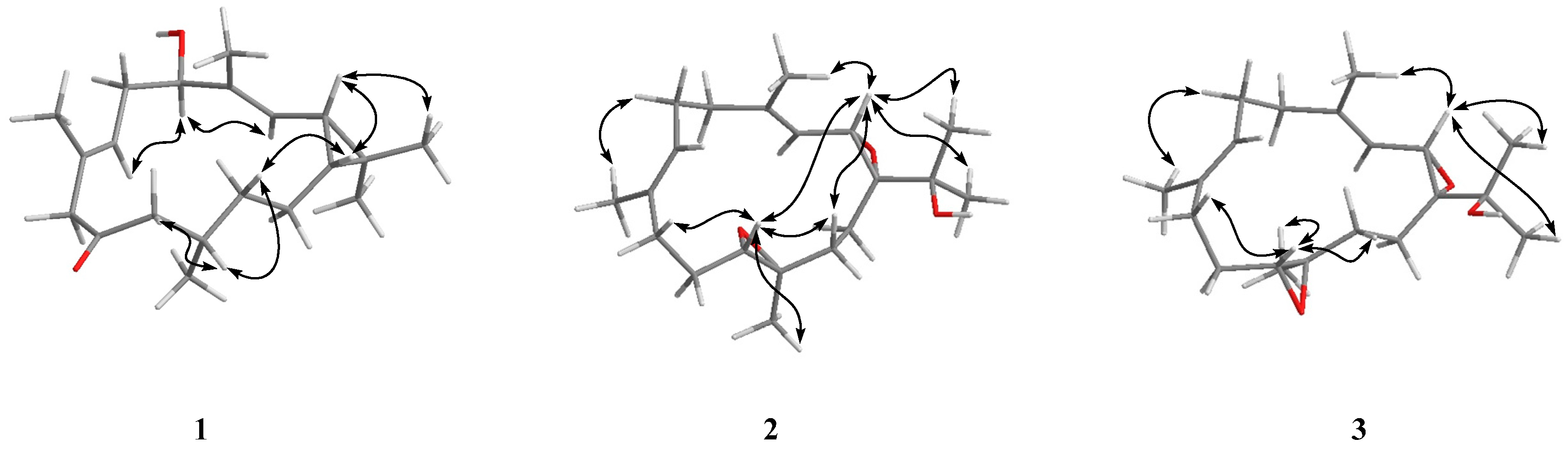
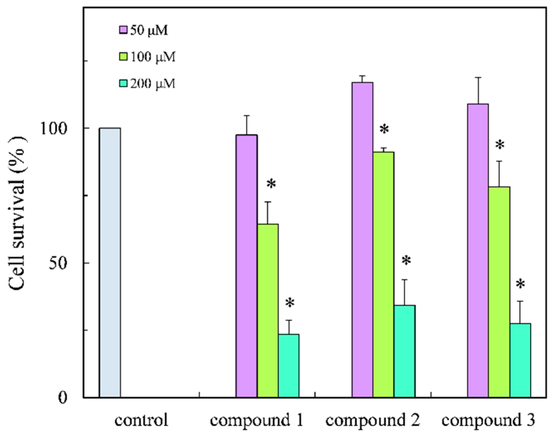
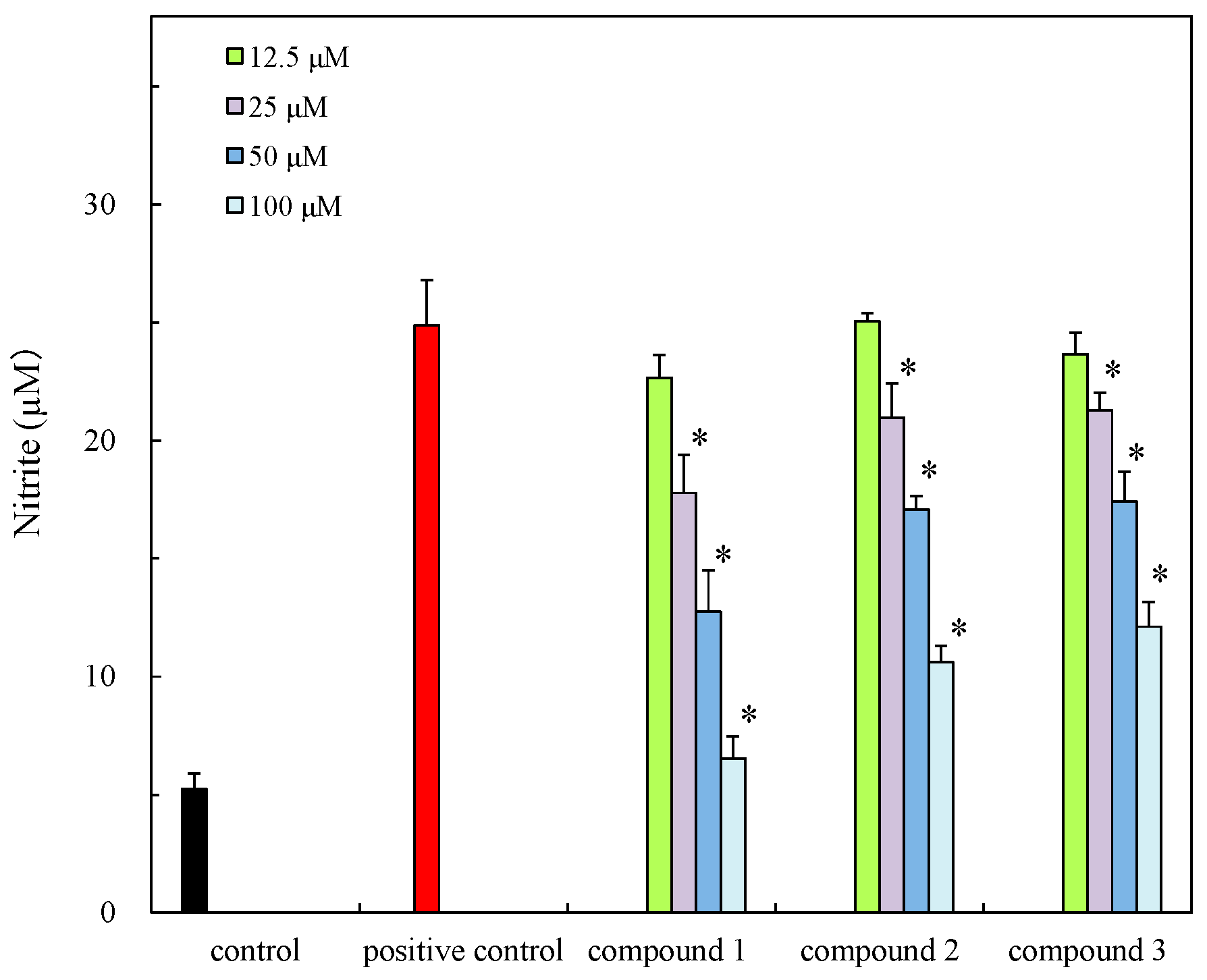
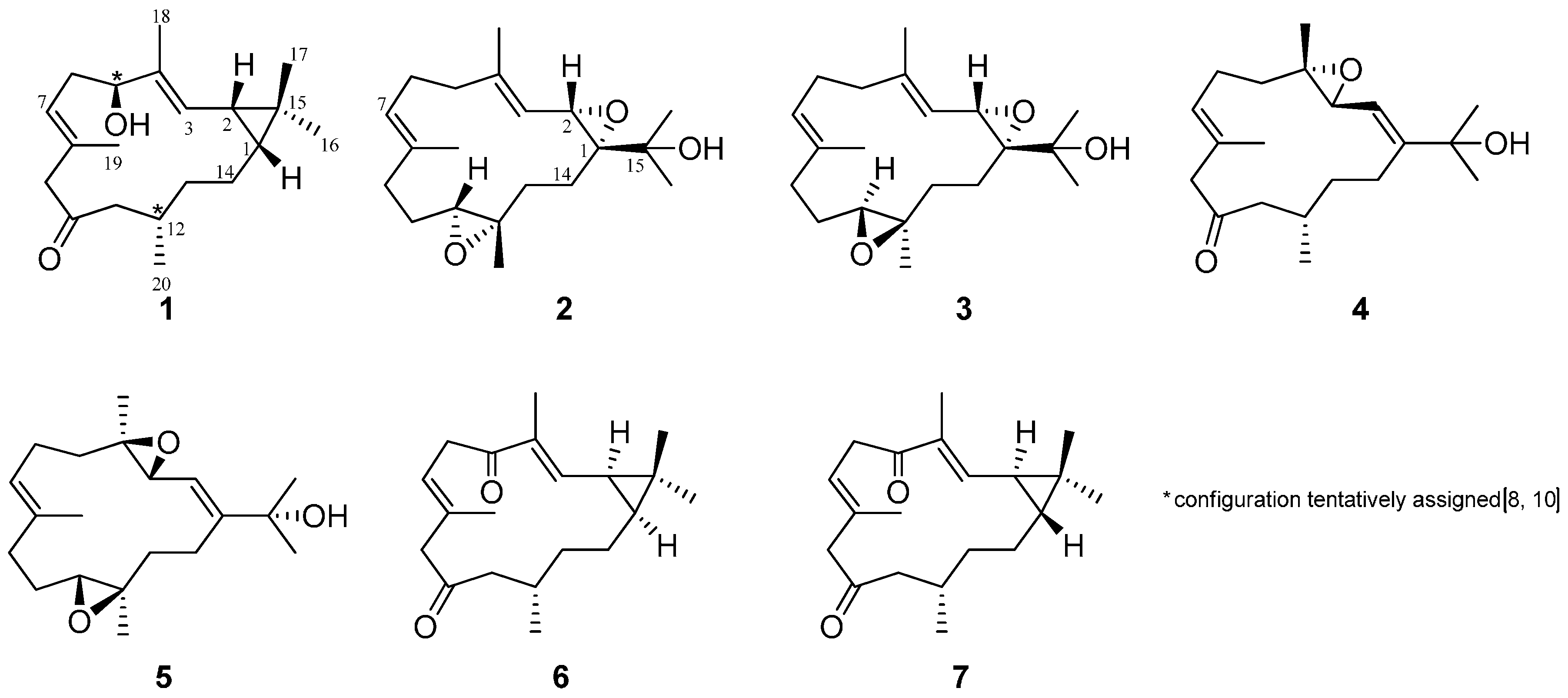
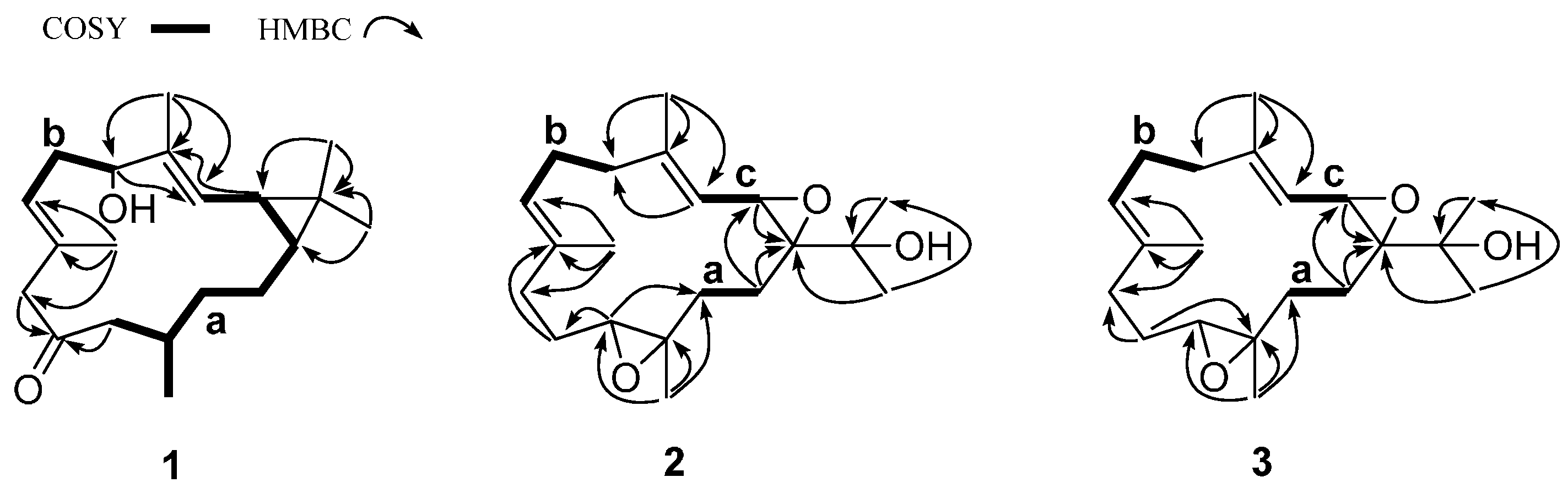
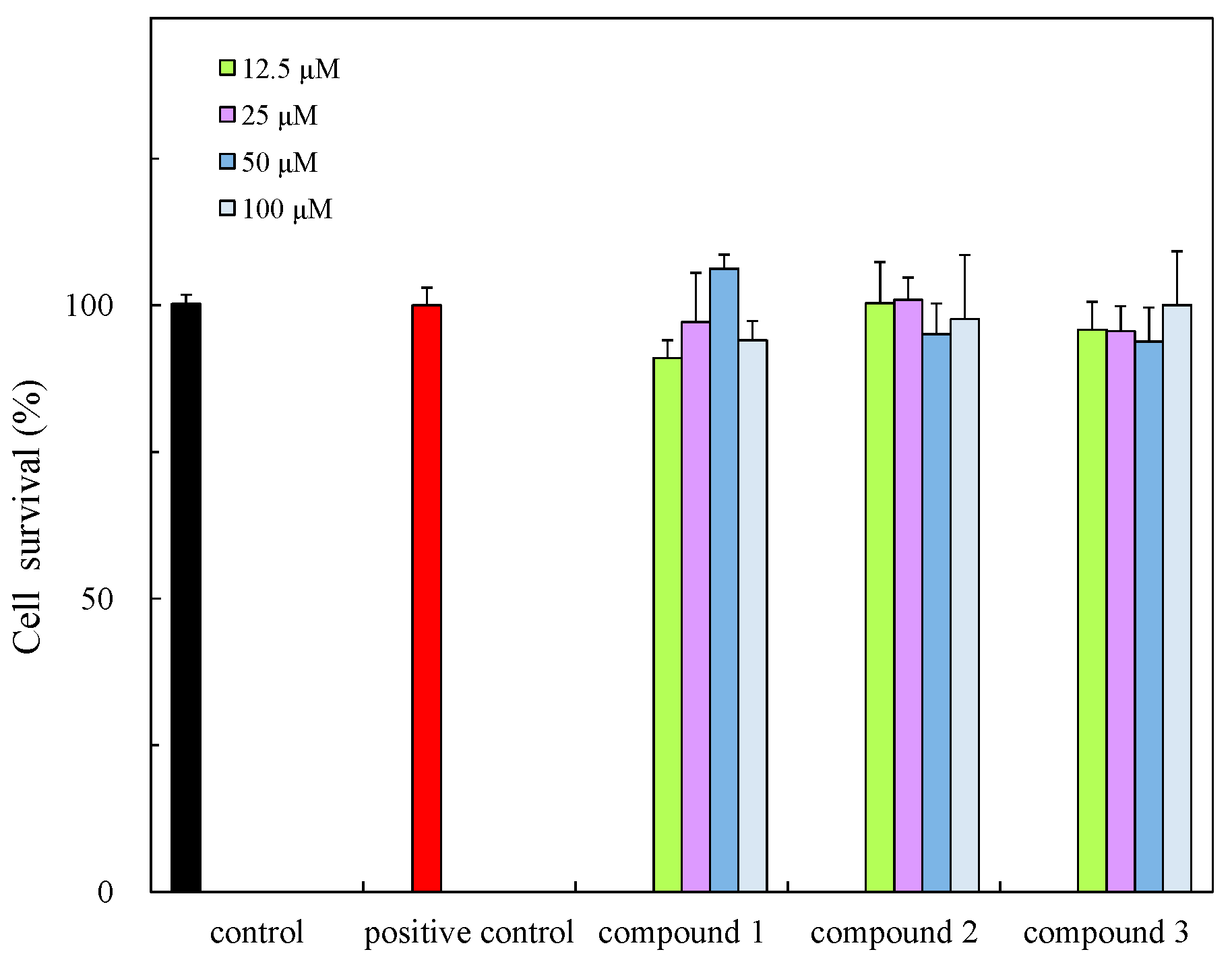
2. Results and Discussion

The Okinawan soft coral, Lobophytum sp. was collected from Irabu Island, Okinawa, and extracted with acetone. The acetone extract was partitioned between ethyl acetate and water. The ethyl acetate portion inhibited the growth of the Gram-positive bacterium Staphylococcus aureus and Gram-negative bacterium Escherichia coli with inhibition zones at 18 and 15 mm at 50 µg/disc, respectively. Repeated chromatographic and HPLC purification of the active crude extract resulted in the isolation of three new metabolites 1 (0.0023%, wet weight), 2 (0.0014%) and 3 (0.0005%) and four known metabolites 4 (0.0039%), 5 (0.0102%) 6 (0.0072%) and 7, 0.0026%) identified by comparison of their NMR data with reported values [8,9,10,11].
The molecular formula of 1 was determined to be C20H32O2 by high-resolution nanospray-ionization MS (HRNSIMS) (m/z 305.2470 [M + H]+, calcd. for C20H33O2, 305.2475), with five degrees of unsaturation. The IR spectrum showed hydroxyl and carbonyl groups (absorption bands at 3279 and 1701 cm−1). 1H- and 13C-NMR data (Table 1, Supplementary Material) suggested it was a diterpenoid and indicated the presence of a ketone (δC 210.6), two trisubstituted double bonds (δC 126.0 (δH 5.09 d, J = 9.5 Hz); 137.1; 124.1 (δH 4.90 t, J = 6.9 Hz); 131.3), one oxygenated carbon atom (δC 79.2 (δH 4.09 dd, J = 4.4, 11.0 Hz)), three sp3 methines (δC 31.4 (δH 0.65 ddd, J = 3.1, 9.0, 11.2 Hz), 25.3 (δH 1.22 dd, J = 9.5, 9.0 Hz), 31.6 (δH 1.88 m)), five sp3 methylenes (δC 33.0 (δH 2.34 m, 2.44 m); 51.9 (δH 3.15 d, J = 14.7 Hz and 2.82 d, J = 14.7 Hz); 52.4 (δH 2.22 d, J = 7.0 Hz); 37.2 (δH 1.15 m); 23.8 (δH 1.59 m, 0.75 m)) and five methyls (δC 15.7 (δH 1.01 s); 29.1 (δH 1.05 s); 10.3 (δH 1.64 s); 17.8 (δH 1.74 s) and 20.4 (δH 0.91 d, J = 6.6 Hz)). On the basis of 1H-1H COSY correlations, the two major spin systems (a: −CH2(11) −CH(12) −CH3(20) −CH2(13) −CH2(14) −CH(1) −CH2(2) −CH2(3) and b: −CH(5) −CH2(6) −CH(7)) were established (Figure 2).
Since compound 1 has three π-bonds, 1 must be bicyclic to satisfy the five degrees of unsaturation requirement. For the four singlet methyls, two were assigned to each of vinyl methyls in the two trisubstituted double bonds, based on heteronuclear multiple bond connectivity (HMBC) correlations (H3-18/C-3, -4, -5; H3-19/C-7, -8, -9) and the NMR chemical shifts; the remaining two were part of a gem-dimethyl group as indicated by HMBC correlations of H3-16/C-1, -15 and H3-17/C-2, -15 and COSY correlation between two cyclopropyl protons (δH 0.65 (ddd, J = 3.1, 9.0, 11.2 Hz), δH 1.22 (dd, J = 9.5, 9.0 Hz)), that indicated a tetrasubstituted cyclopropane ring in molecule 1. An isolated methylene was associated with the ketonic carbonyl and a vinyl methyl (HMBC correlations of H2-9/C-10, -19), situated between C-8 and C-10. In addition, the tetrasubstituted cyclopropane ring associated with partial structure a was shown by HMBC correlations of H3-16/C-1, -15; H3-17/C-1, -2, -15 and H-2/C-4, -15 (Figure 2).
At this point in the structure determination, the partial structures (a with a cyclopropane ring, b, C-4–C-18, C-8–C-19–C-9–C-10 and C-15–C-16–C-17) were identified, but not assembled (Figure 2). HMBC correlations (H3-18/C-3, -4, -5; H3-19/C-7, -8, -9; H2-9, -11/C-10) finally connected these partial structures to give the 14-membered macrocyclic planar structure as a rare casbane-type diterpenoid (Figure 2). The two double bonds at C-3 and C-7, were assigned as E geometry due to the δC values of CH3-18 and CH3-19 (<20 ppm) [27]. The junction of the two rings at carbons C-1/C-2 was suggested to be cis orientation by comparison of the 13C chemical shifts of the geminal methyls [δC 15.7 (C-16) and 29.1 (C-17)] in 1 with those of the known cis-fused casbane diterpenes [10,13,14]. The coupling constant (J = 9.0 Hz) between H-1 (δH 0.65 (ddd, 3.1, 9.0, 11.2 Hz)) and H-2 ((δH 1.22 (dd, J = 9.5, 9.0 Hz)) and an NOE of H-1/H-2 also supported cis configuration of the cyclopropane protons.
The relative stereo structure of 1 was tentatively assigned by 1D Nuclear Overhauser Effect (NOE) experiments (Figure 3) and by comparison of the NMR data for 1 with those reported for congeners of 1 [10,13,14]. In the NOE experiments of 1, irradiation of the H-1 signal revealed NOEs with H-2, and irradiation of the H-2 signal showed NOEs with H-1, H3-17, suggesting these protons were on the same face (Figure 3). The NOEs between H-1 and H-13 and H-12/H-11, -13, suggested that H3-20 was on the side opposite these protons in the molecule. Irradiation of the H-5 signal revealed NOEs with H-3, -7 but not with H-2 and irradiation of the H-3 signal, showed an NOE with H-5 but not H-2. So, H-5 and H-2 could be opposite sides of the molecule. Unfortunately, attempts to prepare MTPA esters for determination of the absolute stereochemistry failed because of its instability and the small quantity of compound 1 available.
The HRNSIMS (m/z 321.2418 [M + H]+, calcd. for C20H33O3, 321.2424) of 2 suggested the molecular formula C20H32O3, which accounted for five degrees of unsaturation. The IR spectrum showed hydroxyl and epoxide functionalities (absorption bands at 3481, 1295 and 1252 cm−1). 1H- and 13C-NMR data (Table 1), coupled with the molecular formula C20H32O3, suggested it was a diterpenoid derivative and indicated the presence of one oxygenated carbon atom (δC 70.1), two epoxides (δC 57.5 (δH 3.75 d, J = 4.2 Hz); δC 68.2 and δC 61.6 (δH 2.71 dd, J = 3.4, 9.1 Hz); δC 61.2), two trisubstituted double bonds (δC 118.8 (δH 5.08 brd, J = 4.2 Hz); δC 141.9 and δC 126.2 (δH 5.15 t, J = 6.1 Hz); δC 133.7), six sp3 methylenes (δC 38.9 (δH 2.27 m); 24.5 (δH 2.24 m); 36.8 (δH 2.23 m, 2.04 m); 24.3 (δH 2.25 m, 1.96 m); 35.2 (δH 1.99 m); 25.4 (δH 1.86 m, 1.34 m)) and five methyls (δC 26.2 (δH 1.25 s); 26.7 (δH 1.31 s); 17.7 (δH 1.70 s); 15.3 (δH 1.64 s) and 17.0 (δH 1.24 s)). Since compound 2 has two π-bonds and two epoxides, 2 must be monocarbocyclic to fulfill the five degrees of unsaturation requirement. Three major spin system (a: −CH2(13)−CH(14), b: −CH(5)−CH(7), and c: −CH(2) −CH(3)), were identified from the 1H-1H COSY correlations (Figure 2). The two epoxides were trisubstituted, based on HMBC correlations (H-2/C-1, H2-14/C-1, -2). For the five methyls, two were assigned to each of vinyl methyls in the two trisubstituted double bonds, based on HMBC correlations (H3-18/C-3, -4, -5 and H3-19/C-7, -8, -9) and the NMR chemical shifts; one was associated with an epoxide at C-12 (HMBC correlations of H3-20/C-11, -12, -13), and the remaining two were part of a gem-dimethyl group, and were associated with another epoxide at C-1 as indicated by HMBC correlations of H3-16/C-1, -15, -17 and H3-17/C-15, -16. On the basis of HMBC correlations (Figure 2), three partial structures (a, b and c) and other fragments could be connected to give the planar structure 2 as a cembrane-type diterpenoid (Figure 2).
The relative configuration of 2 was assigned by detailed analysis of 1D NOE experiments. NOE correlations between H-2/H-11, H-2/H-13, H-2/H3-16, H-2/H3-17, H-2/H3-18, H-11/H-9, H-11/H-13 and H-11/ H3-20 implied that these protons were on the same face (Figure 3). The NOE correlations between H-2/H3-18 and H-6/H3-19, and δC values of CH3-18 and CH3-19 (<20 ppm) [27] suggested that the two double bonds at C-3 and C-7 should be assigned as E geometry.
The molecular formula of 3 (C20H32O3) was the same as 2, as inferred by HRNSIMS (m/z 321.2419 [M + H]+, calcd. for C20H33O3, 321.2424). The 1H- and 13C-NMR spectra (Table 1) of 3 were very similar to those of 2. Extensive analysis of 1D and 2D NMR data, and comparison of the NMR data with those of 2 led to the same planar structure as that of 2. Since the NOEs observed for the portions at C1, C2, C3, C4, C7 and C8 in 3 resembled those described above for 2, both compounds possess identical stereochemistry in these portions. An NOE between H-11/H3-20 in 3, along with no NOE effect on H-2 and H-11 upon irradiation of H-11, suggested that the protons H-11 and H3-20 in 3 were on the same face (the opposite of that found in 2) (Figure 3). Therefore, compounds 3 and 2 were epoxide moiety stereoisomers at C-12.
The isolates were evaluated for antibacterial activity using the paper disc method [28] against S. aureus, S. enterica and E. coli and new isolates also evaluated for cytotoxicity and anti-inflammatory effect in cells (Table 2). The isolates showed weak anti-bacterial activity and new compounds exhibited cytotoxicity against HCT 116 cells (Figure 4) but this was weaker than those of previously reported compounds, for example alcyonolide and its congeners isolated from soft coral Cespitularia sp. were in the IC50 5.85–91.4 µM range [4]. The anti-inflammatory activity of compounds 13 was also evaluated in LPS/IFN-γ-stimulated RAW 264.7 macrophage cells under non-cytotoxic concentration ranges (Figure 5 and Figure 6). The compounds suppressed NO production in a dose dependent manner, indicating the compounds have the anti-inflammatory effect. The inhibition was similar to that of flavonoids, but they were low levels (IC50 (µM), 41.2–74.8) by comparison with alcyonolide congeners (2–8 µM) [29] and marine carotenoids (6.25–25 µM), such as fucoxanthin and fucoxanthinol [30].

3. Experimental Section

3.1. General Procedures

Optical rotation was measured using a JASCO P-1010 polarimeter (JASCO International Co. Ltd., Tokyo, Japan). Nuclear magnetic resonance (NMR) spectra were recorded on an Avance III 500 spectrometer (Bruker, Rheinstetten, Germany) in CDCl3. Chemical shifts and coupling constants were given as δ and Hz, respectively and 1H- and 13C- chemical shifts were referenced to the solvent peaks (δH = 7.26 and δC = 77.24). Infrared (IR) spectra were recorded on a JASCO FT/IR-6100 Fourier Transform Infrared Spectrometer (JASCO International Co. Ltd.). High-resolution mass spectra (HRMS) were obtained on an LTQ Orbitrap hybrid mass spectrometer (Thermo Scientific, Bemen, Germany) equipped with a nanospray ionization (NSI) source. Open column chromatography was performed on Kieselgel 60 (70–230 mesh, Merck, Darmstadt, Germany). High performance liquid chromatography (HPLC) was performed using a COSMOSIL Si60 HPLC column (5 SL, Φ 10 × 250 mm, Nacalai tesque Inc, Osaka, Japan). Analytical thin layer chromatography (TLC) was performed using Kieselgel 60 F254 DC-fertigplatten (Merck). All solvents were reagent grade.

3.2. Animal Materials

The soft coral Lobophytum sp. (220.0 g, wet weight) was collected by hand during low tide from the coast of Irabu Island, Okinawa, Japan, in March 2013, and identified as a Lobophytum sp. A voucher specimen was deposited at University of the Ryukyus (Specimen No. 13033102).

3.3. Extraction and Isolation

The soft coral was transported to the lab and extracted with acetone (2 L × 3). After filtration, extracts were concentrated under reduced pressure to form an acetone extract. The acetone extract was partitioned between H2O (200 mL) and EtOAc (200 mL × 2). The EtOAc part was evaporated in vacuo to give a crude extract (2.41 g) that inhibited the growth of the Gram-positive bacterium Staphylococcus aureus and Gram-negative bacterium Escherichia coli with inhibition zones of 18 and 15 mm, respectively, at 50 µg/disc. The active crude extract was first chromatographed over silica gel to give 19 fractions (hexane/EtOAc/MeOH gradient). On the basis of its 1H-NMR spectrum, fraction 8 was subjected to further purification by HPLC. An aliquot (102.3 mg) of fraction 8 (213.4 mg) was purified by HPLC (a COSMOSIL Si-60 column SiO2) using hexane/EtOAc (7:3) to afford new diterpenoids 1 (2.5 mg), 2 (1.5 mg), 3 (0.6 mg) and known diterpenoids 4 (4.2 mg) and 5 (10.8 mg). An aliquot (44.2 mg) of fraction 5 (130.4 mg) was purified by HPLC using hexane/EtOAc (4:1) to afford known diterpenoids 6 (5.4 mg) and 7 (2.0 mg).
Compound 1: Colorless oil; [ α ] D 31.4 −111.4 (c 0.07 CH3OH); FT/IR νmax (film) 3279, 2921 and 1701 cm−1; 1H-NMR and 13C-NMR data are listed in Table 1; HRNSIMS m/z 305.2470 [M + H]+ (calcd. for C20H33O2, 305.2475).
Compound 2: Colorless oil; [ α ] D 31.5 +12.0 (c 0.05 CH3OH); FT/IR νmax (film) 3481, 2932, 1295 and 1252 cm−1; 1H and 13C-NMR (CDCl3) data are listed in Table 1; HRNSIMS m/z 321.2418 [M + H]+ (calcd. for C20H33O3, 321.2424).
Compound 3: Colorless oil; [ α ] D 31.7 −16.6 (c 0.06 CH3OH); FT/IR νmax (film) 3465, 2930, 1254 and 1166 cm−1; 1H and 13C-NMR (CDCl3) data are listed in Table 1; HRNSIMS m/z 321.2419 [M + H]+ (calcd. for C20H33O3, 321.2424).

3.4. Molecular Mechanics Calculations

Implementation of the MM2 force field [32] in ChemBioOffice Ultra 12.0 software (Cambridge Soft Corporation, Cambridge, MA, USA) was used to calculate molecular models.

3.5. Anti-Bacterial Assay

The paper disk diffusion method [28] was used to evaluate the anti-bacterial activity of compounds 15, using the bacterial strains Staphylococcus aureus, Salmonella enterica and Escherichia coli. The strains were received from the Biological Resource Center (NBRC, Tokyo, Japan), Japan and cultured in an agar medium containing polypeptone (10 g/L distilled water), yeast (2 g/L distilled), MgSO4·7H2O (1 g/L distilled) and agar (15 g/L distilled). The medium was autoclaved and transferred into petri dishes. The bacterial inoculum was evenly spread on the above agar medium. Each methanolic solution of the test compounds was perfused (25 µg/25 µL) to a sterilized disc (Φ 8 mm, Toyo Roshi Kaisha, Ltd., Tokyo, Japan). After the removal of the solvent, the disks containing test compounds were placed on seeded bacterial lawn on the agar surface. The plate was incubated for 2 days at 30 °C and then the inhibition zone sizes were measured.

3.6. Cell Culture

HCT116 human colon cancer cells (ATCC, Manassas, VA, USA) and RAW 264.7 cells (mouse macrophages, American Type Culture Collection) were cultured in DMEM (Gibco-BRL, Life Technologies, South San Francisco, CA, USA) medium (including 10% FBS, 100 U/mL penicillin and 100 μg/mL streptomycin) at 37 °C in a 5% CO2 atmosphere.

3.7. Cell Viability

The MTT assay was used to examine the cytotoxity of compounds 13. Briefly, HCT116 cells were seeded at a density of 5.0 × 10⁵ cells/mL in 96-well plate and cultured for 24 h with or without the test compound. After the culture, MTT (0.05%) was added to each well and incubated for 2 h, and then suspension was removed. Extraction with DMSO (50 μL) was measured at 540 nm with the reference at 655 nm using a microplate reader (BIORAD model 550, BIO-RAD, Hercules, CA, USA).

3.8. Anti-Inflammatory Effect on Nitrite Production on RAW 264.7 Macrophages

The RAW 264.7 cells (2.5 × 10⁶ cells /mL) were treated with the compounds 13 in the presence of LPS (100 ng/mL), L-arginine (2 mM), and IFN-γ (100 U/mL) in 96-well microplate. Cells with or without LPS, IFN-γ and L-arginine were used as the positive control and the control, respectively. After culturing for 17 h, the nitrite concentrations in the medium were determined by previously reported method [31].

3.9. Statistical Analysis

Data were expressed as mean ± SD. Statistical significance (p < 0.01) was analyzed by Student’s t-tests.

4. Conclusions

Seven diterpenoids 17, including three new compounds 13, were isolated from the Okinawan soft coral, Lobophytum sp. Their relative stereostructures were established by spectroscopic analysis (NMR, IR, and MS) and comparisons with similar reported metabolites. The new isolates showed weak antibacterial activity, mild cytotoxicity against human colon cancer cells and showed anti-inflammatory effect in LPS/IFN-γ-stimulated RAW 264.7 macrophage cells.

Supplementary Materials

Supplementary materials can be assessed at: https://www.mdpi.com/1420-3049/21/5/679/s1.

Acknowledgments

We are grateful to the Japanese Government (Ministry of Education, Culture, Sports, Science, and Technology) for funding of the research. The authors thank the IRC, at the University of the Ryukyus for analytical services. The authors are also thankful to Dr. Kevin Wakeman (The University of Tokyo) for editing the manuscript.

Author Contributions

Prodip K. Roy, Runa Ashimine and Katsuhiro Ueda conceived and designed the experiments, performed the experiments, analyzed the data, elucidated the structures and wrote the manuscript. Haruna Miyazato and Junsei Taira carried out the biological assay.

Conflicts of Interest

The authors declare no conflict of interest.

References

  1. Burhan, R.Y.R.; Putri, E.M.M.; Zetra, Y.; Herdhiansyah, R.; Putra, M.Y. Terpenes from soft corals of the genus Lobophytum (Alcyoniidae): Chemistry and biological activities. J. Chem. Pharm. Res. 2014, 6, 585–595. [Google Scholar]
  2. Blunt, J.W.; Copp, B.R.; Keyzers, R.A.; Munro, M.H.G.; Northcote, P.N.T.; Prinsep, M.R. Marine natural products. Nat. Prod. Rep. 2015, 32, 116–211. [Google Scholar] [CrossRef] [PubMed]
  3. Roy, P.K.; Maarisit, W.; Roy, S.R.; Taira, J.; Ueda, K. Cytotoxic alcyonolide congeners from an Okinawan soft coral Cespitularia sp. Heterocycles 2012, 85, 2465–2472. [Google Scholar]
  4. Roy, P.K.; Maarisit, W.; Roy, M.C.; Taira, J.; Ueda, K. Five new diterpenoids from an Okinawan soft coral, Cespitularia sp. Mar. Drugs 2012, 10, 2741–2748. [Google Scholar] [CrossRef] [PubMed]
  5. Roy, P.K.; Roy, M.C.; Taira, J.; Ueda, K. Structure and bioactivity of a trisnorditerpenoid and diterpenoid from an Okinawan soft coral, Cespitularia sp. Tetrahedron Lett. 2014, 55, 1421–1423. [Google Scholar] [CrossRef]
  6. Maarisit, W.; Yano, K.; Miyazato, T.; Roy, P.K.; Taira, J.; Ueda, K. Structures and bioactivities of xenicanes from an Okinawan soft coral Xenia sp. Heterocycles 2015, 91, 505–514. [Google Scholar]
  7. Ueda, K.; Hu, Y. Haterumalide B: A new cytotoxic macrolide from an Okinawan ascidian Lissoclinum sp. Tetrahedron Lett. 1999, 40, 6305–6308. [Google Scholar] [CrossRef]
  8. Ahmed, A.F.; Tai, S.H.; Wen, Z.H.; Su, J.H.; Wu, Y.C.; Hu, W.P.; Sheu, J.H. A C-3 methylated isocembranoid and 10-oxocembranoids from a Formosan soft coral, Sinularia grandilobata. J. Nat. Prod. 2008, 71, 946–951. [Google Scholar] [CrossRef] [PubMed]
  9. Hou, R.S.; Duh, C.Y.; Chiang, M.Y.; Lin, C.N. Sinugibberol, a new cytotoxic cembranoid diterpene from the soft coral Sinularia gibberosa. J. Nat. Prod. 1995, 58, 1126–1130. [Google Scholar] [CrossRef] [PubMed]
  10. Li, Y.; Carbone, M.; Vitale, R.M.; Amodeo, P.; Castelluccio, F.; Sicilia, G.; Mollo, E.; Nappo, M.; Cimino, G.; Guo, Y.W.; et al. Rare casbane diterpenoids from the Hainan soft coral Sinularia depressa. J. Nat. Prod. 2010, 73, 133–138. [Google Scholar] [CrossRef] [PubMed]
  11. Yin, J.; Zhao, M.; Ma, M.; Xu, Y.; Xiang, Z.; Kai, Y.; Dong, J.; Lei, X.; Huang, K.; Yan, P. New casbene diterpenoids from a South China Sea soft coral, Sinularia sp. Mar. Drugs 2013, 11, 455–465. [Google Scholar] [CrossRef] [PubMed]
  12. Robinson, D.R.; West, C.A. Biosynthesis of cyclic diterpenes in extracts from seedlings of Ricinus communis L. I. Identification of diterpene hydrocarbons formed from mevalonate. Biochemistry 1970, 9, 70–79. [Google Scholar] [PubMed]
  13. Chio, Y.H.; Kim, J.; Pezzuto, J.M.; Kinghorn, A.D.; Farnsworth, N.R. Agrostistachin, a novel cytotoxic macrocyclic diterpene from Agrostistachys hookeri. Tetrahedron Lett. 1986, 48, 5795–5798. [Google Scholar] [CrossRef]
  14. Zhang, C.X.; Yan, S.J.; Zhang, G.W.; Lu, W.G.; Su, J.Y.; Zeng, L.M.; Gu, L.Q.; Yang, X.P.; Lian, Y.J. Cytotoxic sterols from the soft coral Nephthea erecta. J. Nat. Prod. 2005, 68, 1087–1089. [Google Scholar] [CrossRef] [PubMed]
  15. Xu, Z.H.; Sun, J.; Xu, R.S.; Qin, G.W. Casbane diterpenoids from Euphorbia ebracteolata. Phytochemistry 1998, 49, 149–151. [Google Scholar] [CrossRef]
  16. Wang, S.K.; Duh, C.Y.; Wu, Y.C.; Wang, Y.; Cheng, M.C.; Soong, K.; Fang, L.S. Studies on Formosan soft corals, II. Cytotoxic cembranolides from the soft coral Lobophytum michaelae. J. Nat. Prod. 1992, 55, 1430–1435. [Google Scholar] [CrossRef] [PubMed]
  17. Wang, S.K.; Duh, C.Y. New cytotoxic cembranolides from the soft coral Lobophytum michaelae. Mar. Drugs 2012, 10, 306–318. [Google Scholar] [CrossRef] [PubMed]
  18. Wang, L.T.; Wang, S.K.; Soong, K.; Duh, C.Y. New cytotoxic cembranolides from the soft coral Lobophytum michaelae. Chem. Pharm. Bull. 2007, 55, 766–770. [Google Scholar] [CrossRef] [PubMed]
  19. Coval, S.J.; Patton, R.W.; Petrin, J.M.; James, L.; Rothofsky, M.L.; Lin, S.; Patel, M.; Reed, J.K.; McPhil, A.T.; Bishop, W.R. A cembranolide diterpene farnesyl protein transferase inhibitor from the marine soft corals Lobophytum cristagalli. Bioorg. Med. Chem. Lett. 1696, 6, 909–912. [Google Scholar] [CrossRef]
  20. Higuchi, R.; Miyamoto, T.; Yamada, K.; Komori, T. Cytotoxic and ichthyotoxic compounds from marine Opisthobranchia and soft coral. Toxicon 1998, 36, 1703–1705. [Google Scholar] [CrossRef]
  21. Matthée, G.F.; König, G.M.; Wright, A.D. Three new diterpenes from the marine soft coral Lobophytum crassum. J. Nat. Prod. 1998, 61, 237–240. [Google Scholar] [CrossRef] [PubMed]
  22. Duh, C.Y.; Wang, S.K.; Huang, B.T.; Dai, C.F. Cytotoxic cembrenolide diterpenes from the Formosan soft coral Lobophytum crassum. J. Nat. Prod. 2000, 63, 884–885. [Google Scholar] [CrossRef] [PubMed]
  23. Cheng, S.Y.; Wang, S.K.; Duh, C.Y. Secocrassumol, a seco-cembranoid from the Dongsha a toll soft coral Lobophytum crissum. Mar. Drugs 2014, 12, 6028–6037. [Google Scholar] [CrossRef] [PubMed]
  24. Lin, S.T.; Wang, S.K.; Cheng, S.Y.; Duh, C.Y. Lobocrasol, a new diterpenoid from the soft coral Lobophytum crassum. Org. Lett. 2009, 14, 3012–3014. [Google Scholar] [CrossRef] [PubMed]
  25. Cheng, S.Y.; Wen, Z.H.; Chiou, S.F.; Hsu, C.H.; Wang, S.K.; Dai, C.F.; Chiang, M.Y.; Duh, C.Y. Durumolides A–E, anti-inflammatory and antibacterial cembranolides from the soft coral Lobophytum durum. Tetrahedron 2008, 64, 9698–9704. [Google Scholar] [CrossRef]
  26. Cheng, S.Y.; Wen, Z.H.; Wang, S.K.; Chiou, S.F.; Hsu, C.H.; Dai, C.F.; Duh, C.Y. Anti-inflammatory cembranolides from the soft coral Lobophytum durum. Bioorg. Med. Chem. 2009, 17, 3763–3769. [Google Scholar] [CrossRef] [PubMed]
  27. Breitmaier, E.; Voelter, W. Carbon-13 NMR Spectroscopy; VCH: New York, NY, USA, 1987; pp. 192–196. [Google Scholar]
  28. Ericsson, H. The paper disc method for determination of bacterial sensitivity to antibiotics studies on the accuracy of the technique. Scand. J. Clin. Lab. Invest 1960, 12, 408–413. [Google Scholar] [CrossRef] [PubMed]
  29. Taira, J.; Tsuchida, E.; Uehara, M.; Kinjyo, Y.; Roy, P.K.; Ueda, K. Dual biological functions of the apoptotic activity and anti-inflammatory effect by alcyonolide congeners from the Okinawan soft coral, Cespitularia sp. Bioorg. Med. Chem. Lett. 2015, 25, 4496–4499. [Google Scholar] [CrossRef] [PubMed]
  30. Taira, J.; Uehara, M. Anti-inflammatory effect of fucoxanthinol as bioavailable marine carotenoid on LPS-stimulated RAW 264.7 macrophages through iNOS suppression and nitrogen radical-scavenging. World J. Pharm. Sci. 2015, 3, 1747–1754. [Google Scholar]
  31. Taira, J.; Nanbu, H.; Ueda, K. Nitric oxide-scavenging compounds in Agrimonia pilosa Ledeb on LPS-induced RAW 264.7 macrophages. Food Chem. 2009, 115, 1221–1227. [Google Scholar] [CrossRef]
  32. Allinger, N.L. Conformational analysis. 130. MM2. A hydrocarbon force field utilizing V1 and V2 torsional terms. J. Am. Chem. Soc. 1977, 99, 8127–8134. [Google Scholar] [CrossRef]
  • Sample Availability: Samples of the compounds 17 are available from the authors.
Figure 1. Chemical structures of compounds 17.
Molecules 21 00679 g001
Figure 2. Partial structures of 13 based on COSY (bold line) and key HMBC correlations (arrow).
Molecules 21 00679 g002
Figure 3. Computer-generated model of 13 using MM2 force calculations and key NOE correlations.
Molecules 21 00679 g003
Figure 4. Cytotoxicity of 13 against HCI116 colon cancer cells. Significance * p < 0.01 was considered statistically significant for control.
Molecules 21 00679 g004
Figure 5. Cytotoxicity of 13 for NO production in LPS/IFN-γ stimulated RAW 264.7 macrophage cells.
Molecules 21 00679 g005
Figure 6. Anti-inflammatory effect of 13 against NO production in LPS-stimulated RAW 264.7 macrophage cells. Significance * p < 0.01 was considered statistically significant for positive control.
Molecules 21 00679 g006
Table 1. 1H- (500 MHz) and 13C-(125 MHz) NMR data for 13 in CDCl3.
C No.123
δH (mult., J/Hz)δCδH (mult., J/Hz)δCδH (mult., J/Hz)δC
10.65 (ddd, 3.1, 9.0, 11.2)31.4 (CH) 68.2 (C) 68.0 (C)
21.22 (dd, 9.5, 9.0)25.3 (CH)3.75 (d, 4.2)57.5 (CH)3.80 (d, 5.8)57.9 (CH)
35.09 (brd, 9.5)126.0 (CH)5.08 (brd, 4.2)118.8 (CH)4.79 (brd, 5.8)121.0 (CH)
4 137.1 (C) 141.9 (C) 140.0 (C)
54.09 (dd, 4.4, 11.0)79.2 (CH)2.27 (m)38.9 (CH2)2.11 (m)38.9 (CH2)
62.44 (m)33.0 (CH2)2.24 (m)24.5 (CH2)2.31 (m)24.8 (CH2)
2.34 (m)
74.90 (dd, 6.9, 6.9)124.1 (CH)5.15 (t, 6.1)126.2 (CH)5.10 (t, 5.2)125.8 (CH)
8 131.3 (C) 133.7 (C) 134.0 (C)
93.15 (d, 14.7)51.9 (CH2)2.23 (m)36.8 (CH2)2.18 (m)36.8 (CH2)
2.82 (d, 14.7) 2.04 (m)
10 210.6 (C)2.25 (m)24.3 (CH2)2.29 (m)23.7 (CH2)
1.96 (m) 2.02 (m)
112.22 (d, 7.0)52.4 (CH2)2.71 (dd, 3.4, 9.1)61.6 (CH)2.59 (dd, 3.3, 10.6)61.9 (CH)
121.88 (m)31.6 (CH) 61.2 (C) 61.4 (C)
131.15 (m)37.2 (CH2)1.99 (m)35.2 (CH2)2.27 (m)35.1 (CH2)
1.96 (m)
141.59 (m)23.8 (CH2)1.86 (m)25.4 (CH2)2.09 (m)24.3 (CH2)
0.75 (m) 1.34 (m)
15 21.0 (C) 70.1 (C) 70.8 (C)
161.01 (s)15.7 (CH3)1.25 (s)26.2 (CH3)1.29(s)25.4 (CH3)
171.05 (s)29.1 (CH3)1.31 (s)26.7 (CH3)1.28 (s)26.5 (CH3)
181.64 (s)10.3 (CH3)1.70 (s)17.7 (CH3)1.70 (s)17.0 (CH3)
191.74 (s)17.8 (CH3)1.64 (s)15.3 (CH3)1.64 (s)14.9 (CH3)
200.91 (d, 6.6)20.4 (CH3)1.24 (s)17.0 (CH3)1.25 (s)16.0 (CH3)
Table 2. Antibacterial activity, cytotoxicity and anti-inflammatory effect of compounds 15.
CompoundAntibacterial Activity aCytotoxicity (IC50, µM)Anti-Inflammatory Effect (IC50, µM)
S. aureusS. entericaE. coliHCT116 cellsRAW 256.7 cells
110N.A b10135.5741.21
291210177.1164.96
391010153.1174.76
410N.A b12N.T cN.T c
510N.A b15N.T cN.T c
Streptomycin sulfate15N.T c13N.T cN.T c
a Inhibition zone in mm at 25 µg/disc, b Not active, and c Not tested [31].

