
Novel Gold Nanoparticles Reduced by Sargassum glaucescens: Preparation, Characterization and Anticancer Activity

 ,
, 
Abstract
:1. Introduction
2. Results
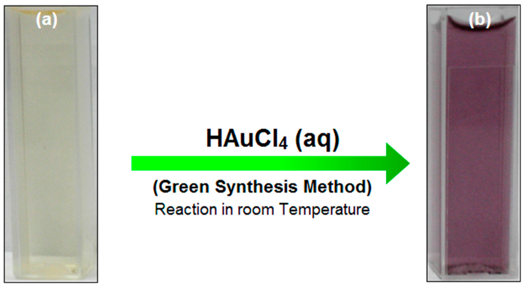
2.1. Preparation of SG-Stabilized AuNPs

2.2. SG-Stabilized AuNPs Characterization
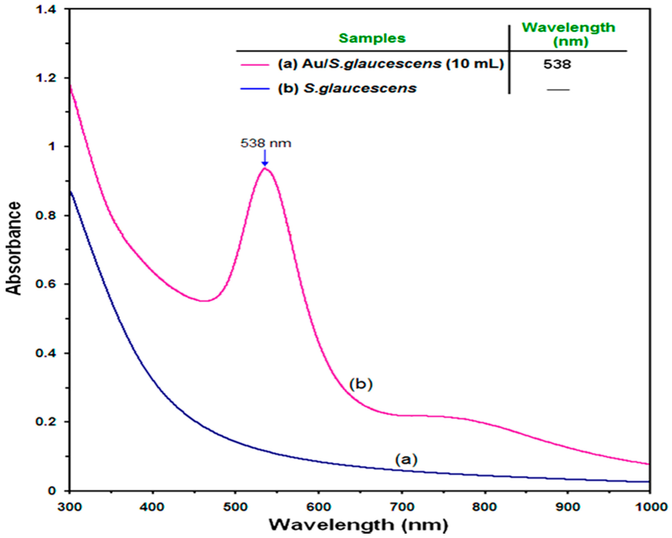
2.2.1. Ultraviolet-Visible (UV-Vis) Spectroscopy Analysis
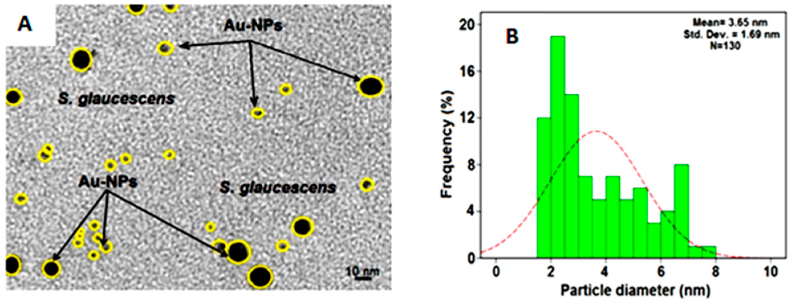
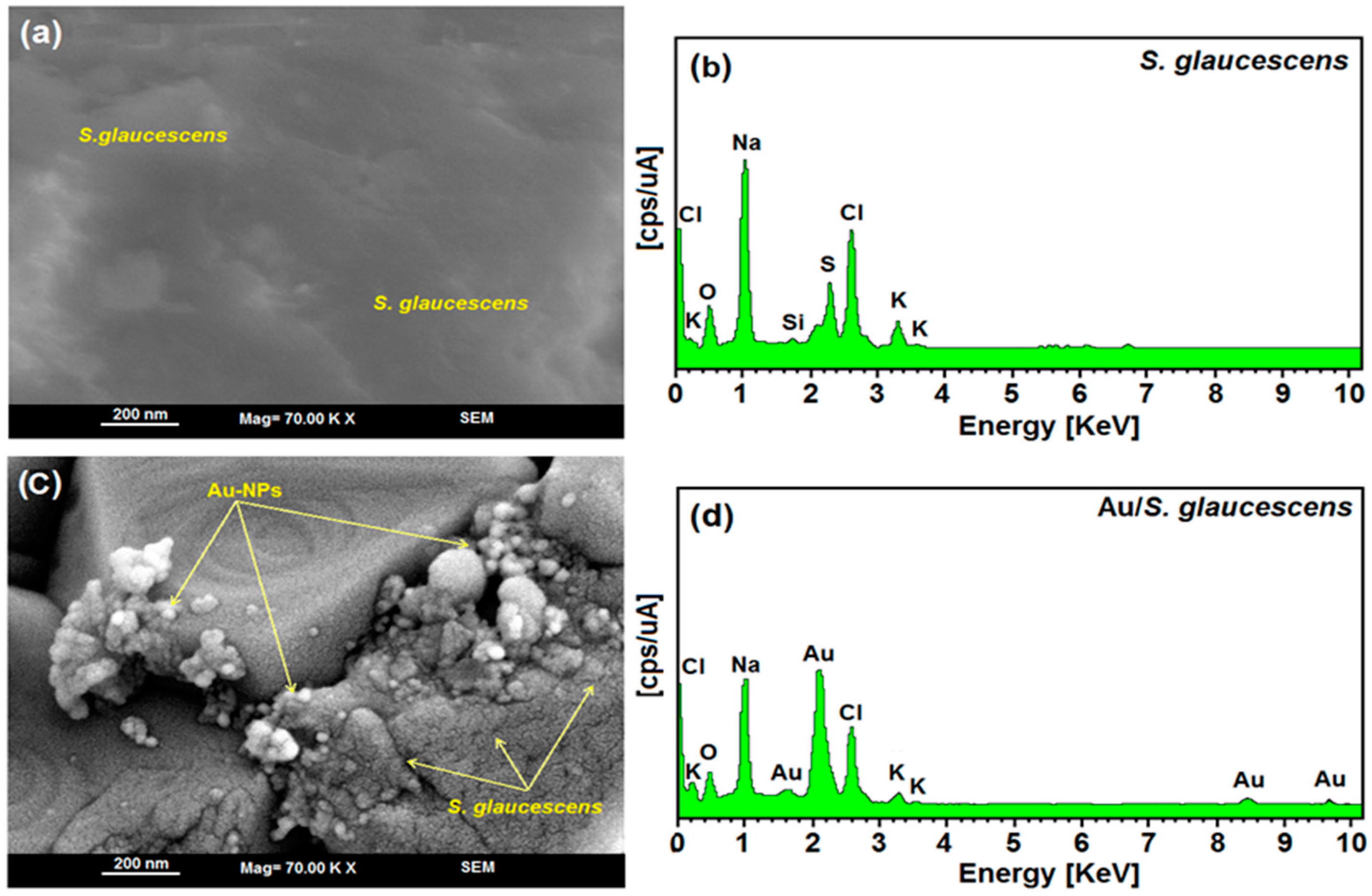
2.2.2. Morphology Study


2.3. Anticancer Activity of SG-Stabilized AuNPs
2.3.1. Cytotoxicity ASSAY

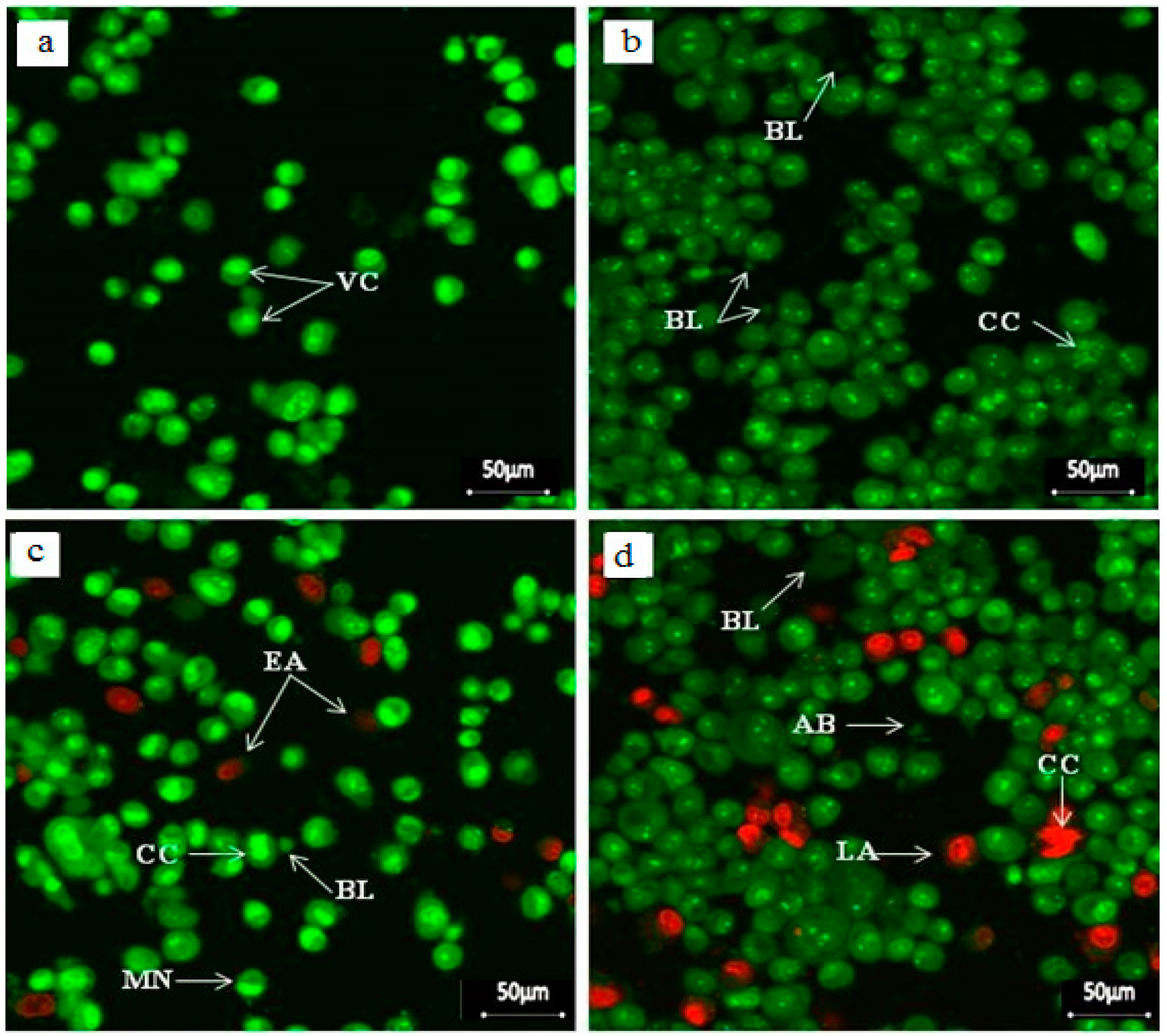
2.3.2. Apoptosis Detection Assay
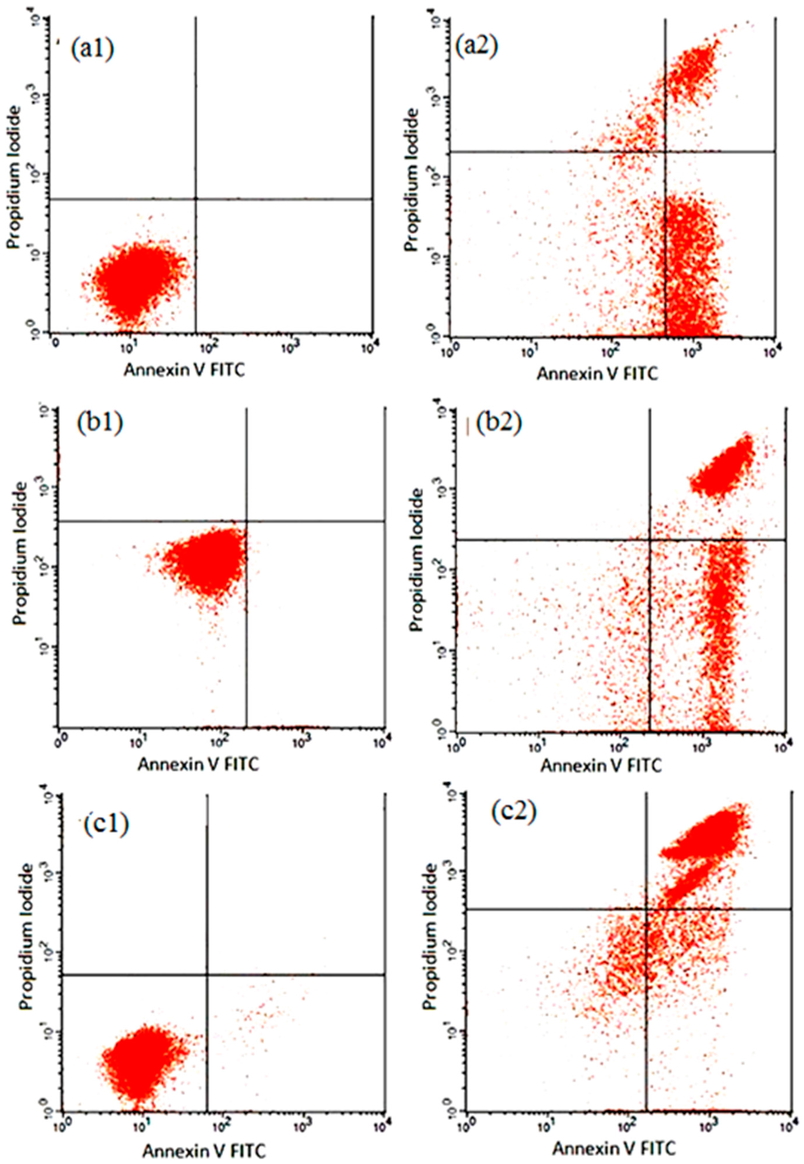
2.3.3. Annexin V-FITC Assay
2.3.4. Cell Cycle Assay

| Cells (%) | ||||||
|---|---|---|---|---|---|---|
| Cell Condition | Control 12 h | Treated 12 h | Control 24 h | Treated 24 h | Control 48 h | Treated 48 h |
| Viable Cells | 95.04 ± 0.71 | 84.59 ± 0.56 | 90.33 ± 0.50 | 68.29 ± 0.86 | 87.18 ± 0.26 | 54.08 ± 1.33 |
| Early Apoptosis | 1.5 ± 0.14 | 5.75 ± 0.61 * | 1.88 ± 0.57 | 9.50 ± 1.38 * | 3.50 ± 2.41 | 14.04 ± 1.52 * |
| Late Apoptosis/Necrosis | 3.5 ± 1.24 | 7.73 ± 0.65 ** | 7.79 ± 0.20 | 22.21 ± 0.72 ** | 9.32 ± 1.25 | 31.88 ± 0.40 ** |
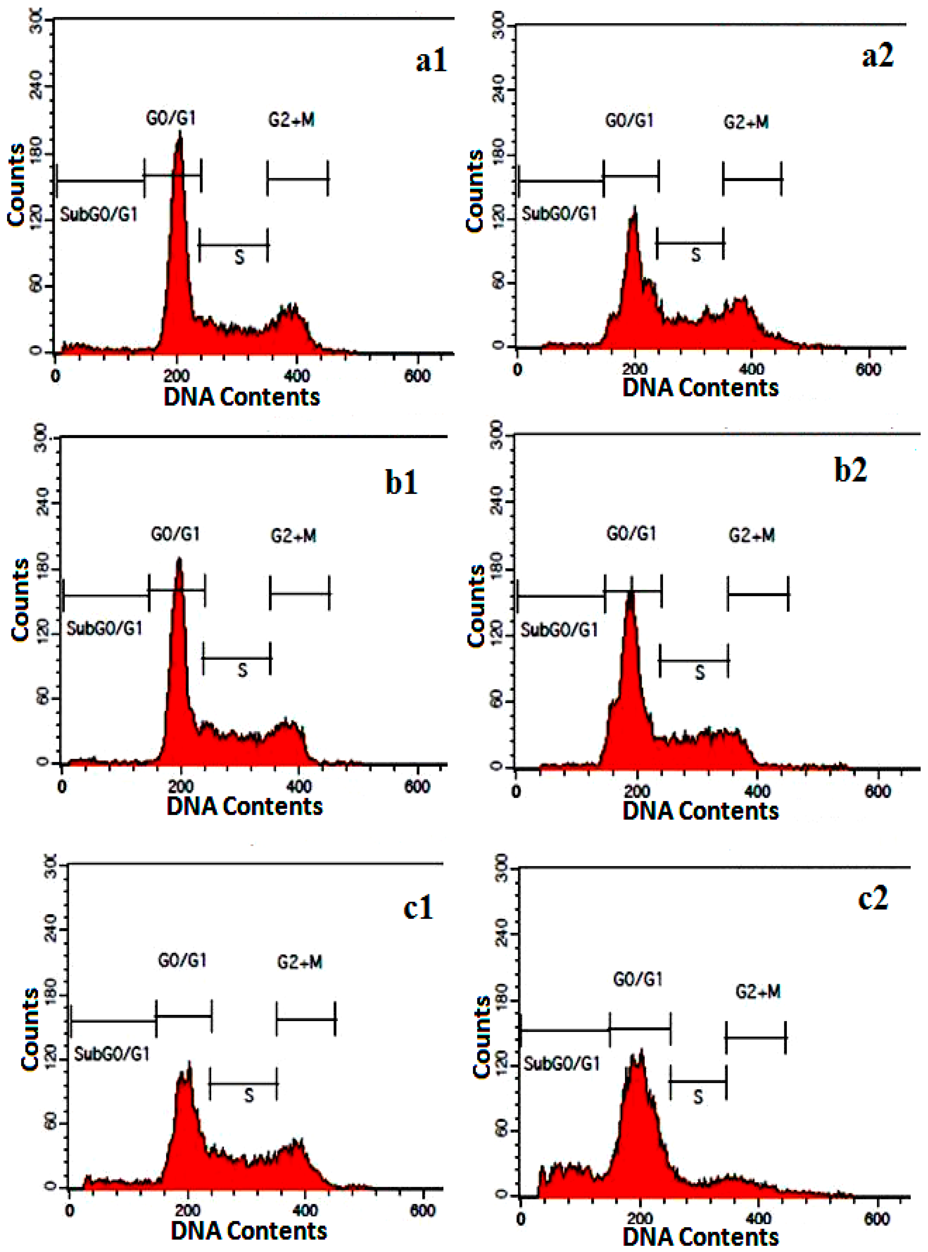
| Cell Cycle Phases | Cells (%) | |||||
|---|---|---|---|---|---|---|
| Control 24 h | Treated 24 h | Control 48 h | Treated 48 h | Control 72 h | Treated 72 h | |
| G0/G1 | 55.25 ± 0.6 | 32.44 ± 0.95 | 51.15 ± 0.25 | 43.61 ± 0.55 | 36.64 ± 0.35 | 46.68 ± 1.68 |
| G2/M | 18.85 ± 0.46 | 19.29 ± 0.11 | 20.16 ± 0.29 | 11.29 ± 0.39 | 25.22 ± 0.25 | 9.06 ± 1.93 |
| Synthesis | 23.91 ± 0.25 | 37.23 ± 0.63 | 28.24 ± 0.86 | 23.93 ± 0.32 | 36.95 ± 0.65 | 20.35 ± 1.18 |
| Sub G0/G1 | 1.98 ± 0.75 | 11.10 ± 0.98 * | 1.20 ± 0.24 | 19.0 ± 0.40 * | 2.02 ± 1.46 | 24.85 ± 1.56 * |
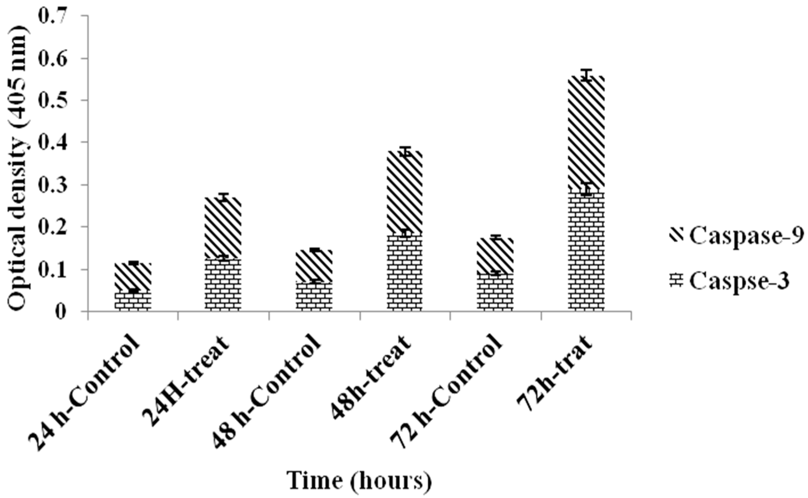
2.3.5. Caspase Assay

| Cells % | ||||||
|---|---|---|---|---|---|---|
| Caspase | Control 24 h | Treated 24 h | Control 48 h | Treated 48 h | Control 72 h | Treated 72 h |
| Caspase-3 | 0.05 ± 0.3 | 0.125 ± 2.2 * | 0.07 ± 0.15 | 0.185 ± 0.5 * | 0.09 ± 1.91 | 0.29 ± 0.55 * |
| Caspase-9 | 0.065 ± 0.27 | 0.145 ± 0.9 * | 0.075 ± 0.75 | 0.195 ± 0.65 * | 0.085 ± 1.33 | 0.27 ± 1.15 * |
3. Discussion
4. Materials and Methods
4.1. Chemicals
4.2. Cell Culture
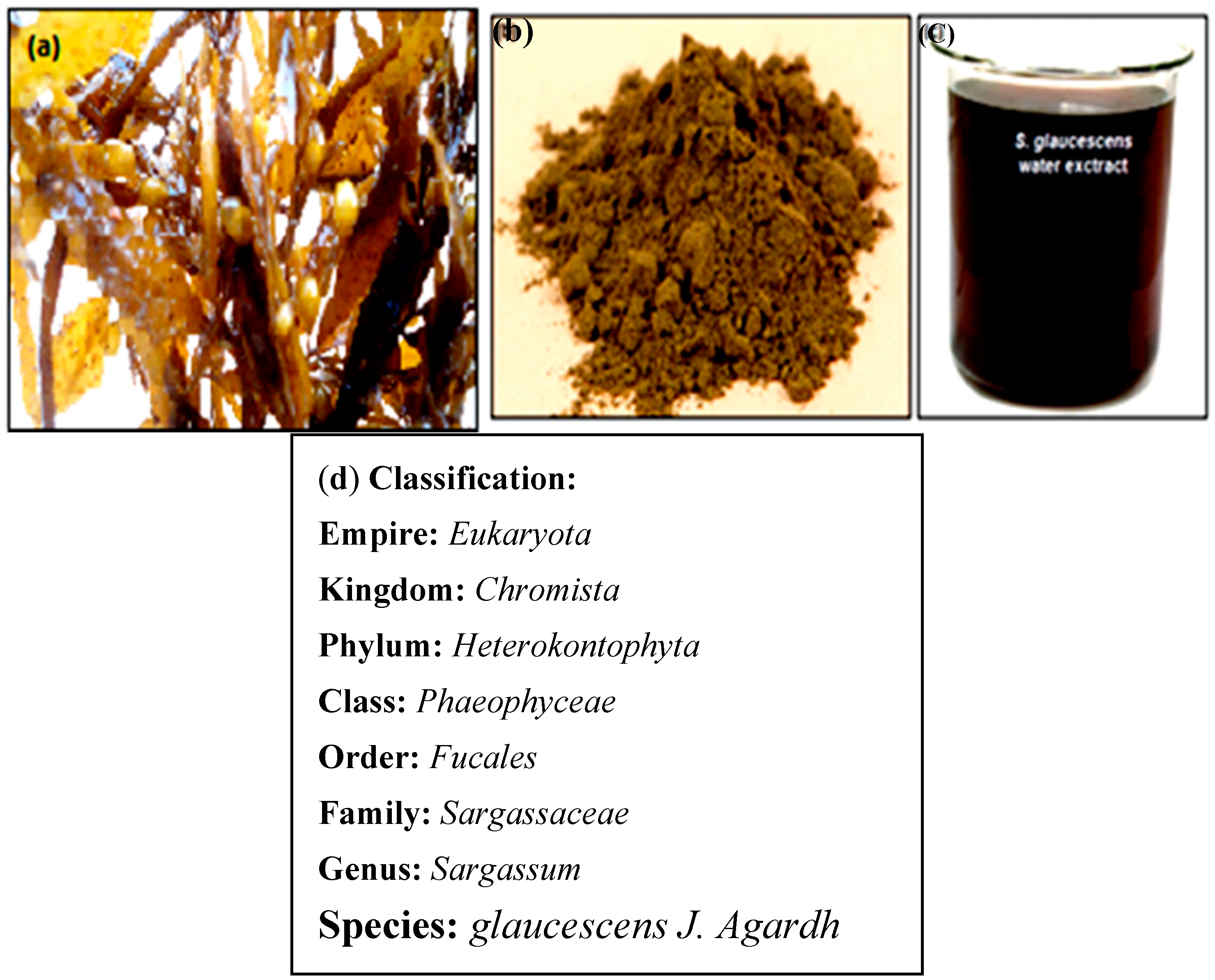
4.3. SG Extract Preparation
4.4. Synthesis of Au/SG Suspension
4.5. Characterization of SG-Stabilized AuNPs
4.6. Determination of Anticancer Activity
4.6.1. Cytotoxicity Assay
4.6.2. Apoptosis Detection Assay
4.6.3. Annexin V-FITC Assay
4.6.4. Cell Cycle Assay
4.6.5. Caspase Assays
4.7. Statistical Analysis
5. Conclusions
Acknowledgments
Author Contributions
Conflicts of Interest
References
- Venkatesh, T.; Reddy, A.K.; Maheswari, J.U.; Dalith, M.D.; Kumar, C.K.A. Nanosuspensions: Ideal approach for the drug delivery of poorly water soluble drugs. Der Pharm. Lett. 2011, 3, 203–213. [Google Scholar]
- Jain, P.K.; El-Sayed, I.H.; El-Sayed, M.A. Au nanoparticles target cancer. Nano Today 2007, 2, 18–29. [Google Scholar] [CrossRef]
- Cai1, W.; Gao, T.; Hong, H.; Sun, J. Applications of gold nanoparticles in cancer nanotechnology. Nanotechnol. Sci. Appl. 2008, 1, 17–32. [Google Scholar]
- Yildirimer, L.; Thanh, N.T.K.; Loizidou, M.; Seifalian, A.M. Toxicology and clinical potential of nanoparticles. Nano Today 2011, 6, 585–607. [Google Scholar] [CrossRef] [PubMed]
- Alivisatos, P. The use of nanocrystals in biological detection. Nat. Biotechnol. 2004, 22, 47–52. [Google Scholar] [CrossRef] [PubMed]
- Burda, C.; Chen, X.; Narayanan, R.; El-Sayed, M. Chemistry and properties of nanocrystals of different shapes. Chem. Rev. 2005, 105, 1025–1102. [Google Scholar] [CrossRef] [PubMed]
- Patra, C.R.; Bhattacharya, R.; Mukhopadhyay, D.; Mukherjee, P. Fabrication of gold nanoparticles for targeted therapy in pancreatic cancer. Adv. Drug Deliv. Rev. 2010, 62, 346–361. [Google Scholar] [CrossRef] [PubMed]
- Kawasaki, E.S.; Player, A. Nanotechnology, nanomedicine, and the development of new, effective therapies for cancer. Nanomed. Nanotechnol. Biol. Med. 2005, 1, 101–109. [Google Scholar] [CrossRef] [PubMed]
- Horton, M.A.; Khan, A. Medical nanotechnology in the UK: A perspective from the London Centre for Nanotechnology. Nanomed. NBM 2006, 2, 42–48. [Google Scholar] [CrossRef] [PubMed]
- Zhao1, X.Q.; Liang, Y.; Hu, Z.Q.; Liu, B.X. Oxidation characteristics and magnetic properties of iron carbide and iron ultrafine particles. J. Appl. Phys. 1996, 80, 5857–5860. [Google Scholar] [CrossRef]
- Antonovych, T.T. Gold nephropathy. Ann. Clin. Lab. Sci. 1981, 11, 386–391. [Google Scholar] [PubMed]
- Thakor, A.S.; Jokerst, J.; Zavaleta, C.; Massoud, T.F.; Gambhir, S.S. Gold Nanoparticles: A Revival in Precious Metal Administration to Patients. Nano Lett. 2011, 11, 4029–4036. [Google Scholar] [CrossRef] [PubMed]
- Shameli, K.; Ahmad, M.B.; Al-Mulla, E.A.J.; Ibrahim, N.A.; Shabanzadeh, P.; Rustaiyan, A.; Abdollahi, Y.; Bagheri, S.; Abdolmohammadi, S.; Usman, M.S.; et al. Green Biosynthesis of Silver Nanoparticles Using Callicarpa maingayi Stem Bark Extraction. Molecules 2012, 17, 8506–8517. [Google Scholar] [CrossRef] [PubMed]
- Manimaran, M.; Jana, N.R. Detection of protein molecules by surface-enhanced Raman spectroscopy-based immunoassay using 2–5 nm gold nanoparticle lables. J. Raman Spectrosc. 2007, 38, 1326–1331. [Google Scholar] [CrossRef]
- Jain, P.K.; El-Sayed, M.A. Plasmonic coupling in noble metal nanostructures. Chem. Phys. Lett. 2010, 487, 153–164. [Google Scholar] [CrossRef]
- Sharma, V.K.; Yngard, R.A.; Lin, Y. Silver nanoparticles: Green synthesis and their antimicrobial activities. Adv. Colloid Interface Sci. 2009, 145, 83–96. [Google Scholar] [CrossRef] [PubMed]
- Nagarajan, S.; Kuppusamy, K.A. Extracellular synthesis of zinc oxide nanoparticle using seaweeds of gulf of Mannar, India. J. Nanobiotechnol. 2013, 11, 1–11. [Google Scholar] [CrossRef] [PubMed]
- Holdt, S.L.; Kraan, S. Bioactive compounds in seaweed: Functional food applications and legislation. J. Appl. Phycol. 2011, 23, 543–597. [Google Scholar] [CrossRef]
- Smit, A.J. Medicinal and pharmaceutical uses of seaweed natural products: A review. J. Appl. Phycol. 2004, 16, 245–262. [Google Scholar] [CrossRef]
- Wijesinghe, W.A.J.P.; Jeon, Y.J. Biological activities and potential cosmeceutical applications of bioactive components from brown seaweeds: A review. Phytochem. Rev. 2011, 10, 431–443. [Google Scholar] [CrossRef]
- Cui, W.; Li, J.; Zhang, Y.; Rong, H.; Lu, W.; Jiang, L. Effects of aggregation and the surface properties of gold nanoparticles on cytotoxicity and cell growth. Nanomed. Nanotechnol. Biol. Med. 2012, 8, 46–53. [Google Scholar] [CrossRef] [PubMed]
- Huang, X.; El-Sayed, M.A. Gold nanoparticles: Optical properties and implementations in cancer diagnosis and photothermal therapy. J. Adv. Res. 2010, 1, 13–28. [Google Scholar] [CrossRef]
- Yang, D.P.; Cui, D.X. Advances and prospects of gold nanorods. Chem. Asian J. 2008, 3, 2010–2022. [Google Scholar] [CrossRef] [PubMed]
- Tiwari, P.; Vig, K.; Dennis, V.; Singh, S. Functionalized Gold Nanoparticles and Their Biomedical Applications. J. Nanomater. 2011, 1, 31–63. [Google Scholar] [CrossRef]
- Tkachenko, A.G.; Xie, H.; Liu, Y.; Coleman, D.; Ryan, J.; Glomm, W.R.; Shipton, M.K.; Franzen, S.; Feldheim, D.L. Cellular trajectories of peptide-modified gold particle complexes: Comparison of nuclear localization signals and peptide transduction domains. Bioconjugate Chem. 2004, 15, 482–490. [Google Scholar] [CrossRef] [PubMed]
- Bastis, N.G.; Sanchez-Tillo, E.; Pujals, S.; Farrera, C.; Kogan, M.J.; Giralt, E.; Celada, A.; Lloberas, J.; Puntesa, V. Peptides conjugated to gold nanoparticles induce macrophage activation. Mol. Immunol. 2009, 46, 743–748. [Google Scholar] [CrossRef] [PubMed]
- Shukla, R.; Bansal, V.; Chaudhary, M.; Basu, A.; Bhonde, R.R.; Sastry, M. Biocompatibility of gold nanoparticles and their endocytotic fate inside the cellular compartment: A microscopic overview. Langmuir 2005, 21, 10644–10654. [Google Scholar] [CrossRef] [PubMed]
- Connor, E.E.; Mwamuka, J.; Gole, A.; Murphy, C.J.; Wyatt, M.D. Gold nanoparticles are taken up by human cells but do not cause acute cytotoxicity. Small 2005, 1, 325–327. [Google Scholar] [CrossRef] [PubMed]
- Namvar, F.; Rahman, H.S.; Mohamad, R.; Rasedee, A.; Yeap, S.K.; Chartrand, M.S.; Azizi, S.; Tahir, P.M. Apoptosis Induction in Human Leukemia Cell Lines by Gold Nanoparticles Synthesized Using the Green Biosynthetic Approach. J. Nanomater. 2015, 2015, 1–10. [Google Scholar] [CrossRef]
- Rahman, H.S.; Rasedee, A.; Abdul, A.B.; Zeenathul, N.A.; Othman, H.H.; Yeap, S.K.; How, C.W.; Wan, N.H. Zerumbone-loaded nanostructured lipid carrier induces G2/M cell cycle arrest and apoptosis via mitochondrial pathway in a human lymphoblastic leukemia cell line. Int. J. Nanomed. 2014, 9, 527–538. [Google Scholar]
- Tsoli, M.; Kuhn, H.; Brandau, W.; Esche, H.; Schmid, G. Cellular uptake and toxicity of Au55 clusters. Small 2005, 1, 841–844. [Google Scholar] [CrossRef] [PubMed]
- Mehta, D.K. British National Formulary, 52nd ed.; Pharmaceutical Press: London, UK, 2006. [Google Scholar]
- Martin, S.; Reutelingsperger, C.; McGahon, A.J.; Rader, J.A.; van Schie, R.; LaFace, D.M.; How, C.W.; Hafiza, W.A. Early redistribution of plasma membrane phosphatidylserine is a general feature of apoptosis regardless of the initiating stimulus: Inhibition by overexpression of Bcl-2 and Abl. J. Exp. Med. 1995, 182, 1545–1556. [Google Scholar] [CrossRef] [PubMed]
- Park, C.; Moon, D.O.; Rhu, C.H.; Choi, B.T.; Lee, W.H.; Kim, G.Y.; Choi, Y.H. β-Sitosterol induces anti-proliferation and apoptosis in human leukemic U937 cells through activation of caspase-3 and induction of Bax/Bcl-2 ratio. Biol. Pharm. Bull. 2007, 30, 1317–1323. [Google Scholar] [CrossRef] [PubMed]
- Green, D.R. Apoptotic pathways: Paper wraps stone blunts scissors. Cell 2000, 102, 1–4. [Google Scholar] [CrossRef]
- Miyoshi, N.; Nakamura, Y.; Ueda, Y.; Abe, M.; Ozawa, Y.; Uchida, K.; Osawa, T. Dietary ginger constituents, galanals A and B, are potent apoptosis inducers in Human T lymphoma Jurkat cells. Cancer Lett. 2003, 199, 113–119. [Google Scholar] [CrossRef]
- Ibrahim, M.Y.; Abdul, A.B.; Ibrahim, T.A.T.; Wahab, S.I.A.; Elhassan, M.M.; Mohan, S. Attenuation of cisplatin-induced nephrotoxicity in rats using zerumbone. Afr. J. Biotechnol. 2012, 9, 4434–4441. [Google Scholar]
- Bradford, M.M. A rapid and sensitive method for the quantitation of microgram quantities of protein utilizing the principle of protein-dye binding. Anal. Biochem. 1976, 72, 248–254. [Google Scholar] [CrossRef]
- Sample Availability: Samples of SG/AuNPs are available from the authors.
© 2016 by the authors. Licensee MDPI, Basel, Switzerland. This article is an open access article distributed under the terms and conditions of the Creative Commons by Attribution (CC-BY) license ( http://creativecommons.org/licenses/by/4.0/).
Share and Cite
Ajdari, Z.; Rahman, H.; Shameli, K.; Abdullah, R.; Abd Ghani, M.; Yeap, S.; Abbasiliasi, S.; Ajdari, D.; Ariff, A. Novel Gold Nanoparticles Reduced by Sargassum glaucescens: Preparation, Characterization and Anticancer Activity. Molecules 2016, 21, 123. https://doi.org/10.3390/molecules21030123
Ajdari Z, Rahman H, Shameli K, Abdullah R, Abd Ghani M, Yeap S, Abbasiliasi S, Ajdari D, Ariff A. Novel Gold Nanoparticles Reduced by Sargassum glaucescens: Preparation, Characterization and Anticancer Activity. Molecules. 2016; 21(3):123. https://doi.org/10.3390/molecules21030123
Chicago/Turabian StyleAjdari, Zahra, Heshu Rahman, Kamyar Shameli, Rasedee Abdullah, Maaruf Abd Ghani, Swee Yeap, Sahar Abbasiliasi, Daniel Ajdari, and Arbakariya Ariff. 2016. "Novel Gold Nanoparticles Reduced by Sargassum glaucescens: Preparation, Characterization and Anticancer Activity" Molecules 21, no. 3: 123. https://doi.org/10.3390/molecules21030123