Article

Two New Oleanane-Type Triterpenoids from Platycodi Radix and Anti-proliferative Activity in HSC-T6 Cells

1
Department of Pharmacy, Changzheng Hospital, Second Military Medical University, 415 Fengyang Road, Shanghai 200003, China
2
School of Pharmacy, Second Military Medical University, 325 Guohe Road, Shanghai 200433, China
*
Author to whom correspondence should be addressed.
Molecules 2012, 17(12), 14899-14907; https://doi.org/10.3390/molecules171214899
Submission received: 9 November 2012 / Revised: 29 November 2012 / Accepted: 30 November 2012 / Published: 13 December 2012

Abstract

:
Two new oleanane-type triterpenoids, named platycodonoids A and B (1, 2), together with five known saponins, including platycodin D (3), deapioplatycodin D (4), 3-O-β-d-glucopyranosyl polygalacic acid (5), 3-O-β-d-glucopyranosyl platycodigenin (6) and polygalacin D (7), were isolated from the roots of Platycodon grandiflorum. On the basis of spectral data and chemical evidence, the structures of the new compounds were elucidated as 2β,3β,23,24-tetrahydroxy-28-nor-olean-12-en-16-one (1) and 2β,3β,23,24-tetrahydroxy-28-nor-olean-12-en-16-one-3-O-β-d-glucopyranoside (2). Compounds 17 were evaluated for their in vitro anti-proliferative activity against the HSC-T6 cell line.

1. Introduction

Platycodon grandiflorum A. DC. is a perennial plant in the family Campanulaceae which grows widely in East Asia. Platycodi Radix, the root of P. grandiflorum, has been used in traditional Oriental medicine as an expectorant for pulmonary disease and a remedy for respiratory disorders [1,2]. Platycodi Radix has recently been reported to exhibit many pharmacological activities, including anti-cancer properties [3,4,5,6,7,8], atopic dermatitis-like skin lesions treating [9,10,11], anti-skin photoaging effects [12], antiobesity and glucose metabolism regulation [13,14,15,16,17], anti-atherosclerotic [18], and anti-hyperlipidemic [19] activities. Chemical investigation of Platycodi Radix revealed that triterpenoid saponins were the main chemical components, and more than 55 triterpenoid saponins have been isolated from Platycodi Radix to date [20,21]. Based on the structures of the aglycones, the triterpenoid saponins are classified into three types: the platycodigenin type, platycogenic acid A lactone type and polygalacic acid type [22]. It has been previously demonstrated that Platycodi Radix showed protective effects against acute ethanol, acetaminophen-, carbon tetrachloride-, thioacetamide and cholestasis-induced hepatotoxicity or hepatic injury in mice and inhibited the progress of hepatic fibrosis in rats [23,24,25,26,27,28,29,30]. In our preliminary pharmacological study, the 70% EtOH extract of Platycodi Radix was also found to exhibit significant protective activities against liver fibrosis in rats. In a continued effort to search for possible hepatoprotective component from this herb, an investigation of Platycodi Radix was undertaken, and this has led to the isolation of two new triterpenoids [an aglycone and its saponin, named platycodonoids A (1) and B (2) (Figure 1)], which possess a rare 28-nor-oleanane-type with a C-16 keto group in the aglycone structure. Besides, five known saponins were isolated and identified as platycodin D (3) [31], deapio-platycodin D (4) [21], 3-O-β-d-glucopyranosyl polygalacic acid (5) [32], 3-O-β-d-glucopyranosyl platycodigenin (6) [33] and polygalacin D (7) [32,34], by comparison of their IR, NMR and MS data with literature values. To the best of our knowledge, it was first time triterpenoids such a platycodonoids A and B having a 28-nor-oleanane-type skeleton are reported from the genus Platycodon.

2. Results and Discussion

The air-dried roots of P. Grandiflorum were extracted three times with 70% EtOH under reflux. The combined extract was chromatographed over a macroporous adsorbing resin column and partitioned as described in the Experimental section. After repeated column chromatography, two new compounds 1 and 2 and five known saponins 37 were isolated and identified.
Compound 1 was obtained as white amorphous powder. Its molecular formula was assigned to be C29H46O5 based on the HRESIMS spectrum (m/z 497.3243 [M+Na]+, calcd. for C29H46O5Na, 497.3243). The IR spectrum exhibited absorptions at 3415, 2947, 1711, and 1385 cm−1 assignable to hydroxyl, methyl, ketone and methylene functions, respectively. The 1H-NMR spectrum of 1 showed the following signals: five tertiary methyl groups at δH 0.78, 0.84, 0.96, 1.14, and 1.63 (each 3H, s); two pairs of germinal oxygenated protons at δH 4.20, 4.21, 4.87, 5.20 (each, d, J = 10.8 Hz); two oxygenated protons at δH 4.39 (d, J = 3.6 Hz) and 4.58 (dt, J = 7.2, 3.6 Hz); and an olefinic proton at δH 5.38 (t, J = 3.6 Hz). The 13C-NMR spectrum revealed 29 carbon signals, which were further classified by DEPT and HSQC experiments as five methyls, ten methylenes (two oxygenated), six methines (two oxygenated), five quaternary carbons, one trisubstituted double bond (δC 123.3 and 142.9), and one keto carbonyl (δC 214.0) (Table 1). The aforementioned data implied that 1 was a nor-oleanane-type triterpenoid with four hydroxyls. In the HMBC spectrum, two pairs of germinal oxygenated protons at δH 4.20, 4.21, 4.87, 5.20 (H2-23, H2-24) showed correlations to C-3 (δC 75.2), C-4 (δC 48.1) and C-5 (δC 48.5), indicating that two hydroxyls were linked at C-23 and C-24. The two remaining hydroxyls were placed at C-2 (δC 72.0) and C-3, which were deduced by the HMBC correlations from the proton H-2 to C-1 (δC 44.7), C-3, and C-4, and from H-3 (δH 4.39) to C-1, C-2, C-3, C-4, C-23 (δC 64.1) and C-24 (δC 64.7) (Figure 2). The small coupling constant (J2,3 = 3.6 Hz) of H-2 and H-3 indicated the axial position of H-3 and the equatorial position of H-2, which was further confirmed by a NOESY correlation between H-2 and H-3. Moreover, the chemical shifts H2-15 at δH 2.55 (d, J = 14.4 Hz), 1.93 (d, J = 14.4 Hz) and C-15 at δC 47.2 were quite different from that reported for oleanane-type triterpenoid [platycoside O, H-15, δH 1.81 (dd, J = 15.0, 3.0 Hz), 2.55 (d, J = 12.0 Hz), C-15 (δC 36.1)] [21]. These results suggested the keto group (δC 214.0) was at C-16, which was confirmed by the HMBC spectrum correlations from H2-15, H-17 (δH 1.65), H-18 (δH 2.79) and H-22 (δH 1.34, 2.14) to C-16 (δC 214.0). The 1H-1H COSY spectrum of 1 indicated the presence of partial structures (Figure 2). Consequently, by comparison with the structure of platycoside O [21], compound 1 was determined to be 2β,3β,23,24-tetrahydroxy-28-nor-olean-12-en-16-one, and named platycodonoid A.
Compound 2 was obtained as white amorphous powder. It gave an [M+Na]+ peak at m/z 659.3728 in HRESIMS spectrum corresponding to the molecular formula of C35H56O10 (calcd. for C35H56O10Na, 659.3771), which showed a unit of C6H10O5 more than that of compound 1. The 1D-NMR spectra of 2 displayed similarities to those of 1, except for an additional sugar unit. An anomic proton signal at δH 5.15 (H-1', d, J = 7.8 Hz), an oxygenated methylene (H2-6') at δH 4.35 (t, J = 6.0 Hz) and 4.59 (d, J = 10.8 Hz), four oxymethine protons in the range δH 3.50−4.50 in the 1H-NMR spectrum suggested that 2 contained a sugar moiety. In accordance, the 13C-NMR spectrum displayed six carbon signals at δC 63.0, 72.0, 75.6, 79.0, 79.0, 106.6. The subsequent acid hydrolysis of 2 gave glucose only, which was analyzed by gas chromatography as glucitol acetate [35]. The linkage of this β-d-glucopyranose (J = 7.8 Hz) (Table 1) was confirmed by the HMBC correlation between H-1' and C-3. Therefore, the structure of platycodonoid B (2) was elucidated as 2β,3β,23,24-tetrahydroxy-28-nor-olean-12-en-16-one 3-O-β-d-glucopyranoside.
The origin of compounds 1 and 2 (Scheme 1) was proposed to be the oleanane-type triterpenoids i (compound 37). Decarboxylation at C-28 would produce a key intermediate ii, which could undergo an oxidation at C-16 to yield iii (1 and 2).
Compounds 17 were evaluated for their anti-proliferative activities against the Hepatic Stellate Cell (HSC)-T6 line using the 3-(4,5-dimethylthiazol-2-yl)-2.5-diphenyltetrazolium bromide (MTT) assay [36]. Colchicine was used as positive control in this study (IC50 value < 10 μM). Among the tested compounds, compounds 1, 3, 4 and 7 were the most potent, showing IC50 values of 5.27, 1.77, 8.24 and 1.04 μM, respectively. The IC50 values for the remaing compounds, 2, 5 and 6, were 69.63, 8150.23, and 13.36 μM, respectively. It was reported that saponins from Platycodi Radix prevented the increase in the serum levels of hepatic enzyme markers (alanine aminotransferase and aspartate aminotransferase) and reduced oxidative stress, such as glutathione content and lipid peroxidation, in the liver in a dose-dependent manner [23,24,25,26,27,28,29,30]. The reason why compounds 1, 3, 4 and 7 delayed the formation of liver fibrosis need to be further studied. In the structure-activity relationship of these oleanane-type triterpenoids, the presence of a free carboxyl functional group at C-28 seemed not to be related to the hepatoprotective activity (compounds 5 and 6). When forming C-28 glycosides, the presence of the glycosides affected the activity, and the number of monosaccharides in the sugar moiety increased the activity (compounds 3, 4 and 7). In contrast, when came to the 28-nor-oleanane-type triterpenoids 1 and 2, the aglycone was more active than its corresponding glycoside.

3. Experimental

3.1. General

Optical rotations were measured with Perkin-Elmer 341 polarimeter. UV and IR spectra were recorded on Shimadzu UV-2550 and Perkin-Elmer 577 (using KBr disks) spectrophotometers, respectively. NMR spectra were acquired on a Bruker Avance III (600 MHz for 1H-NMR, ppm relative to TMS) spectrometer. ESIMS spectra were made on an Agilent 1200 series HPLC and interfaced to an Agilent 6410 triple-quadrupole mass spectrometer equipped with an electrospray ionization source, and HRESIMS spectra were made on an Agilent 1290 series HPLC and interfaced to an Agilent 6538 UHD Accurate-Mass Q-TOF LC/MS (Agilent Corporation, Wilmington, DE, USA). GC-MS was conducted on a Thermo Finnigan Trace GC apparatus using an L-Chirasil-Val column (25 m × 0.32 mm, i.d.). Semi-preparation RP-HPLC isolation was achieved with an Agilent 1200 instrument with refractive index detector (RID) using a YMC 5 μm C8 column (250 mm × 10 mm). Methanol for semi-preparative HPLC was of HPLC-grade (Merck, Darmstadt, Germany). Column chromatography: silica gel (200−300 mesh); macroporous adsorbing resin (D-101, ZTC-1, 0.3−1.2 mm, Tianjin Zhentiancheng Science & Technology Co., Ltd., Tianjin, China); sephadex LH-20 gel (40−70 μm, Amersham Pharmacia Biotech AB, Uppsala, Sweden); silica gel H (Qingdao Haiyang Chemical Co. Ltd., Qingdao, China). All solvents for column chromatography and acid hydrolysis were of analytical grade (Shanghai Chemical Reagents Company, Ltd., Shanghai, China). Spots of compounds on TLC were developed using 10% H2SO4-EtOH solution.

3.2. Plant Material

Platycodi Radix was collected from Taihe, Anhui Province, China, in September 2010 and was identified by Professor Hanming Zhang of School of Pharmacy, Second Military Medical University. A voucher specimen (No. 20100921) was deposited at the Department of Pharmacognosy, School of Pharmacy, Second Military Medical University.

3.3. Extraction and Isolution

Platycodi Radix was air-dried (10 kg) and extracted three times with 70% EtOH (50 L × 3 times) under reflux. The combined extract was concentrated in vacuo and suspended in water. The aqueous layer was chromatographed over a macroporous adsorbing resin column eluting with H2O, 30% EtOH, 60% EtOH and 95% EtOH. The 95% EtOH-eluted fraction (30 g) was applied to a silica gel column (CHCl3-MeOH, 50:1 to 10:1, v/v) and purified by semi-preparative HPLC (MeOH-H2O, 4:1) to give compound 1 (10.4 mg). The 60% EtOH-eluted fraction (120 g) was chromatographed on silica gel column eluting with a CHCl3-MeOH gradient (30:1 to 2:1, v/v) to afford five subfractions (A-E). Subfraction B (20 g) was chromatographed on a silica gel column (CHCl3-MeOH, 10:1 to 5:1, v/v), followed by Sephadex LH-20 column (MeOH-H2O, 1:1), and finally separated by semi-preparative HPLC (MeOH-H2O, 3:2) to yield compound 2 (9.3 mg). By the same procedure, compounds 3 (20.0 mg), 4 (8.7 mg), 5 (11.2 mg), 6 (9.4 mg) and 7 (7.6 mg) were obtained from subfractions C−E.

3.4. Characterization of Compound 1 and Compound 2

Compound 1: white amorphous powder; [ α ] D 22.0 +44.3 (c 0.174, MeOH); IR (KBr) νmax 3396, 2943, 2908, 1705, 1639, 1452, 1429, 1381, 1248, 1074, 1043, 898.7 cm−1; 1H-NMR and 13C-NMR data see Table 1; ESIMS m/z 497.4 [M+Na]+; HRESIMS m/z 497.3243 [M+Na]+ (calcd. for C29H46O5Na, 497.3243).
Compound 2: white amorphous powder; [ α ] D 22.0 +59.9 (c 0.128, MeOH); IR (KBr) νmax 3415, 2947, 1711, 1456, 1433, 1385, 1365, 1134, 1047, 696, 594 cm−1; 1H-NMR and 13C-NMR data see Table 1; ESIMS m/z 659.5 [M+Na]+; HRESIMS m/z 659.3728 [M+Na]+ (calcd. for C35H56O10Na, 659.3771).

3.5. Acid Hydrolysis of Compound 2

Compound 2 (3.0 mg) was refluxed with 1M HCl (dioxane-H2O, 1:1, 2 mL) at 90 °C for 3 h in a water bath. After dioxane was removed, the solution was extracted with EtOAc (2 mL × 3 times). After evaporating to dryness, the monosaccharide portion was analyzed by gas chromatography after conversion of the hydrolysates into corresponding alditol acetates. Only D-glucose was detected. The EtOAc portion was washed with H2O and evaporated to yield the aglycone. The aglycone was identified by TLC together with compound 1.

3.6. In Vitro Inhibitory Activity on Cell Proliferation

Tested compounds 17 were dissolved in DMSO (final concertration, 0.1%). Inhibitory activity of compounds 17 against HSC-T6 cell line was evaluated by the MTT assay [36]. Briefly, cells at the exponential growth phase were harvested and seeded into a flatbottom 96-well plate. A total of 90 μL containing 5 × 10⁴ cells was added to each well of the plate and incubated for 24 h in a 5% humidified CO2 at 37 °C. HSC-T6 cells were treated with vehicle or compounds at concerntration of 0.01, 0.1, 1, 10, 100 and 1000 μg/mL. After 48 h of incubation at 37 °C, 20 μL/well, MTT was then added and the plate was again incubated at 37 °C for 4 h. Reduction of MTT to formazan was measured in an ELISA plate reader at 570 nm. Inhibitory activity of compounds 17 on cell proliferation (% of control) was calculated as 100 × (absorbance of treated compound—absorbance of background light)/(absorbance of control—absorbance of background light). Data were expressed as the mean of the three independent experiment. Colchicin was used as a positive control.

4. Conclusions

In conclusion, the phytochemical investigation of the roots extract of Platycodi Radix afforded two new triterpenoids, platycodonoids A (1) and B (2), together with five known triterpenoids: platycodin D (3), deapio platycodin D (4), 3-O-β-d-glucopyranosyl polygalacic acid (5), 3-O-β-d-glucopyranosyl platycodigenin (6) and polygalacin D (7). The structures of the compounds were elucidated on the basis of spectral analysis and chemical evidence and literature comparisons in the case of the known ones. Compounds 1, 3, 4 and 7 exhibited significant hepatoprotective activities against HSC-T6 cell lines in vitro (IC50 value < 10 μM).

Acknowledgments

We gratefully acknowledge financial support of this work by National Natural Science Foundation of China (81073030) and The National Key Technology R & D Program in the 11th Five year Plan of China (2008BA151B00-2).

References

  1. Chinese Pharmacopoeia Commission. Pharmacopoeia of the People’s Republic of China; Chemical Industry Press: Beijing, China, 2010; Volume 1, pp. 259–260. [Google Scholar]
  2. Hong, D.Y.; Lian, Y.S.; Shen, L.D. Flora Reipublicae Popularis Sinica (Zhongguo Zhiwu Zhi); Science Press: Beijing, China, 1977; Volume 73, Issue 2, p. 77. [Google Scholar]
  3. Yu, M.; Fang, P.H.; Yu, G.F.; Liu, M.X. Research advances on chemical constituents and anti-tumor effect of Radix Platycodi. J. Int. Pharm. Res. 2011, 38, 280–283. [Google Scholar]
  4. Park, D.I.; Lee, J.H.; Moon, S.K.; Kim, C.H.; Lee, Y.T.; Cheong, J.; Choi, B.T.; Choi, Y.H. Induction of apoptosis and inhibition of telomerase activity by aqueous extract from Platycodon grandiflorum in human lung carcinoma cells. Pharmacol. Res. 2005, 51, 437–443. [Google Scholar] [CrossRef] [PubMed]
  5. Lee, K.J.; Shin, D.W.; Chung, Y.C.; Jeong, H.G. Chemopreventive effect of saponins derived from roots of Platycodon grandiflorum on 4-(methyinitrosamino)-1-(3-pyridyl)-1-butanone-induced lung turnorigenesis in A/J mice. Arch. Pharm. Res. 2006, 29, 651–656. [Google Scholar] [CrossRef] [PubMed]
  6. Li, W.; Qi, Y.; Wang, Z.; Zhang, J.; Zhang, W.; Zheng, Y.N. Study on anti-tumor activity of saponins from Platycodi Radix in vitro. Pharmacol. Clin. Chin. Mater. Med. 2009, 25, 37–40. [Google Scholar]
  7. Yu, J.S.; Kim, A.K. Platycodin D induces apoptosis in MCF-7 human breast cancer cells. J. Med. Food 2010, 13, 298–305. [Google Scholar] [CrossRef] [PubMed]
  8. Yu, J.S.; Kim, A.K. Platycodin D induces reactive oxygen species-mediated apoptosis signal-regulating kinase 1 activation and endoplasmic reticulum stress response in human breast cancer cells. J. Med. Food 2012, 15, 691–699. [Google Scholar] [CrossRef] [PubMed]
  9. Kim, M.S.; Hur, Y.G.; Kim, W.G.; Park, B.W.; Ahn, K.S.; Kim, J.J.; Bae, H. Inhibitory effect of Platycodon grandiflorum on T(H)1 and T(H)2 immune responses in a murine model of 2,4-dinitrofluorobenzene-induced atopic dermatitis-like skin lesions. Ann. Allergy Asthma Immunol. 2011, 106, 54–61. [Google Scholar] [CrossRef] [PubMed]
  10. Park, S.J.; Lee, H.A.; Kim, J.W.; Lee, B.S.; Kim, E.J. Platycodon grandiflorus alleviates DNCB-induced atopy-like dermatitis in NC/Nga mice. Indian J. Pharmacol. 2012, 44, 469–474. [Google Scholar] [PubMed]
  11. Choi, J.H.; Han, E.H.; Park, B.H.; Kim, H.G.; Hwang, Y.P.; Chung, Y.C.; Lee, Y.C.; Jeong, H.G. Platycodi Radix suppresses development of atopic dermatitis-like skin lesions. Environ. Toxicol. Pharmacol. 2012, 33, 446–452. [Google Scholar] [CrossRef] [PubMed]
  12. Hwang, Y.P.; Kim, H.G.; Choi, J.H.; Han, E.H.; Kwon, K.I.; Lee, Y.C.; Choi, J.M.; Chung, Y.C.; Jeong, T.C.; Jeong, H.G. Saponins from the roots of Platycodon grandiflorum suppress ultraviolet A-induced matrix metalloproteinase-1 expression via MAPKs and NF-κB/AP-1-dependent signaling in HaCaT cells. Food Chem. Toxicol. 2011, 49, 3374–3382. [Google Scholar] [CrossRef] [PubMed]
  13. Lee, H.; Bae, S.; Kim, Y.S.; Yoon, Y. WNT/β-catenin pathway mediates the anti-adipogenic effect of platycodin D, a natural compound found in Platycodon grandiflorum. Life Sci. 2011, 89, 388–394. [Google Scholar] [CrossRef] [PubMed]
  14. Han, S.; Oh, K.S.; Yoon, Y.; Park, J.S.; Park, Y.S.; Han, J.H.; Jeong, A.L.; Lee, S.; Park, M.; Choi, Y.A.; et al. Herbal extract THI improves metabolic abnormality in mice fed a high-fat diet. Nutr. Res. Pract. 2011, 5, 198–204. [Google Scholar] [CrossRef] [PubMed]
  15. Twiner, E.M.; Liu, Z.; Gimble, J.; Yu, Y.; Greenway, F. Pharmacokinetic pilot study of the antiangiogenic activity of standardized Platycodi Radix. Adv. Ther. 2011, 28, 857–865. [Google Scholar] [CrossRef] [PubMed]
  16. Ahn, Y.M.; Kim, S.K.; Kang, J.S.; Lee, B.C. Platycodon grandiflorum modifies adipokines and the glucose uptake in high-fat diet in mice and L6 muscle cells. J. Pharm. Pharmacol. 2012, 64, 697–704. [Google Scholar] [CrossRef] [PubMed]
  17. Lee, C.E.; Hur, H.J.; Hwang, J.T.; Sung, M.J.; Yang, H.J.; Kim, H.J.; Park, J.H.; Kwon, D.Y.; Kim, M.S. Long-term consumption of Platycodi Radix ameliorates obesity and insulin resistance via the activation of ampk pathways. Evid. Based Complement. Alternat. Med. 2012, 2012, 1–11. [Google Scholar] [CrossRef] [PubMed]
  18. Wu, J.; Yang, G.; Zhu, W.; Wen, W.; Zhang, F.; Yuan, J.; An, L. Anti-atherosclerotic activity of platycodin D derived from roots of Platycodon grandiflorum in human endothelial cells. Biol. Pharm. Bull. 2012, 35, 1216–1221. [Google Scholar] [CrossRef] [PubMed]
  19. Chung, M.J.; Kim, S.H.; Park, J.W.; Lee, Y.J.; Ham, S.S. Platycodon grandiflorum root attenuates vascular endothelial cell injury by oxidized low-density lipoprotein and prevents high-fat diet-induced dyslipidemia in mice by up-regulating antioxidant proteins. Nutr. Res. 2012, 32, 365–373. [Google Scholar] [CrossRef] [PubMed]
  20. Guo, W.J.; Xu, X.D.; Wei, J.H.; Yang, J.S. Advances in studies on triterpenoid saponins of Platycodon grandiflorum. Chin. Pharm. J. 2008, 43, 801–804. [Google Scholar]
  21. Fu, W.W.; Fu, J.N.; Zhang, W.M.; Sun, L.X.; Pei, Y.H.; Liu, P. Platycoside O, a new triterpenoid saponin from the roots of Platycodon grandiflorum. Molecules 2011, 16, 4371–4378. [Google Scholar] [CrossRef] [PubMed]
  22. Fu, W.W.; Dou, D.Q.; Pei, Y.H. Review on chemical components and bioactivities of Platycodon grandiflorum. J. Shenyang Pharm. Univ. 2006, 23, 184–187. [Google Scholar]
  23. Lee, K.J.; Kim, J.Y.; Jung, K.S.; Choi, C.Y.; Chung, Y.C.; Kim, D.H.; Jeong, H.G. Suppressive effects of Platycodon grandiflorum on the progress of carbon tetrachloride-induced hepatic fibrosis. Arch. Pharm. Res. 2004, 27, 1238–1244. [Google Scholar] [CrossRef] [PubMed]
  24. Lee, K.J.; Choi, J.H.; Kim, H.G.; Han, E.H.; Hwang, Y.P.; Lee, Y.C.; Chung, Y.C.; Jeong, H.G. Protective effect of saponins derived from the roots of Platycodon grandiflorum against carbon tetrachloride induced hepatotoxicity in mice. Food Chem. Toxicol. 2008, 46, 1778–1785. [Google Scholar] [CrossRef] [PubMed]
  25. Khanal, T.; Choi, J.H.; Hwang, Y.P.; Chung, Y.C.; Jeong, H.G. Saponins isolated from the root of Platycodon grandiflorum protect against acute ethanol-induced hepatotoxicity in mice. Food Chem. Toxicol. 2009, 47, 530–535. [Google Scholar] [CrossRef] [PubMed]
  26. Kim, T.W.; Lim, J.H.; Song, I.B.; Park, S.J.; Yang, J.W.; Shin, J.C.; Suh, J.W.; Son, H.Y.; Cho, E.S.; Kim, M.S.; et al. Hepatoprotective and anti-hepatitis C viral activity of Platycodon grandiflorum extract on carbon tetrachloride-induced acute hepatic injury in mice. J. Nutr. Sci. Vitaminol. (Tokyo) 2012, 58, 187–194. [Google Scholar] [CrossRef] [PubMed]
  27. Lim, J.H.; Kim, T.W.; Park, S.J.; Song, I.B.; Kim, M.S.; Kwon, H.J.; Cho, E.S.; Son, H.Y.; Lee, S.W.; Suh, J.W.; et al. Protective effects of Platycodon grandiflorum aqueous extract on thioacetamide-induced fulminant hepatic failure in mice. J. Toxicol. Pathol. 2011, 24, 223–228. [Google Scholar] [CrossRef] [PubMed]
  28. Noh, J.R.; Kim, Y.H.; Gang, G.T.; Hwang, J.H.; Kim, S.K.; Ryu, S.Y.; Kim, Y.S.; Lee, H.S.; Lee, C.H. Hepatoprotective effect of Platycodon grandiflorum against chronic ethanol-induced oxidative stress in C57BL/6 mice. Ann. Nutr. Metab. 2011, 58, 224–231. [Google Scholar] [CrossRef] [PubMed]
  29. Kim, T.W.; Lee, H.K.; Song, I.B.; Lim, J.H.; Cho, E.S.; Son, H.Y.; Jung, J.Y.; Yun, H.I. Platycodin D attenuates bile duct ligation-induced hepatic injury and fibrosis in mice. Food Chem. Toxicol. 2012, 51C, 364–369. [Google Scholar] [CrossRef] [PubMed]
  30. Kim, T.W.; Lee, H.K.; Song, I.B.; Kim, M.S.; Hwang, Y.H.; Lim, J.H.; Park, S.J.; Lee, S.W.; Kim, J.W.; Yun, H.I. Protective effect of the aqueous extract from the root of Platycodon grandiflorum on cholestasis-induced hepatic injury in mice. Pharm. Biol. 2012, 50, 1473–1478. [Google Scholar] [CrossRef] [PubMed]
  31. Nikaido, T.; Koike, K.; Mitsunaga, K.; Saeki, T. Two New Triterpenoid Saponins from Platycodon grandiflorum. Chem. Pharm. Bull. 1999, 47, 903–904. [Google Scholar] [CrossRef] [PubMed]
  32. Fu, W.W.; Hou, W.B.; Dou, D.Q.; Hua, H.M.; Gui, M.H.; Fu, R.; Chen, Y.J.; Pei, Y.H. Saponins of polygalacic acid type from Platycodon grandiflorum. Acta Pharm. Sinic 2006, 41, 358–360. [Google Scholar]
  33. Fu, W.W.; Dou, D.Q.; Hou, W.B.; Cheng, B.H.; Liu, F.; Chen, Y.J.; Pei, Y.H.; Takeda, T. Isolation and identification of triterpenoid saponins from Platycodon grandiflorum (Jacq.) A. DC. Chin. J. Med. Chem. 2005, 15, 297–301. [Google Scholar]
  34. Li, L.J.; Liu, Z.H.; Chen, Y.; Tian, J.K. Chemical constituents from roots of Platycodon grandiflorum. Chin. J. Chin. Mater. Med. 2006, 31, 1506–1509. [Google Scholar]
  35. Fox, A.; Morgan, S.L. Analysis of Carbohydrates by GLC and MS; CRC Press: Boca Raton, FL, USA, 1989; pp. 87–107. [Google Scholar]
  36. Mosmann, T. Rapid colorimetric assay for cellular growth and survival: Application to proliferation and cytotoxicity assays. J. Immunol. Methods 1983, 65, 55–63. [Google Scholar] [CrossRef]
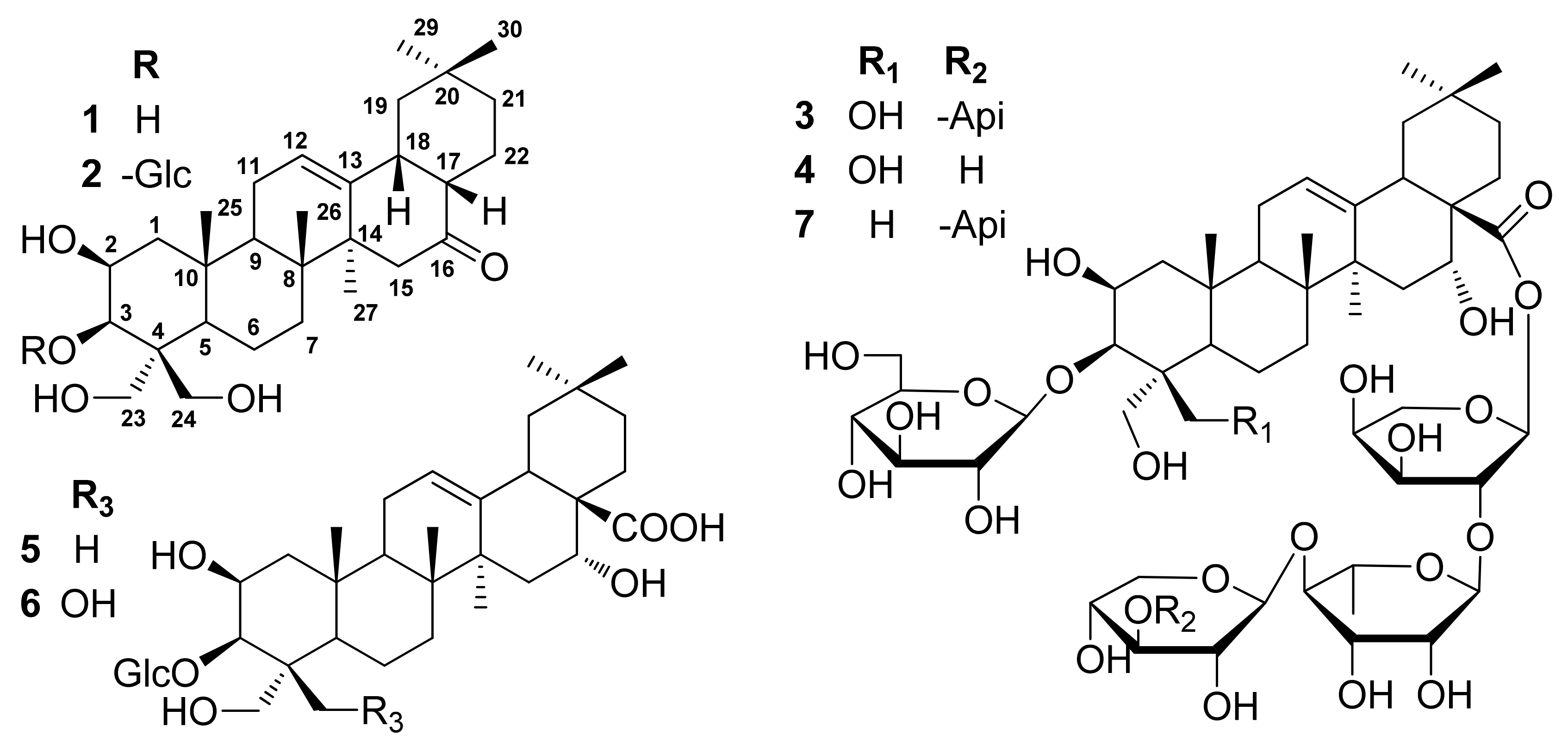
Sample Availability: Samples of the compounds 17 are available from the authors.
Figure 1. Structures of compounds 17.
Molecules 17 14899 g001
Figure 2. Key 1H-1H COSY (bold lines) and HMBC (H→C) correlations of compound 1.
Molecules 17 14899 g002
Scheme 1. The plausible biogenetic origin of compounds 1 and 2.
Molecules 17 14899 sch001
Table 1. 1H-NMR (600 MHz) and 13C-NMR (150 MHz) data for compounds 1 and 2 (pyridine-d5, δH in ppm, J in Hz).
Position12
δCδHδCδH
144.72.33 (1H, dd, 13.8, 3.0), 1.30 (1H, m)45.02.08 (1H, m), 1.57 (1H, m)
272.04.58 (1H, dt, 7.2, 3.6)70.04.76 (1H, m)
375.24.39 (1H, d, 3.6)86.44.61 (1H, brs)
448.1 47.8
548.51.82 (1H, brd, 12.0)48.41.85 (1H, d, 12.0)
619.21.98 (1H, m), 1.83 (1H, brs)19.41.92 (1H, m ), 1.61 (1H, m)
733.51.53 (1H, m), 0.91 (1H, m)33.41.47 (1H, t, 13.8), 1.18 (1H, m)
840.1 40.3
947.32.49 (1H, t, 5.4)47.32.53 (1H, t, 5.4)
1037.3 37.7
1124.22.05 (1H, m), 0.84(1H, m)24.22.04 (1H, m), 0.88 (1H, m)
12123.35.38 (1H, t, 3.6)123.65.42 (1H, brs)
13142.9 142.9
1446.9 47.1
1547.22.55 (1H, d, 14.4), 1.93 (1H, d, 14.4)47.12.58 (1H, d, 14.4), 1.96 (1H, d, 14.4)
16214.0 213.8
1748.11.65 (1H, m)48.31.71 (1H, m)
1845.12.79 (1H, m)45.22.83 (1H, m)
1947.01.40 (1H, t, 13.2), 1.17 (1H, m)47.31.47 (1H, t, 13.2), 1.21(1H, m)
2031.3 31.4
2135.01.09 (2H, m)35.01.09 (2H, m)
2221.52.14 (1H, m), 1.34 (1H, m)21.62.17 (1H, m), 1.38 (1H, m)
2364.14.87 (1H, d, 10.8), 4.20 (1H, d, 10.8)63.84.99 (1H, d, 10.8), 4.11 (1H, d, 11.4)
2464.75.20 (1H, d, 10.8), 4.21 (1H, d, 10.8)63.84.80 (1H, d, 10.8), 4.26 (1H, d, 10.8)
2517.90.96 (3H, s)18.30.99 (3H, s)
2617.61.63 (3H, s)18.01.55 (3H, s)
2727.31.14 (3H, s)27.21.17 (3H, s)
2933.60.78 (3H, s)33.50.85 (3H, s)
3023.70.84 (3H, s)23.80.89 (3H, s)
1' 106.65.15 (1H, d, 7.8)
2' 75.64.05 (1H, t, 8.1)
3' 79.04.18 (1H, m)
4' 72.04.18 (1H, m)
5' 79.03.98 (1H, m)
6' 63.04.59 (1H, d, 10.8), 4.35 (1H, t, 6.0)

