Article

Novel Steroidal Components from the Underground Parts of Ruscus aculeatus L.

1
Dipartimento di Chimica delle Sostanze Naturali, Università degli Studi di Napoli “Federico II”, Via D. Montesano 49, I-80131 Napoli, Italy
2
Dipartimento di Bioscienze e Territorio, Università degli Studi del Molise, Contrada Fonte Lappone, I-86090 Pesche (Isernia), Italy
*
Author to whom correspondence should be addressed.
Molecules 2012, 17(12), 14002-14014; https://doi.org/10.3390/molecules171214002
Submission received: 8 October 2012 / Revised: 16 November 2012 / Accepted: 18 November 2012 / Published: 26 November 2012
(This article belongs to the Section Natural Products Chemistry)

Abstract

:
Two new furostanol saponins 12 and three new sulphated glycosides 3a,b and 4 were isolated from the underground parts of Ruscus aculeatus L., along with four known furostanol and one spirostanol saponins 59 and three free sterols. All of the structures have been elucidated on the basis of spectroscopic data 1D and 2D NMR experiments, MS spectra and GC analyses.

1. Introduction

Ruscus aculeatus L. (Ruscaceae family) is a small evergreen shrub and a widely distributed European plant. The hydroalcoholic extract of its rhizome is commonly used as a vascular preventive and vascular tonic in pharmaceutical preparations [1]. Previous chemical analysis of secondary metabolites have been described in R. aculeatus L. [2,3], R. hypoglossum L. [4], R. colchicus Y. Yeo [5] and R. ponticus Wor. [6]. The steroidal constituents of this herb, including spirostane, furostane and triterpene type, are the main secondary metabolites isolated from the rhizome and leaves [7].
In the present work we performed a phytochemical investigation of the fresh underground parts of Ruscus aculeatus L. Two new furostanol saponins 12 and three new sulphated glycosides 3a,b and 4 were isolated from its methanolic extract along with four known furostanol and one spirostanol glycosides 59 (Figure 1 and Figure 2).
Figure 1. New compounds isolated from Ruscus aculeatus L.
Molecules 17 14002 g001
Figure 2. Known compounds isolated from Ruscus aculeatus L.
Molecules 17 14002 g002
The composition of free sterols was also determined and campesterol, stigmasterol, sitosterol were the major components. All new components are bisdesmosidic saponins with a diglycoside moiety linked at C-1 and a glucose unit linked at C-26. The isolation of sulphated compounds in Ruscus aculeatus L. are previously reported only by Oulad-Ali et al. [8].
Their structures were determined by spectroscopic methods, including 1D and 2D NMR techniques, HRESI-MS, and chemical methods. Herein, we report the isolation and structural elucidation of the new compounds 14.

2. Results and Discussion

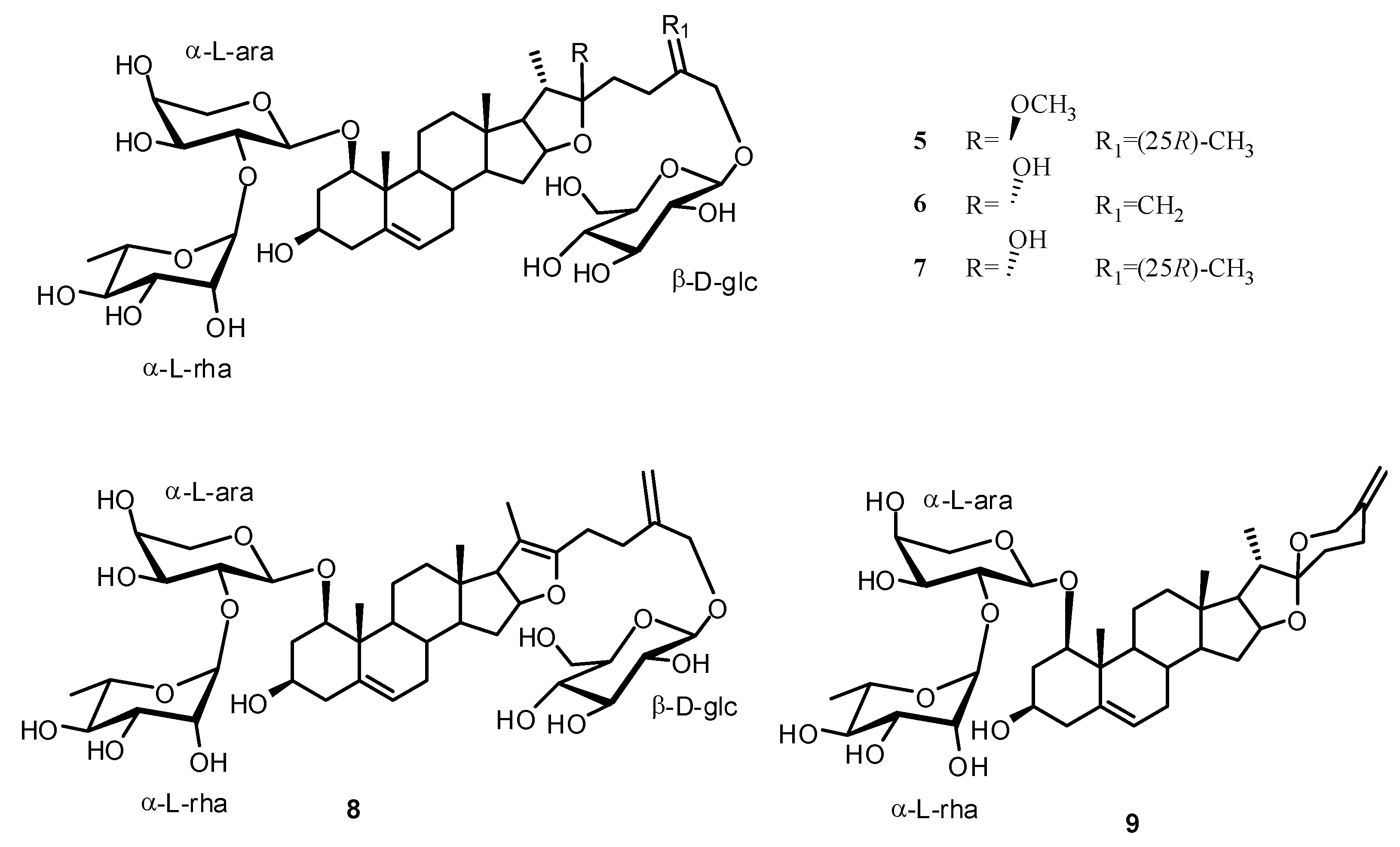
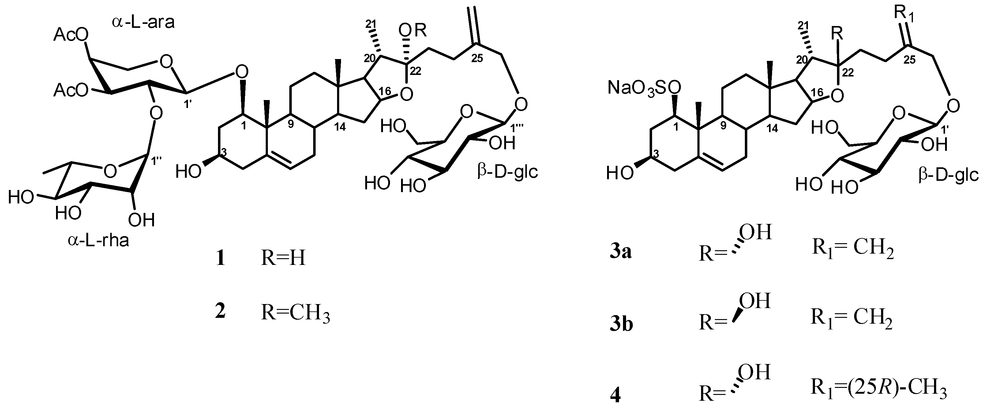
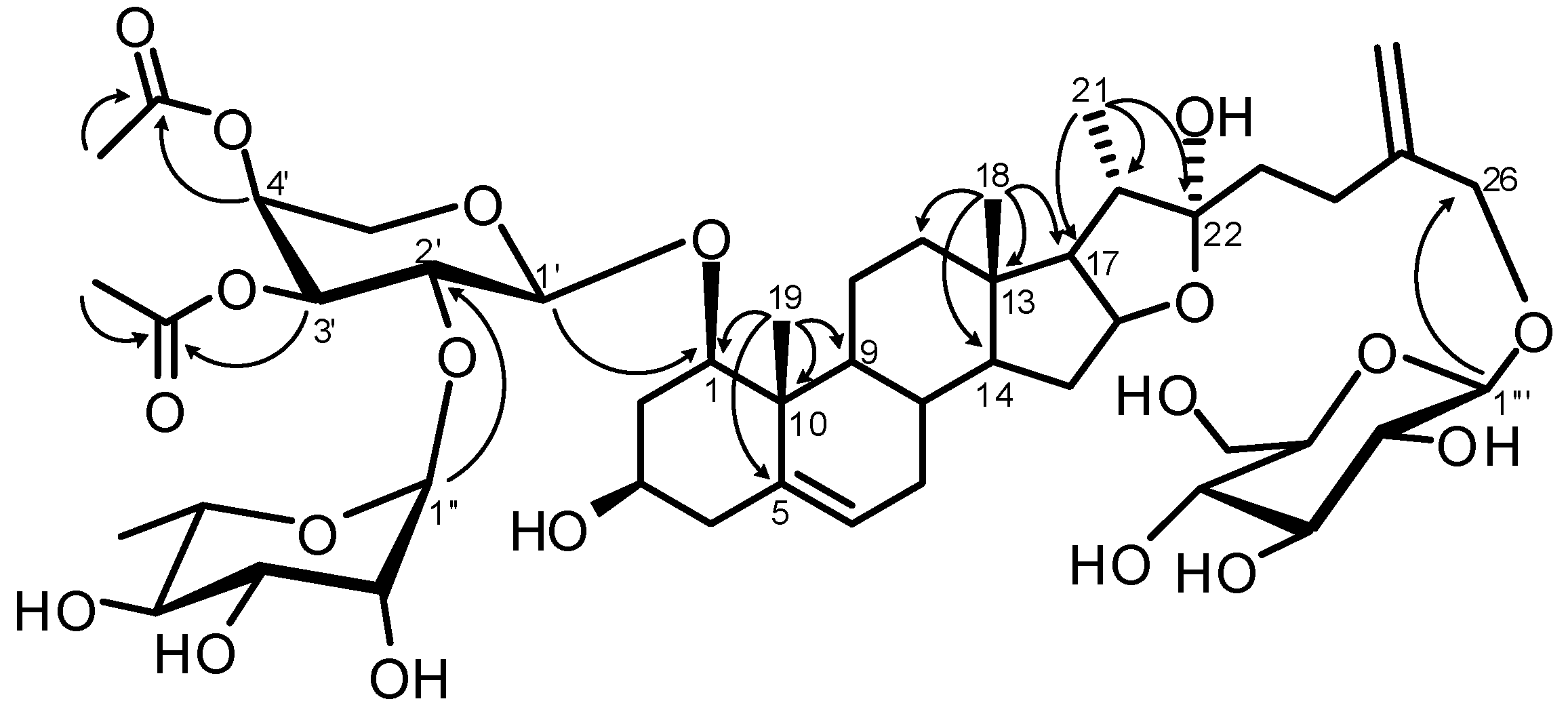
Structure Analysis and Characterization of Compounds 14

Compound 1 was obtained as a white amorphous solid. The molecular formula was determined to be C48H74O20 from the molecular ion peak [M+Na]+ at m/z 993.4698 (calcd. for C48H74O20Na, 993.4671) in the positive HRESI-MS. The analysis of the 1H-NMR spectrum of 1, in combination with HSQC data, showed signals for three anomeric protons δH 4.95/δC 101.7, δH 4.50/δC 100.1 and δH 4.28/δC 103.1, suggesting the presence of three sugar moieties.
In addition, the 1H-NMR spectrum (Table 1) showed signals for two tertiary methyl groups at δH 1.09 and 0.86 (each 3H, s) and one secondary methyl group at δH 1.02 (3H, d, 6.8 Hz), as well as one signal for a trisubstituted double bond (δH 5.55, 1H, br d, 5.6 Hz) and for an exometylene group (δH 5.09 and 4.92 each 1H, br s), three methine proton signals at δH 4.56 (1H, m, H-16), 3.38 (1H, m, H-1) and 3.33 (1H, m, H-3) indicative of secondary alcoholic functions and two methylene proton signals at δH 4.33 and 4.13 (each 1H, d, 12.5 Hz) ascribable to a primary alcoholic function.
The 13C-NMR spectrum showed three secondary alcoholic functions at δC 83.9, 81.8 and 68.6, one primary alcoholic function at δC 72.4 and a hemiacetalic carbon signal at δC 111.7, suggesting the presence of a furostanol skeleton (Table 1). Comparison with literature data and analysis of HSQC and HMBC data revealed a furosta-5,25(27)-diene-1β,3β,22α,26-tetrol moiety. Glycosylation shifts on the aglycone were observed for C-1 (δC 83.9) and C-26 (δC 72.4). The C-22 α-configuration of 1 was assigned on the basis of key ROESY correlations between H-20 (δH 2.14) and the protons H2-23 (δH 1.84) and of the downfield shift of H-16 at δH 4.56 [9].
As concerning the sugar portion, in addition to the carbinol protons, the 1H-NMR spectrum (Table 2) showed signals at δH 2.03 and 2.04 (each 3H, s), ascribable to the methyl groups of two acetyl groups [δC 170.8 and 170.9 (C=O); δC 20.7 and 20.8 (CH3)], and one signal at δH 1.26 (3H, d, 6.3 Hz) indicative of a 6-deoxyhexopyranose unit (Table 2).
The assignment of all protons and carbon chemical shifts of the three sugar units was performed by careful analysis of 2D NMR spectra, including COSY, TOCSY, HSQC and HMBC experiments, allowing the identification of one β-glucopyranosyl (Glc), one α-arabinopyranosyl (Ara) and one α-rhamnopyranosyl (Rha) units. The relatively large JH1-H2 values (7.4–8.0 Hz) indicated a β-orientation for the anomeric center of glucose and an α-orientation for that of arabinose in their pyranose form, whereas a small JH1-H2 coupling (1.2 Hz) indicated the α-configuration of the rhamnopyranosyl unit. The monosaccharides obtained from the acidic hydrolysis of 1 were identified as D-glucose, L-arabinose and L-rhamnose by GC analysis of their chiral derivatives [10].
The position of the acetyl groups at C-3′ and C-4′ of the arabinose unit was suggested by the downfield shift observed for the H-3′ (δH 5.05) and H-4′ (δH 5.30) and for the upfield shift of C-2′ (δC 73.5) and C-5′ (δC 64.5) in comparison with the data reported for the authentic sample, ruscoponticoside E (6), which is known compound also isolated in the present study (Table 2). These evidences were confirmed by the HMBC correlations between the proton signals at δH 5.05 (H-3′) and δH 5.30 (H-4′) with the carbonyl resonances at 170.9 ppm and 170.8 ppm (Figure 3), respectively.
The sequence and interglycosidic linkages among the three sugar units and the aglycone were revealed by HMBC experiment (Figure 3). In the HMBC spectrum, a correlation peak between H-1′′′ of Glc at δH 4.28 and C-26 at δC 72.4, implied that the glucose unit is attached to C-26 of the aglycone, which is a structural feature in plant furostanol saponins. The linkage of the arabinose unit to C-1 of the aglycone was ascertained by the HMBC correlation between H-1′ of arabinose (δH 4.50) and C-1 at δC 83.9. Furthermore the anomeric proton of rhamnose at δH 4.95 was correlated with C-2′ of arabinose at δC 73.5 which supported the proposed sequence of the disaccharidic chain linked at C-1 of the aglycone.
Figure 3. Key HMBC correlations for compound 1.
Molecules 17 14002 g003
Thus the structure of compound 1 was established as 26-O-β-D-glucopyranosylfurosta-5,25(27)-diene-1β,3β,22α,26-tetrol 1-O-[α-L-rhamnopyranosyl-(1′′→2′)-O-(3′,4′-di-O-acetyl)-α-L-arabino- pyranoside].
HRESI-MS of compound 2 showed a pseudomolecular ion peak at m/z 1007.4857 [M+Na]+ corresponding to the molecular formula C49H76O20, which differs from that of compound 1 only in the gain of 14 u.m.a. 1H-NMR [δH 3.16 (3H, s)] and 13C-NMR [δC 113.3 (C-22) and 47.5 (-OCH3)] data suggested compound 2 to be a 22-methoxyfurostanol saponin (Table 1).
A HMBC experiment confirmed this hypothesis and showed a correlation between the methoxy group at δH 3.16 and C-22 (δC 113.3) of the aglycone. The ROESY experiment allowed us to assign the stereochemistry of the ketal carbon C-22. Clear correlations were observed between the methoxy group at δH 3.16 and the H-16 at δH 4.38 and between H-20 (δH 2.22) and H-23a (δH 1.90)/H-23b (δH 1.84) indicating an α-orientation of the methoxy group [3].
Analysis of COSY, HSQC and HMBC experiments revealed that 2 possessed sugar moieties identical to those of 1 (Table 2). Acidic hydrolysis of 2 afforded D-glucose, L-arabinose and L-rhamnose which were confirmed by GC analysis. Thus saponin 2 was elucidated as 26-O-β-D-glucopyranosyl-22α-methoxy-furosta-5,25(27)diene-1β,3β,26-triol 1-O-[α-L-rhamnopyranosyl-(1′′→2′)-O-(3′,4′-di-O-acetyl)-α-L-arabinopyranoside].
Although we have used mild extraction conditions (room temperature) we cannot exclude the possibility that compound 2 is an artifact due to reaction of compound 1 with the extraction solvent (MeOH).
Compound 3a showed the molecular formula of C33H51O13S, deduced by a HRESI-MS measurement (m/z 687.3043, [M−H]). The presence of a sulphate group was indicated by a fragment ion peak at m/z 631 [M−NaSO3+H+Na]+ in the ESI-MS/MS spectrum recorded in a positive ion mode, corresponding to the loss of a SO3Na from the parent ion, and by IR bands at 1245 and 1086 cm−1.
Table 1. 1H- and 13C-NMR data (CD3OD, 500 and 125 MHz) data of the aglycon portions of compounds 1 and 2.
Position12
δHaδCδHaδC
13.38 m83.93.44 dd (11.8, 3.7)83.7
2a2.10 m36.82.10 m37.0
2b1.69 m 1.69 m
33.33 m68.63.35 m69.0
42.23 ovl43.02.21 ovl43.2
5-139.2-139.2
65.55 br d (5.6)125.45.56 br d (5.4)125.6
7a1.97 ovl32.31.96 ovl32.6
7b1.53 ovl1.53 ovl
81.54 ovl33.61.55 ovl33.8
91.25 ovl50.91.25 ovl51.1
10-43.5-43.4
11a2.55 m24.72.56 m25.0
11b1.49 m1.48 m
12a1.68 ovl40.71.70 ovl41.1
12b1.21 m1.21 m
13-41.6-41.4
141.13 m57.21.13 m57.5
15a1.98 ovl32.51.97 ovl32.6
15b1.29 m1.26 ovl
164.56 m81.84.38 m82.4
171.77 m63.71.73 ovl65.0
180.86 s16.70.86 s17.1
191.09 s14.91.12 s15.1
202.14 m40.42.22 ovl41.0
211.02 d (6.8)15.41.04 d (6.8)15.9
22-111.7-113.3
23a1.84 m37.41.90 m32.1
23b1.84 m
242.29 ovl28.32.18 ovl28.5
25-147.4-147.1
26a4.33 d (12.5)72.44.34 d (12.6)72.4
26b4.13 d (12.5)4.13 d (12.6)
27a5.09 br s112.15.08 br s112.2
27b4.92 br s4.93 br s
-OCH3 3.16 s47.5
Ovl: overlapped signals; a Coupling constants are in parentheses and given in Hertz. 1H- and 13C-NMR assignments aided by COSY, HSQC and HMBC experiments.
Table 2. 1H- and 13C-NMR data (CD3OD, 500 and 125 MHz) data of the sugar portions of compounds 1 and 6.
Position1 a6 b
δHcδCδHcδC
α-L-Ara
1′4.50 d (7.4)100.14.26 d (7.1)100.7
2′3.85 ovl73.53.70 ovl75.4
3′5.05 dd (9.7, 3.1)75.73.64 dd (9.5, 3.2)75.7
4′5.30 br s70.63.74 ovl70.5
5′3.89 ovl3.73 ovl64.53.85 dd (12.1, 2.0)3.48 dd (12.1, 3.0)67.2
3′-COCH32.04 s20.8
3′-COCH3 170.9
4′-COCH32.03 s20.7
4′-COCH3 170.8
α-L-Rha
1′′4.95 br d (1.2)101.75.29 br d (1.2)101.3
2′′3.72 ovl72.13.88 ovl72.0
3′′3.62 dd (9.6, 3.3)71.83.69 ovl71.8
4′′3.39 t (9.6)73.83.40 t (9.7)73.8
5′′4.08 m69.84.08 m69.5
6′′1.26 d (6.3)18.31.26 d (6.2)18.0
β-D-Glc (C26)
1′′′4.28 d (7.7)103.14.28 d (7.6)103.0
2′′′3.22 t (8.3)74.93.21 t (8.4)74.9
3′′′3.35 ovl77.93.35 ovl77.9
4′′′3.27 ovl71.53.28 ovl71.4
5′′′3.25 ovl77.73.26 ovl77.6
6′′′3.87 ovl3.66 dd (12.1, 4.5)62.63.87 ovl3.67 dd (12.0, 4.5)62.6
a The chemical shift values of the sugar portion of 2 are identical to those reported for 1; b Data reported for the authentic sample, ruscoponticoside E (6), also isolated in the present study. Ovl: overlapped signals; c Coupling constants are in parentheses and given in Hertz. 1H and 13C assignments aided by COSY, TOCSY, HSQC and HMBC experiments.
Preliminary 1H-NMR analysis of 3a (Table 3) indicated the steroid glycoside nature of the compound. The 1H-NMR spectrum showed three methyl signals: two tertiary (δH 0.86 and 1.10) and one secondary (δH 1.04), and one anomeric proton signal at δH 4.28. Its 13C-NMR spectrum exhibited 33 carbon signals, with 27 being attributable to the aglycone and six attributable to the monosaccharide unit. The 13C-NMR spectrum further showed three secondary alcoholic functions at δC 85.5, 81.8 and 68.6, one primary alcoholic function at δC 72.6 and a hemiacetalic carbon signal at δC 111.1 indicating a furostane nature for the steroidal aglycone of 3a.
Combined analysis of COSY and TOCSY experiments allowed the detection of five spin systems, four belonging to the aglycone moiety and one attributable to the monosaccharide.
Table 3. 1H- and 13C-NMR (CD3OD, 500 and 125 MHz) data of compounds 3a, 3b and 4.
Position3a 3b 4
δHa δC δHa δC δHa δC
14.03 dd (11.9, 3.8)85.54.02 dd (11.9, 3.8)85.44.01 dd (11.8, 4.0)85.6
2a
2b
2.55 m
1.69 m
38.82.56 m
1.69 m
38.72.55 m
1.67 m
38.8
33.43 dddd
(12.0, 12.0, 6.4, 6.4)
68.63.41 dddd
(12.0, 12.0, 6.4, 6.4)
68.63.45 dddd
(12.1, 12.1, 6.5, 6.5)
68.7
42.21 ovl 43.22.23 ovl 43.22.21 m (2H)43.0
5-138.7-139.0-138.8
65.61 br d (5.2)126.55.60 br d (5.2)126.45.60 br d (5.4)126.5
7a
7b
1.98 ovl
1.55 ovl
32.81.99 ovl
1.57 ovl
33.01.96 m
1.54 ovl
32.6
81.55 ovl33.81.56 ovl33.81.54 ovl33.8
91.37 ovl50.61.37 ovl50.71.35 ovl50.7
10-43.7-43.6-43.7
11a
11b
2.36 br d (12.6)
1.52 ovl
24.02.35 br d (12.6)
1.52 ovl
24.02.36 br d (12.4)
1.48 ovl
24.0
12a
12b
1.71 ovl
1.26 m
40.81.70 ovl
1.28 m
40.91.70 ovl
1.24 ovl
40.9
13-41.2-41.4-41.4
141.17 m 57.41.17 m 57.51.18 m 57.5
15a
15b
1.99 ovl
1.32 ovl
32.72.00 ovl
1.33 ovl
32.61.95 ovl,
1.26 ovl
32.7
164.57 m 81.84.38 m 82.24.57 q (5.6)82.0
171.78 ovl64.01.74 ovl64.11.71 ovl64.1
180.86 s16.80.87 s16.90.83 s16.9
191.10 s14.51.10 s14.51.10 s14.6
202.18 ovl40.82.19 ovl40.92.16 ovl41.1
211.04 d (6.7)15.81.01 d (6.7)16.00.99 d (7.1)16.1
22-111.1-113.7-111.3
23a
23b
1.90 ovl
1.86 ovl
37.51.91 ovl
1.85 ovl
37.31.61 ovl31.4
24a
24b
2.22 ovl
2.15 ovl
28.52.21 ovl
2.15 ovl
28.41.57 ovl
1.12 m
28.6
25-147.0-147.31.72 m 34.8
26a
26b
4.34 d (12.5)
4.11 d (12.5)
72.64.33 d (12.4)
4.12 d (12.4)
72.43.74 dd (9.4, 6.5)
3.39 ovl
75.6
27a
27b
5.09 br s
4.93 br s
111.95.10 br s
4.94 br s
111.90.95 d (6.6)17.1
Glucose
1′ 4.28 d (7.8)103.04.27 d (7.8)103.14.24 d (7.7)104.2
2′ 3.22 t (8.5)74.93.20 t (8.5)75.03.19 t (8.4)74.8
3′3.36 ovl77.83.34 ovl77.83.35 ovl77.9
4′3.28 ovl71.53.28 ovl71.43.27 ovl71.5
5′3.27 ovl77.73.26 ovl77.83.26 ovl77.7
6′3.86 d (11.7)
3.66 dd (11.7, 5.2)
62.63.88 d (11.7)
3.65 dd (11.7, 5.2)
62.63.87 d (11.9)
3.68 dd (11.9, 5.1)
62.7
Ovl: overlapped signals; a Coupling constants are in parentheses and given in Hertz. 1H- and 13C- assignments aided by COSY, TOCSY, HSQC and HMBC experiments.
The location of the sulphate group at position-1 was inferred by the downfield shift of the corresponding nuclei (H-1, δH 4.03 and C-1, δC 85.5) [7]. The relative stereochemistry at C-1 and C-3 was evaluated by an accurate coupling constants analysis and by ROESY experiments. In particular, H-1 appeared as a double doublet (11.9 and 3.8 Hz), whereas H-3 appeared as a dddd with two large (ax-ax) and two small (ax-eq) coupling constants. These data pointed to the axial position of both H-1 and H-3 also confirmed by ROESY correlation of H-1 with H-3. The NMR data of side chain from C-22 to C-26, was almost superimposable to the data observed in compound 1 indicating the presence of an exomethylene function 25(27). The C-22 configuration of 3a was assigned as α-configuration and was derived by the ROESY experiment that showed key correlations between H-20 (δH 2.18) and the protons H-23a (δH 1.90)/H-23b (δH 1.86) [3] and on the basis of the downfield shift of H-16 at δH 4.57 [9].
The linkage of the sugar to the C-26 hydroxyl group was shown by the HMBC correlation between the anomeric carbon at δC 103.0 and the two protons at C-26 (δH 4.34 and 4.11). Acidic hydrolysis of 3a afforded D-glucose, which was confirmed by GC analysis. Thus compound 3a was established as 26-O-β-D-glucopyranosyl-furosta-5,25(27)diene-1β,3β,22α,26-tetrol 1-O-sulphate.
The spectral data of glycoside 3b indicated its isomeric relationship with sulphated glycoside 3a. In fact, 3b has the same molecular formula determined by HRESI-MS (See Experimental), and 1H- and 13C-NMR spectra (Table 3) almost identical to those of 3a, differing only in the resonances of the carbon atom C-22 (see Table 3). This, in agreement with previous findings [11], indicated that 3b had the opposite configuration at the hemiacetal carbon 22 (22β−ΟΗ) also supported by the H-16 resonance at δH 4.38.
COSY, HSQC and HMBC experiments showed that 3b was substituted at its C-1 position by a sulphate group and at C-26 by a β-D-glucopyranosyl moiety, thus compound 3b was defined as: 26-O-β-D-glucopyranosyl-furosta-5,25(27)diene-1β,3β,22β,26-tetrol 1-O-sulphate.
The HRESI-MS spectrum of 4 exhibited a pseudomolecular ion peak at m/z 689.3190 [M−H] (calcd. for C33H53O13S, 689.3207) indicating the molecular formula C33H53NaO13S in accordance with 13C NMR data. The 1H- and 13C-NMR data of the aglycone portion of compound 4, in comparison to those of aglycone of 3a, clearly suggested that 4 differs from 3 by the replacement of the exomethylene group with a secondary methyl group at C-27 (δH 0.95, δC 17.1) (Table 3). The absolute configuration of C-25 was deduced to be R based on the difference of chemical shifts (Δab = δA − δB) of the geminal protons H2-26 (Δab = 0.35 ppm). It has been described that Δab is usually ≥0.57 ppm in 25S compounds and ≤0.48 ppm in 25R compounds [12].
The presence of the sulphate group was confirmed after solvolysis in a dioxane-pyridine mixture that afforded a less polar desulphated derivative 4a, which gave a pseudomolecular ion at m/z 615 [M+Na]+. The analysis of NMR spectra showed a high field shift of H-1 at δH 3.34 (vs. δH 4.01) and C-1 at δC 78.6 (vs. δC 85.6), confirming the location of the sulphate at C-1. A moderate upfield shift was observed also for the CH3-19 at δH 1.05 (vs. δH 1.10 in the natural compound). The solvolysis reaction led to the loss of a H2O molecule as determined by ESI-MS data and by appearance in the 1H-NMR spectrum of one allylic methyl group at δH 1.60 assigned to C-21.
The NMR data (COSY, TOCSY, HSQC, HMBC) for the sugar portion, were superimposable with those of compound 3a and 3b also confirmed by acidic hydrolysis and GC sugar analysis. Thus compound 4 was elucidated as (25R),26-O-β-D-glucopyranosyl-furost-5-ene-1β,3β,22α,26-tetrol 1-O-sulphate.
In previous studies on the crude extracts from the rhizome of Ruscus aculeatus L., Oulad-Ali et al. [7] reported the isolation of a compound constitutionally identical to compound 4. The stereochemistry at C-22 was left unassigned. Comparison between the 13C-NMR data of the two compounds evidenced some small but not insignificant differences, pointing to a stereoisomeric relationship.
Five known compounds were additionally isolated, namely ceparoside A (5) [13]; ruscoponticoside E (6) [14]; ceparoside B (7) [13]; 26-O-β-D-glucopyranosyl-furosta-5,20(22), 25(27)-triene-1β,3β,26-triol 1-O-[α-L-rhamnopyranosyl-(1→2)-O-α-L-arabinopyranoside] (8) [15]; and spirosta-5,25(27)-diene-1β,3β-diol 1-O-[α-L-rhamnopyranosyl-(1→2)-O-α-L-arabinopyranoside] (9) [15].
Besides saponins and furostanol glycosides, the hexane extract of the rhizome contains also several minor sterols (campesterol, stigmasterol and sitosterol). The identification has been performed by means of MS spectra and NMR data and comparison with literature data. A previous study on sterol composition of Ruscus aculeatus L. was reported by Dunouau et al. [7].

3. Experimental

3.1. General

High-resolution ESI mass spectrometry (HRESI-MS) was recorded on a Micromass QTOF spectrometer and electrospray ionization mass spectrometry (ESI-MS) experiments were performed on an Applied Biosystem API 2000 triple-quadrupole mass spectrometer. Optical rotations were determined on a Jasko P-2000 polarimeter. NMR spectra were obtained on a Varian Inova 500 NMR spectrometer (1H at 500 MHz and 13C at 125 MHz) equipped with a Sun hardware, δ (ppm), J in Hz, using solvent signal for calibration (13CD3OD at δC 49.0 and residual CD2HOD at δH = 3.31). The Heteronuclear Single-Quantum Coherence (HSQC) spectra were optimized for an average 1JCH of 140 Hz; the gradient-enhanced Heteronuclear Multiple Bond Correlation (HMBC) experiment were optimized for a 3JCH of 8 Hz.
HPLC was performed using a Waters 510 pump equipped with a Rheodyne 7125 injector and a Waters 401 differential refractometer as detector, using a Nucleodur 100-5 C18 column (5 µm, 4.6 mm i.d. × 250 mm); flow rate was 1 mL min−1. Droplet counter-current chromatography (DCCC) was performed on a DCC-A apparatus (Tokyo Rikakikai Co., Tokyo, Japan) equipped with 250 glass-columns.
The GC/MS analysis was carried out with an Agilent Technologies 6890N Network gas chromatograph coupled to an Agilent Technologies 5973 Network quadrupole mass selective spectrometer and provided with a split/splitless injection port. Helium was used as carrier gas at a linear velocity of 40 cm/s. Separation of compounds was performed on a HP-5 MS capillary column (30 m × 0.25 mm, 0.25 µm film thickness, Agilent USA). GC oven temperature was kept constant at 180 °C. The injector temperature was 230 °C. The temperature of the ion source and the transfer line was 250 and 280 °C, respectively. Mass spectra were taken at 70 eV and the mass range was from 40 to 350 amu.

3.2. Plant Material

Selected samples of wild growing plants Ruscus aculeatus L. (Ruscaceae) were collected in May of 2009 in the mountain area of the Tuscany region in Italy. Plants were identified at the Dipartimento di Bioscienze e Territorio, (University of Molise) and a voucher specimen is deposited under No. PGT-58-09 in the Herbarium of University of Molise (Pesche, Isernia). Rhizomes were kept frozen at −20 °C until analyzed.

3.3. Compound Isolation

Underground fresh parts (243 g) were semi-thawed, cut and extracted with MeOH (3 × 700 mL) at room temperature. The combined extracts (56 g) were concentrated and subjected to a modified Kupchan’s [16] partitioning procedure as follows. The MeOH extract was dissolved in 10% aqueous methanol and partitioned against n-hexane to furnish a n-hexane extract (483.8 mg). The water content (% v/v) of the MeOH extract was adjusted to 40% and partitioned against CHCl3, to furnish a CHCl3 extract (3.74 g). The aqueous phase was concentrated to remove MeOH and then extracted with n-BuOH yielding 9.0 g of glassy material.
The CHCl3 extract (1.8 g) was fractionated by DCCC using CHCl3/MeOH/H2O (7:13:8) in the ascending mode (the lower phase was the stationary phase), flow rate 8 ml/min; 4 ml fractions were collected. Fractions were monitored by TLC on SiO2 with CHCl3/MeOH/H2O (80:18:2) as eluent and combined on the basis of their similar TLC retention factors. Three major fractions were obtained and then separated by HPLC on a Nucleodur 100-5 C18 column (5 µm, 4.6 mm i.d × 250 mm): fraction 1 was purified with MeOH/H2O (65:35) as eluent, to afford 6.0 mg of known compound 5; fraction 2 was purified with MeOH/H2O (7:3) to give 1.9 mg of compound 1, 1.5 mg of compound 2. Fraction 3 yielded known compound 9 (35.3 mg).
The n-BuOH extract (2.0 g) was submitted to DCCC with n-BuOH/Me2CO/H2O (3:1:5) in the descending mode (the upper phase was the stationary phase). The obtained fractions were monitored by TLC on Silica gel plates with n-BuOH/OHAc/H2O (12:3:5) and CHCl3/MeOH/H2O (80:18:2) as eluents. Two fractions A and B were obtained and purified by HPLC on a Nucleodur 100-5 C18 column (5 µm, 4.6 mm i.d × 250 mm).
Fraction A (195 mg) was separated with MeOH/H2O (48:52) as eluent (flow rate 1 mL/min) affording 2 mg of compound 3a, 1.9 mg of compound 3b and 2.6 mg of compound 4.
Fraction B (541 mg) was purified by HPLC with MeOH/H2O (48:52) as eluent and contained known compounds 6 (28.8 mg), 7 (6.2 mg) and 8 (2.7 mg).
Compound 1: Amorphous solid. [α]25D −29.7 (c 0.05, MeOH); HRESI-MS m/z 993.4698 [M+Na]+ (calcd. for C48H74O20Na, 993.4671). The 1H- and 13C-NMR spectral data are listed in Table 1, Table 2.
Compound 2: Amorphous solid. [α]25D −7.3 (c 0.15, MeOH); HRESI-MS m/z 1007.4857 [M+Na]+ (calcd. for C49H76O20Na, 1007.4828). The 1H- and 13C-NMR spectral data are listed in Table 1, Table 2.
Compound 3a: Amorphous solid. [α]25D −70.5 (c 0.2, MeOH); HRESI-MS m/z 687.3043 [M−H] (calcd. for C33H51O13S, 687.3050); ESI-MS (+ve ion) m/z 733 [M+Na]+. ESI-MS/MS (+ve ion) m/z 631 [M-NaSO3+H+Na]+. IR νmax (KBr disc)/cm−1 1245, 1086. The 1H- and 13C-NMR spectral data are listed in Table 3.
Compound 3b: Amorphous solid. [α]25D −75.3 (c 0.19, MeOH); HRESI-MS m/z 687.3030 [M−H] (calcd. for C33H51O13S, 687.3050); ESI-MS (+ve ion) m/z 733 [M+Na]+. The 1H- and 13C-NMR spectral data are listed in Table 3.
Compound 4: Amorphous solid. [α]25D −29.2 (c 0.26, MeOH); HRESI-MS m/z 689.3190 [M−H] (calcd. for C33H53O13S, 689.3207). The 1H- and 13C-NMR spectral data are listed in Table 3.
Compound 5: Amorphous solid. [α]25D −28.0 (c 0.60, MeOH); HRESI-MS m/z 925.4782 [M+Na]+ (calcd. for C45H74O18Na, 925.4773). The 1H- and 13C-NMR spectral data are consistent with the published data [12].
Compound 6: Amorphous solid. [α]25D −30.0 (c 0.93, MeOH); HRESI-MS m/z 909.4465 [M+Na]+ (calcd. for C44H70O18Na, 909.4460). The 1H- and 13C-NMR spectral data are consistent with the published data [13].
Compound 7: Amorphous solid. [α]25D −29.4 (c 0.07, MeOH); HR-ESI-MS m/z 911.4623 [M+Na]+ (calcd. for C44H72O18Na, 911.4616). The 1H- and 13C-NMR spectral data are consistent with the published data [12].
Compound 8: Amorphous solid. [α]25D −4.63 (c 0.08, MeOH); HRESI-MS m/z 891.4361 [M+Na]+ (calcd. for C44H68O17Na, 891.4354). The 1H- and 13C-NMR spectral data are consistent with the published data [14].
Compound 9: Amorphous solid. [α]25D −64.0 (c 0.69, MeOH); HRESI-MS m/z 729.3832 [M+Na]+ (calcd. for C38H58O12Na, 729.3826). The 1H- and 13C-NMR spectral data are consistent with the published data [14].

3.4. Solvolysis of Compound 4 Giving 4a

A solution of compound 4 (2.6 mg, 0.0036 mmol) in pyridine (0.5 mL) and dioxane (0.5 mL) was heated at 150 °C for 2 h in a stoppered reaction vial. After the solution was cooled, the mixture was evaporated to dryness and then purified by HPLC on a Nucleodur 100-5 C18 column (5 µm, 4.6 mm i.d. × 250 mm) with MeOH/H2O 8:2, to give 1.7 mg of desulphated compound 4a. Compound 4a: [α]25D −7.8 (c 0.17, MeOH); ESI-MS: 615 [M+Na]+; selected 1H-NMR (CD3OD, 500 MHz) data for compound 4a: 5.55 (1H, br d, 5.4 Hz, H-6), 4.72 (1H, m, H-16), 3.70 (1H, dd, 9.4, 6.5 Hz, H-26a), 3.39 (1H, ovl, H-26b), 3.39 (1H, ovl, H-3), 3.34 (1H, ovl, H-1), 1.60 (3H, s, H3-21), 1.05 (3H, s, H3-19), 0.95 (3H, d, 6.6 Hz, H3-27), 0.72 (3H, s, H3-18).

3.5. Methanolysis of 12: Sugar Analysis

A solution of compounds 12 (0.5 mg) in anhydrous 2 N HCl-MeOH (0.5 mL) was heated at 80 °C in a stoppered reaction vial. After 2 h, the reaction mixture was cooled, neutralized with Ag2CO3, and centrifuged, and the supernatant was taken to dryness under N2. 1-(Trimethylsilyl)imidazole in pyridine was added and left at room temperature for 15 min. The derivatives were analyzed by GC-MS (HP-5MS capillary column, helium carrier, flow 10 mL min−1 oven temperature 150 °C). GC-MS peaks in the sylilated saponin hydrolysate coeluted with those in silylated standards (methyl rhamnosides, methyl arabinosides and methyl glucosides).

4. Conclutions

Two new furostanol saponins 12 and three new sulphated glycosides 3a, 3b and 4 were isolated from the underground parts of Ruscus aculeatus L., along with four known furostanol and one spirostanol saponins 59 and three free sterols. The new compounds add knowledge in the field of isolation and structural characterization of new metabolites from natural sources.

Acknowledgments

MS and NMR spectra were provided by Centro di Servizio Interdipartimentale di Analisi Strumentale (CSIAS), Università di Napoli “Federico II”, Napoli, Italy.

Conflict of Interest

The authors declare no conflict of interest.
  • Sample Availability: Samples of the pure compounds are available from the authors.

References

  1. Hostettmann, K.; Marston, A. Saponins; Cambridge University Press: London, UK, 1995; pp. 302–303. [Google Scholar]
  2. Mimaki, Y.; Kuroda, M.; Kameyama, A.; Yokosuka, A.; Sashida, Y. New steroidal constituents of the underground parts of Ruscus aculeatus and their cytostatic activity on HL-60 cells. Chem. Pharm. Bull. 1998, 46, 298–303. [Google Scholar]
  3. Mari, A.; Napolitano, A.; Perrone, A.; Pizza, C.; Piacente, S. An analytical approach to profile steroidal saponins in food supplements: The case of Ruscus aculeatus. Food Chem. 2012, 134, 461–468. [Google Scholar]
  4. de Combarieu, E.; Falzoni, M.; Fuzzati, N.; Gattesco, F.; Giori, A.; Lovati, M.; Pace, R. Identification of Ruscus steroidal saponins by HPLC-MS analysis. Fitoterapia 2002, 73, 583–596. [Google Scholar]
  5. Perrone, A.; Muzashvili, T.; Napolitano, A.; Skhirtladze, A.; Kemertelidze, E.; Pizza, C.; Piacente, S. Steroidal glycosides from the leaves of Ruscus colchicus: isolation and structural elucidation based on a preliminary liquid chromatography-electrospray ionization tandem mass spectrometry profiling. Phytochemistry 2009, 70, 2078–2088. [Google Scholar]
  6. Napolitano, A.; Muzashvili, T.; Perrone, A.; Pizza, C.; Kemertelidze, E.; Piacente, S. Steroidal glycosides from Ruscus ponticus. Phytochemistry 2011, 72, 651–661. [Google Scholar]
  7. Dunouau, C.; Belle, R.; Oulad-Ali, A.; Anton, R.; David, B. Triterpenes and sterols from Ruscus aculeatus. Planta Med. 1996, 62, 189–190. [Google Scholar]
  8. Oulad-Ali, A.; Guillaume, R.B.; David, B.; Anton, R. Sulphated steroidal derivatives from Ruscus aculeatus. Phytochemistry 1996, 42, 895–897. [Google Scholar] [CrossRef]
  9. Corea, G.; Fattorusso, E.; Lanzotti, V.; Capasso, R.; Izzo, A.A. Antispasmodic saponins from bulbs of red onion, Allium cepa L. var. Tropea. J. Agric. Food. Chem. 2005, 53, 935–940. [Google Scholar]
  10. Hara, S.; Okabe, H.; Mihashi, K. Gas-liquid chromatographic separation of aldose enantiomers as trimethylsilyl ethers of methyl 2-(polyhydroxyalkyl)thiazolidine-4(R)-carboxylates. Chem. Pharm. Bull. 1987, 35, 501–506. [Google Scholar]
  11. Corea, G.; Fattorusso, E.; Lanzotti, V. Saponins and flavonoids of Allium triquetrum. J. Nat. Prod. 2003, 66, 1405–1411. [Google Scholar]
  12. Challinor, V.L.; Piacente, S.; De Voss, J.J. NMR assignment of the absolute configuration of C-25 in furostanol steroidal saponins. Steroids 2012, 77, 602–608. [Google Scholar]
  13. Yuan, L.; Ji, T.F.; Wang, A.G.; Yang, J.B.; Su, Y.L. Two new furostanol saponins from the seeds of Allium cepa L. Chin. Chem. Lett. 2008, 19, 461–464. [Google Scholar]
  14. Bombardelli, E.; Bonati, A.; Gabetta, B.; Mustich, G. Glycosides from rhizomes of Ruscus aculeatus. Fitoterapia 1972, 43, 3–10. [Google Scholar]
  15. Mimaki, Y.; Takaashi, Y.; Kuroda, M.; Sashida, Y.; Nikaido, T. Steroidal saponins from Nolina recurvata stems and their inhibitory activity on cyclic AMP phosphodiesterase. Phytochemistry 1996, 42, 1609–1615. [Google Scholar]
  16. Kupchan, S.M.; Britton, R.W.; Ziegler, M.F.; Sigel, C.W. Bruceantin, a new potent antileukemic simaroubolide from Brucea antidysenterica. J. Org. Chem. 1973, 38, 178–179. [Google Scholar]

