Article

Antioxidant Activities of Melittis melissophyllum L. (Lamiaceae)

Department of Chemistry, Biochemistry and Environmental Protection, Faculty of Science, University of Novi Sad, Trg Dositeja Obradovica 3, 21000 Novi Sad, Serbia
*
Author to whom correspondence should be addressed.
Molecules 2011, 16(4), 3152-3167; https://doi.org/10.3390/molecules16043152
Submission received: 14 March 2011 / Revised: 12 April 2011 / Accepted: 14 April 2011 / Published: 14 April 2011

Abstract

:
Extracts of Melittis melissophyllum leaves in ether, chloroform, ethyl acetate, n-butanol and water were evaporated to dryness and dissolved in 50% ethanol to make 10% (w/v) solutions. The potential protective action of the extracts was assessed by the corresponding in vitro and in vivo tests. In the in vitro experiments extracts were tested as potential scavengers of free radicals (DPPH, O2·, NO, and OH radicals), as well as inhibitors of liposomal peroxidation (LPx). The results obtained show that all extracts (exept n-BuOH extract) are good scavengers of radicals and reduce LPx intensity in liposomes, which points to their protective (antioxidant) activity. In vivo experiments were concerned with antioxidant systems (activities of GSHPx, GSHR, Px, CAT, XOD, GSH content and intensity of LPx) in liver homogenate and blood-hemolysate of experimental animals after their treatment with extracts of M. melissophyllum leaves, or in combination with CCl4. On the basis of the results obtained it can be concluded that the examined extracts have protective (antioxidative) effect and this antioxidative behaviour is more pronounced in liver than in blood-hemolysate. The reason is probably the fact that liver contains other enzymatic systems, which can also participate in the antioxidative mechanism. Of all the extracts the H2O one showed the highest protective activity.

1. Introduction

One of the paradoxes of life on Earth is that, on the one hand, oxygen is necessary for the life of aerobic organisms. On the other hand, increased concentrations of oxygen and especially its reactive metabolites (reactive oxygen species) may lead to the development of numerous diseases. A major source of free radicals in biological systems is molecular oxygen (O2). By interacting with fundamental cell structures and biomolecules, reactive oxygen species (ROS) may lead to a number of physiological and patophysiological disorders such as Alzheimer’s, Parkinson’s, ischemia – reperfusion injury, coronary atherosclerosis, diabetes mellitus, hypertension, and cancer genesis, as well in the aging process [1,2]. To prevent and/or treat the damage caused by oxygen free radicals, which initiate the process of peroxidation of the cell lipid membrane, use is made various substances capable of inhibiting free-radical reactions in the cell.
The changes in the oxidative status or the natural protection system indicate the need for an antioxidative therapy. Active compounds from medicinal herbs (flavonoids, flavolignans, and polyphenols) are natural antioxidants which should be preferably used both for the prevention and antioxidative therapy of various diseases [3]. It is well-known that most spices, especially those belonging to the Lamiaceae family, possess a wide range of biological and pharmacological activities. Since ancient times, they have been used to improve the flavor and the organoleptic properties of different types of food. Furthermore, the use of aromatic plants and spices in phytotherapy is mostly related to different activities of their essential oils, such as antimicrobial, spasmolytic, carminative, hepatoprotective, antiviral, anticarcinogenic, etc. activities [4,5].
Melittis melissophyllum L. (Lamiaceae) leaves contain no more than 0.13% of essential oil which is of complex and variable composition. Among the more than 50 compounds identified to date, citronellal (dominantly the (R) enantiomer), β-caryophyllene, β-caryophyllene oxide, germacrene-D, nerol, geranial, citronellol, and geraniol amount to about 70% of the oil [6]. The composition is similar to that of lemongrass, but balm oil can be identified by its typical pattern of chiral compounds; for example, almost enantiomerically pure (R)-(+)-methyl citronellate is a good indicator of true balm oil. Beside that, leaves contain flavonoids (apigenin, luteolin, and kaempferol), triterpenes, phenolic acids (rosmarinic, caffeic, and chlorogenic acid), sterols and salts [7]. The essential oil exhibits spasmolitic action and acts as a muscle relaxant, sedative, narcotic, antibacterial, and antifungal [8].
In view of the above it was of interest to study the potential in vitro protective action of various extracts of M. melissophyllum, to assess their potential capacity as scavengers of free radicals. Furthermore, extracts of M. melissophyllum leaves were used to study their in vivo effects on some antioxidant systems in the experimental animal liver and blood-hemolysate in combination with and without carbon tetrachloride (CCl4), in order to prove possible antioxidative properties of this, very often used folk medicine remedy (secondary medicaments).

2. Results and Discussion

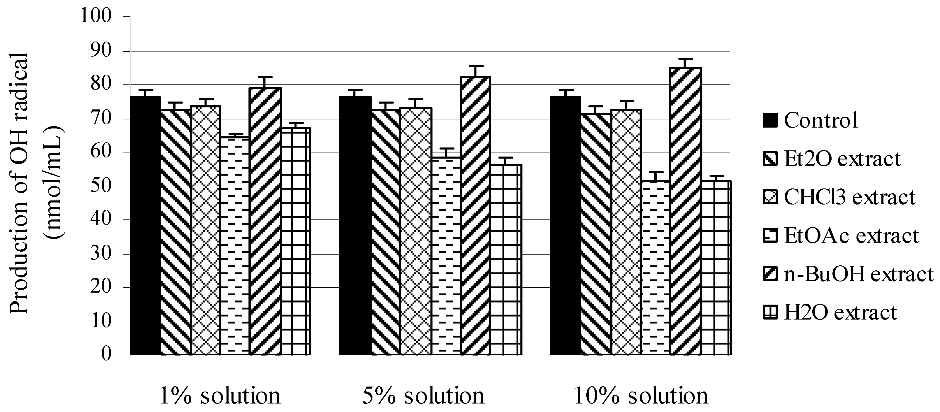
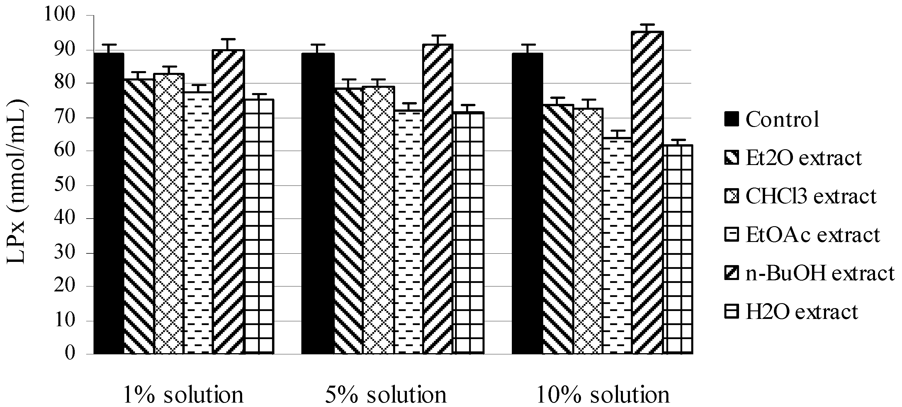
2.1. In vitro experiments

Results of determination of total flavonoids in M. melissophyllum leaves extracts are given in Table 1.
Table 1. Content of total flavonoids (mg/g) in extracts of leaves of M. melissophyllum.
ExtractEt2O CHCl3EtOAc n-BuOH H2O
Leaf 0.791.141.310.411.98
The amount of flavonoids in extracts plays an important role in their antioxidative behavior. The richest in flavonoids proved to be the water extract, while the n-BuOH extract contains the lowest amount of these active substances.
Antiradical activity was observed in the study of M. melissophyllum leaves extracts in different solvents on the content of DPPH, O2· and NO radicals (Table 2), whereby the H2O extract exhibited the strongest inhibitory effect, as the IC50 value was achieved with the lowest concentration.
In the DPPH assay, the ability of the investigated extracts to act as donors of hydrogen atoms or electrons in transformation of DPPH into its reduced form DPPH-H was investigated (Table 2). All of the assessed extracts were able to reduce the stable, purple-colored radical DPPH into yellow-colored DPPH-H reaching 50% of reduction with an IC50 as follows: 9.21 μg/mL for H2O, 11.34 μg/mL for EtOAc, 11.92 μg/mL for CHCl3, 17.21 μg/mL for n-BuOH, and 18.09 μg/mL for Et2O extract. Comparison of the DPPH scavenging activity of the investigated extracts with those expressed by BHT (14.31 μg/mL) showed that H2O, EtOAc, and CHCl3 extracts expressed stronger antioxidant effects. When investigating neutralization of O2· and NO radicals, water extract has also exhibited the greatest ability of their scavenging.
Table 2. IC50 values (μg/mL) of the neutralization of DPPH, O2· andNO radicals with M. melissophyllum extracts.
IC50 (μg/mL)
ExtractEt2OCHCl3EtOAcn-BuOHH2OBHT
DPPH radical18.0911.9211.3417.219.2114.31
O2° radical8.117.9114.4213.266.3810.46
NO radical8.047.428.919.126.178.63
For the neutralization of DPPH, O2· and NO radicals, the most responsible compounds were flavonoids and phenolic acids present in the leaves of M. melissophyllum [9,10], so obtained results can be related to the experiments in which the total amount of flavonoids was determined (Table 1.), which show that water extract of M. melissophyllum leaf contains the largest amounts of total flavonoids. It is well known that some flavonoids and phenolic acids isolated from M. melissophyllum possess certain biological and pharmacological activity [11,12]. For example, apigenin, one of the flavonoids present in M. melissophyllum, was shown to express strong antioxidant effects, increasing the activities of antioxidant enzymes and, related to that, decreasing the oxidative damage to tissues [10,13]. Luteolin is also thought to play an important role in the human body as an antioxidant, a free radical scavenger, an agent in the prevention of inflammation, a promoter of carbohydrate metabolism, and an immune system modulator. These characteristics of luteolin are also believed to play an important part in the prevention of cancer. Multiple research experiments describe luteolin as a biochemical agent that can dramatically reduce inflammation and the symptoms of septic shock [14]. Furthermore, it can be supposed that such antiradical activity is caused, besides flavonoids, also by triterpenoids acids (especially ursolic, oleanolic, and pomolic acid) since nonpolar solvents also exhibited high antiradical potential [15,16,17].
The hydroxyl RSC of the examined M. melissophyllum extracts (1%, 5% and 10%) measured by the deoxyribose assay is shown(Figure 1) The protective effects of the extracts on 2-deoxy-D-ribose were assessed as their ability to remove hydroxyl radicals, formed in Fenton reaction, from the test solution and to prevent its degradation. M. melissophyllum extracts have exhibited different behavior related to production of OH radicals. Ethyl acetate and water extracts at all investigated concentrations expressed OH radical inhibition. A stronger inhibition of OH radical production was expressed by the water extracts, especially the 10% solution (51.2 ± 1.7 nmol/mL) in comparison with the control (76.3 ± 2.1 nmol/mL).
This behaviour of the water extract can be explained by the presence of phenolic acids and flavonoid glycosides [18]. It is known from the literature [19,20] that bastard balm, and especially its leaves, is characterised by a high content of flavonoid glucosides. Luteolin 7-glucoside has been isolated from the M. melissophyllum flower and leaf, whereas of phenolic compounds were detected rosmarinic, caffeic and chlorogenic acid in leaves and ursolic acid in the plant root and leaf [20].
Figure 1. Inhibition of degradation of 2-deoxyribose by different extracts of M. melissophyllum in the deoxyribose assay.
Molecules 16 03152 g001
On the basis of this it can be supposed that the very pronounced protective effect of the H2O and EtOAc extracts of M. melissophyllum is due to the presence of flavonoids and glucosides, namely of luteolin, either being present as free or in the form of its glucosides. The suggested mechanism of flavonoid antioxidative action is as follows: the double bond in position 2,3 is conjugated with the C4-carbonyl group, and free OH groups (C5, C3 and C7) can form chelates with ions of d-elements. Once formed, complex with Fe2+ ion prevents formation of OH· radicals in Fenton’s reaction [21]. It was determined that rosmarinic acid has stronger antioxidant effect than vitamin E. Rosmarinic acid prevents cell damage caused by free radicals and reduces the risk of cancer and atherosclerosis. In contrast to the histamines, rosmarinic acid prevents activation of the immune system cells that cause swelling and fluid collection [22]. n-BuOH extract has prooxidative effects, when other two extracts (etheric and chloroformic) don`t show neither ani-, neither pro-oxidative effect.
The protective effects on lipid peroxidation (LP) of M. melissophyllum extracts have been evaluated using the Fe2+/ascorbate system of induction, by the TBA-assay (Figure 2). Inhibition of LP was determined by measuring the formation of secondary components (mainly MDA) of the oxidative stress, using liposomes as an oxidizable substrate. In general, all of the examined extracts (except n-BuOH extract) expressed strong antioxidant capacity. Protective activity can be explained by present of phenolic acids and their influence on antioxidative capacity of ascorbic acid, which doesn`t show a strong antioxidative effect in lipid phase, but different phenolic compounds can result increase of its antioxidant activity [23]. The largest inhibitory activity, again, was exhibited by water extract. The antioxidant activities of all extracts of M. melissophyllum leaves were dose dependent.
Figure 2. Inhibition of LP in Fe2+/ascorbate system of induction by different extracts of M. melissophyllum in the TBA assay.
Molecules 16 03152 g002
The n-BuOH extract shows a prooxidative effect that is increased by increasing concentration of added extract. It can be supposed that compounds with polar groups were extracted by n-BuOH, and are present in high concentration in the extract. It is notable that molecules which show antioxidant activity, when they are present in high concentration, might behave as prooxidants [24], so n-BuOHextract of Melittis melissophyllum L. leaves probably have this kind of activity.

2.2. In vivo experiments

The represented antioxidant activity results show that extracts of examined plant species, specially H2O extract, are efficient in the protection of tissues and cells from oxidative stress. Anyway, according to variations in regard to antoxidant activity of tested by different in vitro models, there are also requiste in vivo tests that would confirm antioxidant activity. In vivo tests are also necessary because a lot of plant phenols are biotransformed during their active metabolism. The experimental animals were given 1 mL/kg of 2% of Et2O, CHCl3, EtOAc, n-BuOH or H2O extract (i.p.) of M. melissophyllum leaves for 7 days. After 7 days, the animals were sacrificed. In the liver homogenate and blood-hemolysate of sacrificed animals the following biochemical parameters were determined: LPx intensity, content of GSH and activities of GSHR, GSHPx, Px, XOD and CAT (Table 3 and Table 5). In Table 4 and Table 6 the results of the same parameters obtained after pretreatment of experimental animals with the examined M. melissophyllum extracts, followed by a single dose of carbon tetrachloride (CCl4) as a well-known radical generator are presented.
As can be seen from Table 3. the EtOAc extract decreased the GSHPx activity; Et2O extract caused its increase, while the other three extracts caused no essential changes of this parameter. The EtOAc extract decreased the GSH content compared with control. All permanent extracts increased the index of GSH, but only the increase caused by treatment with H2O extract are statistically remarkable. Since about 95% of glutathione in liver is reduced and only 5% is oxidized [25], this increase in the GSH index by applying by M. melissophyllum L. leaves extracts (except EtOAc extract) probably derives from some compounds that contains a free SH-group, regardless if these compounds are present in extract or secondary biomolecules from these extracts have influenced an increase in the synthesis of GSH or cysteine. Treatment with the Et2O, CHCl3, and H2O extracts yielded an increase in GSHR activity, whereas the EtOAc extract caused a statistically significant decrease of this enzyme, which was in agreement with the action of this extract on GSH.
Table 3. Effect of extracts of M. melissophyllum leaves on the biochemical parameters in the liver homogenate.
ParameterControlExtract
Et2O CHCl3EtOAc n-BuOH H2O
GSHPx 3.43 ± 0.173.96 ± 0.16a3.32 ± 0.182.84 ± 0.21a3.37 ± 0.153.46 ± 0.18
GSH2.61 ± 0.132.76 ± 0.112.86 ± 0.142.12 ± 0.17a2.87 ± 0.223.49 ± 0.13a
GSHR3.96 ± 0.184.82 ± 0.21a5.36 ± 0.25a2.98 ± 0.14 a3.96 ± 0.195.52 ± 0.23a
Px 4.38 ± 0.134.81 ± 0.17 a4.93 ± 0.21 a3.89 ± 0.13 a5.11 ± 0.25 a4.87 ± 0.15 a
LPx7.19 ± 0.237.36 ± 0.217.91 ± 0.19a6.71 ± 0.16 a7.12 ± 0.236.19 ± 0.27 a
CAT4.41 ± 0.163.83 ± 0.17 a5.03± 0.19a3.20 ± 0.15 a4.52 ± 0.115.49 ± 0.13a
XOD5.27 ± 0.176.17 ± 0.23a6.02 ± 0.16a4.23 ± 0.16 a5.11 ± 0.224.17 ± 0.19 a
T-test a p ≤ 0.05 n = 5; x ± SD. Content of GSH is expressed in nmol GSH/mg of protein. Activities of GSHPx, GSHR, Px, CAT and XOD are expressed in nmol/mg of protein x min−1. Intensity of LPx is expressed in nmol malondialdehyde/mg of protein.
On the other hand, all the extracts (except the EtOAc extract) produced a statistically significant increase in Px activity. In addition to the very important role of peroxidase in the oxidative stress there are literature data on some other actions of peroxidases. Thus, some plant peroxidases oxidize phenols to phenoxy radicals to form polymers and enable their removal from industrial wastewaters [26]. Having in mind the results presented in Table 1 and Figure 1 it might be interesting to test the aqueous extract of M. melissophyllum as a biological marker. As compared with control, intensity of LPx is statistically saignificant reduced during the treatment with ethylacetate and water extracts of M. melissophyllum leaves. The result derived by treatment with ethylacetate and water extracts is in according with amounts got in vitro experiment (Figure 2). Using CHCl3 extract leads to a significant increase of LPx intensity, whereas the other two extracts had no effect on this parameter. The CAT increased in the treatments with CHCl3 and H2O extracts, and decreased in the treatment with the Et2O and EtOAc extract. n-BuOH extract caused no essential changes of CAT with respect to control. An increased XOD value was observed only in the treatments with the Et2O and CHCl3 extracts.
Table 4 presents the values of biochemical parameters obtained for the liver homogenate of animals treated with extracts of M. melissophyllum leaves and CCl4. In comparison with the control (animals received only physiological solution), treatment with CCl4 yielded a significant decrease in activities of all the enzymes (GSHPx, GSHR, Px, CAT, XOD), as well as of GSH content, the only increase being in the intensity of LPx. In combination with CCl4 the extracts exhibited different effects on GSHPx: while the CHCl3 and H2O extracts showed a statistically significant increase, the EtOAc extract decreased the activity of this enzyme. The Et2O and n-BuOH extracts in combination with CCl4 had no effect on the GSHPx activity.
Table 4. Effect of M. melissophyllum leaves extracts and CCl4 on the liver homogenate biochemical parameters.
ParameterControl + CCl4Extract + CCl4
Et2O CHCl3EtOAcn-BuOHH2O
GSHPx 2.12 ± 0.172.31 ± 0.152.61 ± 0.18a1.62 ± 0.23a2.18 ± 0.242.58 ± 0.18a
GSH2.26 ± 0.172.18 ± 0.122.35 ± 0.191.86 ± 0.12 a1.92 ± 0.15 a2.49 ± 0.25
GSHR2.53 ± 0.213.46 ± 0.28a4.25 ± 0.24a2.08 ± 0.15a2.26 ± 0.263.07 ± 0.26a
Px 3.47 ± 0.183.05 ± 0.17a2.97 ± 0.25a2.84 ± 0.21a3.06 ± 0.233.01 ± 0.24a
LPx8.91 ± 0.297.12 ± 0.21a7.06 ± 0.24 a6.92 ± 0.17a6.98 ± 0.24 a6.81 ± 0.24a
CAT2.08 ± 0.172.17 ± 0.222.47 ± 0.251.02 ± 0.12a2.10 ± 0.141.44 ± 0.18a
XOD4.61 ± 0.253.69 ± 0.23a5.38 ± 0.21a 3.02 ± 0.19a3.98 ± 0.17 a2.39 ± 0.14 a
T-test a p ≤ 0.05 n = 5; x ± SD. Content of GSH is expressed in nmol GSH/mg of protein. Activities of GSHPx, GSHR, Px, CAT and XOD are expressed in nmol/mg of protein x min−1. Intensity of LPx is expressed in nmol malondialdehyde/mg of protein.
Treatment with CCl4 caused no significant reduction of GSH content compared with that seen in untreated animals. Combined treatment with the extracts and CCl4 had a different effect on the GSH content in the liver homogenate. While the EtOAc and n-BuOH extracts caused a decrease, the Et2O, CHCl3 and H2O extracts showed no effect on this parameter. The Et2O, CHCl3 and H2O extracts in combination with CCl4 increased the GSHR activity in the liver homogenate and the EtOAc extract reduced it. On the other hand, the n-BuOH extract did not influence this parameter. Bearing in mind the fact that the CCl4-induced oxidative injuries of the liver require a high consumption of GSH, which is regenerated via the GSHR activity, it can be concluded that the extracts showed no protective effect, as they did not cancel out the effect of treatment with CCl4. All the extracts (except n-BuOH one) in combination with CCl4 yielded a statistically significant decrease in Px, the EtOAc extract exhibiting the strongest effect. All extracts of M. melissophyllum leaves combine with CCl4 have showed a statistically significant decrease of LPx intensity, and this behavior of the extract probably results from the presence of secondary biomolecules like flavonoids and phenolic acids. Handa et al. [27] determined that secondary biomolecules such as flavonoids, xanthones and tannins in combination with CCl4 have protective effects on liver. Phenolic components present in M. melissophyllum leaves (rutin, luteolin, kvercetin) are known as strong inhibitors of CCl4-induced LP [28]. Flavonoids could affect the initiation phase of lipid peroxidation, where they influence the metabolism of CCl4, they scavenge the free radicals, or they decrease the microsomal enzyme systems that are claimed for CCl4 metabolism [29]. In continuation of this process, flavonoids can scavenge lipoperoxides and their radicals or they can act as chelating agents for Fe2+ ion, and in this way can stop Fenton reactions [30]. On the basis of these results, we can conclude that all of extracts of M. melissophyllum leaves showed protection effect in relation to the CCl4-induced lipid peroxidation. The EtOAc and H2O extracts in combination with CCl4 yielded a statistically significant decrease in CAT activity. Administration of CCl4 significantly decreased the activity of XOD (4.61 ± 0.25 nmol/mg of protein x min-1) compared with the untreated animals (5.27 ± 0.17 nmol/mg of protein x min-1). Further, the Et2O, EtOAc, n-BuOH and H2O extracts in combination with CCl4 significantly lowered the activity of XOD. On the contrary, CHCl3 extract significantly increased XOD activity. Some recent studies point to the relationship between elevated XOD activity and oxidative stress in hypertension and the production of oxygen radicals in diabetes [31]. However, allopurinol, a XOD inhibitor known in clinical practice, reduces oxidative stress in diabetes [32], interacting with some peroxy radical species, such as Cl3OO. It can be supposed that the active constituents present in Et2O, EtOAc, n-BuOH and H2O extracts act similarly, reducing the activity of this enzyme.
Like those given in Table 3, the results in Table 5 show that all the extracts caused a statistically significant decrease in blood GSHPx activity. All extracts reduced the GSH content in the hemolysate too, the decrease being statistically significant. The activity of GSHR decreased after treatment with Et2O, EtOAc and n-BuOH extracts.
Table 5. Effect of extracts of M. melissophyllum leaves on the biochemical parameters in blood hemolysate.
ParameterControlExtract
Et2OCHCl3EtOAc n-BuOHH2O
GSHPx 5.94 ± 0.224.81 ± 0.16a4.95 ± 0.19a2.77 ± 0.23a4.79 ± 0.16a3.89 ± 0.16a
GSH6.78 ± 0.126.22 ± 0.14a6.13 ± 0.17a5.56 ± 0.21a3.96 ± 0.21a4.37 ± 0.19 a
GSHR7.67 ± 0.246.17 ± 0.24a7.87 ± 0.275.56 ± 0.17a6.81 ± 0.19a7.72 ± 0.28
Px 3.72 ± 0.173.39 ± 0.213.69 ± 0.252.94 ± 0.19a3.46 ± 0.273.65 ± 0.18
LPx4.81 ± 0.244.59 ± 0.283.78 ± 0.17a2.96 ± 0.13a4.74 ± 0.194.07 ± 0.24a
CAT4.28 ± 0.264.77 ± 0.18a3.75 ± 0.19a3.18 ± 0.16a4.11 ± 0.183.86 ± 0.23
XOD4.76 ± 0.295.92 ± 0.31a5.44 ± 0.26a5.58 ± 0.19a5.41 ± 0.27a5.48 ± 0.24a
T-test a p ≤ 0.05 n = 5; x ± SD. Content of GSH is expressed in μmol GSH/mL erythrocytes; Activities of GSHPx, GSHR, Px, CAT and XOD are expressed in nmol/mL erythrocytes x min−1; Intensity of LPx is expressed in nmol malondialdehyde/mL erythrocytes.
As for the Px activity, all extracts reduced the activity of this enzyme, the difference being statistically significant only with the EtOAc extract. Three extracts, CHCl3, EtOAc and H2O, induced a significant decrease of LPx intensity, while the Et2O and n-BuOH ones decreased the level of this enzyme insignificantly. A statistically significant decrease in CAT activity was produced by the CHCl3 and EtOAc extracts. On the other hand, all extracts caused a statistically significant increase in XOD activity. XOD is an enzyme which is present in measurable amounts only in liver and jejunum. Otherwise, because of different irregularities in liver, this enzyme should be cleared by the circulation. Consequently, an increase of XOD in the blood could be an indicator of liver damage [33]. Nowadays, increase of XOD activity is related to amplification of oxidative stress and production of free radicals [34]. From the results obtained during this research, we can conclude that the high level of XOD in the experimental animals blood-hemolysate that were treated by M. melissophyllum leave extracts is probably consequent of liver damage, so these extracts showed hepatotoxic effects. High production of oxygen radicals (priority O2·) and H2O2 probably influenced the increased production of XOD. We must bear in mind that only one of two forms of XOD is related to high production of oxygen radicals, while the other form has a completely different metabolic way, a mechanism proposed by Kisher et al. [35]. According to this mechanism, results obtained from this research could be interpreted otherwise. Particulary, the other form of XOD leads to the formation of NADH+H+, which could be included in the respiratory chain with a view to synthesize ATP, that could be interpreted as the ultimate eventual protective effect of M. melissophyllum leave extracts.
In Table 6 the results of biochemical parameters measured in the blood-hemolysate of animals treated with extracts of M. melissophyllum leaves and CCl4 are presented. The activity of glutathione peroxidase (GSHPx) in the blood of animals treated with CCl4 (Table 6) was increased compared with the control (Table 5). However, only the administration of EtOAc and n-BuOH extracts significantly decreased the activity of GSHPx. Other administered extracts did not cause notable changes.
Table 6. Effect of extracts of M. melissophyllum leaves and CCl4 on the biochemical parameters in blood hemolysate.
ParameterControl + CCl4Extract + CCl4
Et2O CHCl3EtOAc n-BuOHH2O
GSHPx 6.08 ± 0.175.83 ± 0.215.77 ± 0.244.69 ± 0.21a4.94 ± 0.27a5.91 ± 0.33
GSH 5.21 ± 0.134.84 ± 0.19a3.97 ± 0.24a4.08 ± 0.17a3.03 ± 0.18a3.86 ± 0.24a
GSHR6.12 ± 0.295.76 ± 0.285.87 ± 0.224.12 ± 0.18a6.17 ± 0.296.58 ± 0.32
Px 4.08 ± 0.223.27 ± 0.16a4.86 ± 0.24a3.15 ± 0.23 a3.04 ± 0.18 a4.11 ± 0.29
LPx5.11 ± 0.245.31 ± 0.174.92 ± 0.213.02 ± 0.24a5.17 ± 0.252.98 ± 0.12a
CAT3.74 ± 0.243.19 ± 0.23a3.24 ± 0.19a3.07 ± 0.25a2.98 ± 0.18a3.13 ± 0.28a
XOD6.19 ± 0.316.08 ± 0.264.96 ± 0.24a5.11 ± 0.17a6.97 ± 0.25a5.23 ± 0.21a
T-test a p ≤ 0.05 n = 5; x ± SD. Content of GSH is expressed in μmol GSH/mL erythrocytes; Activities of GSHPx, GSHR, Px, CAT and XOD are expressed in nmol/mL erythrocytes x min−1; Intensity of LPx is expressed in nmol malondialdehyde/mL erythrocytes.
The amount of GSH from the blood of non-CCl4 treated animals was compered with CCl4 treated animals (5.21 ± 0.13 vs. 6.78 ± 0.12), and it was observed that the amount of GSH is one of the important secondary biomolecules, that take a part in different detoxification processes in organism. Not only “radical“ GSH already in conjugation with toxic metabolites protects organism. From the results presented it is obvious that CCl4 decreased the values of GSHR (6.12 ± 0.29 nmol/mL erythrocytes x min−1), compared with the levels in the control group (7.67 ± 0.24 nmol/mL erythrocytes x min−1). The results of GSHR activity were significantly lower only in the combination of CCl4 with EtOAc extract (4.12 ± 0.18 nmol/mL erythrocytes x min−1). Also, treatment with Et2O and CHCl3 extracts caused a decrease of GSHR activity, but not notably. Treatment with CCl4, compared with the control animals (Table 5 and Table 6) did not cause notable differences in the activity of Px. Application of three extracts (Et2O, EtOAc or n-BuOH) with CCl4, caused a statistically significant decrease of Px. On the other hand, the CHCl3 extract significantly increased the activity of Px in combination with CCl4. Similar to the values of Px, values of LPx showed an insignificant increase of activity in the blood of animals treated with CCl4 (Table 6), compared with the control (Table 5). A clear ‘protective’ effect was seen in experimental animals administered H2O extract and CCl4 compared with untreated animals. Furthermore, EtOAc extract also significantly decreased the activity of LPx, while Et2O, CHCl3 and n-BuOH extracts did not change notably the levels of lipid peroxidation. In all previous research works (Figure 2, Table 3, Table 4, Table 5), H2O extract showed the best protective effect on LPx. Already it was noticed that these protective properties probably come from secondrary biomolecules such as the flavonoids and phenolic acids are. Administration of CCl4 caused a decrease in the values of CAT activity in blood of experimental animals compared with the control. Combined treatment of experimental animals with CCl4 and all of examined extracts reduced the activity of this enzyme, especially in the case of the n-BuOH extract. The level of activity of xanthine oxidase (XOD) was increased in the blood of experimental animals after administration of CCl4 (Table 6) with respect to the untreated ones (Table 5). On the contrary, lower activities of XOD were obtained after combined treatment of CCl4 with CHCl3, EtOAc or H2O extracts. However, only the n-BuOH extract exhibited a significant increase of XOD activity.

3. Experimental

3.1. General

Leaves of cultivated Melittis melissophyllum L., Lamiaceae were collected in May 2008 in the city of Vrdnik, Republic of Serbia. A voucher specimen of the collected leaves (Melittis melissophyllum L. 1753 subsp. melissophyllum, No 2-1810, Serbia, Fruška Gora, Vrdnik (UTM 34T DQ 1 09), 17.05.2008., det.: Goran Anačkov) was confirmed and deposited at the Herbarium of the Department of Biology and Ecology (BUNS Herbarium), Faculty of Natural Sciences, University of Novi Sad.
The plant leaves were dried in air and ground in a mixer. An amount of 200 g of the finely powdered material was extracted three times with 4 L of 70% methanol (MeOH) during a 24-h period. After removal of MeOH under reduced pressure, the aqueous phase was successively extracted with four solvents of increasing polarity, namely ether (Et2O), chloroform (CHCl3), ethylacetate (EtOAc) and n-butanol (n-BuOH). The extraction was carried out until a colourless extract was obtained. The residue was the aqueous extract. All of five extracts (Et2O, CHCl3, EtOAc, n-BuOH, and H2O) were evaporated to dryness and then dissolved in 50% ethanol to make 10% (w/v) solutions. These solutions, either as such or in diluted state, were used in the subsequent experiments.

3.2. In vitro experiments

Determination of total flavonoids was conducted by a colorimetric method based on the property of flavonoids and flavon glycosides to build metal complexes with aluminum ions [36]. Absorbance of analyzed solutions is measured at λ = 430 nm.
The DPPH assay was performed as described before [37], following the transformation of the DPPH radical to its reduced, neutral form (DPPH-H). The samples of all extracts of M. melissophyllum leaves were investigated in concentrations of 2.50–25.00 μg/mL. The same procedure was repeated with tert-butylated hydroxytoluene (BHT) as a positive control. For each sample five replicates were recorded. The disappearance of DPPH was measured spectrophotometrically at 515 nm.
Superoxide anion radicals were generated in the system xanthine/xanthine-oxidase, and the quantity of O2· was determined by the nitrite method [38] with modifications. The samples of all extracts of M. melissophyllum leaves were investigated in concentrations of 2.50–25.00 μg/mL. The same procedure was repeated with BHT as a positive control. For each sample five replicates were recorded. The intensity of color was measured spectrophotometrically (λ = 550 nm).
Production of NO radicals was determined spectrophotometrically. NO radical generated from sodium-nitropruside (SNP) reacts with oxygen in water solution at a physiological pH to give nitrite ions. Concentration of nitrite anions was determined using Griess reagent [39,40]. At room temperature nitrite ions react with Griess reagent and form purple complex. The samples of all extracts of M. melissophyllum leaves were investigated in concentrations of 2.50–25.00 μg/mL. The intensity of color, which is the function of the nitrite concentrations, was measured spectrophotometrically (λ = 546 nm). For each sample, five replicates were recorded.
The percentage of RSC for each radical was calculated using the following equation:
RSC(%) = 100 × (Ablank-AsampleAblank)
From the obtained RSC values, the IC50 values, which represented the concentrations of the essential oils that caused 50% neutralization, were determined by linear regression analysis.
Scavenging capacity of the M. melissophyllum leaves extracts for hydroxyl radicals was determined by monitoring the chemical degradation of 2-deoxy-D-ribose [41]. The reaction was initiated by hydroxyl radicals obtained in Fenton’s reaction [3], which yields products that react with thiobarbituric acid (TBA test). The obtained products, among which malondialdehyde (MDA) is the most important, are determined by a spectrophotometric method at 532 nm. The absorbance of the resulting solutions and the blank (with same chemicals, except sample) was recorded. For the experiment, three concentrations of extracts of bastard leaves were prepared (10%, 5% and 1% solution). Five replicates were performed for each sample.
The absorbance read at the end of the experiment was used for the calculation of the percentage inhibition of deoxyribose degradation by the examined extracts:
I(%) = 100 × (Ablank-AsampleAblank)
The extent of LP was determined by measuring the color of the adduct produced in the reaction between 2-thiobarbituric acid (TBA) and malondialdehyde (MDA), as an oxidation product in the peroxidation of membrane lipids, by the TBA assay [3,42,43]. The commercial preparation of liposomes ‘PRO-LIPO S’ (Lucas-Meyer) pH = 5–7 was used as a model system of biological membranes. The liposomes, 225–250 nm in diameter, were obtained by dissolving the commercial preparation in demineralized water (1:10), in an ultrasonic bath. For the experiment, three concentrations of extracts of M. melissophyllum leaves were prepared (10%, 5% and 1% solution). Five replicates were performed for each sample.
The percentage of LP inhibition was calculated by the following equation:
I(%) = (A0A1) ⁄ A0 × 100
where A0 was the absorbance of the control reaction (full reaction, without the test compound) and A1 was the absorbance in the presence of the inhibitor.

3.3. In vivo antioxidant activity

This investigation was conducted on sexually mature white laboratory mice (both sexes), type BALB/C, with an average body weight of 20–25 grams. Animal care and all experimental procedures were conducted in accordance with the Guide for the Care and Use of Laboratory Animal Resources, edited by Commission of Life Sciences, National Research Council (USA). Experimental animals were bred in the vivarium at the Department of Pharmacology, Toxicology and Clinical Pharmacology, Medical Faculty, University of Novi Sad, Serbia. Animals were kept in standard plexiglass cages (room temperature 21 ± 1 °C; humidity 55 ± 1.5%, with 12 h light period). They were fed standard laboratory animals feed, produced by the Veterinary Institute in Zemun. Animals were given free access to food and fluid (water). Animals were randomly assigned into six groups, consisting of 10 animals each. The animals of control group were given intraperitoneally (i.p.) physiological solution, the animals of 5 experimental groups received at the same time (i.p., 1.0 mL/kg) one of five M. melissophyllum leaves extracts. After 7 days five animals from each group were sacrificed to determine the biochemical liver and blood parameters. The remaining five animals of each group were treated (i.p.) with CCl4 in olive oil (1:1, 2.0 mL/kg) and sacrificed 24 h later, to determine the same biochemical parameters.
Examined biochemical parameters were measured in blood hemolysate and liver homogenate. Liver was homogenized in a Potter homogenizer with TRIS-HCl/sucrose in a ratio of 1:3 at 4 °C. Obtained homogenate was filtered. The following biochemical parameters were analyzed in blood hemolysate and liver homogenate: extent of lipid peroxidation (LPx) was determined after Buege and Aust [43], peroxidase (Px) activity was measured after Simon et al. [44], catalase activity (CAT) after Beers and Sizer [45]. Glutathione peroxidase (GSHPx) activity was evaluated as described by Chin et al. [46], glutathione reductase (GSHR) after Glatzle et al. [47], xanthine oxidase (XOD) after Bergmayer [48], content of reduced glutathione (GSH) after Kapetanović and Mieyal [49]. The total protein content in liver was determined after Gornall et al. [50].

3.4. Chemicals

Thiobarbituric acid (TBA), xanthine, xanthine-oxidase, ethylenediaminetetraacetic acid (EDTA), 2,2-diphenyl-1-picrylhydrazil (DPPH) were obtained from Sigma Chemicals (St. Louis, MO, USA). 2-Deoxy-D-ribose was purchased from Aldrich. N-(1-Naphthyl)-ethylenediamine dihydrochloride (NEDA), n-hexane, DTNB, and reduced glutathione were obtained from Merck (Darmstadt, Germany). tert-butylated hydroxytoluene was obtained from Fluka, AG (Buchs, Switzerland). The commercial preparation of liposomes “PRO-LIPO S” was purchased from Lucas-Meyer (Hamburg, Germany). All chemicals used were of analytical grade.

3.5. Statistical analysis

Results of biochemical analyses are presented as mean value ± standard deviation (S.D.). The differences between control and test groups were analyzed using the Student t-test (significant difference at p ≤ 0.05 confidence level).

4. Conclusions

In conclusion, the results of the antioxidant effects of the investigated extracts of M. melissophyllum, obtained with different methods of assessment, point out very strong protective activities, both as free radical scavengers and LP inhibitors. In particular, the H2O extract showed the strongest effect in the neutralization of free radicals, as the IC50 value was achieved at the lowest concentration. Also, a very strong protective activity of the EtOAc and H2O extracts in lipid peroxidation processes was recorded, which means that they may have a protective role in oxidative stress. Based on the experimental results, the strongest protective effect in in vitro experiments was shown by the H2O extract. Furthermore, M. melissophyllum leaves extracts exhibited different activities in relation to the investigated biochemical parameters. The results obtained indicate toxicity of CCl4, probably due to the radicals involved in its metabolism. Although the in vitro testing, the H2O extract showed the best protective properties, in in vivo experiments the best protective effect was shown by the EtOAc extract.

Acknowledgments

This work was supported by the Ministry of Science and Environmental Protection of the Republic of Serbia (Project No. 172058) and by the Provincial Secretariat for Science and Technological Development, Autonomous Province of Vojvodina, Republic of Serbia.

Conflict of Interest

The authors declare no conflict of interest.

References and Notes

  1. Halliwell, B. Antioxidants and Human Disease: A general Introduction. Nutr. Rev. 1997, 55, 44–52. [Google Scholar] [CrossRef]
  2. Shirwaikar, A.; Rajendran, K.; Kumar, C.D. In vitro antioxidant studies of Annoasquamosa Linn. Leaves. Leaves. Ind. J. Exp. Biol. 2004, 42, 803–807. [Google Scholar]
  3. Halliwell, B.; Gutteridge, J.M.C. Free Radicals in Biology and Medicine; Clarendon Press: Oxford, UK, 1986; pp. 183–189. [Google Scholar]
  4. Blumenthal, M. The Complete German Commission E Monographs; American Botanical Council: Austin, TX, USA, 1999. [Google Scholar]
  5. Bruneton, J. Pharmacognosy, Phytochemistry, Medicinal Plants, 2nd ed; Intercept Ltd.: London, UK, 1999. [Google Scholar]
  6. Velasco-Negueruela, A.; Sanz, J.; Perez-Alonso, M.J.; Pala-Paul, J. The volatile components of the aerial parts of Melittis melissophyllum L. Bot. Complutensis 2004, 28, 133–136. [Google Scholar]
  7. Skrzypczak-Pietraszek, E.; Hensel, A. Polysaccharides from Melittis melissophyllum L. herb and callus. Pharmazie 2000, 55, 768–771. [Google Scholar]
  8. Skrzypczak, E.; Skrzypczak, L. The tissue culture and chemical analysis of Melittis melissophyllum L. Acta Hort. 1993, 330, 263–267. [Google Scholar]
  9. Nuutila, A.M.; Puupponen-Pimia, R.; Aarni, M.; Oksman-Caldentey, K.M. Comparison of antioxidant activities of onion and garlic extracts by inhibition of lipid peroxidation and radical scavenging activity. Food Chem. 2003, 81, 485–493. [Google Scholar] [CrossRef]
  10. Fejes, S.; Blazovics, A.; Lemberkovics, E.; Petri, G.; Szoke, E.; Kery, A. Free radical scavenging and membrane protective effects of methanol extracts from Anthriscus cerefolium L. (Hoffm.) and Petroselinum crispum (Mill.) Nym. ex A. W. Hill. Phytother. Res. 2000, 14, 362–365. [Google Scholar] [CrossRef]
  11. Fraga, B. Natural sesquiterpenoids. Nat. Prod. Rep. 2003, 20, 392–413. [Google Scholar] [CrossRef]
  12. Fischer, H.; Lu, T.; Cantrell, L.; Castaneda-Acosta, J.; Quijano, L.; Franzblau, G. Antimycobacterial evaluation of germacranolides. Phytochemistry 1988, 49, 559–564. [Google Scholar]
  13. Nielsen, S.E.; Young, J.F.; Daneshvar, B.; Lauridsen, S.T.; Knuthsen, P.; Sandström, B.; Dragsted, L.O. Effect of parsley (Petroselinum crispum) intake on urinary apigenin excretion, blood antioxidant enzymes and biomarkers for oxidative stress in human subject. Br. J. Nutr. 1999, 8, 447–455. [Google Scholar]
  14. Mann, J. Secondary Metabolism, 2nd ed; Oxford University Press: Oxford, UK, 1992; pp. 279–280. [Google Scholar]
  15. Balanehru, S.; Nagarajan, B. Protective effect of oleanolic acid and ursolic acid against lipid peroxidation. Biochem. Int. 1991, 24, 981–990. [Google Scholar]
  16. Kitani, K.; Kanai, S.; Ivy, G.O.; Carillo, M.C. Pharmacological modifications of endogenous antioxidant enzymes with special reference to the effects of deprenyl: A possible antioxidant strategy. Mech. Age. Dev. 1999, 111, 211–221. [Google Scholar] [CrossRef]
  17. Zhang, Z.; Chang, Q.; Zhu, M.; Huang, Y.; Ho, W.K.; Chen, Z. Characterization of antioxidants present in hawthorn fruits. J. Nutr. Biochem. 2001, 12, 144–152. [Google Scholar] [CrossRef]
  18. Liu, J. Pharmacology of oleanolic acid and ursolic acid. J. Ethnopharmacol. 1995, 49, 57–68. [Google Scholar] [CrossRef]
  19. Wichtl, M. Herbal Drugs and Phytopharmaceuticals; Scientific Publisher: Stuttgart, Germany, 1994; p. 446. [Google Scholar]
  20. Williams, C.A.; Goldstone, A.; Greenham, J. Flavonoids, cinnamic-acid, and coumarins from the different tissues and medicinal preparations of Taraxacum officinale. Phytochemistry 1996, 42, 121–127. [Google Scholar] [CrossRef]
  21. Cock, C.; Samman, S. Flavonoids chemistry metabolism, cardioprotectioeffects and dietary sources. J. Nutr. Biochem. 1996, 7, 66–76. [Google Scholar] [CrossRef]
  22. Shetty, K. Biotechnology to harness the benefits of dietary phenolics; focus on Lamiaceae. Asia Pacific J. Clin. Nutr. 1997, 6, 162–171. [Google Scholar]
  23. Doba, T.; Burton, G.W.; Ingold, K.U. Antioxidant and co-oxidant activity of vitamin C. The effects of vitamin C, either alone or in the presence of vitamin E or a water-soluble vitamin E analogue, upon the peroxidation of aqueous multimalleral phospholipid liposomes. Biochim. Biophys. Acta 1985, 835, 298–303. [Google Scholar] [CrossRef]
  24. Decker, E.A. Phenolics: prooxidants or antioxidants? Nutr. Rev. 1997, 55, 396–407. [Google Scholar] [CrossRef]
  25. Biaglow, J.E.; Varnes, M.E.; Epp, E.R.; Clark, E.P.; Tuttle, S.; Held, K.D. Cellular protection against damage by hydroperoxides: Role of glutatione. In Oxygen Radicals in Biology and Medicine; Simic, M.G., Taylor, K.A., Ward, J.F., von Sonntag, C., Eds.; Plenum Press: New York, NY, USA, 1989; pp. 567–573. [Google Scholar]
  26. Yu, J.; Taylor, K.E.; Zou, H.; Biswas, N.; Bewtra, J.K. Phenol conversion and dimeric intermediates in horseradish peroxidase-catalysed phenol removal from water. Environ. Sci. Technol. 1994, 28, 2154–2160. [Google Scholar] [CrossRef]
  27. Handa, S.S.; Sharma, A.; Chakraborti, K.K. Natural products in plants as liver protecting drugs. Fitoterapia 1986, 57, 307–310. [Google Scholar]
  28. Cholbi, M.R.; Paya, M.; Alcaraz, M.J. Inhibitory effect of phenolic compounds on CCl4-induced microsomal lipid peroxidation. Experimentia 1991, 47, 195–198. [Google Scholar] [CrossRef]
  29. Sousa, R.L.; Marletta, M.A. Inhibition of cytochrome P-450 activity in rat liver microsomes by the natural occuring flavonoid, quercetin. Arch. Biochem. Biophys. 1985, 240, 345–348. [Google Scholar] [CrossRef]
  30. Rekka, E.; Kourounakis, P.N. Effect of hydroxyethyl rutoside and related compounds on lipid peroxidation and free radical scavening activity. Some structural aspects. J. Pharmac. Pharmacol. 1991, 43, 486–490. [Google Scholar] [CrossRef]
  31. Swei, A.; Suzuki, H.; Parks, D.A.; Delano, F.A.; Schmid-Schonbein, G.W. Mechanism of oxygen free radical formation in experimental forms of hypertension. INABIS 1998, 1, 837. [Google Scholar]
  32. Desco, M.; Asensi, M.; Marquez, R.; Martinez-Valls, J.; Vento, M.; Pallardo, F.V.; Sastre, J.; Viña, J. Xanthine oxidase is involved in free radical production in type 1 diabetes: protection by allopurinol. Diabetes 2002, 51, 1118–1124. [Google Scholar] [CrossRef]
  33. Giler, S.; Sperling, O.; Brosh, S.; Urca, I.; de Vries, A. Serum xanthine oxidase in jaundice. Clin. Chim. Acta 1975, 63, 37–40. [Google Scholar] [CrossRef]
  34. Jang, S.; Kelley, K.W.; Johnson, R.W. Luteolin reduces IL-6 production in microglia by inhibiting JNK phosphorylation and activation of AP-1. Proc. Natl. Acad. Sci. USA 2008, 105, 7534–7539. [Google Scholar] [CrossRef]
  35. Kisker, C.; Schindelin, H.; Rees, D.C. Molybdenum-cofactor-containig enzymes: structure and mechanism. Annu. Rev. Biochem. 1997, 66, 233–267. [Google Scholar] [CrossRef]
  36. Gruenwald, J.; Brendler, T.; Jaenicke, C. PDRc for Herbal Medicines; Jaenicke, C., Ed.; Medical Economics Co: Montvale, NJ, USA, 2000; pp. 271-275, 719-725. [Google Scholar]
  37. Sanchez-Moreno, C.; Larrauri, A.; Saura-Calixto, F. A procedure to measure their anti-radical efficiency of polyphenols. J. Sci. Food Agric. 1998, 76, 270–2768. [Google Scholar] [CrossRef]
  38. Cos, P.; Ying, L.; Callome, M.; Hu, P.; Cimanga, K.; Van Poel, B.; Pieters, L.; Vlietinick, J.; Van den Berghe, D. Structure-activity relationship and classification of flavonoids as inhibitors of Xanthine oxidase and superoxide scavengers. J. Nat. Prod. 1998, 61, 71–76. [Google Scholar] [CrossRef]
  39. Green, C.; Wagner, A.; Glogowski, J.; Skipper, I.; Wishnok, S.; Tannenbaum, R. Analysis of nitrat, nitrit and [15N]nitrit in biological fluids. Anal. Biochem. 1982, 126, 131–138. [Google Scholar]
  40. Babu, H.; Shylesh, S.; Padikkala, J. Antioxidant and hepatoprotective effect of Acanthu silicifolius. Fitoterapia 2001, 72, 272–277. [Google Scholar] [CrossRef]
  41. Cheesman, H.; Beavis, A.; Eserbauer, H. Hydroxyl-radical-induced-iron-catalysed degration of 2-deoxyribose. Biochem. J. 1988, 252, 649–653. [Google Scholar]
  42. Fukuzawa, K.; Seko, T.; Minami, K.; Terao, J. Dynamics of iron-ascorbate-induced lipid peroxidation in charged and uncharged phospholipid vesicles. Lipids 1993, 28, 497–503. [Google Scholar] [CrossRef]
  43. Buege, A.J.; Aust, D.S. Methods in Enzymology; Fleischer, S., Parker, L., Eds.; Academic Press: New York, NY, USA, 1988; pp. 302–310. [Google Scholar]
  44. Simon, L.M.; Fatrai, Z.; Jonas, D.E.; Matkovics, B. Study of metabolism enzymes during the development of Phaseolus vulgaris. Biochem. Physiol. Plant 1974, 166, 389–393. [Google Scholar]
  45. Beers, R.F.J.; Sizer, J.W. Spectrophotometric method for measuring of breakdown of hydrogen peroxide by Catalase. J. Biol. Chem. 1950, 195, 133–140. [Google Scholar]
  46. Chin, P.T.Y.; Stults, F.H.; Tappel, A.L. Purification of rat lung soluble Glutathione Peroxidase. Biochem. Biophys. Acta 1976, 445, 558–660. [Google Scholar] [CrossRef]
  47. Glatzle, D.; Vuillenmir, K.; Weber, F.; Decker, K. Glutathione reductase test with whole blood a convenient procedure for the assessment of the riboflavine status in human. Experientia 1974, 30, 665–667. [Google Scholar] [CrossRef]
  48. Bergmayer, U.H. Methoden Der Enzymatischen Analyse; Verlag Chemie: Weinheim, Germany, 1970. [Google Scholar]
  49. Kapetanović, I.M.; Mieyal, I.I. Inhibition of acetaminophen induced hepatotoxicity by phenacetin and its alkoxy analogs. J. Pharmacol. Exp. Ther. 1979, 209, 25–30. [Google Scholar]
  50. Gornall, H.G.; Nardwall, C.L. Estimation of total protein in tissue homogenate. J. Biol. Chem. 1949, 177, 751–756. [Google Scholar]
  • Sample Availability: Samples of the compounds are not available from the authors.

