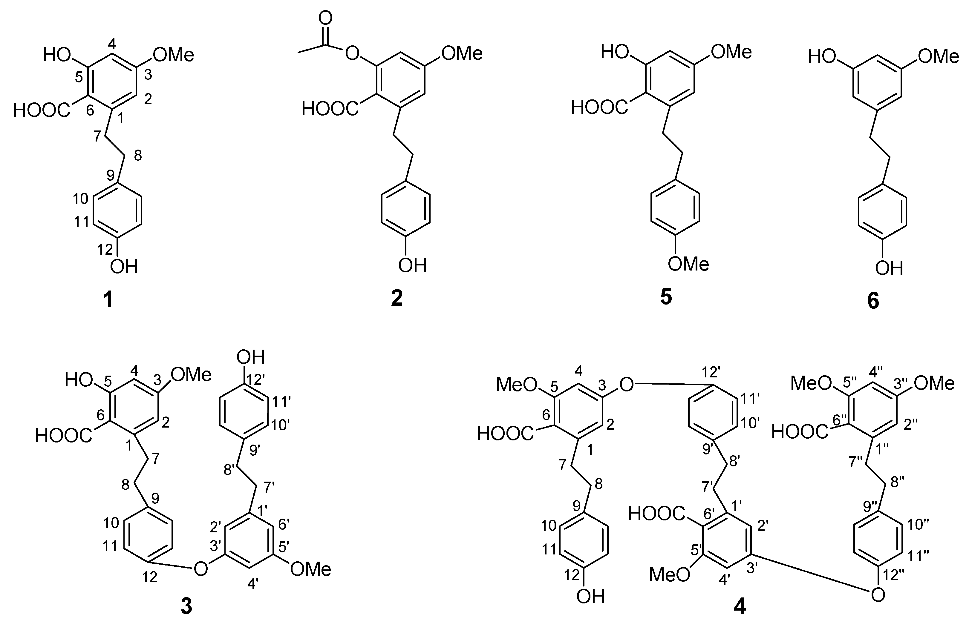
2.1. Structure elucidation of compounds 1-6
The ESIMS spectrum of
1 showed a peak at
m/z 311 corresponding to the [M+Na]
+ ion adduct and indicating the molecular formula C
16H
16O
5.
13C-NMR DEPT confirmed the presence of 16 carbon atoms (
Table 1 and
Table 2). The spectral data of compound
1 showed a close similarity to those of notholaenic acid [
3]. The NMR spectrum of compound
1 indicates that the molecule consists of one
p-disubstituted benzene ring (δ
H 6.82 d, 2H,
J = 8.5 Hz; δ
H 7.11 d, 2H,
J = 8.5 Hz) and one tetrasubstituted benzene ring (δ
H 6.32, 6.26 br s). In addition, the
1H-NMR signals indicated the presence of one -OMe group (δ
H 3.80) and a -CH
2-CH
2- bond (δ
H 3.16 and 2.79) (
Table 1). ESIMS/MS data further pointed to a carboxyl group, as a mass fragment
m/z 267 [(M+Na)-44]
+ was observed. The chemical shift of the -CH
2 groups suggest their position as a linkage between the two benzene rings, thus leading to a dihydrostilbene skeleton, which is in accordance with the UV spectrum and literature data for bibenzyls [
3,
12]. On the other hand, the base peak in the ESIMS, appearing at
m/z 107, can be explained by the fragment -CH
2-C
6H
4OH; and a peak at
m/z 181 shows the second stable portion of the original molecule. Thus, the second benzene ring thus bears the carboxyl group, one methoxyl group, as a well as an -OH group. The chemical shift of the two protons of this ring excludes their proximity to the carboxylic acid function. The relative position of substituents on the second benzene ring could be assigned by the observation of correlations in the HMBC and 1D-ROESY spectra [
12]. HMBC correlations were observed between -OMe and C-3, C-2 and C-4, between H-7 and C-2, C-6, C-9. The position of the -OH and -OMe groups could be assigned by 1D-ROESY spectra, correlation peaks were detected between the signals of H-7 and H-2 and between H-2 and -OMe. From the foregoing evidence, the structure of compound
1 was established as 5,12-dihydroxy-3-methoxy-dibenzyl-6-carboxylic acid.
Compound
2, C
18H
18O
6, showing in ESIMS spectrum an ion at
m/z 353 [M+Na]
+, had to be an acetyl derivative of compound
1 (δ
H 1.95, s, 3H; δ
C 172.0) according to
1H- and
13C-NMR spectra. The analysis of the
1H,
1H-COSY, HSQC and HMBC spectra allowed the assignment of all
1H- and
13C-NMR signals (
Table 1 and
Table 2) [
13,
14]. Thus, structure of compound
2 was established as 5-acetyloxy-12-hydroxy-3-methoxybibenzyl-6-carboxylic acid.
A molecular formula of C31H30O7 was determined for bisbibenzyl 3 by ESIMS, showing a pseudomolecular ion peak at m/z 537 [M+Na]+ and from the 13C- and 13C-DEPT NMR spectra which afforded a total 31 resonances corresponding to four sp3 methylenes, 13 sp2 methines and 11 quaternary carbons including six carbons bearing oxygen atoms and one carboxylic group.
In the
1H-NMR spectrum of
3, two 1,4-disubstituted benzene rings (a set of ring A signals: δ
H 6.83 and 7.14, and ring C signals: δ
H 6.81 and 7.09), one 1,3,5 trisubstituted ring B [δ
H 6.26, 6.24 and 6.18, br s] and one 1,3,5,6 tetrasubstituted ring D (δ
H 6.16 and 6.28) whose presence was reinforced by 1D TOCSY and COSY, HSQC, HMBC experiments were apparent (
Table 1 and
Table 2) [
15,
16]. The
1H-NMR spectrum of
3 also displayed the signals of four benzyl methylenes at δ
H 3.18, 3.20, 2.86, 2.82 corresponding to the
13C NMR signals at δ
C 38.0, 39.0, 38.6, 38.1 which are characteristic for bisbibenzyl derivatives. The arrangement of the substituents on the four benzene rings was established by HMBC experiments. Correlation peaks were observed between H-11’ and C-12’, C-9’ between H-11 and C-3’, C-12, C-9 between H-2 and C-7, C-6, C-3 between H-8 and C-10, C-1. From the foregoing evidence, the structure of compound
3 was established as 12-
O-[3’-(5’-methoxy-12’-hydroxy)-bibenzyl]-5-hydroxy-3-methoxybibenzyl-6-carboxylic acid.
The molecular formula C
49H
46 O
13 was assigned to compound
4 as shown by its ESIMS data ([M+ Na]
+ m/z 865) in combination with the
13C-NMR spectral data. Compound
4 showed protons and carbons for six benzene rings, three of them 1,4-disubstituited and three of them 1,3,5,6-tetrasubstituted [
17,
18]. The
1H- and
13C-NMR data of
4 showed the signals of a trisbibenzyl derivative, and results from the oxidative combination of three bisbenzyl units. Assignment of the
1H- and
13C-NMR signals for each unit could be easy achieved by comparison with data of the compound
1 (
Table 1 and
Table 2) and by 1D TOCSY, HSQC and HMBC data.
From this evidence compound
4 had to be a trimeric derivative of compound
1. The bonds between the three units were obtained from long range coupling of H-4 to C-3, C-6, C-2 respectively, between H-11’ and C-9’, C-10’, C-3 between H-2’ and C-4’, C-7’, C-6’ between H-11’’ and C-3’, C-9’’, C-12’’ (
Table 1 and
Table 2). Thus the structure of
4 was elucidated as 3-
O-{12’-[12’’-
O-(3’’,5’’-dimethoxy-6’’-carboxybibenzyl)]-5’-methoxy-6’-carboxybibenzyl}-12-hydroxy-5-methoxybibenzyl-6-carboxylic acid. The two known bibenzyl derivatives were identified as notholaenic acid (
5) [
3], and 3,12-dihydroxy-5-methoxybibenzyl (
6) [
19,
20], by detailed MS and NMR analyses and comparison with literature data.


 ,
, 

