Article

Screening of Agelasine D and Analogs for Inhibitory Activity against Pathogenic Protozoa; Identification of Hits for Visceral Leishmaniasis and Chagas Disease

1
Department of Chemistry, University of Oslo, P.O.Box 1033, Blindern, N-0315 Oslo, Norway
2
University of Antwerp, Laboratory of Microbiology, Parasitology and Hygiene, Faculty of Pharmaceutical, Biomedical and Veterinary Sciences, Universiteitsplein 1, B-2610 Antwerp, Belgium
3
Institute for Tropical Medicine, Nationalestraat 155, B-2000 Antwerp, Belgium
*
Author to whom correspondence should be addressed.
Molecules 2009, 14(1), 279-288; https://doi.org/10.3390/molecules14010279
Submission received: 3 December 2008 / Revised: 29 December 2008 / Accepted: 4 January 2009 / Published: 8 January 2009
(This article belongs to the Special Issue Neglected Diseases: Medicinal Chemistry and Natural Product Chemistry)

Abstract

:
There is an urgent need for novel and improved drugs against several tropical diseases caused by protozoa. The marine sponge (Agelas sp.) metabolite agelasine D, as well as other agelasine analogs and related structures were screened for inhibitory activity against Plasmodium falciparum, Leishmania infantum, Trypanosoma brucei and T. cruzi, as well as for toxicity against MRC-5 fibroblast cells. Many compounds displayed high general toxicity towards both the protozoa and MRC-5 cells. However, two compounds exhibited more selective inhibitory activity against L. infantum (IC50 <0.5 μg/mL) while two others displayed IC50 <1 μg/mL against T. cruzi in combination with relatively low toxicity against MRC-5 cells. According to criteria set up by the WHO Special Programme for Research & Training in Tropical Diseases (TDR), these compounds could be classified as hits for leishmaniasis and for Chagas disease, respectively. Identification of the hits as well as other SAR data from this initial screening will be valuable for design of more potent and selective potential drugs against these neglected tropical diseases.

Graphical Abstract

Introduction

Several so-called neglected diseases, e.g. illnesses that disproportionally affect poor and marginalized populations and for which satisfactory treatment is not available, partly due to lack of interest in drug development, are due to protozoal infections. Major killers in developing countries include malaria [1,2], visceral leishmaniasis or kala-azar [3,4], African sleeping sickness [4,5] and American sleeping sickness or Chagas disease [6]. Several factors limit the utility of existing drugs in areas were they are really needed, for instance high cost, poor compliance, drug resistance, low efficacy and toxicity [7]. Current drugs used to treat Trypanosoma infections are unsatisfactory with respect to safety [6] and an increasing number of malaria cases are caused by Plasmodium falciparum resistant to first-line drugs [1,2]. Hence, there is a constant need for development of novel antiprotozoal drugs, and one strategy in search for new hits and leads is screening of natural products, including those found in marine organisms [8].
Agelasines are isolated from marine sponges (Agelas sp) [9,10,11,12,13,14,15,16,17]. We have completed the first syntheses of agelasine D [18,19], agelasine E [20] and ent-agelasine F [21] as well as several synthetic analogs [18,19,20,22,23,24,20,22]. It has been shown that many of these compounds possess a broad spectrum of biological activities; including cytotoxicity towards cancer cell lines [19,22,23], antibacterial- [19,20,21,22,23], antifungal [23] and antifouling activities [25]. Recently, we also demonstrated that some agelasine analogs displayed antiprotozoal activity (Acanthamoeba castellanii and A. polyphaga) [22]. Hence we chose to screen agelasine D and some agelasine analogs and structurally related compounds for activity against pathogenic protozoa causing the above mentioned tropical neglected diseases. The structures of the compounds studied are shown in Figure 1. Compounds 8 and 9 may be regarded as analogs of agelasimines, another class of purine-containing natural products isolated from Agelas sp. [26,27].

Results and Discussion

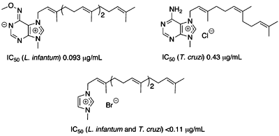
Antiprotozoal activities

The marine sponge metabolite agelasine D (2c) was screened for in vitro activity against the pathogenic protozoa P. falciparum, Leishmania infantum, Trypanosoma cruzi, and Trypanosoma brucei (Table 1). To assess selectivity of action, cytotoxicity against MRC-5 fibroblast cells was also evaluated. A higher activity was found for agelasine D (2c; IC50 0.29 μg/mL = 0.63 μM) against P. falciparum than what was recently reported for agelasines J (IC50 6.6 μM), K (IC50 8.3 μM), and L (IC50 18 μM) [17]. The selectivity index for antimalarial action [SI; IC50(MRC-5 fibroblast)/IC50(P. falciparum)] was 23, and agelasine D (2c) displayed significant inhibitory action also against the other parasites examined. These results encouraged us to examine the antiprotozoal activities of a number of agelasine analogs 19 in search for more potent and selective compounds. The results are presented in Table 1. Compounds with high cytotoxicity and related aspecificity across the different models were not titrated down to the exact IC50 as they may never become relevant hits for further follow-up.
Figure 1. Structures of the studied agelasine analogs 1-9.
Molecules 14 00279 g001
For an antimalaria hit, the WHO Special Programme for Research & Training in Tropical Diseases (TDR) defines an activity criterion to be IC50 <0.2 μg/mL with SI >20 [28]. Only compound 1d, with IC50 = 0.10 μg/mL and SI = 20, met this requirement. However, this compound also showed comparable levels of activity against the other parasite species, suggesting aspecific action. A few other analogues [1a1c, 1f, 1j, 1m1o, in addition to 2c (agelasine D)] displayed IC50 values against P. falciparum in the range 0.2 – 1.0 μg/mL, while compounds 39 were in general only marginally active.
Compound 1d was found to be a potent in vitro inhibitor of L. infantum with an IC50 value of 0.093 μg/mL and SI = 22. This compound meets the definition of a hit according to TDR (IC50 <0.5 μg/mL and SI >20) [28]. Compound 5 also classifies as a hit. We have previously shown that compound 1d displays a rather broad spectrum of antibacterial activities, incl. inhibition of Mycobacterium tuberculosis [19,23], whereas the imidazole 5 were reasonably active against Staphyllococcus aureus, but not against Escherichia coli and M. tuberculosis [23]. Compounds 1f and 1j appeared also as potent inhibitors of L. infantum, but unfortunately these compounds were equally toxic to MRC-5 cells. Compounds 34 and 69 were in general only weakly active against L. infantum.
Quite a few of the agelasine analogs inhibited T. cruzi growth, as they displayed IC50 values <1 μg/mL [28], but particularly compounds 1 also exhibited profound cytotoxicity towards MRC-5 cells, which is also the host cell in the T. cruzi model. Nevertheless, compounds 2a and 5, with SI >50, could be classified as hits according to the definition given above. Agelasine analog 2a is of special interest, since this compound, in contrast to the imidazole 5, displays low inhibitory activity against the other protozoa. In addition, previous studies have revealed the compound 2a is virtually inactive against bacteria (S. aureus, E. coli and M. tuberculosis) [23]. Moderate toxicity (SI = 10 – 30) and IC50 against T. cruzi <1 μg/mL were found also for compounds 1d, 3f and 9.
None of the compounds examined qualified as a hit with respect to T. b. brucei (IC50 <0.2 μg/mL and SI >100) [28]. The majority of active compounds were far too toxic. The most interesting results were found for compound 5 with IC50 <0.11 μg/mL and SI >69.

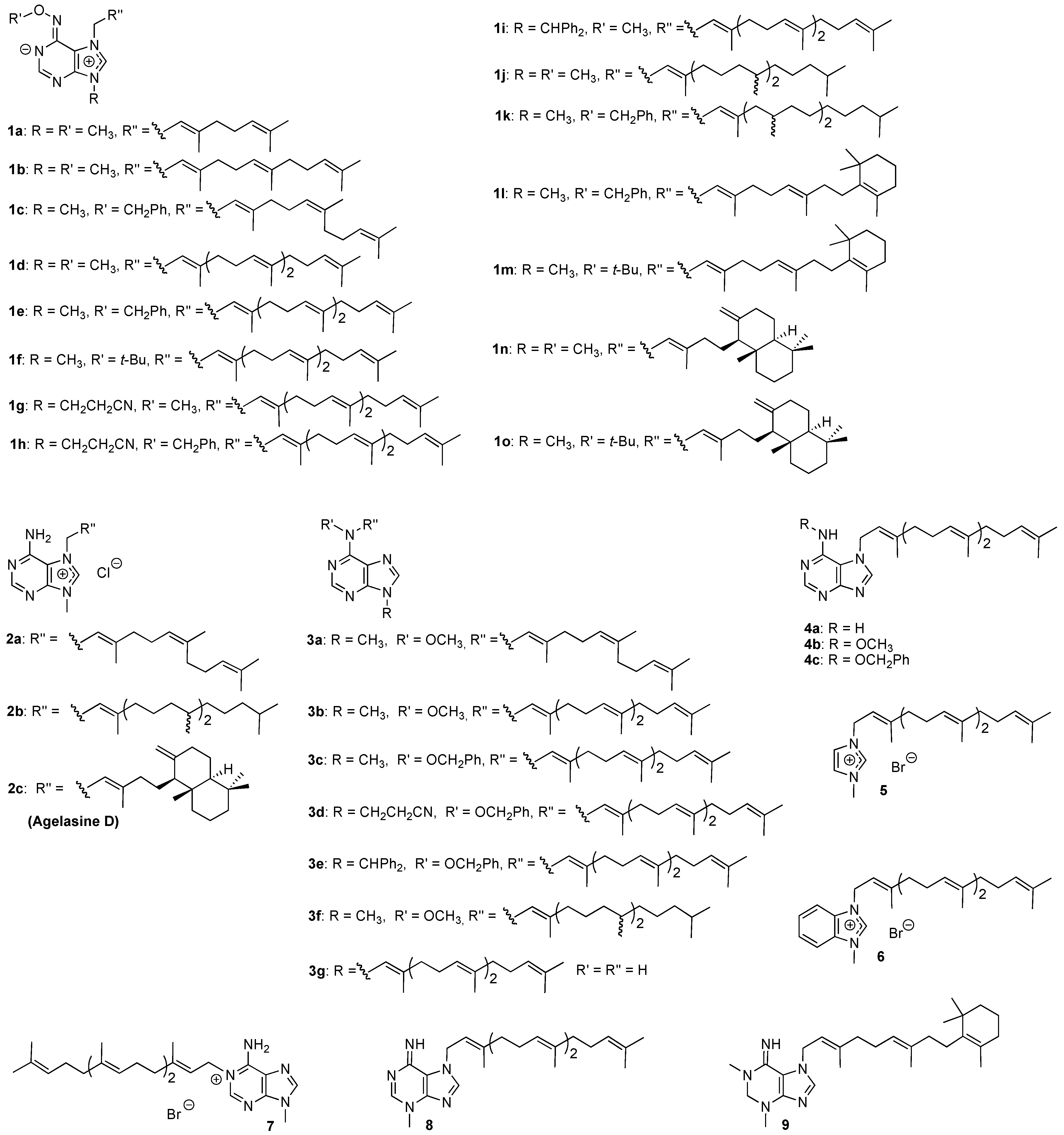
Structure – activity relationships

From the results in Table 1, it can be seen that type 1 compounds (Figure 1) generally exhibit a broad spectrum of antiprotozoal activities, but in an aspecific manner, since many of these compounds are equally toxic towards MRC-5 cells. Compounds with a monoterpene- (eg. 1a), or sesquiterpene (1b and 1c) derived side-chain in the purine 7-position appear to be more toxic, compared to some of the compounds with longer side-chains. Among the diterpenoids, a phytyl substituent (1j and 1k) and the β-cyclocitral derived side-chain found in 1l and 1m, also results in aspecific activity. The most interesting compounds in this class have a geranylgeranyl side-chain or the same diterpenoid substituent as found in agelasine D (2c). Compounds 1d and 1n, both carrying a methoxy group at N6 and a methyl group at N-9, were identified as antileishmanial hits. A trend seems to be that changing the N6 substituents from methoxy to a benzyloxy- (compounds 1c, 1e, 1h, 1k, and 1l) or tert-butoxy group (1f, 1m, and 1o) generally results in compounds with enhanced selectivity towards Trypanosoma sp., compared to the other parasites, but this modification does not result in significant reduction of MRC-5 cytotoxicity. Similar results were obtained when the N-9 substituent differed from methyl (1g1i).
Agelasine D and analogs 2 have a primary amino group in the purine 6-position, instead of the alkoxyamino group found in compounds 1. This generally results in reduced cytotoxicity and improved selectivity for antiprotozoal activity. Agelasine analog 2a is a quite selective hit with respect to inhibition of T. cruzi. Since only three compounds of this class (2a2c) were included in this initial screening, few other conclusions regarding SAR can be drawn at this point.
Table 1. Activity of compounds 19 against P. falciparum, L. infantum, T. cruzi and T. b. brucei, as well as MRC-5 fibroblast cells.
Compound No. aP. falciparumL. infantumT. cruziT. b. bruceiMRC-5
IC50μg/mLb(<0.2 μg/mL)cSId(>20)c IC50μg/mLe(<0.5 μg/mL)cSId(>20)cIC50μg/mf(<1.0 μg/mL)cSId(>50)cIC50 μg/mLg(<0.2 μg/mL)cSId(>100)cIC50 μg/mL
1a0.461.97.5<1<0.079>111.9<10.86
1b0.63<10.77<1<0.096>3.60.201.80.35
1c0.74<10.99<1<0.11>2.8<0.11>2.80.31
1d0.10 200.093220.11 180.23 8.72.0
1e4.2<15.4<11.42.30.29113.2
1f0.261.00.27<1<0.12>2.2<0.12>2.20.26
1g5.3<112<10.81<11.0<10.49
1h10<118<13.01.21.72.13.5
1i3.5<11.2<11.3<10.293.20.92
1j0.30<10.23<1<0.11>1.90.111.90.21
1k7.3<11.3<1<0.13>2.2<0.13>2.20.28
1l1.3<112<10.143.8<0.13>4.10.53
1m0.69<10.99<1<0.12>4<0.12>40.48
1n0.29 160.637.10.49 9.20.30154.5
1o0.94<14.0<1<0.12>3.8<0.12>3.80.45
2a2.9>9.0>26_0.43>6013>2>26
2b0.96<12.9<1<0.12>6.30.233.30.75
2c (Agelasine D)0.29 231.5 4.54.5 1.50.907.46.7
3a101.1>26<12.54.42.34.811
3b3.93.1>29<13.63.32.64.612
3c1.8<12.7<1<0.13>2.4<0.13>2.40.31
3d2.31.1>36<12.21.11.22.12.5
3e3.56>44<14.53.31.31215
3f>29<1>29<10.773.52.71.02.7
3g>26_9.8>2.70.97>272.9>9.0>26
4a9.4<1>26<12.31.13.5<12.6
4b>28<1>28<10.287.50.892.42.1
4c25<1>33<11.51.03.8<11.5
50.977.8<0.11>69<0.11>69<0.11>697.6
61.02.02.5>69<0.12>170.19112.0
7101.6>32>69111.54.13.916
83.37>6913>690.19120.633.72.3
9>28_>28_3.2>8.814>2>28
(a) The structures can be found in Figure 1; (b) Chloroquine 0.04 μg/mL; (c) Activity/safety criteria for an antiprotozoal hit according to TDR [28]; (d) SI = IC50(fibroblast)/IC50(parasite); (e) Miltefosine 0.24 μg/mL; (f) Benznidazole 0.25 μg/mL; (g) Melarsoprol 0.005 μg/mL.
Compounds 3a3e are neutral isomers of compounds 1. In general, they exhibit rather low antiprotozoal activities, with exception of compound 3c, which is a potent but non-selective inhibitor of Trypanosoma sp. 9-Geranylgeranyladenine 3f was a quite selective T. cruzi inhibitor with only moderate toxicity.
N-9 Dealkylation of compounds 1 and 2 results in derivatives with the general structure 4. This modification was particularly detrimental for antiplasmodial and antileishmania activity, while antitrypanosomal activities were also somewhat reduced and toxicity towards MRC-5 cells was virtually unchanged or even increased (cf. 4c with 1e).
The more interesting compound among the miscellaneous structures 59 is the imidazole derivative 5. Compared to the agelasine analogs 1d1f, the whole pyrimidine ring is removed. This modification results in a less toxic compound with a broad antiprotozoal spectrum. It is worth noting that the benzimidazole derivative 6 is more toxic (MRC-5) and less active against L. infantum compared to the corresponding imidazole 5.

Conclusions

Agelasine D and several agelasine analogs and related structures were screened for inhibitory activity against P. falciparum, L. infantum, T. b. brucei and T. cruzi, as well as for cytotoxicity on MRC-5 fibroblast cells. Many compounds displayed high general toxicity. Nevertheless, two compounds (1d and 5) were identified as hits for leishmaniasis and two (2a and 5) for Chagas disease. Identification of the hits as well as other SAR data from this initial screening will be valuable for design of more potent and selective potential drugs against these neglected tropical diseases.

Experimental

Compounds

All compounds studied were synthesized as described before; 1a, 1d, and 3b [20], 1b-1c, 1e-1k, 2a-2b, 3a, 3c-3g, 4-8 [23], 1l-1m, 9 [22], 1n-1o, 2c [19]. Stock solutions were prepared in 100% DMSO at 20 mg/mL.

Test plate production

The experiments were performed in 96-well plates (Greiner) at four-fold dilutions in a dose-titration range of 64 μg/mL to 0.25 μg/mL. Dilutions were carried out by a programmable precision robotic station (BIOMEK 2000, Beckman, USA). Each plate also contained medium-controls (blanks: 0% growth), infected untreated controls (negative control: 100% growth) and reference controls (positive control). Tests were run in duplicate in two independent experiments.

Biological screening tests

The integrated panel of microbial screens for the present study and the standard screening methodologies were adopted as have been described before [29]. Compounds with high cytotoxicity and related aspecificity across the different protozoa models were not titrated down to the exact IC50.

Antiplasmodial activity

The chloroquine-suceptible P. falciparum GHA-strain was used. Parasites were cultured in human erythrocytes A+ at 37 °C under a low oxygen atmosphere (3% O2, 4% CO2, and 93% N2) in a modular incubation chamber [30]. The culture medium was RPMI-1640, supplemented with 10% human serum. Two hundred microliters of infected human red blood cells suspension (1% parasitemia, 2% hematocrit) were added to each well of the plates with test compounds and incubated for 72 h. After incubation, test plates were frozen at -20 °C. Parasite multiplication was measured by the Malstat method [31]. One hundred microliters of Malstat reagent were transferred in a new plate and mixed with 20 μL of the hemolysed parasite suspension for 15 minutes at room temperature. After addition of 20 μL NBT/PES solution and 2 h incubation in the dark, the absorbance was spectrophotometrically read at 655 nm (Biorad 3550-UV microplate reader). Percentage growth inhibition was calculated compared to the negative blanks.

Antitrypanosomal activity

T. b. brucei: Trypomastigotes of T. b. brucei Squib-427 strain (suramin-sensitive) were cultured at 37 °C and 5% CO2 in Hirumi-9 medium [32], supplemented with 10% fetal calf serum (FCS). Assays were performed by adding 1.5×10⁴ trypomastigotes/well. After 72 h incubation, parasite growth was assessed fluorimetrically by adding resazurin [33] for 24 h at 37 °C. Fluorescence was measured using a GENios Tecan fluorimeter (excitation 530 nm, emission 590 nm).
T. cruzi: Tulahuen CL2 strain (nifurtimox-sensitive) was maintained on MRC-5 cells in minimal essential medium (MEM) supplemented with 20 mM L-Glutamine, 16.5 mM sodium hydrogen carbonate and FCS (5%) at 37 °C and 5% CO2. To determine in vitro anti-trypanosomal activity, 4×10³ MRC-5 cells and 4×10⁴ parasites were added to each well of test plate with compound. After incubation at 37 °C for 7 days, parasite growth was assessed by adding of β-galactosidase substrate, chlorophenol red β-D-galactopyranoside [34] for 4 h at 37 °C. The color reaction was read at 540 nm and absorbance values were expressed as a percentage of the blank controls.

Antileishmanial activity

Leishmania infantum amastigotes (MHOM/ET 67) were collected from an infected donor hamster and used to infect primary peritoneal mouse macrophages. To determine in vitro antileishmanial activity, 3×10⁴ macrophages were seeded in each well of a 96-well plate. After 48 h outgrowth, 5×10⁴ amastigotes/well were added and incubated for 2 h at 37 °C. Pre-diluted compounds were subsequently added and the plates were further incubated for 120 h at 37 °C and 5% CO2. Parasite burdens were determined microscopically after Giemsa staining and expressed as a percentage of the blank controls without compound.

Cytotoxicity assay

MRC-5 SV2 cells, human fetal lung fibroblast, were cultivated in MEM, supplemented with L-glutamine (20 mM), 16.5 mM sodium hydrogen carbonate and 5% FCS at 37 °C and 5% CO2. For the assay, 104 MRC-5 cells/well were seeded onto the test plates containing the pre-diluted compounds and incubated at 37 °C and 5% CO2 for 72 h. Cell viability was determined after addition of resazurin.

Acknowledgements

The Norwegian Research Council is greatly acknowledged for scholarships to A.V. and A.P. The screens were performed in a centre receiving financial support from the UNDP/World Bank/WHO Special Programme for Research and Training in Tropical Diseases (TDR).

References and Notes

  1. Gelb, M.H. Drug discovery for malaria: a very challenging and timely endeavor. Curr. Opin. Chem. Biol. 2007, 11, 440–445. [Google Scholar] [CrossRef]
  2. Tuteja, R. Malaria – an overview. FEBS J. 2007, 274, 4670–4679. [Google Scholar] [CrossRef]
  3. Croft, S.L.; Barrett, M.P.; Urbina, J.A. Chemotherapy of trypanosomiases and leishmaniasis. Trends Parasitol. 2005, 21, 508–512. [Google Scholar] [CrossRef]
  4. Chappuis, F.; Sundar, S.; Hailu, A.; Ghalib, H.; Rijal, S.; Peeling, R.W.; Alvar, J.; Boerlaert, M. Visceral leishmaniasis: what are the needs for diagnosis, treatment and control? Nature. Rev. Microbiol. 2007, 5, S7–S16. [Google Scholar] [CrossRef]
  5. Barrett, M.P.; Boykin, D.W.; Brun, R.; Tidwell, R.R. Human African trypanosomiasis: pharmacological re-engagement with a neglected disease. J. Pharmacol. 2007, 152, 1155–1171. [Google Scholar]
  6. Teixeira, A.R.L.; Nitz, N.; Giumaro, M.C.; Gomes, C.; Santos-Buch, C.A. Chagas disease. Postgrad. Med. J. 2006, 82, 788–798. [Google Scholar] [CrossRef]
  7. Nwaka, S.; Hudson, A. Innovative lead discovery strategies for tropical diseases. Nature Rev. Drug. Discov. 2006, 5, 941–955. [Google Scholar] [CrossRef]
  8. Plubrukarn, A. Marine invertebrates: a diverted approach towards the treatment of tropical diseases. Recent. Prog. Med. Plants 2006, 11, 439–461. [Google Scholar]
  9. Capon, R.J.; Faulkner, D.J. Antimicrobial metabolites from a Pacific sponge, Agelas sp. J. Am. Chem. Soc. 1984, 106, 1819–1822. [Google Scholar] [CrossRef]
  10. Nakamura, H.; Wu, H.; Ohizumi, Y.; Hirata, Y. Agelasine-A –B, -C and –D, novel bicyclic diterpenoids with a 9-methyladeninium unit possessing inhibitory effects on Na,K-ATPase isolated from the Okinawian sea sponge Agelas sp. Tetrahedron Lett. 1984, 25, 2989–2992. [Google Scholar] [CrossRef]
  11. Wu, H.; Nakamura, H.; Kobayashi, J.; Ohizumi, Y. Agelasine-E and –F, novel monocyclic diterpenoids with a 9-methyladeninium unit possessing inhibitory effects on Na,K-ATPase isolated from the Okinawian sea sponge Agelas nakamurai Hoshino. Tetrahedron Lett. 1984, 25, 3719–3722. [Google Scholar] [CrossRef]
  12. Wu, H.; Nakamura, H.; Kobayashi, J.; Kobayashi, M.; Ohizumi, Y.; Hirata, Y. Structures of agelasines, diterpenes having a 9-methyladeninium chromophore isolated from the Okinavian marine sponge Agelas nakamurai Hoshino. Bull. Chem. Soc. Jpn. 1986, 59, 2495–2504. [Google Scholar] [CrossRef]
  13. Ishida, K.; Ishibashi, M.; Shigemori, H.; Sasaki, T.; Kobayashi, J. Agelasine G, a new antileukemic alkaloid from the Okinavian marina sponge Agelas sp. Chem. Pharm. Bull. 1992, 40, 766–767. [Google Scholar] [CrossRef]
  14. Hattori, T.; Adachi, K.; Shizuri, Y. New agelasine compound from the marine sponge Agelas mauretania as an antifouling substance against macroalgae. J. Nat. Prod. 1997, 60, 411–413. [Google Scholar] [CrossRef]
  15. Fu, X.; Schmitz, F.J.; Tanner, R.S.; Kelly-Borges, M. Agelasines H and I, 9-methyladenine-containing diterpenoids from Agelas sponge. J. Nat. Prod. 1998, 61, 548–550. [Google Scholar] [CrossRef]
  16. Iwagawa, T.; Kaneko, M.; Okamura, H.; Nakatani, M.; van Soest, R.W.M. New alkaloids from the Papua New Guinean sponge Agelas nakamurai. J. Nat. Prod. 1998, 61, 1310–1312. [Google Scholar] [CrossRef]
  17. Appenzeller, J.; Michi, G.; Martin, M.-T.; Gallard, J.-F.; Menou, J.-L.; Boury-Esnault, N.; Hooper, J.; Petek, S.; Chevalley, S.; Valentin, A.; Zaparucha, A.; Al-Mourabit, A.; Debitus, C. Agelasines J, K, and L from the Solomon Islands marine sponge Agelas cf. mauritiana. J. Nat. Prod. 2008, 71, 1451–1454. [Google Scholar] [CrossRef]
  18. Utenova, B.T.; Gundersen, L.-L. Synthesis of (+)-agelasine D from (+)-manool. Tetrahedron Lett. 2004, 45, 4233–4235. [Google Scholar] [CrossRef]
  19. Vik, A.; Hedner, E.; Charnock, C.; Samuelsen, Ø.; Larsson, R.; Gundersen, L.-L.; Bohlin., L. (+)-Agelasine D: improved synthesis and evaluation of antibacterial and cytotoxic activities. J. Nat. Prod. 2006, 69, 381–386. [Google Scholar] [CrossRef]
  20. Bakkestuen, A.K.; Gundersen, L.-L.; Petersen, D.; Utenova, B.T.; Vik, A. Synthesis and antimycobacterial activity of agelasine E and analogs. Org. Biol. Chem. 2005, 3, 1025–1033. [Google Scholar] [CrossRef]
  21. Proszenyák, Á; Brændvang, M.; Charnock, C.; Gundersen, L.-L. The first synthesis of ent-Agelasine F. Tetrahedron 2009, 65, 194–199. [Google Scholar] [CrossRef]
  22. Proszenyák, Á; Charnock, C.; Hedner, E.; Larsson, R.; Bohlin, L.; Gundersen, L.-L. Synthesis and antimicrobial and antineoplastic activities of agelasine and agelasimine analogs with a β-cyclocitral derived substituent. Arch. Pharm. Chem. Life Sci. 2007, 340, 625–634. [Google Scholar] [CrossRef]
  23. Vik, A.; Hedner, E.; Charnock, C.; Tangen, L.W.; Samuelsen, Ø.; Larsson, R.; Bohlin., L.; Gundersen, L.-L. Antimicrobial and cytotoxic activity of agelasine and agelasimine analogs. Bioorg. Med. Chem. 2007, 15, 4016–4037. [Google Scholar] [CrossRef]
  24. Roggen, H.; Gundersen, L.-L. Synthetic studies directed towards agelasine analogs. Synthesis, tautomerism, and alkylation of 2-substituted N-methoxy-9-methyl-9H-purin-6-amines. Eur. J. Org. Chem. 2008, 5099–5106. [Google Scholar] [CrossRef]
  25. Sjögren, M.; Dahlström, M.; Hedner, E.; Jonsson, P.R.; Vik, A.; Gundersen, L.-L.; Bohlin, L. Antifouling activity of the sponge metabolite agelasine D and synthesized analogs on Balanus improvisus. Biofouling 2008, 24, 251–258. [Google Scholar] [CrossRef]
  26. Fathi-Afshar, R.; Allen, T.M. Biological active metabolites from Agelas mauretania. Can. J. Chem. 1988, 66, 45–50. [Google Scholar] [CrossRef]
  27. Fathi-Afshar, R.; Allen, T.M. Some pharmacological activities of novel adenine-related compounds isolated from a marine sponge Agelas mauretania. Can. J. Physiol. Pharmacol. 1989, 67, 276–281. [Google Scholar] [CrossRef]
  28. TDR, Lead discovery for drugs, Business Plan 2008-2013. 2007; BL 3.
  29. Cos, P.; Vlietinck, A.J.; Berghe, D.V.; Maes, L. Anti-infective potential of natural products: How to develop a stronger in vitro ‘proof-of-concept’. J. Ethnopharmacol. 2006, 106, 290–302. [Google Scholar] [CrossRef]
  30. Trager, W.; Jensen, J.B. Human malaria parasites in continuous culture. Science 1976, 193, 673–675. [Google Scholar]
  31. Makler, M.T.; Ries, J.M.; Williams, J.A.; Bancroft, J.E.; Piper, R.C.; Gibbins, B.L.; Hinrichs, D.J. Parasite lactate dehydrogenase as an assay for Plasmodium falciparum drug sensitivity. Am. J. Trop. Med. Hyg. 1993, 48, 739–741. [Google Scholar]
  32. Hirumi, H.; Hirumi, K. Continuous cultivation of Trypanosoma brucei blood stream forms in a medium containing a low concentration of serum protein without feeder cell layers. J. Parasitol. 1989, 75, 985–989. [Google Scholar] [CrossRef]
  33. Raz, B.; Iten, M.; Grether-Buhler, Y.; Kaminsky, R.; Brun, R. The Alamar Blue asssay to determine drug sensitivity of African trypanosomes (T. b. rhodesiense, T. b. gambiense) in vitro. Acta Trop. 1997, 68, 139–147. [Google Scholar] [CrossRef]
  34. Buckner, F.S.; Verlinde, C.L.; La Flamme, A.C.; Van Voorhis, W.C. Efficient technique for screening drugs for activity against Trypanosoma cruzi using parasites expressing beta-galactosidase. Antimicrob. Agents Chemother. 1996, 40, 2592–2597. [Google Scholar]
  • Sample Availability: Some samples are available from the corresponding author.

