Article

A New Method for the Determination of Nucleic Acid Using an Eu3+– nicotinic Acid Complex as a Resonance Light Scattering Probe

1
Microscale Science Institute, Department of Chemistry, Weifang University, Weifang 261061, P.R. China
2
Key Laboratory of Colloid and Interface Chemistry (Shandong University), Ministry of Education, School of Chemistry and Chemical Engineering, Shandong University, Jinan 250100, P.R. China
*
Author to whom correspondence should be addressed.
Molecules 2009, 14(1), 10-18; https://doi.org/10.3390/molecules14010010
Submission received: 24 November 2008 / Revised: 13 December 2008 / Accepted: 16 December 2008 / Published: 23 December 2008
(This article belongs to the Special Issue Nucleic Acids)

Abstract

:
This study found that in Tris-HCl buffer, the resonance light scattering (RLS) intensity of the Eu3+-nicotinic acid system can be greatly enhanced by nucleic acids and the enhanced intensity is proportional to the concentration of nucleic acid in the range of 7×10⁻⁸-1×10⁻⁵ g∙mL-1 for fsDNA, and its detection limit is 2×10⁻⁸ g∙mL-1. Based on this, a new method for the determination of nucleic acids is proposed. Synthetic and actual samples are determined satisfactorily. The interaction mechanism is also studied. It is thought that nucleic acid can bind with the Eu3+-nicotinic acid complex through electrostatic attraction and thus form a large Eu3+-nicotinic acid-nucleic acid complex.

Introduction

Nucleic acids have an important function in life processes, so they are often used as a reference for measurements of other components in biological samples. However, direct use of the intrinsic fluorescence and ultraviolet absorption of nucleic acids for their determination has been severely limited by low sensitivity [1]. As a result some probes based on the interactions between nucleic acids and extrinsic reagents have been employed in the determination of nucleic acids using spectral methods [2]. Among these spectral methods, the resonance light scattering (RLS) technique has seen strong interest among chemists and biochemists since Pasternack et al.’s pioneering work [3] using a common spectrofluorometer. Later on, Huang et al. established a new method to determine the nucleic acid content by the RLS technique [4,5]. This method has been used extensively in recent years. Up to now, most of the probes applied in the determination of nucleic acids are organic dyes [2,6,7,8,9,10,11,12,13], thanks to their aggregation on DNA, whereas the use of metal ion complexes as RLS probes is rare [14,15]. To the best of our knowledge, there are no reports on the study of nucleic acids using the Eu3+-nicotinic acid complex as an RLS probe. In this study, it was found that nucleic acids could enhance the RLS intensity of Eu3+-nicotinic acid. Based on this, a new method for the determination of nucleic acid is proposed. The interaction mechanism is also discussed.

Results and Discussion

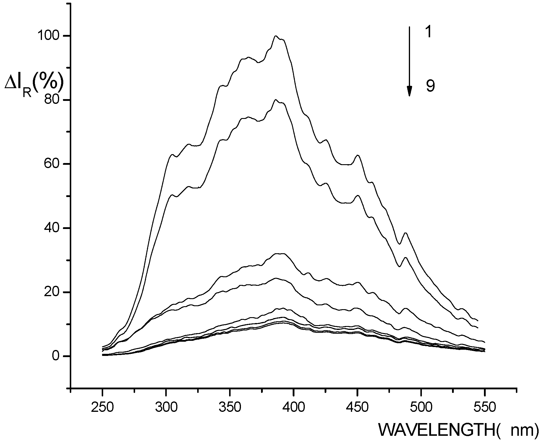
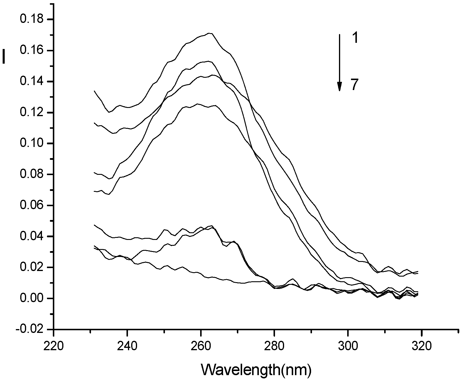
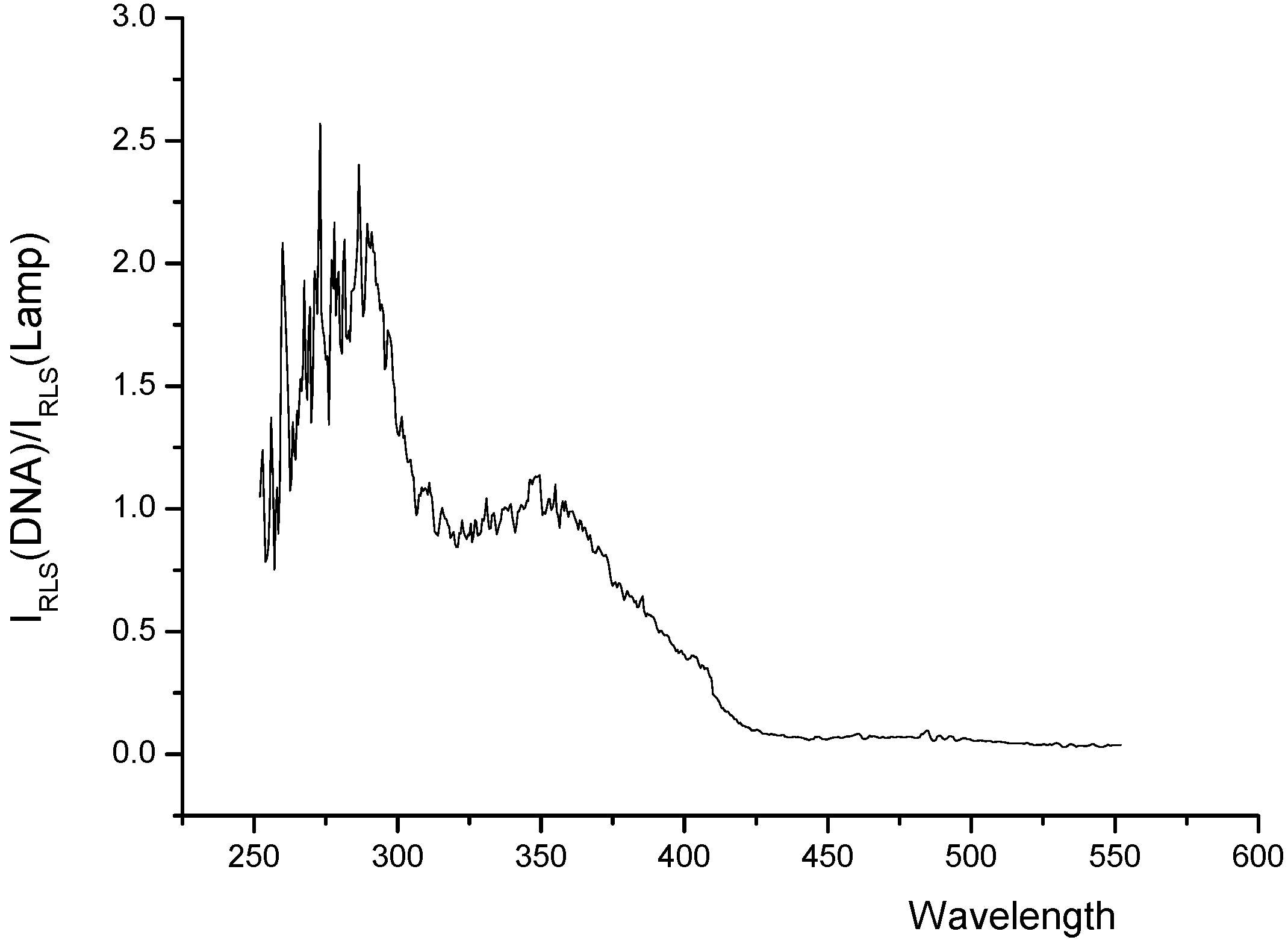
RLS spectra of the systems

The apparent RLS spectra of Eu3+–nicotinic acid-fsDNA (1), Eu3+–nicotinic acid-yRNA (2), Eu3+–nicotinic acid (3), Eu3+ (4), nicotinic acid (5), fsDNA (6), yRNA (7), nicotinic acid- fsDNA (8) and nicotinic acid- yRNA (9) systems are shown in Figure 1. It can be seen that in Tris–HCl buffer (pH 8.1), all the systems have the same RLS peak at about 387 nm. But the RLS intensity of the Eu3+–nicotinic acid system can be enhanced by nucleic acids, which indicates that there is the interaction between nucleic acid and Eu3+–nicotinic acid complex.
Figure 1. Apparent RLS profiles of Eu3+-nicotinic acid with DNA and RNA.
Molecules 14 00010 g001
(1) Eu3+-nicotinic acid-fsDNA; (2) Eu3+-nicotinic acid-yRNA; (3) Eu3+-nicotinic acid; (4) Eu3+; (5) nicotinic acid; (6) fsDNA; (7) yRNA; (8) nicotinic acid-fsDNA; (9) nicotinic acid- yRNA. Conditions used were nicotinic acid: 5.0×10⁻⁷ M; Eu3+: 2.8×10⁻⁴ M; Tris-HCl (0.05 M): pH=8.1; nucleic acids (fsDNA, yRNA): 1.0×10⁻⁶ g∙mL-1.

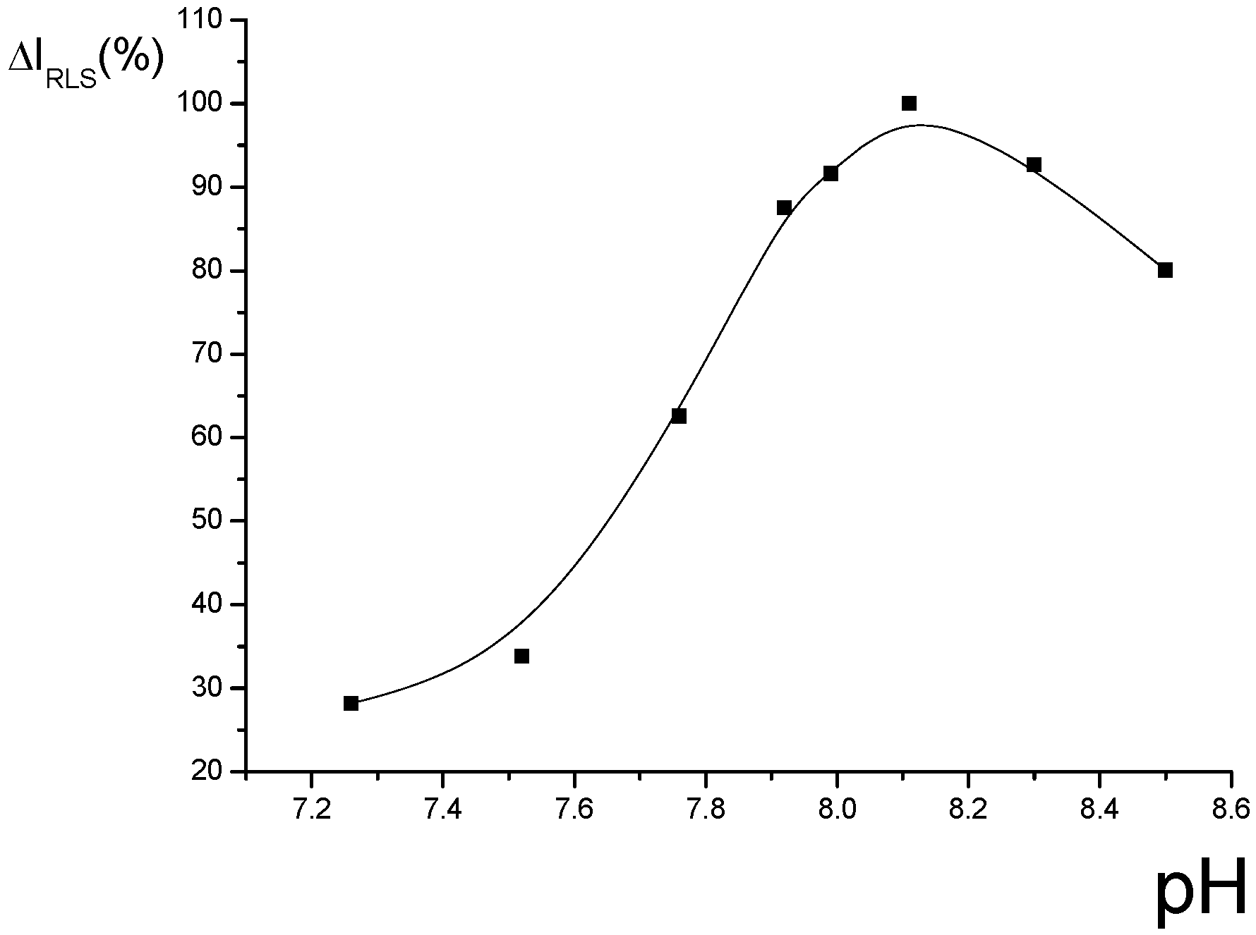
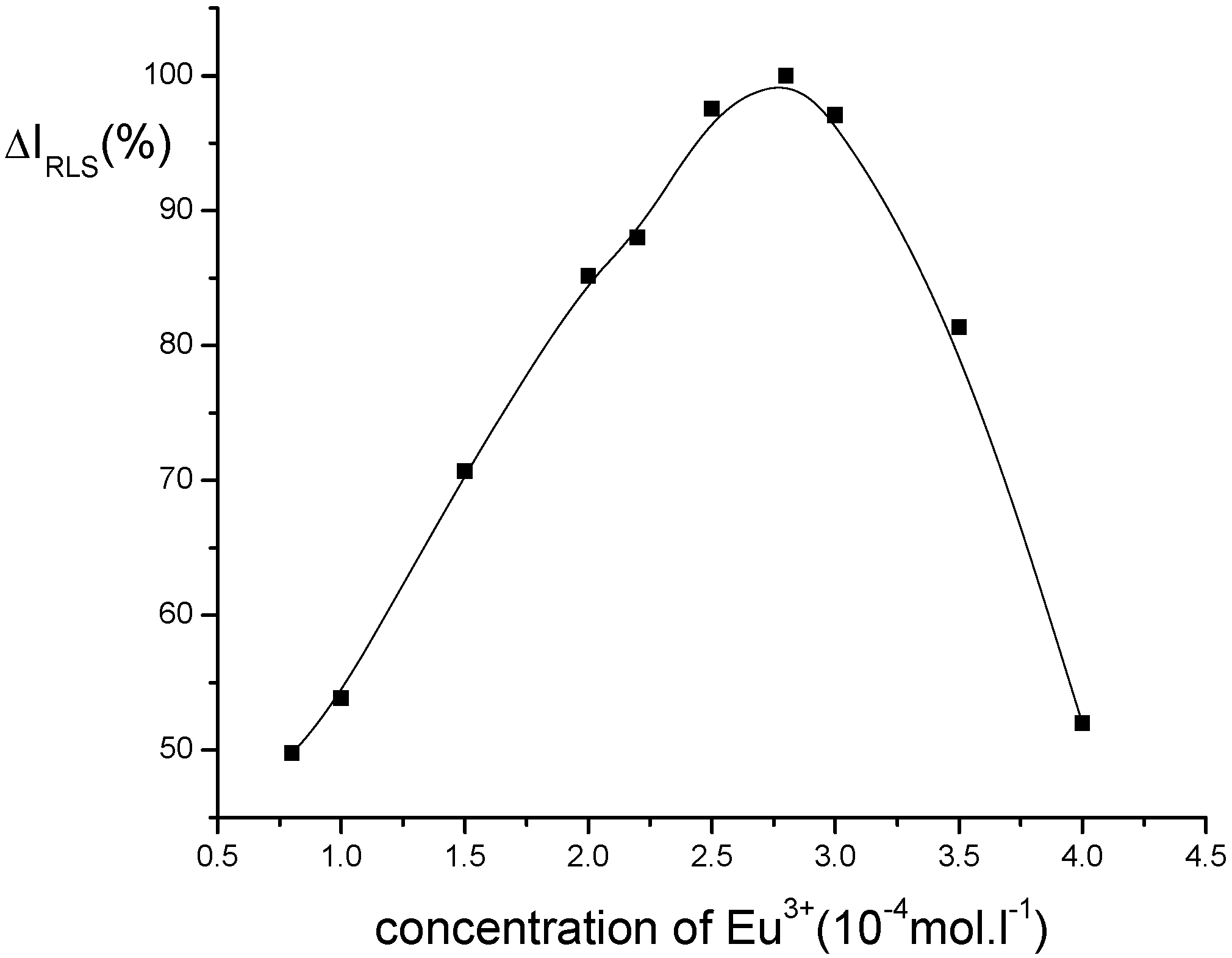
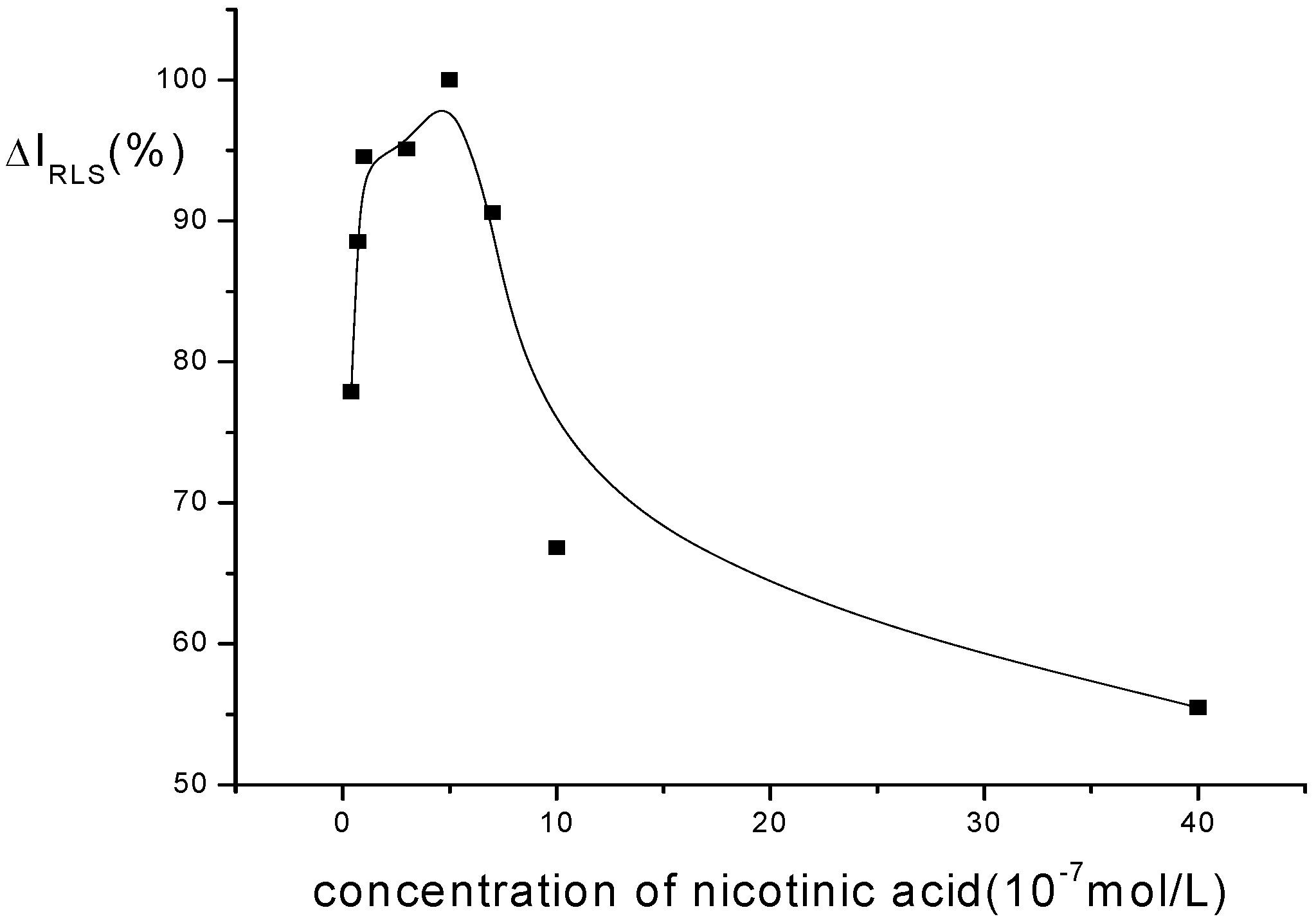
Optimization of conditions for RLS measurements

The effects of pH and buffer solution: The effect of pH on the ΔIR of the system was tested and is shown in Figure 2. The results indicate that the ΔIR reaches a maximum when the pH is about 8.1, so this value was fixed for further research. The effect of the buffers on the ΔIR of this system are shown in Table 1. The results indicate that different buffers also have large effect on the ΔIR of the system, and 0.55 ml of Tris-HCl ( 0.05 M, pH=8.1) is the most suitable buffer.
Figure 2. Effect of pH on RLS intensity of Eu3+-nicotinic acid-fsDNA.
Molecules 14 00010 g002
Conditions: nicotinic acid: 5.0×10⁻⁷ M; Eu3+ : 2.8×10⁻⁴ M; fsDNA: 1.0×10⁻⁶ g∙mL-1.
Table 1. The choice of buffer solutions.
BuffersTris-HClHMTA*-HClNaH2PO4-K2HPO4 NH4Cl-NH3 NaH2PO4-Citric acid
ΔIR(%)10050.912.845.9 49.3
* HMTA- Hexamethylene tetramine; Conditions used were pH: 8.10; nicotinic acid: 5.0×10⁻⁷ M; fsDNA: 1.0×10⁻⁶ g∙mL-1; Eu3+: 2.8×10⁻⁴ M.
The choice of rare earth ions and the effect of Eu3+ concentration: Different rare earth ions can influence the RLS intensity of the system, and the results are shown in Table 2. It is indicated that Eu3+ is suitable for this system. The effect of Eu3+ concentration on the ΔIR of this system is shown in Figure 3. It can be seen that the ΔIR is the strongest when the concentration of Eu3+ is 2.8×10⁻⁴ M. Therefore, 2.8×10⁻⁴ M Eu3+ was chosen for this research.
Table 2. Effect of different rare earth ions on RLS intensity of Eu3+-nicotinic acid-fsDNA.
M3+Eu3+Tb3+Y3+Nd3+Al3+Dy3+Gd3+
IR(%)10046.499.054.666.668.265.0
Conditions: Tris-HCl (0.05 M): pH=8.1; nicotinic acid: 5.0×10⁻⁷ M; fsDNA: 1.0×10⁻⁶ g.mL-1; M3+: 2.8×10⁻⁴ M.
Figure 3. Effect of Eu3+ concentration on RLS intensity of Eu3+-nicotinic acid-fsDNA.
Molecules 14 00010 g003
Conditions: nicotinic acid: 5.0×10⁻⁷ M; Tris-HCl(0.05 M): pH=8.1; fsDNA: 1.0×10⁻⁶ g∙mL-1.
The effect of nicotinic acid concentration: The effect of nicotinic acid concentration on the ΔIR is shown in Figure 4. It can be seen that the ΔIR of the system reaches the maximum when the concentration of nicotinic acid is 5.0×10⁻⁷ M. Thus, 5.0×10⁻⁷ M nicotinic acid is chosen in this research.
Figure 4. Effect of nicotinic acid concentration on RLS intensity of Eu3+-nicotinic acid-fsDNA.
Molecules 14 00010 g004
Conditions: Eu3+: 2.8×10⁻⁴ M; Tris-HCl(0.05 M): pH=8.1; fsDNA: 1.0×10⁻⁶ g∙mL-1.
Addition order and stability of this system: The effect of the addition order on the ΔIR was tested. The result indicates that the order of nicotinic acid, fsDNA, Eu3+ and Tris-HCl buffer is the best. Under the optimum condition, the effect of time on the fluorescence intensity is studied. The result shows that the ΔIR of the system reaches a maximum within 15 min after all the reagents are added and remains stable for 3 h.
Effect of foreign substances: The interference of foreign substances is tested for 1.0×10⁻⁶ g∙mL-1 fsDNA. The results show that within ±5% relative error, 92-fold of NaCl, 60-fold of KCl and CuCl2, 35-fold of MgCl2 do not interfere with to the determination of fsDNA. Fourteen-fold ZnSO4, 7-fold CaCl2, Al(NO3)3, dextrose do not interfere with the determination of fsDNA. Two-fold FeCl3, citric acid is an obvious interferent in the determination of fsDNA.

Analytical application

Calibration curve and detection limit: Under the optimum conditions defined here, the calibration graph for nucleic acid was obtained. The result indicates that there is a linear relationship between the ΔIR of this system and the concentration of fsDNA in the range from 7.0×10⁻⁸-1.0×10⁻⁵ g∙mL-1, and the detection limit (S/N=3) is 2.0×10⁻⁸ g∙mL-1. A comparison with other RLS probes is shown in Table 3. Although the sensitivity of this probe for the determination of nucleic acid is not high, it has lower toxicity.
Table 3. Different probes of nucleic acid by RLS.
ProbeNucleic acidDetection limit (10-9g∙mL-1)References
TAPP*1ctDNA/fsDNA/yRNA4.1/4.6/6.7[4]
Safranine TctDNA/fsDNA/yRNA13.2/39.8/61.1[5]
Neutral RedctDNA/fsDNA/yRNA48.2/35.2/205[6]
BCB*2ctDNA/fsDNA/yRNA118/112/434[7]
Methyl GreenctDNA/fsDNA/yRNA7.8/2.6/9.9[9]
Azur ActDNA/fsDNA19.9/12.6[11]
Crystal VioletctDNA/fsDNA/yRNA13.8/36.8/69[8]
Methyl VioletDNA100[10]
Congo RedfsDNA/ctDNA/yRNA0.019/ 0.89/1.2[12]
Methyl greenfsDNA2.6/7.8/9.9[13]
OA*3–Eu3+fsDNA/ctDNA/yRNA0.02/0.011/0.01[14]
Eu3+-TTA-PhenfsDNA/yRNA/ctDNA0.03/0.006/0.002[15]
1. TAPP: a,b,g,d-tetrakis [4-(trimethylammoniumyl) phenyl] porphine; 2. BCB: brilliant cresol blue; 3. OA: oxolinic acid.
Sample determination: Considering the effects of foreign substances on the RLS intensity of the system, the standard addition method was used for the determination of DNA in synthetic and cucumber samples. The synthetic sample contained fsDNA (1.0×10⁻⁶ g/mL), Na+ (4.0×10⁻⁶ g/mL), K+ (2.0×10⁻⁶ g/mL), Mg+ (2.0×10⁻⁶ g/mL) and Cl- (1.0×10⁻⁵ g/mL). The result indicates that the recovery ratio and relative standard deviation of this method are 97.3% and 4.7%, respectively. At the same time, cucumber samples were also determined. The recovery ratio was 107.8% and the relative standard deviation is 4.7%. So the accuracy and precision of this method are satisfactory.
Interaction mechanism: The light-scattering spectrum not only depends on the nature of the system, but also reflects the characteristics of the instrument [16]. In order to eliminate the effect of the instrument on the light scattering spectrum, the corrected light-scattering spectra of the systems are recorded by using the correction technique [17] and is shown in Figure 5. It can be seen from this figure that the scattering peak of the Eu3+-nicotinic acid-fsDNA system at about 260 nm is located within the absorption bands of both nicotinic acid and fsDNA in the range of 240–280 nm (Figure 6). Therefore, the light scattering of the studied system is ascribed to the resonance light scattering [3], and is considered to be due to the synergetic resonance caused by the absorption of both nicotinic acid and fsDNA.
Figure 5. The corrected RLS profiles of Eu3+-nicotinic acid-fsDNA system.
Molecules 14 00010 g005
Lamp and Eu3+-nicotinic acid-fsRNA. Conditions used were nicotinic acid: 5.0×10⁻⁷ M; Eu3+ : 2.8×10⁻⁴ M; Tris-HCl (0.05 M): pH=8.1; nucleic acids (fsDNA, yRNA): 1.0×10⁻⁶ g∙mL-1.
Figure 6. Absorption spectra.
Molecules 14 00010 g006
(1) nicotinic acid-Eu3+-fsDNA; (2) nicotinic acid-fsDNA; (3) Eu3+-fsDNA; (4) fsDNA; (5) nicotinic acid-Eu3+; (6) nicotinic acid; (7) Eu3+. Conditions used were nicotinic acid: 5.0×10⁻⁷ M; Eu3+: 2.8×10⁻⁴ M; Tris-HCl (0.05 M): pH=8.1; nucleic acids (fsDNA): 1.0×10⁻⁶ g∙mL-1.
Figure 1 also shows that when Eu3+ is added to nicotinic acid system, the RLS intensity of the system is enhanced. This indicates that there is interaction between Eu3+ and nicotinic acid, and a complex forms. After the addition of nucleic acids, the RLS intensity of Eu3+ complex is greatly enhanced, which indicates that there is a further interaction between fsDNA and the Eu3+ complex forming a larger complex, resulting in the enhancement of the RLS intensity of the system. It is considered that Eu3+ complex is positively charged, and can bind with phosphate groups in the nucleic acids through electrostatic force and thus form a larger Eu3+-nicotinic acid-fsDNA complex.

Conclusions

We have found that nucleic acids could greatly enhance the RLS intensity of Eu3+-nicotinic acid. The enhanced intensity is in proportion to the concentration of fsDNA in the range of 7×10⁻⁸-1×10⁻⁵ g∙mL-1, with a detection limit of 2×10⁻⁸g∙mL-1. The synthetic and actual samples are satisfactorily determined. The interaction mechanism is also studied. It is considered that nucleic acid can bind with the complex of Eu3+-nicotinic acid through electrostatic attraction and form a large Eu3+-nicotinic acid-nucleic acid complex. The study is helpful for us to explore the interaction mechanism between metal ion complex and nucleic acid.

Experimental

General

Chemicals: Stock solutions of RNA (1.0×10⁻⁴ g∙ml-1) and DNA (1.0×10⁻⁴ g∙ml-1) were prepared by dissolving commercial yeast RNA (yRNA) (Institution Biochemistry, Chinese Academy of Sciences) and fish sperm DNA (fsDNA, Sigma Co., USA) in 0.05 M sodium chloride solution. These stocks needed to be stored at 0-4 o C. The working solution was prepared by diluting the stock solution to the proper concentration. The purity of DNA was checked by measuring the ratio of the absorbance of 260 nm to that of 280 nm. Stock standard solution of Eu3+ (2.0 x 10⁻³ M) was prepared by dissolving 0.3520 g of the corresponding europium oxide (99.99 %, Shanghai Yuelong Chemical Plant, P.R. China) in hydrochloric acid (10 mL) and then diluting to 100 mL with water. Stock standard solution (1.0 x 10⁻³ M) of nicotinic acid (Sinopharm Chemical Reagent Co., Ltd, P.R. China) was made by dissolving nicotinic acid (0.0308 g) in water (250 mL). A 0.05 M Tris–HCl buffer solution was prepared by dissolving appropriate Tris in water and adjusting the pH with HCl. All the chemicals used were of analytical reagents grade and doubly deionized water was used throughout.
DNA extraction: The extraction process of DNA in cucumber is as follows: after the cucumber sample is carefully ground in liquid nitrogen, a 0.2000 g sample is accurately weighed and dissolved in cell extraction solution (600 μL), then incubated in a water-bath at 65 °C for 20 min. The same volume of CHCl3 is then added and mixed well, and the mixture then centrifuged for 10 min at 12,000 rpm and the supernatant taken. This is repeated until no protein can be seen. The supernants are combined and the same volume of isopropyl alcohol is added and the DNA is then precipitated in an ice-bath for 30 min and centrifuged for 10 min at 12,000 rpm. The supernatant is discarded before washing the DNA with ethanol (70%). The extracted DNA is dissolved in 0.05 M sodium chloride solution (1.5 mL) and stored at 0-4°C.
Apparatus: All RLS spectra were recorded on a LS-55 spectrofluorimeter (PE) in a 1 cm quartz cell. The ultraviolet absorption spectra were performed with a UV-4100 spectrophotometer (Hitachi, Japan) equipped with 1 cm quartz cells. All pH measurements were made with a Delta 320-s pH meter (Mettler Toledo, Shanghai).
Analytical procedure: To a dry 10 mL test-tube, solutions were added as the following order: nicotinic acid (0.5 mL), definite standard nucleic acid (or sample solution), Eu3+ (1.4 mL, 2.00×10⁻³ M) and Tris-HCl buffer (pH 8.1, 0.55 mL). The mixture was diluted to 10.0 mL with water and allowed to stand for 15 min. All RLS spectra were obtained by scanning simultaneously the excitation and emission monochromators (namely, λ = 0 nm) from 270 to 580 nm. The intensity of RLS was measured at λ = 387.0 nm in a 1.00 cm quartz fluorescence cell with slit width of 10 nm for both the excitation and emission. The enhanced RLS intensity of the Eu3+–nicotinic acid system by nucleic acids is represented as ΔIR = IR - IR0. Here IR and IR0 are the RLS intensity of the system with and without nucleic acids, respectively.

Acknowledgements

This work was supported by Natural Science Foundations of China (20575035) and Shandong Province (Y2003B02), and by Visiting Scholar Foundation of Key Lab in the University.

References and Notes

  1. Udenfriend, S.; Zaltzman, P. Fluorescence characteristics of purines pyrimines and their derives. Anal. Biochem. 1962, 3, 49–59. [Google Scholar] [CrossRef]
  2. Wang, Y.B.; Yang, J.H.; Wu, X.; Li, L.; Sun, S.N.; Su, B.Y. Progress of the Spectral Probe for Nucleic Acids. Anal. Lett. 2003, 36, 2063–2094. [Google Scholar] [CrossRef]
  3. Pasternack, R.F.; Bustamante, C.; Collings, P.J.; Giannetteo, A.; Gibbs, E.J. Porphyrin assemblies on DNA as studied by a resonance light-scattering technique. J. Am. Chem. Soc. 1993, 115, 5393–5399. [Google Scholar] [CrossRef]
  4. Huang, C.Z.; Li, K.A.; Tong, S.Y. Determination of nucleic acids by a resonance light-scattering technique with a,b,g,d-tetrakis [4-(trimethylammoniumyl) phenyl] porphine. Anal. Chem. 1996, 68, 2259–2263. [Google Scholar] [CrossRef]
  5. Huang, C.Z.; Li, Y.F.; Liu, X.D. Determination of nucleic acids at nanogram levels with safranine T by a resonance light-scattering technique. Anal. Chim. Acta 1998, 375, 89–97. [Google Scholar] [CrossRef]
  6. Wang, Y.T.; Zhao, F.L.; Li, K.A.; Tong, S.Y. Molecular spectroscopic study of DNA binding with neutral red and application to assay of nucleic acids. Anal. Chim. Acta 1999, 396, 75–81. [Google Scholar] [CrossRef]
  7. Wang, Y.T.; Zhao, F.L.; Li, K.A.; Tong, S.Y. Molecular spectroscopy study of the reaction of nucleic acids with brilliant cresol blue. Spectrochim. Acta. Part A. 2000, 56, 1827–1833. [Google Scholar] [CrossRef]
  8. Zhang, W.J.; Xu, H.P.; Wu, S.Q.; Chen, X.G.; Hu, Z.D. Determination of nucleic acids with crystal violet. Analyst 2001, 126, 513–517. [Google Scholar] [CrossRef]
  9. Liu, R.T.; Yang, J.H.; Wu, X.; Sun, C.X. Simple and sensitive determination of nucleic acid by Rayleigh light scattering technique with methyl green-CTMAB. Indian J. Chem. 2001, 40A, 1121–1123. [Google Scholar]
  10. Liu, Y.; Ma, C.Q.; Li, K.A.; Xie, F.C.; Tong, S.Y. Rayleigh light scattering study on the reaction of nucleic acids and methyl violet. Anal. Biochem. 1999, 268, 187–192. [Google Scholar] [CrossRef]
  11. Li, Y.F.; Huang, C.Z.; Huang, X.H.; Li, M. Determination of DNA by its enhancement effect of resonance light scattering by azur A. Anal. Chim. Acta 2001, 429, 311–319. [Google Scholar] [CrossRef]
  12. Wu, X.; Wang, Y.B.; Yang, J.H.; Sun, S.N. Determination of nucleic acids at nanogram level using resonance light scattering technique with Congo Red. Spectrochim. Acta 2005, 61A, 361–366. [Google Scholar]
  13. Liu, R.T.; Yang, J.H.; Wu, X.; Sun, C.X. Simple and sensitive determination of nucleic acid by Rayleigh light scattering technique with methyl green-CTMAB. Indian J. Chem. 2001, 40A, 1121–1123. [Google Scholar]
  14. Wu, X.; Sun, S.N.; Yang, J.H.; Wang, M,Q.; Liu, L.Y.; Guo, C.Y. Study on the interaction between nucleic acid and Eu3+-oxolinic acid and the determination of nucleic acid using the Resonance Light Scattering technique. Spectrochim. Acta 2005, 62A, 896–901. [Google Scholar]
  15. Jia, Z.; Yang, J.H.; Wu, X.; Sun, C.X.; Liu, S.F.; Wang, F. The sensitive determination of nucleic acids using resonance light scattering quenching method. Spectrochim. Acta Part A. 2006, 64A, 555–559. [Google Scholar]
  16. Collins, F.S.; Patrinos, A.; Jordan, E.; Chakravi, A.; Gesteland, R.; Walters, L. New goals for the U.S. Human Genome Project. Science 1998, 282, 682. [Google Scholar]
  17. McPhie, P. Resonance light scattering profiles must be corrected for instrument performance. Anal. Biochem. 2006, 348, 157–159. [Google Scholar] [CrossRef]
  • Sample Availability: Samples of the synthetic and cucumber are available from authors.

