14 pages, 1418 KB  
Review
Reactive Oxygen Species in Plants: From Source to Sink
by Sheikh Mansoor, Owais Ali Wani, Jafar K. Lone, Sweeta Manhas, Navneet Kour, Pravej Alam, Ajaz Ahmad and Parvaiz Ahmad
Antioxidants 2022, 11(2), 225; https://doi.org/10.3390/antiox11020225 - 25 Jan 2022
Cited by 545 | Viewed by 23315
Abstract
Reactive oxygen species (ROS, partial reduction or derivatives of free radicals) are highly reactive, dangerous and can cause oxidative cell death. In addition to their role as toxic by-products of aerobic metabolism, ROS play a role in the control and regulation of biological [...] Read more.
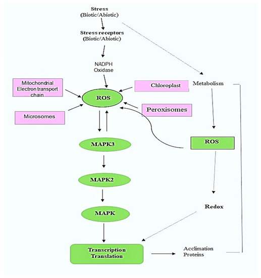
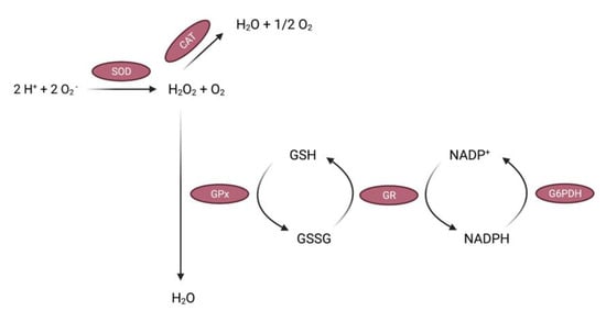
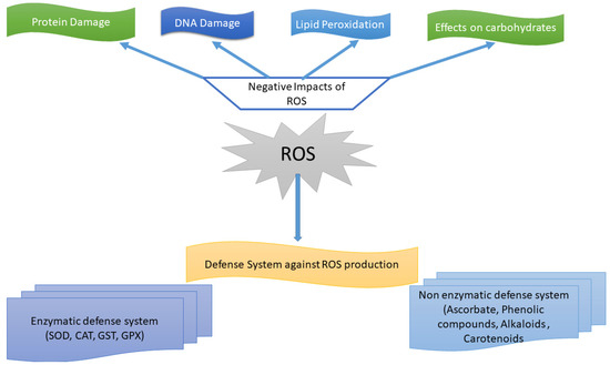
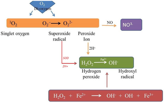
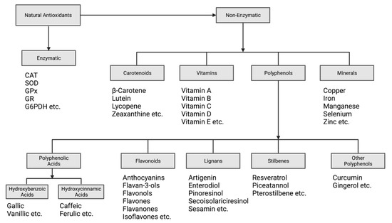
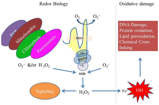
Reactive oxygen species (ROS, partial reduction or derivatives of free radicals) are highly reactive, dangerous and can cause oxidative cell death. In addition to their role as toxic by-products of aerobic metabolism, ROS play a role in the control and regulation of biological processes such as growth, the cell cycle, programmed cell death, hormone signaling, biotic and abiotic stress reactions and development. ROS always arise in plants as a by-product of several metabolic processes that are located in different cell compartments, or as a result of the inevitable escape of electrons to oxygen from the electron transport activities of chloroplasts, mitochondria and plasma membranes. These reactive species are formed in chloroplasts, mitochondria, plasma membranes, peroxisomes, apoplasts, the endoplasmic reticulum and cell walls. The action of many non-enzymatic and enzymatic antioxidants present in tissues is required for efficient scavenging of ROS generated during various environmental stressors. The current review provides an in-depth look at the fate of ROS in plants, a beneficial role in managing stress and other irregularities. The production sites are also explained with their negative effects. In addition, the biochemical properties and sources of ROS generation, capture systems, the influence of ROS on cell biochemistry and the crosstalk of ROS with other signaling molecules/pathways are discussed. Full article
Show Figures

Figure 1
9 pages, 1714 KB  
Article
On the Scavenging Ability of Scutellarein against the OOH Radical in Water and Lipid-like Environments: A Theoretical Study
by Maciej Spiegel, Tiziana Marino, Mario Prejanò and Nino Russo
Antioxidants 2022, 11(2), 224; https://doi.org/10.3390/antiox11020224 - 25 Jan 2022
Cited by 27 | Viewed by 4180
Abstract
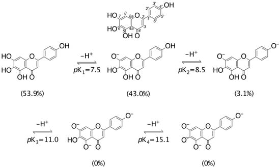
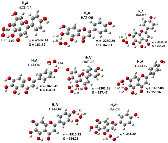
The antioxidant capability of scutellarein, a flavonoid extracted from different plants of the Scutellaria family, was computationally predicted by considering its reaction with the OOH radical in both lipid-like and water environments. The pKa and equilibrium behavior in the aqueous phase were also [...] Read more.
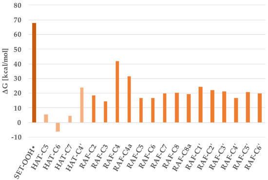
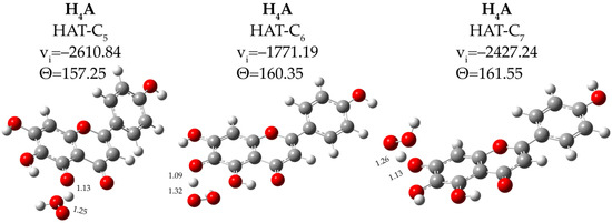
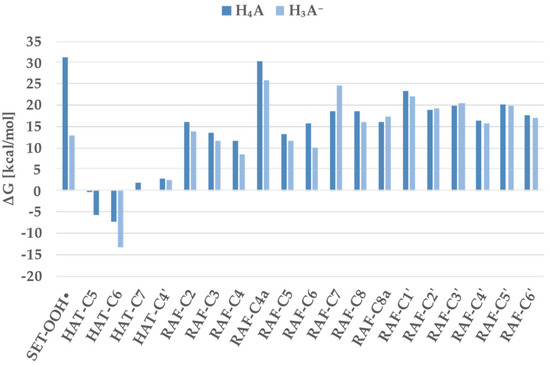
The antioxidant capability of scutellarein, a flavonoid extracted from different plants of the Scutellaria family, was computationally predicted by considering its reaction with the OOH radical in both lipid-like and water environments. The pKa and equilibrium behavior in the aqueous phase were also calculated. Different reaction mechanisms involving the most populated species were considered. The work was performed by using the density functional level of theory. The individual, total, and fraction-corrected total rate constants were obtained. The results show that scutellarein has scavenging power against the hydroperoxyl radical similar to that of Trolox, which is generally used as a reference antioxidant. Full article
(This article belongs to the Section ROS, RNS and RSS)
Show Figures

Figure 1
16 pages, 6977 KB  
Article
Broadening the Biocatalytic Toolbox—Screening and Expression of New Unspecific Peroxygenases
by Sebastian Bormann, Harald Kellner, Johanna Hermes, Robert Herzog, René Ullrich, Christiane Liers, Roland Ulber, Martin Hofrichter and Dirk Holtmann
Antioxidants 2022, 11(2), 223; https://doi.org/10.3390/antiox11020223 - 24 Jan 2022
Cited by 24 | Viewed by 7245
Abstract
Unspecific peroxygenases (UPOs) catalyze the selective transfer of single oxygen atoms from peroxides to a broad range of substrates such as un-activated hydrocarbons. Since specific oxyfunctionalizations are among the most-desired reactions in synthetic chemistry, UPOs are of high industrial interest. To broaden the [...] Read more.
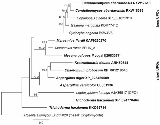
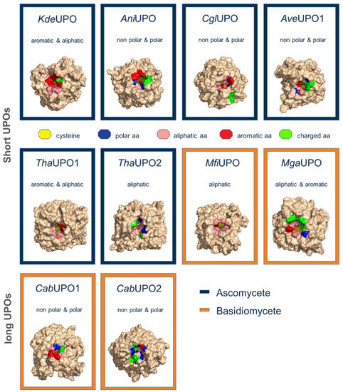
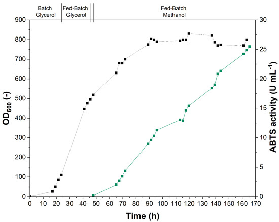
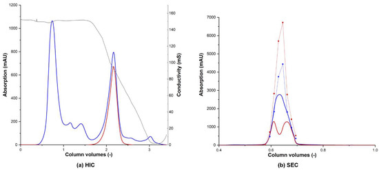
Unspecific peroxygenases (UPOs) catalyze the selective transfer of single oxygen atoms from peroxides to a broad range of substrates such as un-activated hydrocarbons. Since specific oxyfunctionalizations are among the most-desired reactions in synthetic chemistry, UPOs are of high industrial interest. To broaden the number of available enzymes, computational and experimental methods were combined in this study. After a comparative alignment and homology modelling, the enzymes were expressed directly in P. pastoris. Out of ten initially selected sequences, three enzymes (one from Aspergillus niger and two from Candolleomyces aberdarensis) were actively expressed. Cultivation of respective expression clones in a bioreactor led to production titers of up to 300 mg L−1. Enzymes were purified to near homogeneity and characterized regarding their specific activities and pH-optima for typical UPO substrates. This work demonstrated that directed evolution is not necessarily required to produce UPOs in P. pastoris at respective titers. The heterologous producibility of these three UPOs will expand the toolbox of available enzymes and help to advance their synthetic application. Full article
(This article belongs to the Special Issue Dream Peroxygenases)
Show Figures

Figure 1
28 pages, 5669 KB  
Article
Calycosin Alleviates Paraquat-Induced Neurodegeneration by Improving Mitochondrial Functions and Regulating Autophagy in a Drosophila Model of Parkinson’s Disease
by Hitesh Singh Chaouhan, Xin Li, Kuo-Ting Sun, I-Kuan Wang, Tung-Min Yu, Shao-Hua Yu, Kuen-Bao Chen, Wei-Yong Lin and Chi-Yuan Li
Antioxidants 2022, 11(2), 222; https://doi.org/10.3390/antiox11020222 - 24 Jan 2022
Cited by 41 | Viewed by 7399
Abstract
Parkinson’s disease (PD) is the second most common age-related neurodegenerative disorder with limited clinical treatments. The occurrence of PD includes both genetic and environmental toxins, such as the pesticides paraquat (PQ), as major contributors to PD pathology in both invertebrate and mammalian models. [...] Read more.
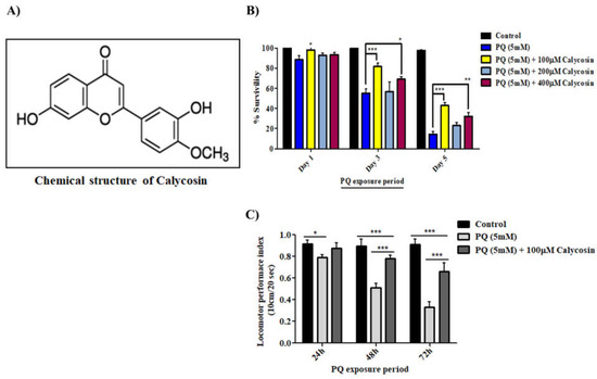
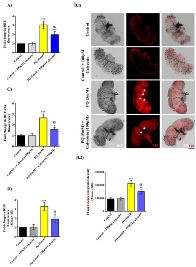
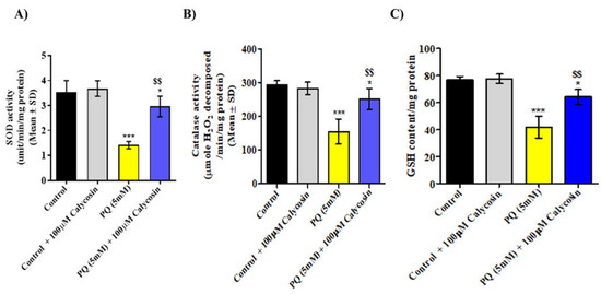
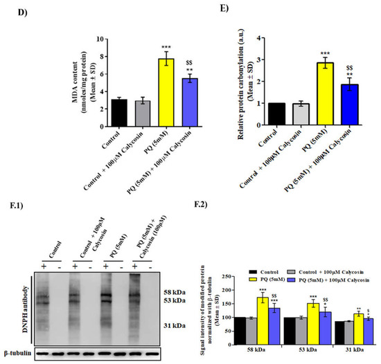
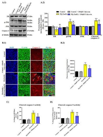
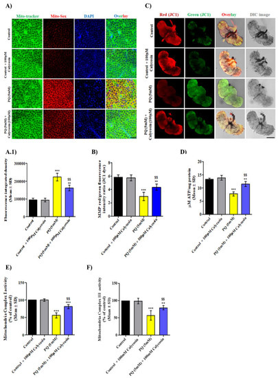
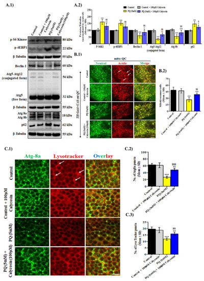
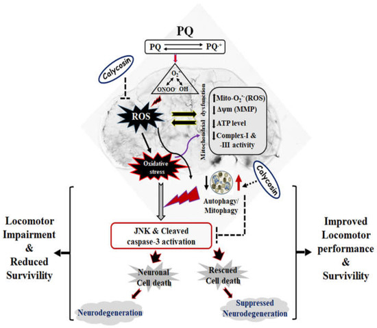
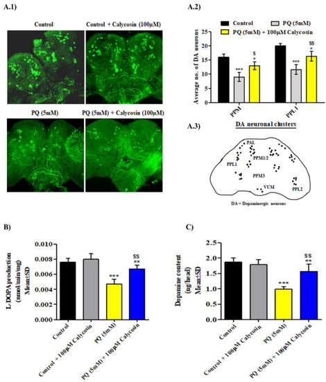
Parkinson’s disease (PD) is the second most common age-related neurodegenerative disorder with limited clinical treatments. The occurrence of PD includes both genetic and environmental toxins, such as the pesticides paraquat (PQ), as major contributors to PD pathology in both invertebrate and mammalian models. Calycosin, an isoflavone phytoestrogen, has multiple pharmacological properties, including neuroprotective activity. However, the paucity of information regarding the neuroprotective potential of calycosin on PQ-induced neurodegeneration led us to explore whether calycosin can mitigate PD-like phenotypes and the underlying molecular mechanisms. We used a PQ-induced PD model in Drosophila as a cost-effective in vivo screening platform to investigate the neuroprotective efficacy of natural compounds on PD. We reported that calycosin shows a protective role in preventing dopaminergic (DA) neuronal cell death in PQ-exposed Canton S flies. Calycosin-fed PQ-exposed flies exhibit significant resistance against PQ-induced mortality and locomotor deficits in terms of reduced oxidative stress, loss of DA neurons, the depletion of dopamine content, and phosphorylated JNK-caspase-3 levels. Additionally, mechanistic studies show that calycosin administration improves PQ-induced mitochondrial dysfunction and stimulates mitophagy and general autophagy with reduced pS6K and p4EBP1 levels, suggestive of a maintained energy balance between anabolic and catabolic processes, resulting in the inhibition of neuronal cell death. Collectively, this study substantiates the protective effect of calycosin against PQ-induced neurodegeneration by improving DA neurons’ survival and reducing apoptosis, likely via autophagy induction, and it is implicated as a novel therapeutic application against toxin-induced PD pathogenesis. Full article
(This article belongs to the Special Issue Antioxidants in Human Health and Disease)
Show Figures

Figure 1
19 pages, 3352 KB  
Article
Hyperoside and Quercitrin in Houttuynia cordata Extract Attenuate UVB-Induced Human Keratinocyte Cell Damage and Oxidative Stress via Modulation of MAPKs and Akt Signaling Pathway
by Nattakan Charachit, Amonnat Sukhamwang, Pornngarm Dejkriengkraikul and Supachai Yodkeeree
Antioxidants 2022, 11(2), 221; https://doi.org/10.3390/antiox11020221 - 24 Jan 2022
Cited by 44 | Viewed by 8538
Abstract
Ultraviolet radiation is a major environmental harmful factor on human skin. In this paper, we investigate the potential mechanism of Houttuynia cordata extract on UVB-induced HaCaT keratinocyte cell death and inflammation. We found that Houttuynia cordata ethyl acetate extract fraction (HC-EA) protected against [...] Read more.
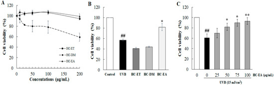
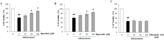
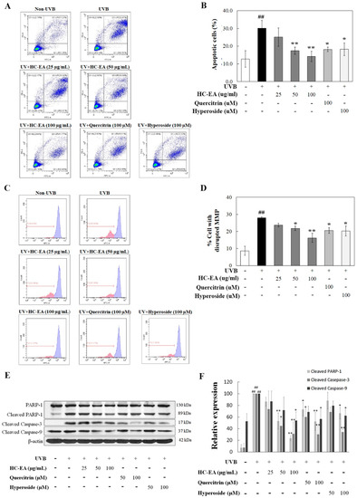
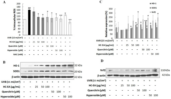
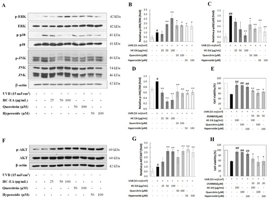
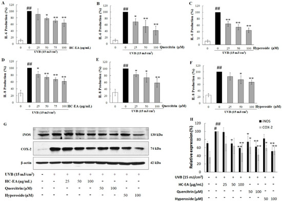
Ultraviolet radiation is a major environmental harmful factor on human skin. In this paper, we investigate the potential mechanism of Houttuynia cordata extract on UVB-induced HaCaT keratinocyte cell death and inflammation. We found that Houttuynia cordata ethyl acetate extract fraction (HC-EA) protected against UVB-induced cell damage. The HPLC results indicate that quercitrin and hyperoside are the major polyphenolics in HC-EA and are responsible for providing protection against UVB-induced cell death. These responses were associated with the regulation of caspase-9 and caspase-3 activation, which rescued HaCaT cells from UVB-induced apoptosis. In addition, HC-EA, quercitrin, and hyperoside attenuated UVB-induced inflammatory mediators, including IL-6, IL-8, COX-2, and iNOS. Furthermore, the treatment of cells with HC-EA and its active compounds abolished intracellular ROS and increased levels of heme oxygenase-1 and superoxide dismutase. UVB-induced ROS production mediated Akt and mitogen activated protein kinases (MAPKs) pathways, including p38, ERK, and JNK. Our results show HC-EA, quercitrin, and hyperoside decreased UVB-induced p38 and JNK phosphorylation, while increasing ERK and Akt phosphorylation. MAPKs and Akt mediated cell survival and death were confirmed by specific inhibitors to Akt and MAPKs. Thus, HC-EA, which contains quercitrin and hyperoside, protected keratinocyte from UVB-induced oxidative damage and inflammation through the modulation of MAPKs and Akt signaling. Full article
Show Figures

Figure 1
15 pages, 3241 KB  
Article
Structure–Activity Relationships and Transcriptomic Analysis of Hypoxia-Inducible Factor Prolyl Hydroxylase Inhibitors
by Andrey A. Poloznikov, Sergey V. Nikulin, Dmitry M. Hushpulian, Anna Yu. Khristichenko, Andrey I. Osipyants, Andrey F. Asachenko, Olga V. Shurupova, Svyatoslav S. Savin, Sue H. Lee, Irina N. Gaisina, Gregory R. J. Thatcher, Anthony Narciso, Eric P. Chang, Sergey V. Kazakov, Nancy Krucher, Vladimir I. Tishkov, Bobby Thomas and Irina G. Gazaryan
Antioxidants 2022, 11(2), 220; https://doi.org/10.3390/antiox11020220 - 24 Jan 2022
Cited by 7 | Viewed by 6508
Abstract
To evaluate the differences in action of commercially available 2-oxoglutarate mimetics and “branched-tail” oxyquinoline inhibitors of hypoxia-inducible factor prolyl hydroxylase (HIF PHD), the inhibitors’ IC50 values in the activation of HIF1 ODD-luciferase reporter were selected for comparative transcriptomics. Structure–activity relationship and computer [...] Read more.
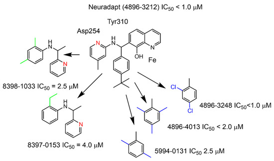
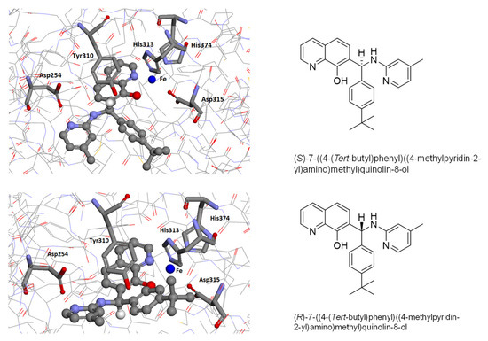
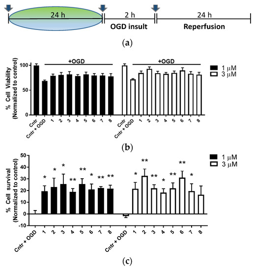
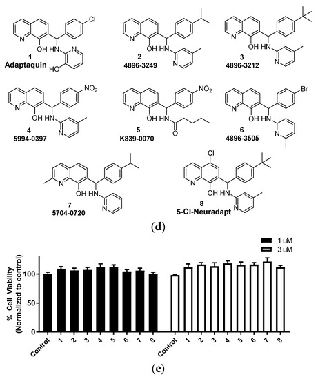
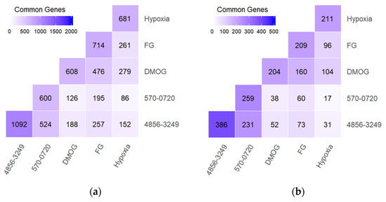
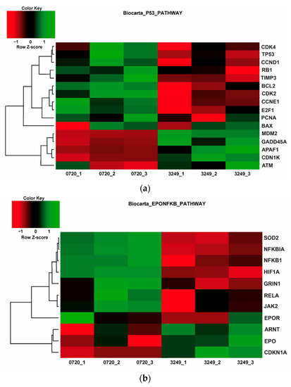
To evaluate the differences in action of commercially available 2-oxoglutarate mimetics and “branched-tail” oxyquinoline inhibitors of hypoxia-inducible factor prolyl hydroxylase (HIF PHD), the inhibitors’ IC50 values in the activation of HIF1 ODD-luciferase reporter were selected for comparative transcriptomics. Structure–activity relationship and computer modeling for the oxyquinoline series of inhibitors led to the identification of novel inhibitors, which were an order of magnitude more active in the reporter assay than roxadustat and vadadustat. Unexpectedly, 2-methyl-substitution in the oxyquinoline core of the best HIF PHD inhibitor was found to be active in the reporter assay and almost equally effective in the pretreatment paradigm of the oxygen-glucose deprivation in vitro model. Comparative transcriptomic analysis of the signaling pathways induced by HIF PHD inhibitors showed high potency of the two novel oxyquinoline inhibitors (#4896-3249 and #5704-0720) at 2 μM concentrations matching the effect of 30 μM roxadustat and 500 μM dimethyl oxalyl glycine in inducing HIF1 and HIF2-linked pathways. The two oxyquinoline inhibitors exerted the same activation of HIF-triggered glycolytic pathways but opposite effects on signaling pathways linked to alternative substrates of HIF PHD 1 and 3, such as p53, NF-κB, and ATF4. This finding can be interpreted as the specificity of the 2-methyl-substitute variant for HIF PHD2. Full article
(This article belongs to the Special Issue Oxidative Stress in Neurons)
Show Figures

Figure 1
12 pages, 1913 KB  
Article
Role of Lung P450 Oxidoreductase in Paraquat-Induced Collagen Deposition in the Lung
by Nataliia Kovalchuk, Joseph L. Jilek, Laura S. Van Winkle, Nathan J. Cherrington and Xinxin Ding
Antioxidants 2022, 11(2), 219; https://doi.org/10.3390/antiox11020219 - 24 Jan 2022
Cited by 7 | Viewed by 4087
Abstract
Paraquat (PQ) is an agrochemical known to cause pulmonary fibrosis. PQ-induced collagen deposition in the lung is thought to require enzymatic formation of PQ radicals, but the specific enzymes responsible for this bioactivation event in vivo have not been identified. We tested the [...] Read more.
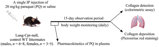
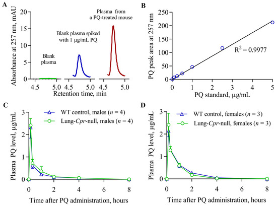
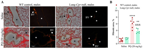
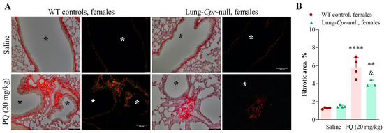
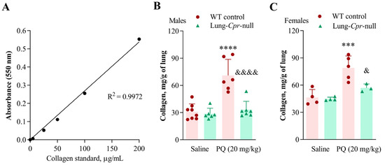
Paraquat (PQ) is an agrochemical known to cause pulmonary fibrosis. PQ-induced collagen deposition in the lung is thought to require enzymatic formation of PQ radicals, but the specific enzymes responsible for this bioactivation event in vivo have not been identified. We tested the hypothesis that lung P450 oxidoreductase (POR or CPR) is important in PQ-induced lung fibrosis in mice. A lung-Cpr-null mouse model was utilized, which undergoes doxycycline-induced, Cre recombinase-mediated deletion of the Por gene specifically in airway Club cells and alveolar type 2 cells in the lung. The lungs of lung-Cpr-null mice and their wild-type littermates were collected on day 15 after a single intraperitoneal injection of saline (control) or PQ (20 mg/kg). Lung tissue sections were stained with picrosirius red for detection of collagen fibrils. Fibrotic lung areas were found to be significantly smaller (1.6-fold for males and 1.4-fold for females) in PQ-treated lung-Cpr-null mice than in sex- and treatment-matched wild-type mice. The levels of collagen in lung tissue homogenate were also lower (1.4–2.3-fold; p < 0.05) in PQ-treated lung-Cpr-null mice compared to PQ-treated wild-type mice. In contrast, plasma PQ toxicokinetic profiles were not different between sex-matched wild-type and lung-Cpr-null mice. Taken together, these results indicate that lung POR plays an important role in PQ-induced pulmonary fibrosis. Full article
(This article belongs to the Special Issue Redox Regulation in Pulmonary Diseases)
Show Figures

Figure 1
18 pages, 2054 KB  
Article
In Vitro Evaluation of Biological Activities of Canes and Pomace Extracts from Several Varieties of Vitis vinifera L. for Inclusion in Freeze-Drying Mouthwashes
by Anca Pop, Catalina Bogdan, Ionel Fizesan, Sonia Iurian, Rahela Carpa, Cecilia Bacali, Laurian Vlase, Daniela Benedec and Mirela L. Moldovan
Antioxidants 2022, 11(2), 218; https://doi.org/10.3390/antiox11020218 - 24 Jan 2022
Cited by 18 | Viewed by 4551
Abstract
In this study, the biological activities of four extracts from Vitis vinifera by-products: two pomace extracts, white (WPE) and red (RPE), a canes extract (CE), and their combination (CoE), were evaluated, to be included in freeze-drying mouthwashes formulations. The cytocompatibility and anticancerous potential [...] Read more.
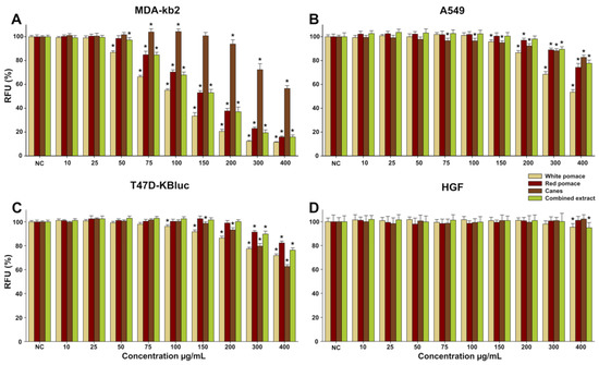
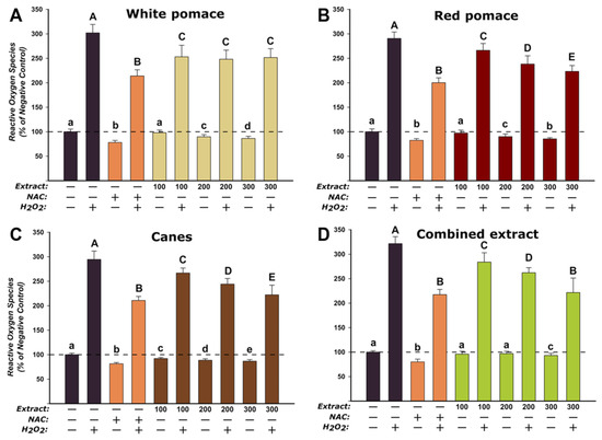
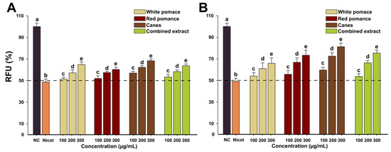
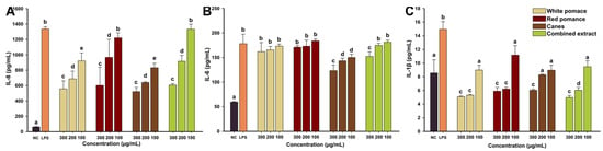
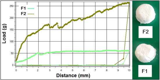
In this study, the biological activities of four extracts from Vitis vinifera by-products: two pomace extracts, white (WPE) and red (RPE), a canes extract (CE), and their combination (CoE), were evaluated, to be included in freeze-drying mouthwashes formulations. The cytocompatibility and anticancerous potential of the four extracts were tested on three cancerous cell lines, as well as the cytoprotective activity against nicotine-induced cytotoxicity and the antioxidant potential determined on a human gingival fibroblasts (HGF) cell line. Additionally, the anti-inflammatory activity and the antimicrobial activity against several microorganisms from the oral microbiome were tested. Freeze-dried mouthwashes with CoE were prepared and characterized, both as lyophilizates and after reconstitution. The four tested extracts showed the highest cytotoxicity on MDA-kb2 cell line. The antioxidant potential was demonstrated for WPE, RPE, CE, and CoE, both in non-stimulated and H2O2 stimulated conditions. The four extracts reduced the levels of proinflammatory cytokines (IL-6, IL-8, and IL-1β) in a dose-dependent manner, confirming their anti-inflammatory activity. The antimicrobial activity of tested extracts was shown against pathogenic bacteria from the oral microbiome. Mouthwashes of CoE with poloxamer-407, xylitol, and different ratios of mannitol were prepared by freeze-drying leading to porous formulations with interesting mechanical properties and reconstitution times. Full article
(This article belongs to the Special Issue Natural Compounds from Plants with Antioxidant Properties)
Show Figures

Figure 1
11 pages, 1099 KB  
Review
Anti-Osteoporotic Mechanisms of Polyphenols Elucidated Based on In Vivo Studies Using Ovariectomized Animals
by Yoshimi Niwano, Hidetsugu Kohzaki, Midori Shirato, Shunichi Shishido and Keisuke Nakamura
Antioxidants 2022, 11(2), 217; https://doi.org/10.3390/antiox11020217 - 23 Jan 2022
Cited by 17 | Viewed by 5925
Abstract
Polyphenols are widely known for their antioxidant activity, i.e., they have the ability to suppress oxidative stress, and this behavior is mediated by the autoxidation of their phenolic hydroxyl groups. Postmenopausal osteoporosis is a common health problem that is associated with estrogen deficiency. [...] Read more.
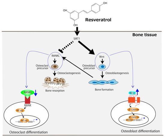
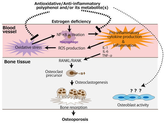
Polyphenols are widely known for their antioxidant activity, i.e., they have the ability to suppress oxidative stress, and this behavior is mediated by the autoxidation of their phenolic hydroxyl groups. Postmenopausal osteoporosis is a common health problem that is associated with estrogen deficiency. Since oxidative stress is thought to play a key role in the onset and progression of osteoporosis, it is expected that polyphenols can serve as a safe and suitable treatment in this regard. Therefore, in this review, we aimed to elucidate the anti-osteoporotic mechanisms of polyphenols reported by in vivo studies involving the use of ovariectomized animals. We categorized the polyphenols as resveratrol, purified polyphenols other than resveratrol, or polyphenol-rich substances or extracts. Literature data indicated that resveratrol activates sirtuin 1, and thereafter, suppresses osteoclastogenic pathways, such as the receptor activator of the nuclear factor kappa B (RANK) ligand (RANKL) pathway, and promotes osteoblastogenic pathways, such as the wingless-related MMTV integration site pathway. Further, we noted that purified polyphenols and polyphenol-rich substances or extracts exert anti-inflammatory and/or antioxidative effects, which inhibit RANKL/RANK binding via the NF-κB pathway, resulting in the suppression of osteoclastogenesis. In conclusion, antioxidative and anti-inflammatory polyphenols, including resveratrol, can be safe and effective for the treatment of postmenopausal osteoporosis based on their ability to regulate the imbalance between bone formation and resorption. Full article
(This article belongs to the Section Health Outcomes of Antioxidants and Oxidative Stress)
Show Figures

Figure 1
15 pages, 1142 KB  
Article
Fermented Biomass of Arthrospira platensis as a Potential Food Ingredient
by Polona Jamnik, Nik Mahnič, Aleksandra Mrak, Lea Pogačnik, Barbara Jeršek, Alberto Niccolai, Jasmina Masten Rutar, Nives Ogrinc, Larisa Dušak, Blaž Ferjančič, Mojca Korošec, Ana Cerar, Borut Lazar, Urša Lovše, Tjaša Pungert, Primož Fabjan and Nataša Poklar Ulrih
Antioxidants 2022, 11(2), 216; https://doi.org/10.3390/antiox11020216 - 23 Jan 2022
Cited by 32 | Viewed by 7610
Abstract
Lactic acid fermentation (LAF) is known to improve nutritional properties and functionality and to extend the shelf life of foods. We studied the LAF of Arthrospira platensis as the sole substrate using Lactobacillus plantarum as the starter culture. Fermented (FB) and non-fermented broth [...] Read more.
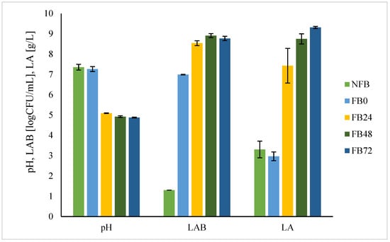
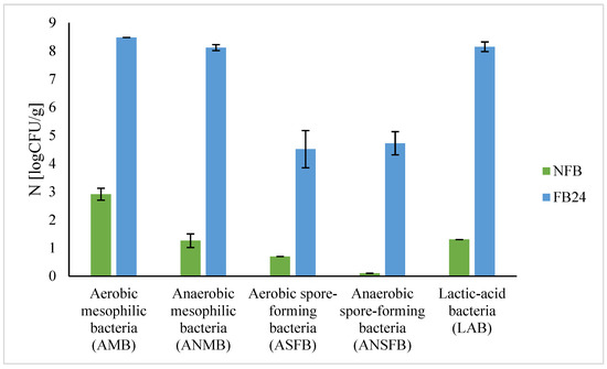
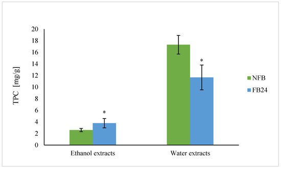
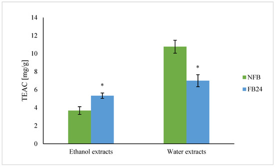
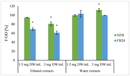
Lactic acid fermentation (LAF) is known to improve nutritional properties and functionality and to extend the shelf life of foods. We studied the LAF of Arthrospira platensis as the sole substrate using Lactobacillus plantarum as the starter culture. Fermented (FB) and non-fermented broth (NFB) were analysed by means of pH, lactic acid bacteria (LAB) count, lactic acid concentration, microbiological safety, and nutritional composition. Additionally, water and ethanol extracts were prepared on which total phenolic content, DPPH radical scavenging activity, and cellular antioxidant activity were determined. The maximum increase in LAB count and lactic acid concentration and drop in pH was observed in the first 24 h of fermentation. Total phenolic content and DPPH radical scavinging activity of ethanol extracts increased after fermentation compared with NFB. Ethanol extracts of FB have been shown as a potential source of antioxidants, which efficiently lowered oxidation level in the cells of yeast Saccharomyces cerevisiae, as well as the oxidative damage of lipids. Additionally, the level of non-protein nitrogen increased, indicating higher protein bioavailability, and fat content decreased in comparison with NFB. No presence of pathogenic bacteria and low pH indicate enhancement of FB microbiological stability. Therefore, inclusion of fermented A. platensis into food products could lead to added-value foods based on microalgae. Full article
(This article belongs to the Special Issue Antioxidants and Bioactive Compounds in Fermented Foods)
Show Figures

Figure 1
27 pages, 3484 KB  
Article
Pharmacokinetics and Pharmacodynamics of Key Components of a Standardized Centella asiatica Product in Cognitively Impaired Older Adults: A Phase 1, Double-Blind, Randomized Clinical Trial
by Kirsten M. Wright, Melissa Bollen, Jason David, Alex B. Speers, Mikah S. Brandes, Nora E. Gray, Armando Alcázar Magaña, Christine McClure, Jan F. Stevens, Claudia S. Maier, Joseph F. Quinn and Amala Soumyanath
Antioxidants 2022, 11(2), 215; https://doi.org/10.3390/antiox11020215 - 23 Jan 2022
Cited by 43 | Viewed by 14683
Abstract
Centella asiatica is reputed in Eastern medicine to improve cognitive function in humans. Preclinical studies have demonstrated that aqueous extracts of C. asiatica improve cognition in mouse models of aging and Alzheimer’s disease (AD) through the modulation of mitochondrial biogenesis and nuclear factor-erythroid-2-related [...] Read more.
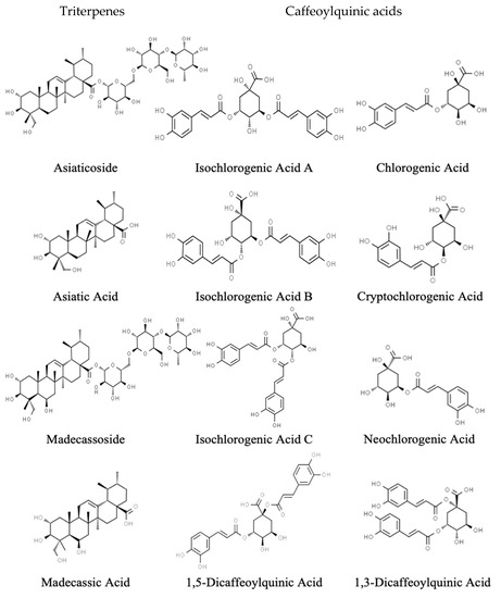
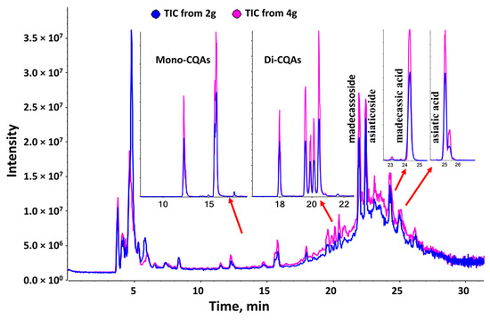
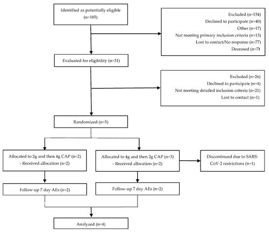
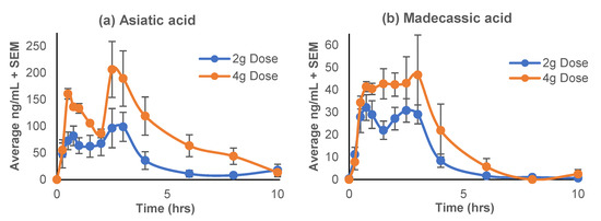
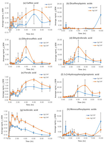
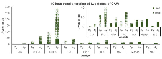
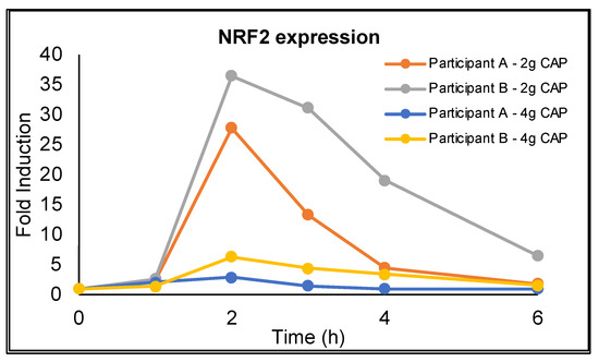
Centella asiatica is reputed in Eastern medicine to improve cognitive function in humans. Preclinical studies have demonstrated that aqueous extracts of C. asiatica improve cognition in mouse models of aging and Alzheimer’s disease (AD) through the modulation of mitochondrial biogenesis and nuclear factor-erythroid-2-related factor 2 (Nrf2)-dependent antioxidant response genes. This randomized, double-blind, crossover Phase I trial explored the oral bioavailability and pharmacokinetics of key compounds from two doses (2 g and 4 g) of a standardized C. asiatica aqueous extract product (CAP), over 10 h, in four mildly demented older adults on cholinesterase inhibitor therapy. The analysis focused on triterpenes (TTs) and caffeoylquinic acids (CQAs), which are known to contribute to C. asiatica’s neurological activity. The acute safety of CAP and the effects on NRF2 gene expression in peripheral blood mononuclear cells were evaluated. Single administration of 2 g or 4 g of CAP was safe and well-tolerated. The TT aglycones, asiatic acid and madecassic acid, were identified in plasma and urine, while the parent glycosides, asiaticoside and madecassoside, although abundant in CAP, were absent in plasma and had limited renal excretion. Similarly, mono- and di-CQAs showed delayed absorption and limited presence in plasma or urine, while the putative metabolites of these compounds showed detectable plasma pharmacokinetic profiles and urinary excretion. CAP elicited a temporal change in NRF2 gene expression, mirroring the TT aglycone’s pharmacokinetic curve in a paradoxical dose-dependent manner. The oral bioavailability of active compounds or their metabolites, NRF2 target engagement, and the acute safety and tolerability of CAP support the validity of using CAP in future clinical studies. Full article
Show Figures

Figure 1
22 pages, 3851 KB  
Article
Inokosterone from Gentiana rigescens Franch Extends the Longevity of Yeast and Mammalian Cells via Antioxidative Stress and Mitophagy Induction
by Yanan Liu, Qian Liu, Danni Chen, Akira Matsuura, Lan Xiang and Jianhua Qi
Antioxidants 2022, 11(2), 214; https://doi.org/10.3390/antiox11020214 - 23 Jan 2022
Cited by 12 | Viewed by 4044
Abstract
In the present study, replicative lifespan and chronological lifespan assays of yeast were used to double-screen antiaging compounds from Gentiana rigescens Franch, a Chinese herb medicine. Inokosterone from G. rigescens Franch extended not only the replicative lifespan of K6001 yeast but also the [...] Read more.
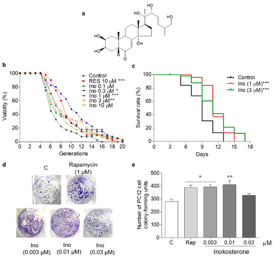
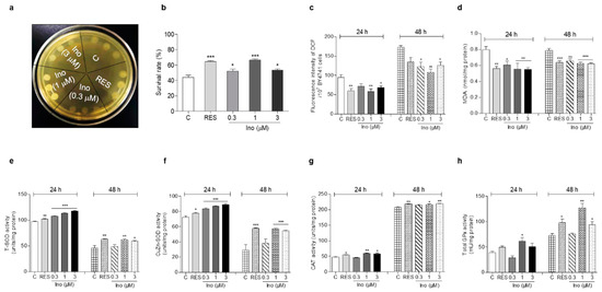
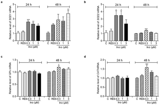
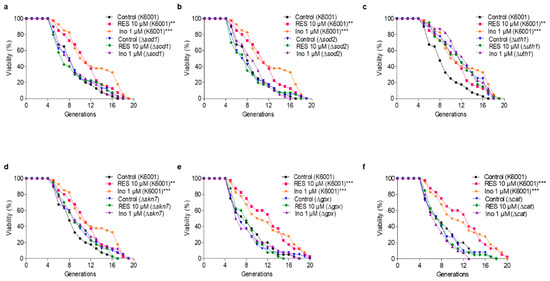
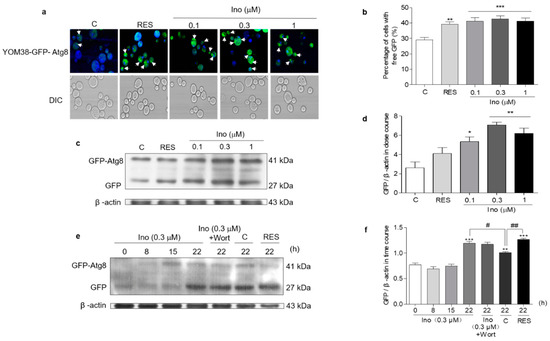
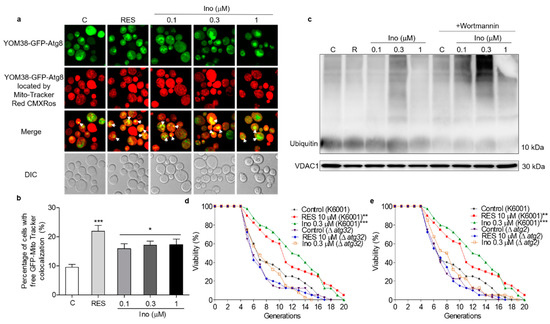
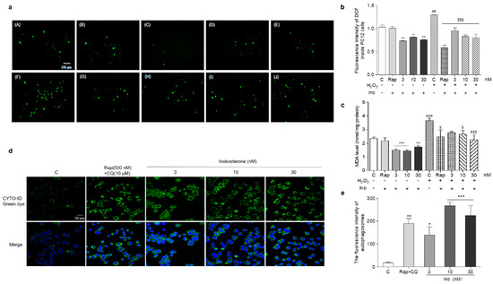
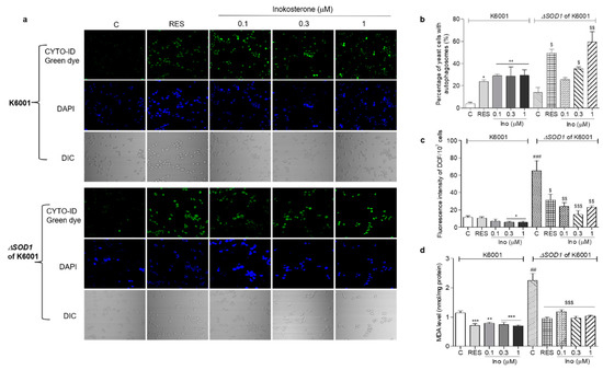
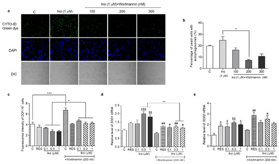
In the present study, replicative lifespan and chronological lifespan assays of yeast were used to double-screen antiaging compounds from Gentiana rigescens Franch, a Chinese herb medicine. Inokosterone from G. rigescens Franch extended not only the replicative lifespan of K6001 yeast but also the chronological lifespan of YOM36 yeast. Furthermore, it can enhance the survival ability of mammalian cells. In order to understand the mechanism of action of this compound, this study focused on antioxidative stress and autophagy when performing the analysis. The increased cell survival rate under oxidative stress conditions, antioxidant enzyme activity and gene expression were observed in the inokosterone-treated groups. Meanwhile, the reactive oxygen species (ROS) and lipid peroxidation of yeast were obviously decreased. Additionally, the macroautophagy and mitophagy in YOM38-GFP-ATG8 yeast were increased upon inokosterone treatment, respectively. At the same time, the cleavage-free GFP from GFP-ATG8 in the cytoplasm and the ubiquitin of the mitochondria at the protein level were markedly enhanced after incubation with inokosterone. Furthermore, we investigated the effect of inokosterone on antioxidative stress and autophagy in mammalian cells, and the relationship between ROS and autophagy. The ROS, malondialdehyde (MDA) were significantly decreased, and the autophagosomes in mammalian cells were obviously increased after inokosterone treatment. The autophagosomes in ∆sod1 yeast with a K6001 background had no obvious changes, and the ROS and MDA of ∆sod1 yeast were increased compared with K6001 yeast. The increase of autophagosomes and the reduction of ROS and MDA in ∆sod1 yeast were observed after treatment with inokosterone. Meanwhile, the reduction of the ROS level and the increase of the SOD1 gene expression of K6001 yeast lacking autophagy were observed after treatment with inokosterone. In order to indicate whether the genes related to antioxidant enzymes and autophagy were involved in the antiaging effect of inokosterone, mutants of K6001 yeast were constructed to conduct a lifespan assay. The replicative lifespans of ∆sod1, ∆sod2, ∆uth1, ∆skn7, ∆gpx, ∆cat, ∆atg2, and ∆atg32 of K6001 yeast were not affected by inokosterone. These results suggest that inokosterone exerted an antiaging activity via antioxidative stress and increased autophagy activation; autophagy affected the ROS levels of yeast via the regulation of SOD1 gene expression. Full article
(This article belongs to the Section Natural and Synthetic Antioxidants)
Show Figures

Figure 1
38 pages, 1636 KB  
Review
Naturally Occurring Antioxidant Therapy in Alzheimer’s Disease
by Andrila E. Collins, Tarek M. Saleh and Bettina E. Kalisch
Antioxidants 2022, 11(2), 213; https://doi.org/10.3390/antiox11020213 - 23 Jan 2022
Cited by 83 | Viewed by 15520
Abstract
It is estimated that the prevalence rate of Alzheimer’s disease (AD) will double by the year 2040. Although currently available treatments help with symptom management, they do not prevent, delay the progression of, or cure the disease. Interestingly, a shared characteristic of AD [...] Read more.
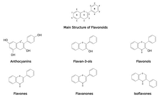
It is estimated that the prevalence rate of Alzheimer’s disease (AD) will double by the year 2040. Although currently available treatments help with symptom management, they do not prevent, delay the progression of, or cure the disease. Interestingly, a shared characteristic of AD and other neurodegenerative diseases and disorders is oxidative stress. Despite profound evidence supporting the role of oxidative stress in the pathogenesis and progression of AD, none of the currently available treatment options address oxidative stress. Recently, attention has been placed on the use of antioxidants to mitigate the effects of oxidative stress in the central nervous system. In preclinical studies utilizing cellular and animal models, natural antioxidants showed therapeutic promise when administered alone or in combination with other compounds. More recently, the concept of combination antioxidant therapy has been explored as a novel approach to preventing and treating neurodegenerative conditions that present with oxidative stress as a contributing factor. In this review, the relationship between oxidative stress and AD pathology and the neuroprotective role of natural antioxidants from natural sources are discussed. Additionally, the therapeutic potential of natural antioxidants as preventatives and/or treatment for AD is examined, with special attention paid to natural antioxidant combinations and conjugates that are currently being investigated in human clinical trials. Full article
(This article belongs to the Special Issue Antioxidants in Age-Related Diseases and Anti-Aging Strategies)
Show Figures

Figure 1
11 pages, 2501 KB  
Article
Nrf2 Is Required for Optimal Alveolar-Macrophage-Mediated Apoptotic Neutrophil Clearance after Oxidant Injury
by Narsa M. Reddy, Chandra Mohan Tamatam, Ankireddy Aparna and Sekhar P. Reddy
Antioxidants 2022, 11(2), 212; https://doi.org/10.3390/antiox11020212 - 22 Jan 2022
Cited by 14 | Viewed by 5081
Abstract
Recognition and clearance of apoptotic cells by phagocytes (also known as efferocytosis), primarily mediated by macrophages, are essential to terminate lung inflammatory responses and promote tissue repair after injury. The Nrf2 transcription factor is crucial for cytoprotection and host defense. Previously, we showed [...] Read more.
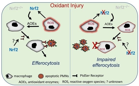
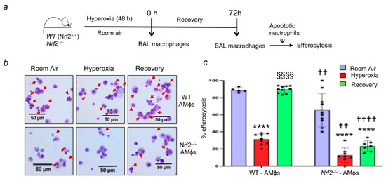
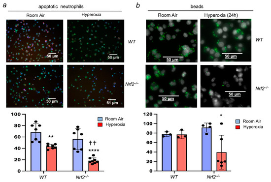
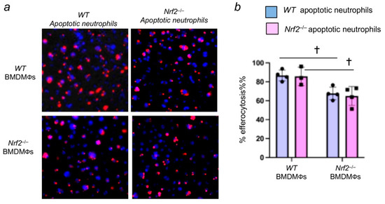
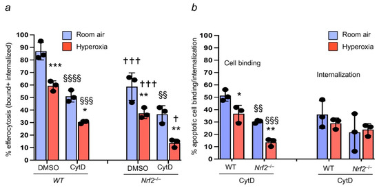
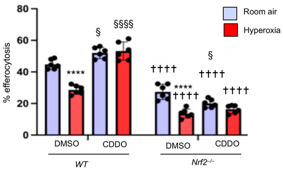
Recognition and clearance of apoptotic cells by phagocytes (also known as efferocytosis), primarily mediated by macrophages, are essential to terminate lung inflammatory responses and promote tissue repair after injury. The Nrf2 transcription factor is crucial for cytoprotection and host defense. Previously, we showed sustained neutrophilic lung inflammation in Nrf2-deficient (Nrf2−/−) mice after hyperoxia-induced lung injury in vivo, but the mechanisms underlying this abnormal phenotype remain unclear. To examine whether Nrf2 regulates apoptotic neutrophil clearance, we used the alveolar macrophages (AMФs) and bone-marrow-derived macrophages (BMDMФs) of wild-type (WT) and Nrf2−/− mice. We found that the efferocytic ability of AMФ was impaired in hyperoxia-exposed mice’s lungs, but the effect was more pronounced in Nrf2−/− mice. Importantly, AMФ-mediated efferocytosis remained impaired in Nrf2−/− mice recovering from injury but was restored to the basal state in the wild-type counterparts. Hyperoxia affected apoptotic neutrophil binding, not internalization, in both WT and Nrf2−/− BMDMФs, but the effect was more significant in the latter cells. Augmenting Nrf2 activity restored hyperoxia attenuated efferocytosis in WT, but not in Nrf2−/− macrophages. However, the loss of Nrf2 in neutrophils affected their uptake by WT macrophages. Collectively, these results demonstrate that Nrf2 is required for optimal macrophage-mediated efferocytosis and that activating Nrf2 may provide a physiological way to accelerate apoptotic cell clearance after oxidant injury. Full article
(This article belongs to the Special Issue NRF2 in Health and Diseases)
Show Figures

Graphical abstract

19 pages, 1112 KB  
Article
Disclosing the Antioxidant and Neuroprotective Activity of an Anthocyanin-Rich Extract from Sweet Cherry (Prunus avium L.) Using In Vitro and In Vivo Models
by Monica Filaferro, Alessandro Codeluppi, Virginia Brighenti, Francesca Cimurri, Ana María González-Paramás, Celestino Santos-Buelga, Davide Bertelli, Federica Pellati and Giovanni Vitale
Antioxidants 2022, 11(2), 211; https://doi.org/10.3390/antiox11020211 - 22 Jan 2022
Cited by 25 | Viewed by 5847
Abstract
In this study, an autochthonous variety of sweet cherry (Prunus avium L.), namely “Moretta di Vignola”, was processed to prepare extracts rich in polyphenols, which were characterized by high-performance liquid chromatography (HPLC) separation coupled to UV/DAD and ESI-MSn analysis. Then, a [...] Read more.
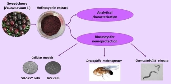
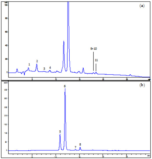
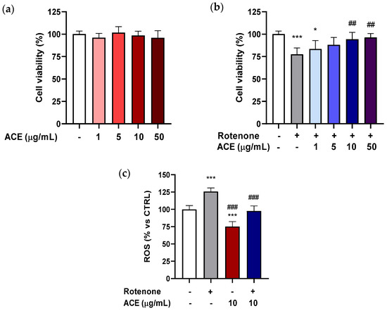
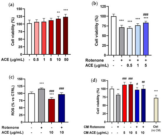
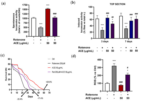
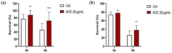
In this study, an autochthonous variety of sweet cherry (Prunus avium L.), namely “Moretta di Vignola”, was processed to prepare extracts rich in polyphenols, which were characterized by high-performance liquid chromatography (HPLC) separation coupled to UV/DAD and ESI-MSn analysis. Then, a sweet cherry anthocyanin-rich extract (ACE) was prepared, fully characterized and tested for its activity against Parkinson’s disease (PD) in cellular (BV2 microglia and SH-SY5Y neuroblastoma) and in Drosophila melanogaster rotenone (ROT)-induced model. The extract was also evaluated for its antioxidant activity on Caenorhabditis elegans by assessing nematode resistance to thermal stress. In both cell lines, ACE reduced ROT-induced cell death and it decreased, alone, cellular reactive oxygen species (ROS) content while reinstating control-like ROS values after ROT-induced ROS rise, albeit at different concentrations of both compounds. Moreover, ACE mitigated SH-SY5Y cell cytotoxicity in a non-contact co-culture assay with cell-free supernatants from ROT-treated BV-2 cells. ACE, at 50 µg/mL, ameliorated ROT (250 μM)-provoked spontaneous (24 h duration) and induced (after 3 and 7 days) locomotor activity impairment in D. melanogaster and it also increased survival and counteracted the decrease in fly lifespan registered after exposure to the ROT. Moreover, heads from flies treated with ACE showed a non-significant decrease in ROS levels, while those exposed to ROT markedly increased ROS levels if compared to controls. ACE + ROT significantly placed the ROS content to intermediate values between those of controls and ROT alone. Finally, ACE at 25 µg/mL produced a significant increase in the survival rate of nematodes submitted to thermal stress (35 °C, 6–8 h), at the 2nd and 9th day of adulthood. All in all, ACE from Moretta cherries can be an attractive candidate to formulate a nutraceutical product to be used for the prevention of oxidative stress-induced disorders and related neurodegenerative diseases. Full article
(This article belongs to the Collection Advances in Antioxidant Ingredients from Natural Products)
Show Figures

Graphical abstract