Cells 2022, 11(10), 1706; https://doi.org/10.3390/cells11101706 - 20 May 2022
Cited by 24 | Viewed by 5049
Abstract
Long-term exercise-induced metabolic adaptations occupy a central position in exercise-afforded cardiac benefits. Emerging evidence suggests that branched-chain amino acid (BCAA) catabolic defect contributes to cardiac dysfunction in multiple cardiometabolic diseases. However, the role of BCAA catabolism in exercise-afforded cardiac benefits remains unknown. Here,
[...] Read more.
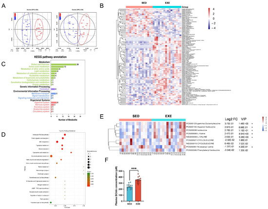
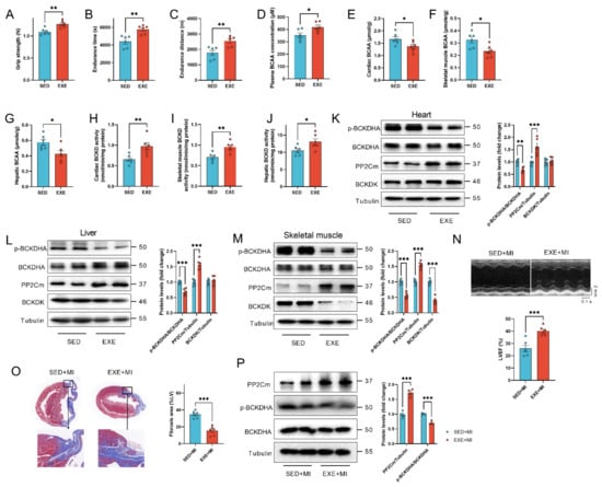
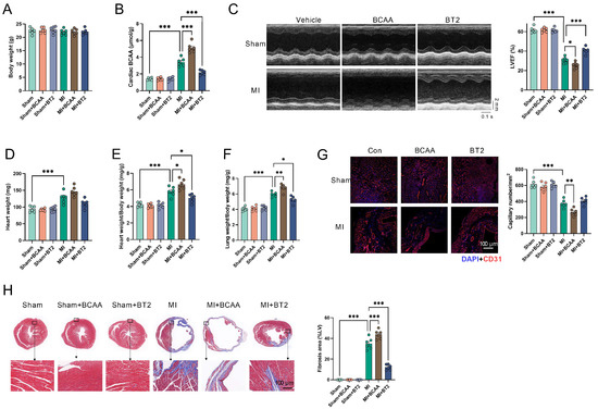
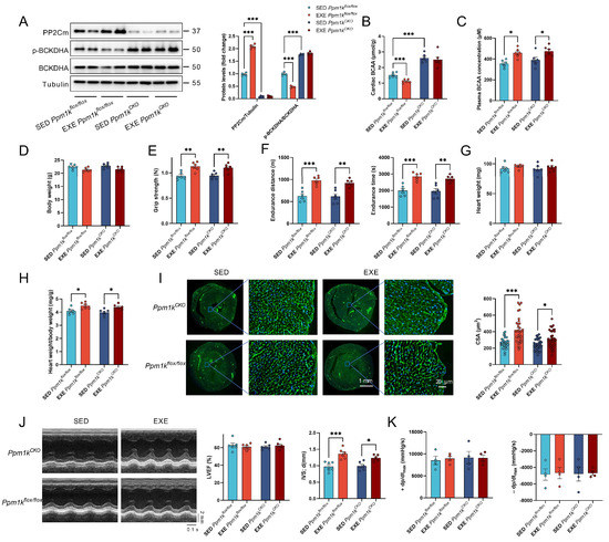
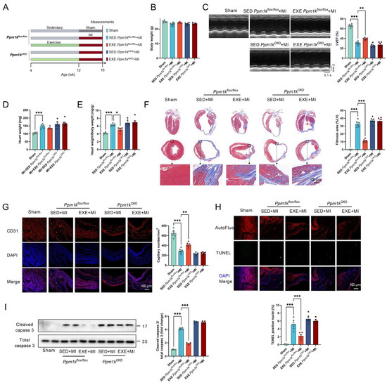
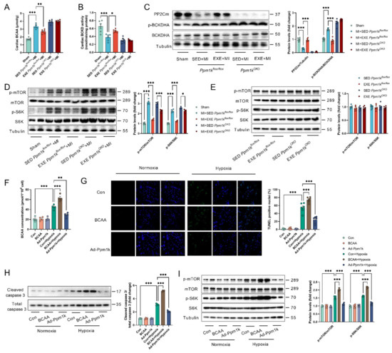
Long-term exercise-induced metabolic adaptations occupy a central position in exercise-afforded cardiac benefits. Emerging evidence suggests that branched-chain amino acid (BCAA) catabolic defect contributes to cardiac dysfunction in multiple cardiometabolic diseases. However, the role of BCAA catabolism in exercise-afforded cardiac benefits remains unknown. Here, we show that exercise improves BCAA catabolism and thus reduce cardiac vulnerability to myocardial ischemic injury. Exercise increased circulating BCAA levels in both humans (male adolescent athletes) and mice (following an 8-week swimming intervention). It increased the expression of mitochondrial localized 2C-type serine-threonine protein phosphatase (PP2Cm), a key enzyme in regulating BCAA catabolism, and decreased BCAA accumulation in mouse hearts, indicating an increase in BCAA catabolism. Pharmacological promotion of BCAA catabolism protected the mouse heart against myocardial infarction (MI) induced by permanent ligation of the left descending coronary artery. Although cardiac-specific PP2Cm knockout showed no significant effects on cardiac structural and functional adaptations to exercise, it blunted the cardioprotective effects of exercise against MI. Mechanistically, exercise alleviated BCAA accumulation and subsequently inactivated the mammalian target of rapamycin in MI hearts. These results showed that exercise elevated BCAA catabolism and protected the heart against myocardial ischemic injury, reinforcing the role of exercise in the promotion of cardiac health.
Full article
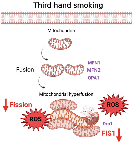
(This article belongs to the Special Issue Mitochondrial Dysfunction in Aging and Metabolic Diseases)
►
Show Figures

Graphical abstract


