Cancers 2022, 14(18), 4468; https://doi.org/10.3390/cancers14184468 - 15 Sep 2022
Cited by 15 | Viewed by 4552
Abstract
Functional imaging with diffusion-weighted imaging (DWI) is a complementary tool to conventional diagnostic magnetic resonance imaging sequences. It is being increasingly investigated to predict tumor response and assess tumor recurrence. We elucidate the specific technical modifications of DWI preferred for gynecological imaging, including
[...] Read more.
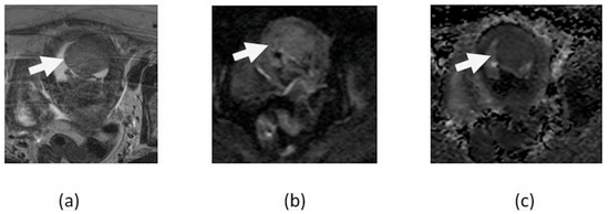
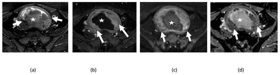
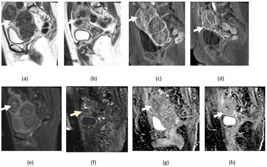
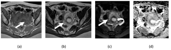
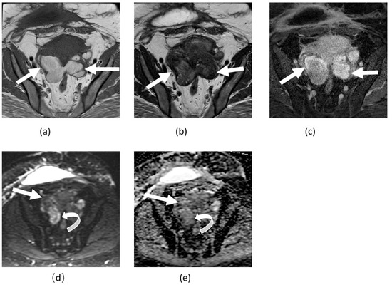
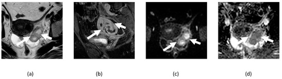
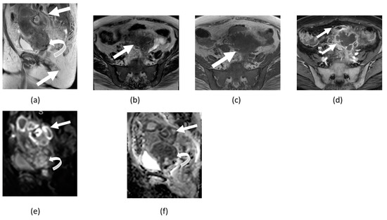
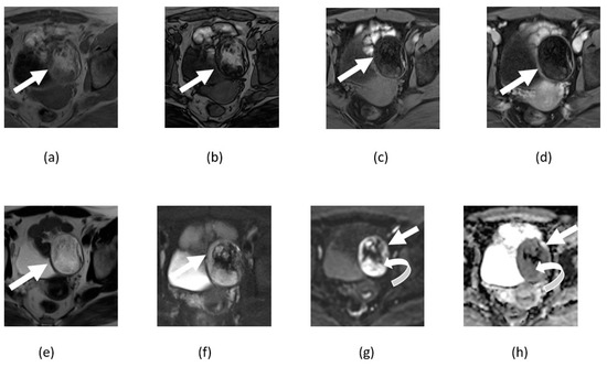
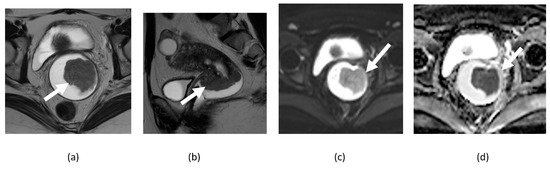
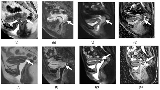
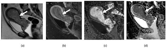
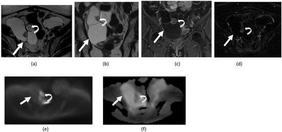
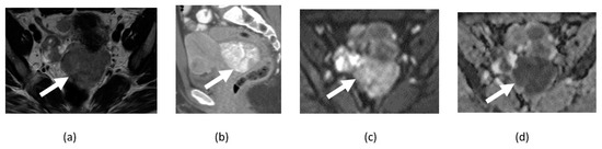
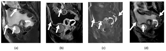
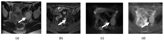
Functional imaging with diffusion-weighted imaging (DWI) is a complementary tool to conventional diagnostic magnetic resonance imaging sequences. It is being increasingly investigated to predict tumor response and assess tumor recurrence. We elucidate the specific technical modifications of DWI preferred for gynecological imaging, including the different b-values and planes for image acquisition. Additionally, we discuss the problems and potential pitfalls encountered during DWI interpretation and ways to overcome them. DWI has a wide range of clinical applications in malignant and non-malignant gynecological conditions. It provides supplemental information helpful in diagnosing and managing tubo-ovarian abscess, uterine fibroids, endometriosis, adnexal torsion, and dermoid. Similarly, DWI has diverse applications in gynecological oncology in diagnosis, staging, detection of recurrent disease, and tumor response assessment. Quantitative evaluation with apparent diffusion coefficient (ADC) measurement is being increasingly evaluated for correlation with various tumor parameters in managing gynecological malignancies aiding in preoperative treatment planning. Newer advanced DWI techniques of diffusion tensor imaging (DTI) and whole body DWI with background suppression (DWIBS) and their potential uses in pelvic nerve mapping, preoperative planning, and fertility-preserving surgeries are briefly discussed.
Full article
(This article belongs to the Special Issue Gynecologic Cancers: Imaging Updates and Advances)
►
Show Figures
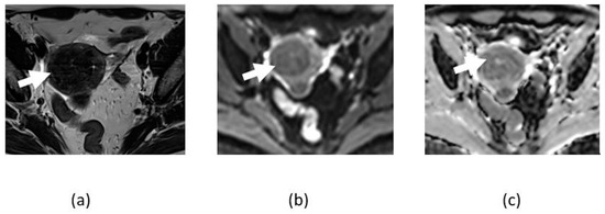
Figure 1


