by
Shahinda S. R. Alsayed 1,* and Hendra Gunosewoyo 1,2,*
1
Curtin Medical School, Faculty of Health Sciences, Curtin University, Bentley, Perth, WA 6102, Australia
2
Curtin Health Innovation Research Institute, Faculty of Health Sciences, Curtin University, Bentley, Perth, WA 6102, Australia
Int. J. Mol. Sci. 2023, 24(6), 5202; https://doi.org/10.3390/ijms24065202 - 8 Mar 2023
Cited by 162 | Viewed by 47495
Abstract
Mycobacterium tuberculosis (M. tb), the causative agent of TB, is a recalcitrant pathogen that is rife around the world, latently infecting approximately a quarter of the worldwide population. The asymptomatic status of the dormant bacteria escalates to the transmissible, active form
[...] Read more.
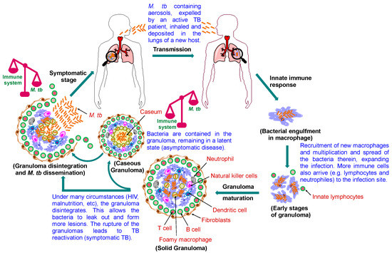
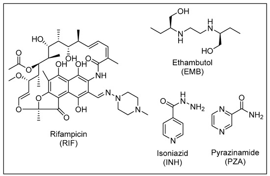
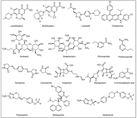
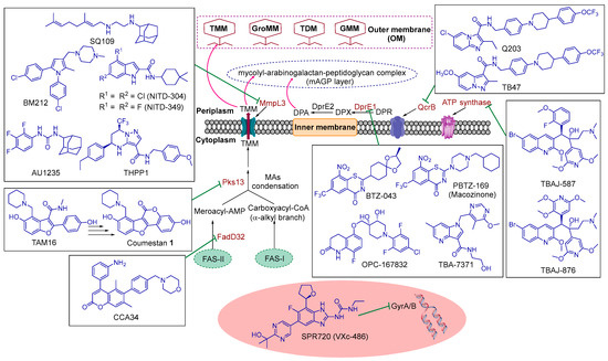
Mycobacterium tuberculosis (M. tb), the causative agent of TB, is a recalcitrant pathogen that is rife around the world, latently infecting approximately a quarter of the worldwide population. The asymptomatic status of the dormant bacteria escalates to the transmissible, active form when the host’s immune system becomes debilitated. The current front-line treatment regimen for drug-sensitive (DS) M. tb strains is a 6-month protocol involving four different drugs that requires stringent adherence to avoid relapse and resistance. Poverty, difficulty to access proper treatment, and lack of patient compliance contributed to the emergence of more sinister drug-resistant (DR) strains, which demand a longer duration of treatment with more toxic and more expensive drugs compared to the first-line regimen. Only three new drugs, bedaquiline (BDQ) and the two nitroimidazole derivatives delamanid (DLM) and pretomanid (PMD) were approved in the last decade for treatment of TB—the first anti-TB drugs with novel mode of actions to be introduced to the market in more than 50 years—reflecting the attrition rates in the development and approval of new anti-TB drugs. Herein, we will discuss the M. tb pathogenesis, current treatment protocols and challenges to the TB control efforts. This review also aims to highlight several small molecules that have recently been identified as promising preclinical and clinical anti-TB drug candidates that inhibit new protein targets in M. tb.
Full article
(This article belongs to the Special Issue Latest Review Papers in Molecular Pathology, Diagnostics, and Therapeutics 2023)
▼
Show Figures
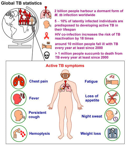
Figure 1


