Int. J. Mol. Sci. 2021, 22(4), 1658; https://doi.org/10.3390/ijms22041658 - 7 Feb 2021
Cited by 9 | Viewed by 5188
Abstract
The deepest evolutionary branches of the trypsin/chymotrypsin family of serine proteases are represented by the digestive enzymes of the gastrointestinal tract and the multi-domain proteases of the blood coagulation and complement system. Similar to the very old digestive system, highly diverse cleavage specificities
[...] Read more.
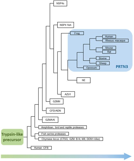
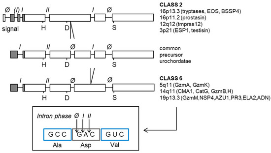
The deepest evolutionary branches of the trypsin/chymotrypsin family of serine proteases are represented by the digestive enzymes of the gastrointestinal tract and the multi-domain proteases of the blood coagulation and complement system. Similar to the very old digestive system, highly diverse cleavage specificities emerged in various cell lineages of the immune defense system during vertebrate evolution. The four neutrophil serine proteases (NSPs) expressed in the myelomonocyte lineage, neutrophil elastase, proteinase 3, cathepsin G, and neutrophil serine protease 4, collectively display a broad repertoire of (S1) specificities. The origin of NSPs can be traced back to a circulating liver-derived trypsin-like protease, the complement factor D ancestor, whose activity is tightly controlled by substrate-induced activation and TNFα-induced locally upregulated protein secretion. However, the present-day descendants are produced and converted to mature enzymes in precursor cells of the bone marrow and are safely sequestered in granules of circulating neutrophils. The potential site and duration of action of these cell-associated serine proteases are tightly controlled by the recruitment and activation of neutrophils, by stimulus-dependent regulated secretion of the granules, and by various soluble inhibitors in plasma, interstitial fluids, and in the inflammatory exudate. An extraordinary dynamic range and acceleration of immediate defense responses have been achieved by exploiting the high structural plasticity of the trypsin fold.
Full article
(This article belongs to the Special Issue Biocatalysis: Mechanisms of Proteolytic Enzymes)
►
Show Figures
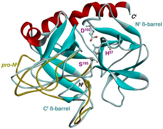
Figure 1


