1
Laboratory of Genome Science, Biosignal Genome Resource Center, Institute for Molecular and Cellular Regulation, Gunma University, Gunma 371-8512, Japan
2
Division of Molecular and Cellular Medicine, National Cancer Center Research Institute, 5-1-1, Tsukiji, Chuo-ku, Tokyo 104-0045, Japan
3
Laboratory of Epigenetics, Institute for Protein Research, Osaka University, 3-2 Yamadaoka, Suita, Osaka 565-0871, Japan
Int. J. Mol. Sci. 2013, 14(7), 14647-14658; https://doi.org/10.3390/ijms140714647 - 12 Jul 2013
Cited by 123 | Viewed by 11685
Abstract
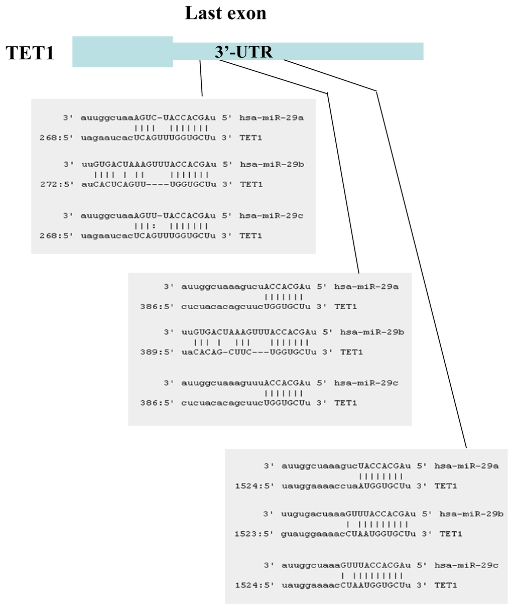
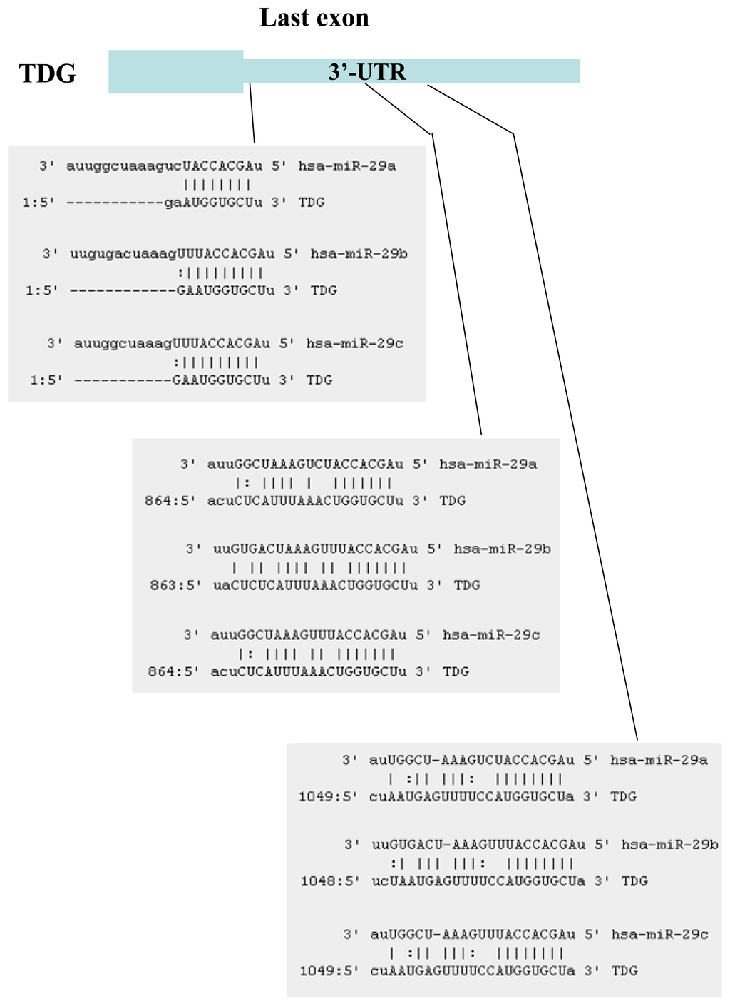
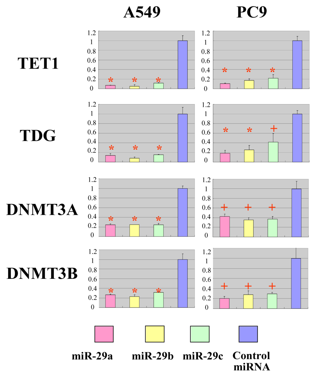
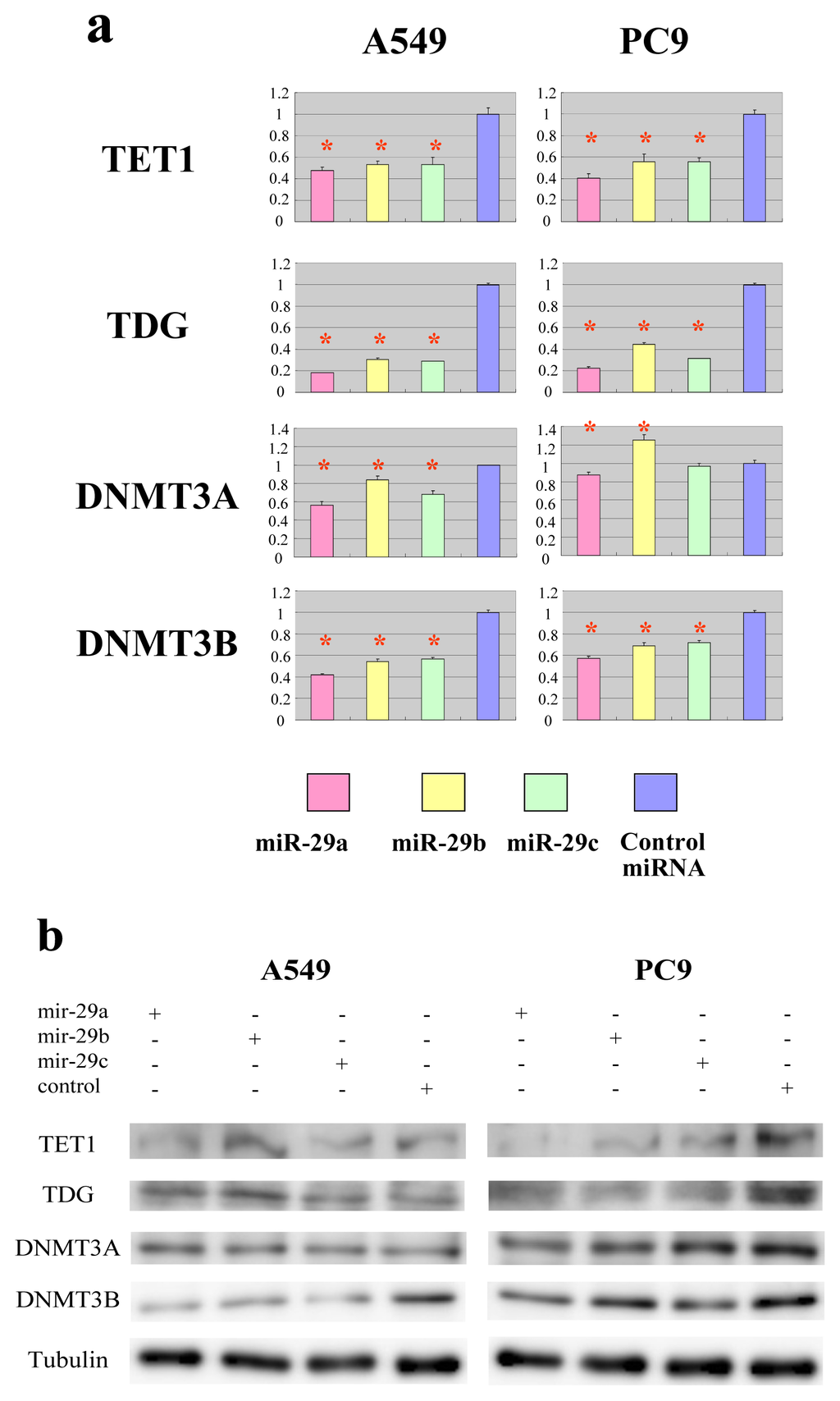
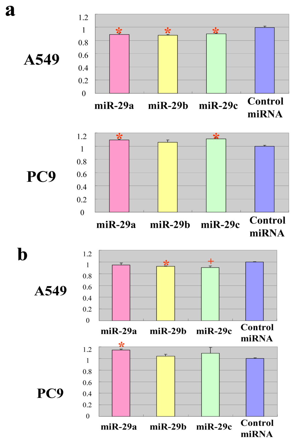
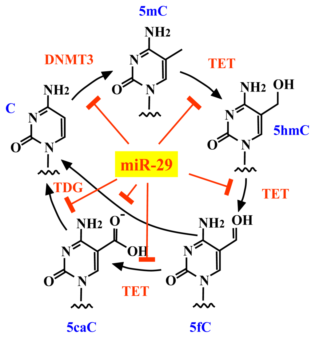
Members of the microRNA-29 (miR-29) family directly target the DNA methyltransferases, DNMT3A and DNMT3B. Disturbances in the expression levels of miR-29 have been linked to tumorigenesis and tumor aggressiveness. Members of the miR-29 family are currently thought to repress DNA methylation and suppress
[...] Read more.
Members of the microRNA-29 (miR-29) family directly target the DNA methyltransferases, DNMT3A and DNMT3B. Disturbances in the expression levels of miR-29 have been linked to tumorigenesis and tumor aggressiveness. Members of the miR-29 family are currently thought to repress DNA methylation and suppress tumorigenesis by protecting against de novo methylation. Here, we report that members of the miR-29 family repress the activities of DNA methyltransferases and DNA demethylases, which have opposing roles in control of DNA methylation status. Members of the miR-29 family directly inhibited DNA methyltransferases and two major factors involved in DNA demethylation, namely tet methylcytosine dioxygenase 1 (TET1) and thymine DNA glycosylase (TDG). Overexpression of miR-29 upregulated the global DNA methylation level in some cancer cells and downregulated DNA methylation in other cancer cells, suggesting that miR-29 suppresses tumorigenesis by protecting against changes in the existing DNA methylation status rather than by preventing de novo methylation of DNA.
Full article
(This article belongs to the Special Issue Advances in Cancer Diagnosis)
▼
Show Figures


