Biomonitoring of Trace Elements in Subjects Living Near a Hazardous Waste Incinerator: Concentrations in Autopsy Tissues

 ,
,  and
and
Abstract
:1. Introduction
2. Materials and Methods
2.1. Sampling
2.2. Chemical Analysis
2.3. Statistics
3. Results
4. Discussion
5. Conclusions
Author Contributions
Funding
Acknowledgments
Conflicts of Interest
References
- Eurostat. Waste Statistics; European Commission: Brussels, Belgium, 2020; Available online: https://ec.europa.eu/eurostat/statistics-explained/index.php?title=Waste_statistics (accessed on 20 January 2020).
- Rovira, J.; Mari, M.; Nadal, M.; Schuhmacher, M.; Domingo, J.L. Environmental monitoring of metals, PCDD/Fs and PCBs as a complementary tool of biological surveillance to assess human health risks. Chemosphere 2010, 80, 1183–1189. [Google Scholar] [CrossRef]
- Granero, S.; Llobet, J.M.; Schuhmacher, M.; Corbella, J.; Domingo, J.L. Biological monitoring of environmental pollution and human exposure to metals in Tarragona, Spain. I. Levels in hair of school children. Trace Elem. Electrol. 1998, 15, 39–43. [Google Scholar]
- Llobet, J.M.; Granero, S.; Schuhmacher, M.; Corbella, J.; Domingo, J.L. Biological monitoring of environmental pollution and human exposure to metals in Tarragona, Spain. II. Levels in autopsy tissues. Trace Elem. Electrol. 1998, 15, 44–49. [Google Scholar]
- Llobet, J.M.; Granero, S.; Torres, A.; Schuhmacher, M.; Domingo, J.L. Biological monitoring of environmental pollution and human exposure to metals in Tarragona, Spain. III. Blood levels. Trace Elem. Electrol. 1998, 15, 76–80. [Google Scholar]
- Llobet, J.M.; Schuhmacher, M.; Domingo, J.L. Observations on metal trends in soil and vegetation samples collected in the vicinity of a hazardous waste incinerator under construction (1996–1998). Toxicol. Environ. Chem. 2000, 77, 119–129. [Google Scholar] [CrossRef]
- Schuhmacher, M.; Domingo, J.L.; Llobet, J.M.; Kiviranta, H.; Vartiainen, T. PCDD/F concentrations in milk of non-occupationally exposed women living in southern Catalonia, Spain. Chemosphere 1999, 38, 995–1004. [Google Scholar] [CrossRef]
- Schuhmacher, M.; Domingo, J.L.; Llobet, J.M.; Lindström, G.; Wingfors, H. Dioxin and dibenzofuran concentrations in blood of a general population from Tarragona, Spain. Chemosphere 1999, 38, 1123–1133. [Google Scholar] [CrossRef]
- Schuhmacher, M.; Domingo, J.L.; Llobet, J.M.; Lindstrom, G.; Wingfors, H. Dioxin and dibenzofuran concentrations in adipose tissue of a general population from Tarragona, Spain. Chemosphere 1999, 38, 2475–2487. [Google Scholar] [CrossRef]
- Sirot, V.; Traore, T.; Guérin, T.; Noël, L.; Bachelot, M.; Cravedi, J.P.; Mazur, A.; Glorennec, P.; Vasseur, P.; Jean, J.; et al. French infant total diet study: Exposure to selected trace elements and associated health risks. Food Chem. Toxicol. 2018, 120, 625–633. [Google Scholar] [CrossRef]
- González, N.; Calderón, J.; Rúbies, A.; Timoner, I.; Castell, V.; Domingo, J.L.; Nadal, M. Dietary intake of arsenic, cadmium, mercury and lead by the population of Catalonia, Spain: Analysis of the temporal trend. Food Chem. Toxicol. 2019, 132, 110721. [Google Scholar] [CrossRef]
- González, N.; Marquès, M.; Nadal, M.; Domingo, J.L. Occurrence of environmental pollutants in foodstuffs: A review of organic vs. conventional food. Food Chem. Toxicol. 2019, 125, 370–375. [Google Scholar] [CrossRef] [PubMed]
- Domingo, J.L.; Schuhmacher, M.; Granero, S.; Llobet, J.M. PCDDs and PCDFs in food samples from Catalonia, Spain. An assessment of dietary intake. Chemosphere 1999, 38, 3517–3528. [Google Scholar] [CrossRef]
- Llobet, J.M.; Granero, S.; Schuhmacher, M.; Corbella, J.; Domingo, J.L. Biological monitoring of environmental pollution and human exposure to metals in Tarragona, Spain. IV. Estimation of the dietary intake. Trace Elem. Electrol. 1998, 15, 136–141. [Google Scholar]
- Nadal, M.; García, F.; Schuhmacher, M.; Domingo, J.L. Metals in biological tissues of the population living near a hazardous waste incinerator in Catalonia, Spain: Two decades of follow-up. Environ. Res. 2019, 176, 108578. [Google Scholar] [CrossRef] [PubMed]
- Nadal, M.; Mari, M.; Schuhmacher, M.; Domingo, J.L. Monitoring dioxins and furans in plasma of individuals living near a hazardous waste incinerator: Temporal trend after 20 years. Environ. Res. 2019, 173, 207–211. [Google Scholar] [CrossRef] [PubMed]
- Schuhmacher, M.; Mari, M.; Nadal, M.; Domingo, J.L. Concentrations of dioxins and furans in breast milk of women living near a hazardous waste incinerator in Catalonia, Spain. Environ. Int. 2019, 125, 334–341. [Google Scholar] [CrossRef] [PubMed]
- Esplugas, R.; Mari, M.; Marquès, M.; Schuhmacher, M.; Domingo, J.L.; Nadal, M. Biomonitoring of trace elements in hair of schoolchildren living near a hazardous waste incinerator - A 20 years follow-up. Toxics 2019, 7, 52. [Google Scholar] [CrossRef] [Green Version]
- Esplugas, R.; Serra, N.; Marquès, M.; Schuhmacher, M.; Nadal, M.; Domingo, J.L. Trace elements in blood of the population living near a hazardous waste incinerator in Catalonia, Spain. Biol. Trace Elem. Res. 2020. [Google Scholar] [CrossRef]
- Garcia, F.; Ortega, A.; Domingo, J.L.; Corbella, J. Accumulation of metals in autopsy tissues of subjects living in Tarragona County, Spain. J. Environ. Sci. Health A 2001, 36, 1767–1786. [Google Scholar] [CrossRef]
- Bocio, A.; Nadal, M.; Garcia, F.; Domingo, J.L. Monitoring metals in the population living in the vicinity of a hazardous waste incinerator: Concentrations in autopsy tissues. Biol. Trace Elem. Res. 2005, 106, 41–50. [Google Scholar] [CrossRef]
- Mari, M.; Nadal, M.; Schuhmacher, M.; Barbería, E.; García, F.; Domingo, J.L. Human exposure to metals: Levels in autopsy tissues of individuals living near a hazardous waste incinerator. Biol. Trace Elem. Res. 2014, 159, 15–21. [Google Scholar] [CrossRef] [PubMed]
- Kim, J.; Seo, S.; Kim, Y.; Kim, D.H. Review of carcinogenicity of hexavalent chrome and proposal of revising approval standards for an occupational cancers in Korea. Ann. Occup. Environ. Med. 2018, 30, 7. [Google Scholar] [CrossRef] [PubMed]
- Proctor, D.M.; Suh, M.; Campleman, S.L.; Thompson, C.M. Assessment of the mode of action for hexavalent chromium-induced lung cancer following inhalation exposures. Toxicology 2014, 325, 160–179. [Google Scholar] [CrossRef] [PubMed]
- Nadal, M.; Schuhmacher, M.; Domingo, J.L. Metal pollution of soils and vegetation in an area with petrochemical industry. Sci. Total Environ. 2004, 321, 59–69. [Google Scholar] [CrossRef] [PubMed]
- Domingo, J.L.; García, F.; Nadal, M.; Schuhmacher, M. Autopsy tissues as biological monitors of human exposure to environmental pollutants. A case study: Concentrations of metals and PCDD/Fs in subjects living near a hazardous waste incinerator. Environ. Res. 2017, 154, 269–274. [Google Scholar] [CrossRef]
- Dudek-Adamska, D.; Lech, T.; Konopka, T.; Kościelniak, P. Chromium in postmortem material. Biol. Trace Elem. Res. 2018, 186, 370–378. [Google Scholar] [CrossRef] [Green Version]
- Akerstrom, M.; Barregard, L.; Lundh, T.; Sallsten, G. Relationship between mercury in kidney, blood, and urine in environmentally exposed individuals, and implications for biomonitoring. Toxicol. Appl. Pharm. 2017, 320, 17–25. [Google Scholar] [CrossRef]
- Perelló, G.; Nadal, M.; Domingo, J.L. Dietary exposure to metals by adults living near a hazardous waste incinerator in Catalonia, Spain: Temporal trend. Trace Elem. Electrol. 2015, 32, 133–141. [Google Scholar] [CrossRef]
- Laniyan, T.A.; Adewumi, A.J. Health risk assessment of heavy metal pollution in groundwater around an exposed dumpsite in Southwestern Nigeria. J. Health Pollut. 2019, 9, 191210. [Google Scholar]


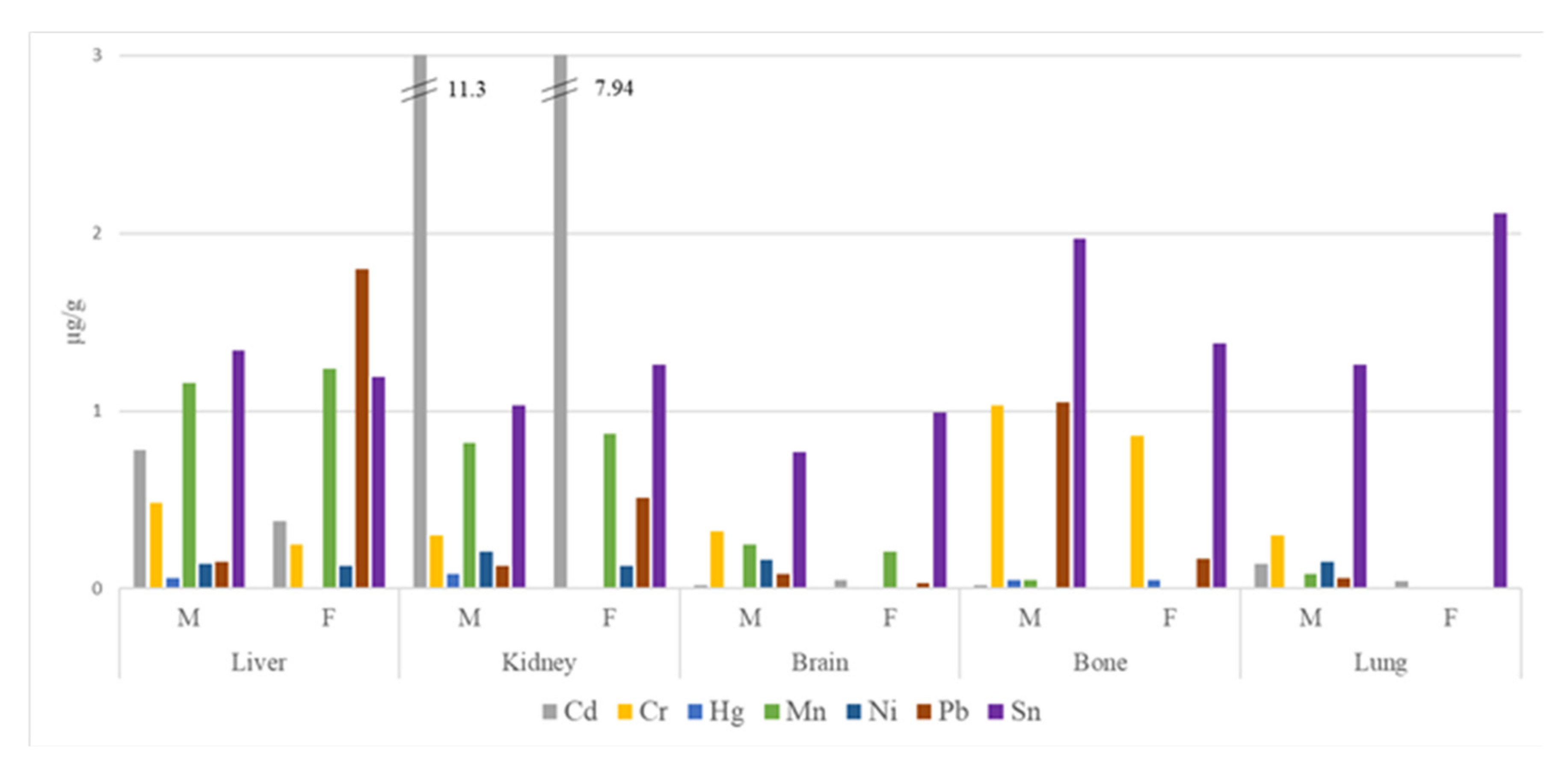
| Tissue | Mean | ± | St. Dev. | Median | Min | Max | Detection Rate (%) | |
|---|---|---|---|---|---|---|---|---|
| LIVER | As | <0.10 | − | ND | ND | 0 | ||
| Be | <0.10 | − | ND | ND | 0 | |||
| Cd | 0.76 | ± | 0.61 | 0.64 | 0.13 | 2.77 | 100 | |
| Cr | 0.47 | ± | 0.17 | 0.54 | <0.50 | 0.74 | 65 | |
| Hg | 0.06 | ± | 0.04 | 0.05 | <0.10 | 0.23 | 5 | |
| Mn | 1.16 | ± | 0.28 | 1.18 | 0.62 | 1.83 | 100 | |
| Ni | 0.14 | ± | 0.06 | 0.13 | <0.25 | 0.35 | 10 | |
| Pb | 0.23 | ± | 0.42 | 0.08 | <0.025 | 1.80 | 80 | |
| Sn | 1.33 | ± | 0.52 | 1.21 | 0.88 | 3.30 | 100 | |
| Tl | <0.025 | − | ND | ND | 0 | |||
| V | <0.50 | − | ND | ND | 0 | |||
| KIDNEY | As | <0.10 | − | ND | ND | 0 | ||
| Be | <0.10 | − | ND | ND | 0 | |||
| Cd | 11.10 | ± | 8.17 | 8.06 | 0.83 | 44.35 | 100 | |
| Cr | 0.29 | ± | 0.11 | 0.25 | <0.50 | 0.58 | 15 | |
| Hg | 0.08 | ± | 0.09 | 0.05 | <0.10 | 0.34 | 15 | |
| Mn | 0.82 | ± | 0.23 | 0.87 | 0.32 | 1.22 | 100 | |
| Ni | 0.21 | ± | 0.23 | 0.13 | <0.25 | 1.08 | 15 | |
| Pb | 0.15 | ± | 0.17 | 0.09 | <0.025 | 0.65 | 85 | |
| Sn | 1.04 | ± | 0.28 | 1.08 | 0.70 | 1.94 | 100 | |
| Tl | <0.025 | − | ND | ND | 0 | |||
| V | <0.50 | − | ND | ND | 0 | |||
| BRAIN | As | <0.10 | − | ND | ND | 0 | ||
| Be | <0.10 | − | ND | ND | 0 | |||
| Cd | 0.02 | ± | 0.02 | 0.01 | <0.025 | 0.06 | 30 | |
| Cr | 0.32 | ± | 0.12 | 0.25 | <0.50 | 0.58 | 25 | |
| Hg | <0.10 | − | ND | ND | 0 | |||
| Mn | 0.25 | ± | 0.15 | 0.21 | 0.16 | 0.86 | 100 | |
| Ni | 0.16 | ± | 0.11 | 0.13 | <0.25 | 0.58 | 10 | |
| Pb | 0.08 | ± | 0.12 | 0.02 | <0.025 | 0.47 | 50 | |
| Sn | 0.78 | ± | 0.25 | 0.72 | 0.54 | 1.60 | 100 | |
| Tl | <0.025 | − | ND | ND | 0 | |||
| V | <0.50 | − | ND | ND | 0 | |||
| BONE | As | <0.10 | − | ND | ND | 0 | ||
| Be | <0.10 | − | ND | ND | 0 | |||
| Cd | 0.02 | ± | 0.02 | 0.01 | <0.025 | 0.07 | 30 | |
| Cr | 1.02 | ± | 0.24 | 1.00 | 0.63 | 1.52 | 100 | |
| Hg | <0.10 | − | ND | ND | 0 | |||
| Mn | 0.05 | ± | 0.04 | 0.03 | <0.05 | 0.16 | 40 | |
| Ni | <0.25 | − | ND | ND | 0 | |||
| Pb | 1.00 | ± | 1.33 | 0.54 | <0.025 | 5.39 | 95 | |
| Sn | 1.94 | ± | 0.64 | 1.73 | 1.07 | 3.51 | 100 | |
| Tl | <0.025 | − | ND | ND | 0 | |||
| V | <0.50 | − | ND | ND | 0 | |||
| LUNG | As | <0.10 | − | ND | ND | 0 | ||
| Be | <0.10 | − | ND | ND | 0 | |||
| Cd | 0.13 | ± | 0.18 | 0.04 | <0.025 | 0.62 | 65 | |
| Cr | 0.30 | ± | 0.12 | 0.25 | <0.50 | 0.62 | 15 | |
| Hg | <0.10 | − | ND | ND | 0 | |||
| Mn | 0.08 | ± | 0.04 | 0.07 | <0.05 | 0.15 | 75 | |
| Ni | 0.15 | ± | 0.07 | 0.13 | <0.25 | 0.40 | 10 | |
| Pb | 0.05 | ± | 0.11 | 0.01 | <0.025 | 0.42 | 35 | |
| Sn | 1.31 | ± | 0.33 | 1.20 | 0.91 | 2.11 | 100 | |
| Tl | <0.025 | − | ND | ND | 0 | |||
| V | <0.50 | − | ND | ND | 0 |
| Tissue | % Variation | |||||||
|---|---|---|---|---|---|---|---|---|
| 1998 | 2003 | 2007 | 2013 | 2019 | 1998–2019 | 2013–2019 | ||
| LIVER | As | <0.05 | <0.05 | 0.07 | <0.05 | <0.10 | - | - |
| Be | <0.02 | <0.05 | <0.03 | <0.05 | <0.10 | - | - | |
| Cd | 0.95 | 1.36 | 0.8 | 1.38 | 0.76 | −20 *** | −45 ** | |
| Cr | 0.26 | <0.25 | 0.63 | 0.66 | 0.47 | 81 | −29 | |
| Hg | 0.2 | 0.14 | 0.14 | <0.05 | 0.06 | −70 * | - | |
| Mn | 1.28 | 1.07 | 0.99 | 1.45 | 1.16 | −9 | −20 | |
| Ni | 0.09 | <0.1 | 0.07 | <0.10 | 0.14 | 56 | - | |
| Pb | 2.56 | 0.3 | 0.35 | 0.18 | 0.23 | −91 *** | 28 | |
| Sn | 5.06 | 0.19 | 0.07 | <0.05 | 1.33 | −74 | - | |
| Tl | <0.02 | <0.01 | <0.01 | <0.03 | <0.025 | - | - | |
| V | <0.12 | <0.25 | <0.25 | <0.10 | <0.50 | - | - | |
| KIDNEY | As | <0.05 | <0.05 | 0.06 | <0.05 | <0.10 | - | - |
| Be | <0.02 | <0.05 | <0.03 | <0.05 | <0.10 | - | - | |
| Cd | 17.52 | 17.46 | 14.72 | 21.15 | 11.1 | −37 | −48 | |
| Cr | 0.09 | <0.25 | 0.42 | 0.66 | 0.29 | 222 *** | −56 ** | |
| Hg | 0.33 | 0.23 | 0.3 | 0.15 | 0.08 | −76 * | −47 * | |
| Mn | 1.01 | 0.74 | 0.78 | 1.09 | 0.82 | −19 | −25 | |
| Ni | <0.01 | <0.10 | <0.05 | <0.10 | 0.21 | - | - | |
| Pb | <0.02 | 0.06 | 0.77 | 0.1 | 0.15 | - | 50 | |
| Sn | 1.66 | 0.17 | 0.05 | <0.05 | 1.04 | −37 * | - | |
| Tl | <0.02 | <0.01 | <0.01 | <0.03 | <0.025 | - | - | |
| V | <0.12 | <0.25 | <0.25 | <0.10 | <0.50 | - | - | |
| BRAIN | As | <0.05 | <0.05 | <0.05 | <0.05 | <0.10 | - | - |
| Be | <0.02 | <0.05 | <0.03 | <0.05 | <0.10 | - | - | |
| Cd | 0.03 | 0.02 | 0.32 | <0.05 | 0.02 | −33 | - | |
| Cr | 0.22 | <0.25 | 0.45 | 0.57 | 0.32 | 45 | −44 | |
| Hg | <0.05 | <0.05 | 0.1 | <0.05 | <0.10 | - | - | |
| Mn | 0.22 | 0.03 | 0.24 | 0.33 | 0.25 | 14 | −24 | |
| Ni | <0.01 | <0.10 | 0.36 | <0.05 | 0.16 | - | - | |
| Pb | 1.41 | 0.06 | 0.1 | <0.05 | 0.08 | −94 *** | - | |
| Sn | 1.32 | 0.09 | 0.03 | <0.05 | 0.78 | −41 | - | |
| Tl | <0.02 | <0.01 | <0.01 | <0.05 | <0.025 | - | - | |
| V | <0.12 | <0.25 | 0.28 | <0.05 | <0.50 | - | - | |
| BONE | As | 0.06 | <0.05 | 0.19 | <0.05 | <0.10 | - | - |
| Be | <0.02 | <0.05 | 0.03 | <0.05 | <0.10 | - | - | |
| Cd | 0.04 | 0.05 | 0.04 | <0.03 | 0.02 | −50 ** | - | |
| Cr | 0.51 | <0.25 | 1.39 | 1.38 | 1.02 | 100 *** | −26 | |
| Hg | <0.05 | <0.05 | 0.05 | <0.05 | <0.10 | - | - | |
| Mn | 0.06 | <0.03 | 0.25 | 0.13 | 0.05 | −17 | −62 * | |
| Ni | 0.64 | 1.16 | 1.53 | <0.10 | <0.25 | - | - | |
| Pb | 3.99 | 2.11 | 2.66 | 1.39 | 1 | −75 *** | −28 | |
| Sn | 7.4 | 0.34 | 0.31 | 0.17 | 1.94 | −74 *** | 1041 *** | |
| Tl | <0.02 | <0.01 | <0.01 | <0.03 | <0.025 | - | - | |
| V | <0.12 | <0.25 | <0.25 | <0.10 | <0.50 | - | - | |
| LUNG | As | <0.05 | <0.05 | 0.14 | <0.05 | <0.10 | - | - |
| Be | <0.02 | <0.05 | <0.03 | <0.05 | <0.10 | - | - | |
| Cd | 0.42 | 0.18 | 0.27 | 0.26 | 0.13 | −69 | −50 | |
| Cr | 0.33 | 0.25 | 0.58 | 0.64 | 0.3 | −9 | −53 * | |
| Hg | <0.05 | <0.05 | <0.05 | <0.05 | <0.10 | - | - | |
| Mn | 0.13 | 0.04 | 0.3 | 0.21 | 0.08 | −38 | −62 ** | |
| Ni | 0.08 | 0.12 | 0.07 | <0.10 | 0.15 | 88 ** | - | |
| Pb | 2.27 | 0.13 | 0.08 | 0.05 | 0.05 | −98 *** | 0 | |
| Sn | 2.16 | 0.2 | 0.07 | <0.05 | 1.31 | −39 | - | |
| Tl | <0.02 | <0.01 | <0.01 | <0.03 | <0.025 | - | - | |
| V | <0.12 | <0.25 | 0.58 | <0.10 | <0.50 | - | - | |
| Tissue | <35 years (n = 3) | 35–65 years (n = 10) | >65 years (n = 7) | |||||||
|---|---|---|---|---|---|---|---|---|---|---|
| Mean | ± | St. Dev. | Mean | ± | St. Dev. | Mean | ± | St. Dev. | ||
| LIVER | As | <0.10 | <0.10 | <0.10 | ||||||
| Be | <0.10 | <0.10 | <0.10 | |||||||
| Cd | 0.99 | ± | 0.53 | 0.55 | ± | 0.34 | 0.96 | ± | 0.86 | |
| Cr | 0.47 | ± | 0.19 | 0.46 | ± | 0.19 | 0.48 | ± | 0.16 | |
| Hg | <0.10 | <0.10 | 0.08 | |||||||
| Mn | 1.32 | ± | 0.45 | 1.06 | ± | 0.24 | 1.24 | ± | 0.25 | |
| Ni | 0.13 | ± | 0.00 | 0.14 | ± | 0.05 | 0.16 | ± | 0.08 | |
| Pb | 0.23 | ± | 0.28 | 0.33 | ± | 0.58 | 0.08 | ± | 0.09 | |
| Sn | 2.03 | ± | 1.20 | 1.20 | ± | 0.30 | 1.21 | ± | 0.11 | |
| Tl | <0.025 | <0.025 | <0.025 | |||||||
| V | <0.50 | <0.50 | <0.50 | |||||||
| KIDNEY | As | <0.10 | <0.10 | <0.10 | ||||||
| Be | <0.10 | <0.10 | <0.10 | |||||||
| Cd | 5.50 | ± | 3.54 | 12.98 | ± | 8.99 | 10.81 | ± | 7.94 | |
| Cr | 0.33 | ± | 0.15 | 0.28 | ± | 0.10 | 0.29 | ± | 0.11 | |
| Hg | 0.15 | ± | 0.17 | 0.08 | ± | 0.09 | <0.10 | |||
| Mn | 1.04 | ± | 0.34 | 0.84 | ± | 0.20 | 0.69 | ± | 0.22 | |
| Ni | <0.25 | 0.25 | ± | 0.31 | 0.18 | ± | 0.14 | |||
| Pb | 0.06 | ± | 0.10 | 0.20 | ± | 0.22 | 0.11 | ± | 0.08 | |
| Sn | 0.85 | ± | 0.18 | 1.15 | ± | 0.31 | 0.96 | ± | 0.22 | |
| Tl | <0.025 | <0.025 | <0.025 | |||||||
| V | <0.50 | <0.50 | <0.50 | |||||||
| BRAIN | As | <0.10 | <0.10 | <0.10 | ||||||
| Be | <0.10 | <0.10 | <0.10 | |||||||
| Cd | 0.03 | ± | 0.02 | 0.02 | ± | 0.02 | 0.02 | ± | 0.01 | |
| Cr | 0.34 | ± | 0.15 | 0.33 | ± | 0.14 | 0.29 | ± | 0.10 | |
| Hg | <0.10 | <0.10 | <0.10 | |||||||
| Mn | 0.22 | ± | 0.05 | 0.29 | ± | 0.20 | 0.21 | ± | 0.03 | |
| Ni | 0.13 | ± | 0.00 | 0.17 | ± | 0.14 | 0.16 | ± | 0.09 | |
| Pb | 0.11 | ± | 0.11 | 0.05 | ± | 0.04 | 0.12 | ± | 0.19 | |
| Sn | 0.73 | ± | 0.10 | 0.79 | ± | 0.32 | 0.80 | ± | 0.17 | |
| Tl | <0.025 | <0.025 | <0.025 | |||||||
| V | <0.50 | <0.50 | <0.50 | |||||||
| LUNG | As | <0.10 | <0.10 | <0.10 | ||||||
| Be | <0.10 | <0.10 | <0.10 | |||||||
| Cd | 0.10 | ± | 0.16 | 0.16 | ± | 0.21 | 0.10 | ± | 0.14 | |
| Cr | 0.37 | ± | 0.21 | 0.28 | ± | 0.08 | 0.30 | ± | 0.14 | |
| Hg | <0.10 | <0.10 | <0.10 | |||||||
| Mn | 0.10 | ± | 0.03 | 0.07 | ± | 0.05 | 0.08 | ± | 0.04 | |
| Ni | <0.25 | 0.15 | ± | 0.09 | 0.15 | ± | 0.06 | |||
| Pb | <0.025 | 0.06 | ± | 0.13 | 0.07 | ± | 0.10 | |||
| Sn | 1.30 | ± | 0.30 | 1.26 | ± | 0.33 | 1.38 | ± | 0.36 | |
| Tl | <0.025 | <0.025 | <0.025 | |||||||
| V | <0.50 | <0.50 | <0.50 | |||||||
| BONE | As | <0.10 | <0.10 | <0.10 | ||||||
| Be | <0.10 | <0.10 | <0.10 | |||||||
| Cd | <0.025 | 0.03 | ± | 0.02 | 0.02 | ± | 0.01 | |||
| Cr | 0.70 | ± | 0.06 | 1.06 | ± | 0.31 | 1.11 | ± | 0.12 | |
| Hg | <0.10 | <0.10 | <0.10 | |||||||
| Mn | 0.05 | ± | 0.02 | 0.04 | ± | 0.07 | 0.06 | ± | 0.05 | |
| Ni | 0.13 | ± | 0.00 | 0.13 | ± | 0.00 | 0.13 | ± | 0.00 | |
| Pb | 0.06 | ± | 0.07 | 0.43 | ± | 0.34 | 2.23 | ± | 1.69 | |
| Sn | 2.39 | ± | 0.69 | 1.80 | ± | 0.36 | 1.94 | ± | 0.61 | |
| Tl | <0.025 | <0.025 | <0.025 | |||||||
| V | <0.50 | <0.50 | <0.50 | |||||||
© 2020 by the authors. Licensee MDPI, Basel, Switzerland. This article is an open access article distributed under the terms and conditions of the Creative Commons Attribution (CC BY) license (http://creativecommons.org/licenses/by/4.0/).
Share and Cite
García, F.; Marquès, M.; Barbería, E.; Torralba, P.; Landin, I.; Laguna, C.; Domingo, J.L.; Nadal, M. Biomonitoring of Trace Elements in Subjects Living Near a Hazardous Waste Incinerator: Concentrations in Autopsy Tissues. Toxics 2020, 8, 11. https://doi.org/10.3390/toxics8010011
García F, Marquès M, Barbería E, Torralba P, Landin I, Laguna C, Domingo JL, Nadal M. Biomonitoring of Trace Elements in Subjects Living Near a Hazardous Waste Incinerator: Concentrations in Autopsy Tissues. Toxics. 2020; 8(1):11. https://doi.org/10.3390/toxics8010011
Chicago/Turabian StyleGarcía, Francisco, Montse Marquès, Eneko Barbería, Pilar Torralba, Inés Landin, Carlos Laguna, José L. Domingo, and Martí Nadal. 2020. "Biomonitoring of Trace Elements in Subjects Living Near a Hazardous Waste Incinerator: Concentrations in Autopsy Tissues" Toxics 8, no. 1: 11. https://doi.org/10.3390/toxics8010011