Article

Anti-Haemophilus Activity of Selected Essential Oils Detected by TLC-Direct Bioautography and Biofilm Inhibition

1
Department of Pharmacognosy, Faculty of Pharmacy, University of Pécs, H-7624 Pécs, Hungary
2
Institute of Pharmaceutical Technology and Biopharmacy, Faculty of Pharmacy, University of Pécs, H-7624 Pécs, Hungary
3
Department of Microbiology, Faculty of Science and Informatics, University of Szeged, H-6726 Szeged, Hungary
4
Department of Medical Microbiology and Immunology, Medical School, University of Pécs, H-7624 Pécs, Hungary
5
Institute of Pharmacognosy, Faculty of Pharmacy, Semmelweis University, H-1085 Budapest, Hungary
6
Department of Genetics and Molecular Biology, Institute of Biology, Faculty of Sciences, University of Pécs, H-7624 Pécs, Hungary
7
Department of Food Engineering, Faculty of Engineering, University of Szeged, H-6724 Szeged, Hungary
*
Author to whom correspondence should be addressed.
Molecules 2019, 24(18), 3301; https://doi.org/10.3390/molecules24183301
Submission received: 7 August 2019 / Revised: 5 September 2019 / Accepted: 10 September 2019 / Published: 11 September 2019
(This article belongs to the Section Natural Products Chemistry)

Abstract

:
Essential oils (EOs) are becoming increasingly popular in medical applications because of their antimicrobial effect. Direct bioautography (DB) combined with thin layer chromatography (TLC) is a screening method for the detection of antimicrobial compounds in plant extracts, for example, in EOs. Due to their lipophilic character, the common microbiological assays (etc. disk diffusion) could not provide reliable results. The aim of this study was the evaluation of antibacterial and anti-biofilm properties of the EO of cinnamon bark, clove, peppermint, thyme, and their main components against Haemophilus influenzae and H. parainfluenzae. Oil in water (O/W) type Pickering nano-emulsions stabilized with silica nanoparticles from each oil were prepared to increase their water-solubility. Samples with Tween80 surfactant and absolute ethanol were also used. Results showed that H. influenzae was more sensitive to the EOs than H. parainfluenzae (except for cinnamon bark oil). In thin layer chromatography-direct bioautography (TLC-DB) the ethanolic solutions of thyme oil presented the best activity against H. influenzae, while cinnamon oil was the most active against H. parainfluenzae. Pickering nano-emulsion of cinnamon oil inhibited the biofilm formation of H. parainfluenzae (76.35%) more efficiently than samples with Tween80 surfactant or absolute ethanol. In conclusion, Pickering nano-emulsion of EOs could inhibit the biofilm production effectively.

1. Introduction

Essential oils (EOs) have been widely used for antimicrobial, medicinal and cosmetic purposes. In the European Union, these plant extracts can be found in foods (as flavorings), perfumes (as fragrances) and pharmaceuticals (as active ingredients) [1,2]. The significance of the EOs and their components as antimicrobial substances are increasing, due to antibiotic-resistant pathogens [3,4]. EOs may represent the richest available reservoir of novel therapeutics [5]. However, the reliability of the common antimicrobial assays used for EOs is questionable because of their non-water soluble property [6].
Direct bioautography (DB) combined with thin layer chromatography (TLC) is a rapid and sensitive screening method for the detection of antimicrobial compounds. Test microorganism is capable of growing directly on the TLC plate, so each step of the assay is performed on the sorbent. Similar to the widely used antimicrobial screening methods (e.g., broth macro- and microdilution), thin layer chromatography-direct bioautography (TLC-DB) should be carried out under controlled conditions, since the experimental parameters (for example, solvents, sample application, resolution of compounds, type of test microorganism, incubation time) may influence the result [7]. This assay is capable of testing multicomponent and lipophilic extracts, e.g., EOs. The applicability of bioautography to detect antimicrobial compounds effective against plant and human pathogenic bacteria has been reported in the literature [8,9,10]. However, there is only a few studies in which respiratory pathogens were included in TLC-DB method [11,12]. According to the data of the World Health Organization (WHO), lower respiratory tract infections are responsible for 5% (3.1 million people) of deaths worldwide [13]. EOs offer effective treatment in the respiratory tract infections because of their volatility and antibacterial effect. We tested other respiratory tract pathogens, such as Streptococcus species and Pseudomonas aeruginosa, too [14,15]. The mode of action of EOs is not fully understood, but the prevention of the bacterial biofilm formation may be suggested. Therefore, we decided to study the biofilm inhibition potential of our EO samples, including respiratory tract pathogens into our experiments.
A biofilm comprises any group of microorganisms in which cells stick to each other and often also to a surface. These adherent cells become embedded within a slimy extracellular matrix that is composed of extracellular polymeric substances (EPS) [16,17]. Biofilms have been found to be involved in a wide variety of microbial infections (e.g., bacterial vaginosis, urinary tract infections, catheter infections, middle-ear infections) in the body, by one estimate 80% of all infections. About 80% of cystic fibrosis patients have a chronic lung infection, caused mainly by P. aeruginosa growing in a non-surface attached biofilms [18]. Infections associated with the biofilm growth usually are challenging to eradicate. It is mostly due to the fact that mature biofilms display tolerance towards antibiotics and the immune response [19]. Most of the publications focus on the inhibition of bacterial biofilm produced by foodborne or dental pathogens [20,21]. Therefore, it was worth involving the pathogens of respiratory tract infections in the studies, in which the effect of our EO samples on biofilm formation produced by respiratory tract bacteria was examined.
Therefore, the aim of this study was the evaluation of antibacterial properties of the EO of cinnamon bark (Cinnamomum verum J. Presl.), clove (Syzygium aromaticum (L.) Merr. And Perry), peppermint (Mentha x piperita L.), thyme (Thymus vulgaris L.), and their main components (trans-cinnamaldehyde, eugenol, menthol, and thymol) against the Gram-negative bacteria, Haemophilus influenzae and H. parainfluenzae using TLC-DB. Furthermore, the biofilm inhibition of different formulation of our EO samples was also performed. The chemical composition of the EOs was measured by gas-chromatography-mass spectrometry (GC-MS).

2. Results

2.1. Chemical Composition of EOs

Chemical analyses of EOs were performed by GC-FID and GC-MS techniques. Identified compounds and percentage evaluation of the oils are shown in Table 1.
Eugenol (78.8%) was the main component in the EO of clove. Cinnamaldehyde (63.7%) was the main component in the oil of cinnamon bark. Menthol (50.4%) was the characteristic compound in the peppermint EO. In the thyme oil thymol (39.8%) was identified as the main constituent.

2.2. TLC-DB

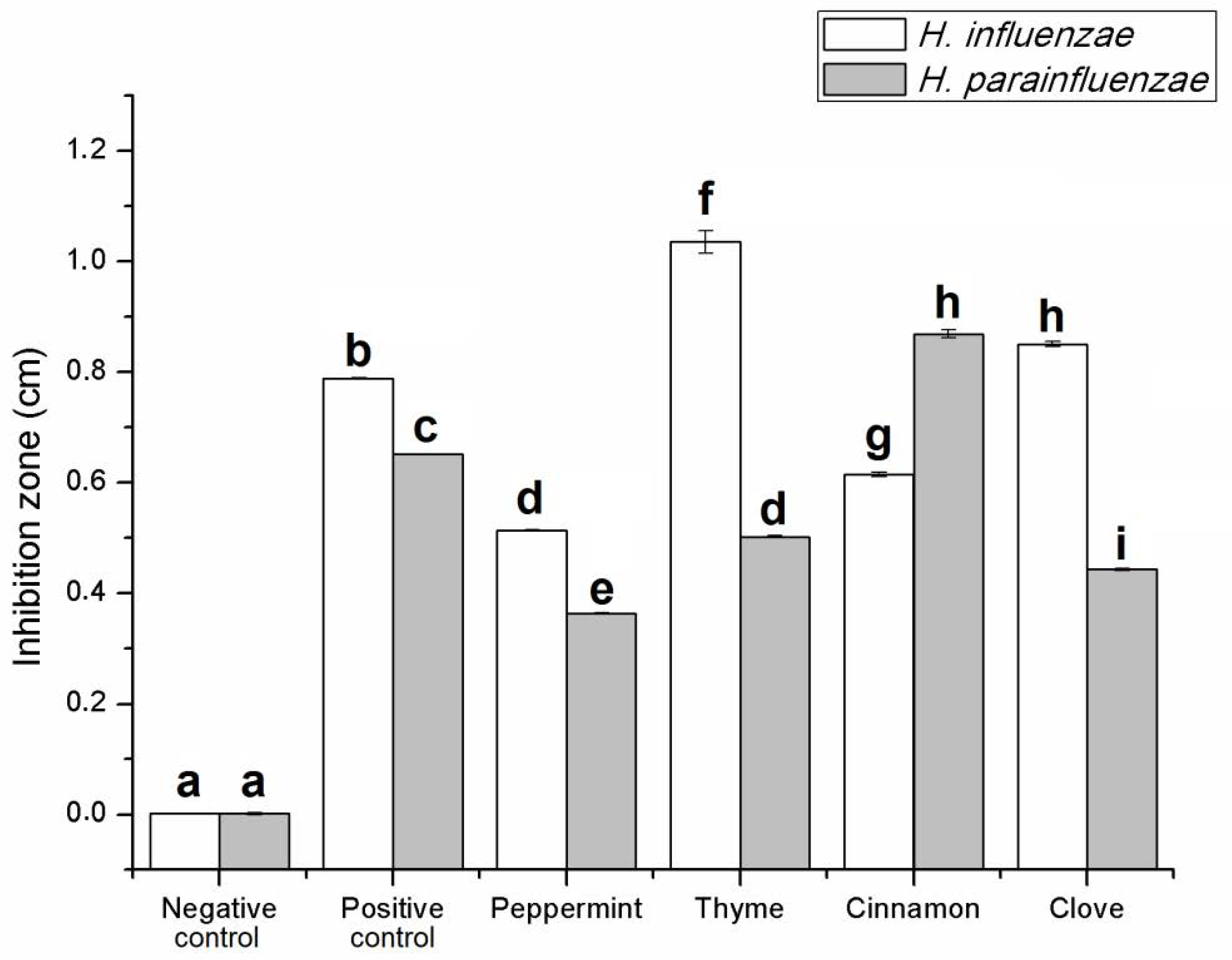
2.2.1. Antibacterial Activity of EOs

In the TLC-DB method, the activity of the EOs without and with separation was tested against H. influenzae and H. parainfluenzae. In the case of activity of the EOs without separation, the development with mobile phase was not prepared; therefore, the activity of the “total” extract (EO) was examined [22]. Figure 1 shows the activity of the EOs without TLC separation. The diameter of the inhibition zones was expressed in cm. From the stock solution of EOs 1 µL was applied (equivalent to 0.2 mg undiluted EO) on the TLC plate. The H. influenzae was more sensitive to the EOs than H. parainfluenzae (except for cinnamon bark oil). Absolute ethanol as negative control did not inhibit the growth of both bacteria. The 0.2 µL solution of the antibiotic sample (amikacin, equivalent to 0.05 mg antibiotic) was effective against both Haemophilus strains. Ethanolic solutions of thyme oil presented the best activity in case of H. influenzae, while cinnamon bark oil was the most potent against H. parainfluenzae. Peppermint showed moderate activity in case of both pathogens (0.51 cm against H. influenzae and 0.31 cm against H. parainfluenzae). In our TLC-DB assay, the tested EO samples did not show more effective activity than the positive control, but their combination (antibiotic and EO) might be the aims of the following assays.

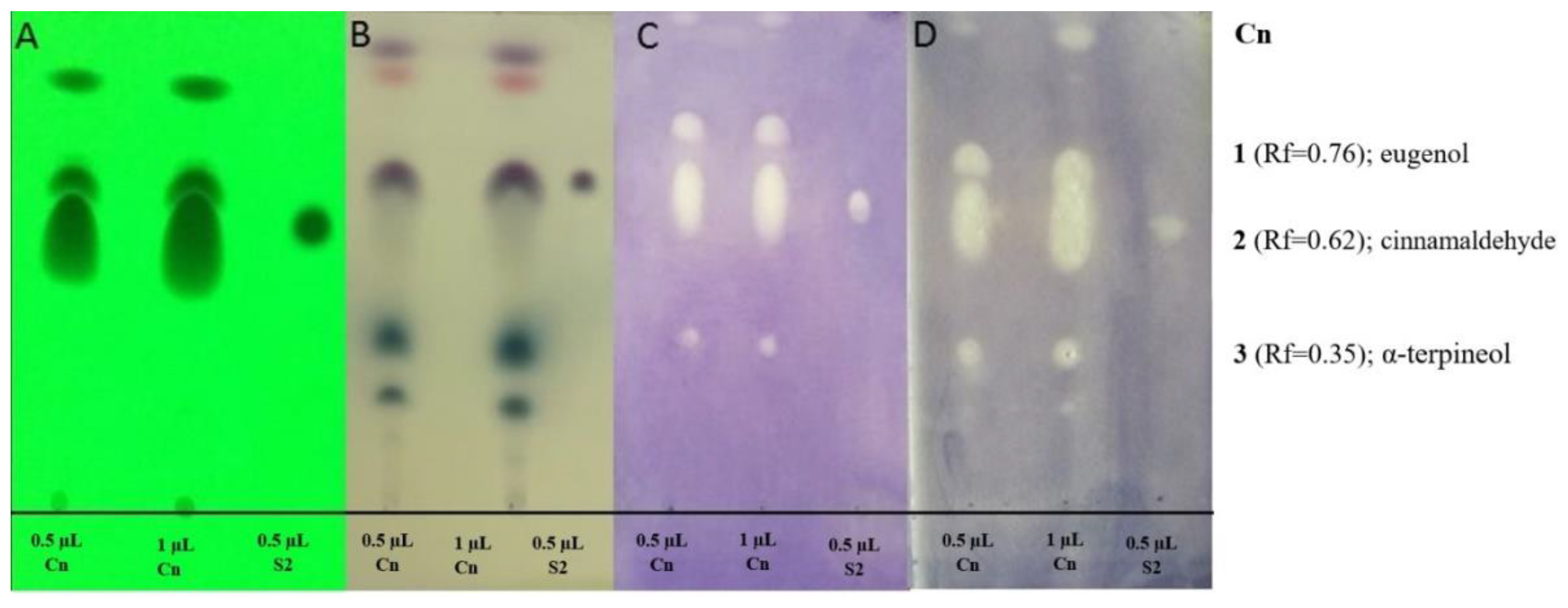
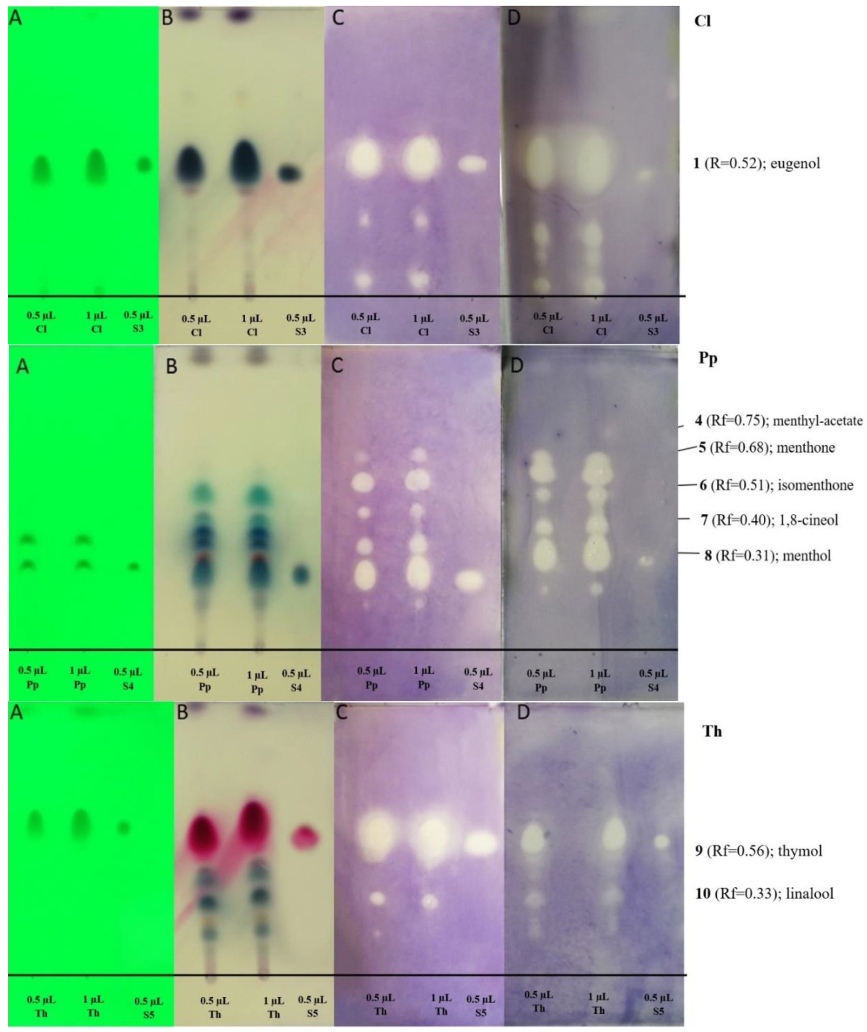
2.2.2. Antibacterial Activity of Main Components of EOs by TLC-DB Method

Generally, the antibacterial activity of the EOs seems to be associated with their most abundant compounds, but the effect of the minor compounds should also be taken into consideration. In the oil of cinnamon bark, cinnamic aldehyde and eugenol components, as well as their standards, showed activity in the case of both bacteria (Figure 2). Moreover, α-terpineol (Rf = 0.35) in the EO of cinnamon showed activity against both bacteria. A-Terpineol was identified according to the GC-MS result and Wagner and Bladt [23]. Eugenol, as the main compound of the clove oil (Rf = 0.52), was active against both Haemophilus strains (Figure 2). In the peppermint oil, several compounds had antibacterial activity at the tested concentration. Menthol (Rf = 0.31) in the peppermint oil and the standard of menthol inhibited the growth of bacteria (Figure 2). Other active compounds of peppermint oil include 1,8-cineole, isomenthon, menthon, and isomenthyl acetate (according to GC-MS and Wagner and Bladt [23]. In the oil of thyme, thymol-carvacrol and the standard, thymol had antibacterial activity (Figure 2). At Rf = 0.33, linalool was identified as an active compound according to GC-MS result and Wagner and Bladt [23].

2.3. Preparation and Characterization of Pickering Nano-Emulsions of EOs

The preparation of the stable Pickering nano-emulsions has been described before [24]. We had considered the emulsion to be stable when the droplet size did not change for at least 24 h; creaming, sedimentation and disproportionation did not occur. The droplet size and stability of Pickering and conventional emulsions of EOs can be seen in Table 2. The Pickering emulsions are more stable than conventional emulsions, the difference in stability is most obvious in a case of peppermint EO; its Pickering emulsion form remains stable for at least five months, while its conventional emulsion is stable for only one month. In the case of clove EO, there is no difference in stability between the Pickering and conventional emulsions; they remained stable for only two weeks. Conventional emulsions are stabilized with Tween 80, while Pickering nano-emulsions were stabilized with silica nanoparticles with a mean size of 20 nm and surface modified with ethyl groups (20ET).

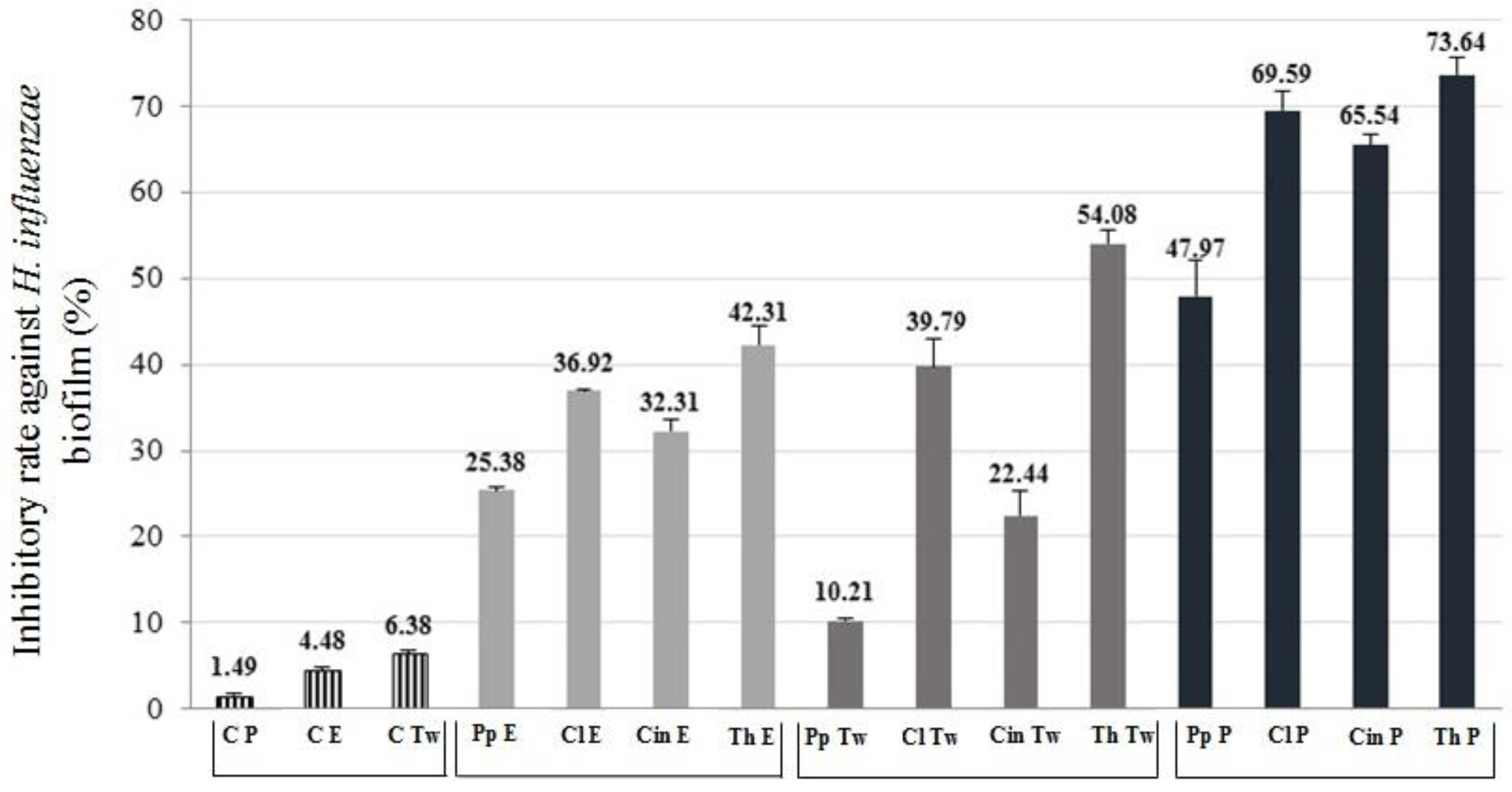
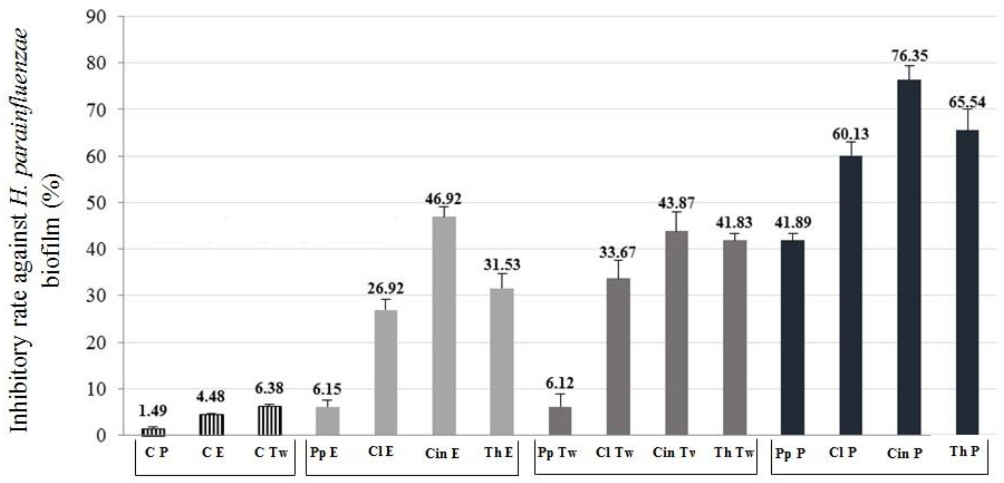
2.4. Anti-Biofilm Activity

We examined the inhibitory effect of the EOs (half of the MIC concentration) according to a previous study [25]. Three different formulations of the EOs were tested. The anti-biofilm formation activity of the EOs was calculated and demonstrated in the term of inhibitory rate according to the following equation: Inhibitory rate = (1−S/C) × 100% (C and S were defined as the average absorbance of control and sample groups respectively) [26]. Our results showed that not only the EOs samples with Tween80 surfactant had an antibacterial effect, but also the EO samples with absolute ethanol and their Pickering nano-emulsion forms inhibited the biofilm formation. Among the controls, Pickering nano-emulsion without EO showed the lowest activity (1.49% inhibitory rate) (Figure 3 and Figure 4). It should be highlighted that the Pickering nano-emulsions were the most effective form of EOs against biofilms. The Pickering nano-emulsion of thyme oil showed the highest inhibitory rate (73.64%) against H. influenzae. Cinnamon oil in Pickering nano-emulsion form inhibited the biofilm formation of H. parainfluenzae (76.35%) most effectively. Among the different formulations, the EO samples with Tween80 showed the lowest activity against biofilm formation. Moreover, in the case of both bacteria, the peppermint oil was the least effective EO among the investigated oil samples. The results of biofilm inhibition assay were in harmony with the results of TLC-DB assay, thyme and cinnamon oils were the most effective among the investigated oils, besides clove also showed potent activity against these respiratory bacteria.

3. Discussion

Plants produced a wide variety of secondary metabolites that exhibited antimicrobial activity against a variety of pathogens (bacteria, fungi, and viruses) [27,28,29]. Several suggestions, (or hypothesis) can be found in the literature about their antifungal, and antibacterial mode of action, but some of them need clarification. In this study, the antibacterial and anti-biofilm effects of clove, cinnamon, thyme, and peppermint oils were investigated against H. influenzae and H. parainfluenzae. The most accepted mechanism of EOs revealed that they could disrupt cell wall and cytoplasmic membrane, leading to lysis and leakage of intracellular compounds [12,30,31,32]. Many bacteria disclosed a high sensitivity to EOs, especially H. influenzae, Stenotrophomonas maltophilia, Streptococcus pneumoniae, S. pyogenes, and S. agalactiae. Cinnamon, thyme and clove oils showed the strongest inhibitory activity against several bacteria, including H. influenzae, S. pyogenes and S. agalactiae, even against multi-resistant strains [11]. However, the common antibacterial assays (e.g., disk diffusion) are not an appropriate method for non-water soluble extracts and compounds. Therefore, in this study, we focused on the antibacterial potential and anti-biofilm activity of clove, cinnamon, thyme and peppermint oils against H. influenzae (DSM 4690) and H. parainfluenzae (DSM 8978) using TLC-DB and biofilm inhibitory assays.
TLC-DB is a directly combined application of an analytical method with an in situ bioassay that allows rapid identification of the active compound or compounds in a complex mixture. To the best of our knowledge, we optimized this technique first using Haemophilus species. The TLC-DB was optimized for two Haemophilus species, but it is necessary to note that attention should be paid to the parameters (e.g., incubation time, the composition of agar for growing the bacterium, etc.) of TLC-DB, which was also confirmed in a previous study [7].
Fabio et al. described that Haemophilus species were sensitive to EOs in the following order: Thyme, cinnamon, clove, eucalyptus, sage, and lavender. We determined the highest activity of the thyme, cinnamon, and clove against H. influenzae and H. parainfluenzae, which was in parallel with the previous observations [12,14].
Our findings showed similar results with Houdkova and co-workers’s [33] and Inouye and co-workers’ [12] results regarding the activity of menthol, menthone and their derivatives, because these EO components are highly responsible for the anti-Haemophilus activity. In our research, TLC-DB was optimized with Haemophilus species, which is an appropriate assay to detect the antimicrobial activity of EO main compounds. We demonstrated the anti-Haemophilus effect of cinnamaldehyde, thymol, menthol, and eugenol. Moreover, some minor components (menthone, isomenthyl acetate, 1,8-cineole, α-terpineole, and linalool) contributed to the antibacterial activity.
In the last decade, the role of natural products derived from medicinal plants for interfering pathogenic biofilms has gained increased attention by the researches [34,35,36,37,38,39,40]. The individual components of the EOs clearly had antibacterial properties, although the mechanism is poorly understood. Therefore, the effect of EOs used in our study on biofilm formation of the two Haemophilus species was also investigated. Some previous studies from the literature have already described that cinnamon oil inhibited the biofilm of following pathogens as well, Campylobacter jejuni, Enterobacter aerogenes, E. coli, L. monocytogenes, P. aeruginosa, Salmonella enteritidis, and S. aureus [41,42,43,44] reported the inhibitory effect of menthol, menthone, pulegone, 1,8-cineole, terpinen-4-ol against the biofilm formation of Salmonella typhimurium, E. coli, Micrococcus luteus, S. aureus. Clove and thyme oil also showed strong anti-biofilm activity against several Gram-positive (e.g., S. aureus, Brochothrix thermosphacta, Lactobacillus rhamnosus, L. monocytogenes, B. subtilis, L. innocua) and Gram-negative (e.g., C. jejuni, E. aerogenes, E. coli, P. fluorescens, S. enteritidis) pathogens as well [45,46,47,48,49,50]. The biofilm inhibition of Haemophilus species has been only screened with synthetic products: Cefotaxime [51], 1,2,4-triazole-ciprofloxacin [52], garenoxacin [53]. The effect of cinnamon, thyme, clove and peppermint oil against Haemophilus species has not been tested earlier.
In the pharmaceutical technology, the formulation of the products and the water solubility of the active ingredients are highly important. In our previous experiment, three different formulations of EOs were prepared used absolute ethanol, Tween80, and Pickering nano-emulsion. Among the EOs, thyme and cinnamon produced the highest inhibitory rates and their Pickering nano-emulsions were the most effective formula [24]. This study showed that the nanotechnological formulated samples had pronounced anti-biofilm effect compared to the non-formulated EOs samples. The samples with absolute ethanol and Tween80 surfactant resulted that the biomass of Haemophilus biofilm decreased by half, but using the Pickering-emulsions the biomass of biofilm decreased to one third. Using Tween80 surfactant, it is discernible the decrease of biomass compared to the BHI control. In this case, we cannot exclude the antibacterial effect of the surfactant. The absolute ethanol itself has not strong antibacterial effect, but the EO samples with absolute ethanol resulted in the least biomass reduction.

4. Materials and Methods

4.1. Essential Oils and Their Components

The EO of clove (Batch number: H7352/1602), cinnamon bark (Batch number: I3201/1609), peppermint (Batch number: H7101/1601), and thyme (Batch number: H3981/1509) were obtained from a Hungarian company (AROMAX Zrt., Budapest, Hungary). Their chemical composition was determined by GC-MS. The main components of the EOs (eugenol, trans-cinnamaldehyde, menthol and thymol) were bought from Sigma-Aldrich (Budapest, Hungary).

4.2. GC-FID and GC-MS

One microliter of EO samples, diluted in ethanol (10 µL/mL), was injected in split mode, the injector temperature was 250 °C, and the split ratio was 1:50. The analyses were carried out with an Agilent 6890N/5973N GC-MSD (Santa Clara, CA, USA) system equipped with an Agilent SLB-5MS capillary column (30 m × 250 µm × 0.25 µm). The GC oven temperature was increased at a rate of 8 °C/min from 60 °C (3 min isothermal) to 250 °C, with a final isotherm at 250 °C for 1 min. High purity helium was used as carrier gas at 1.0 mL/min (37 cm/s) in constant flow mode. The mass selective detector (MSD) was equipped with a quadrupole mass analyser and was operated in electron ionization mode at 70 eV in full scan mode (41–500 amu at 3.2 scan/s). The data were evaluated using MSD ChemStation D.02.00.275 software (Agilent). The identification of the compounds was carried out by comparing retention times, linear retention indexes, and recorded spectra with the data of authentic standards, and the NIST 2.0 library was also used. The GC-FID were made using a Fisons GC 8000 gas chromatograph (Carlo Erba, Italy). An Rt-β-DEXm (Restek) capillary column, 30 m × 0.25 mm i.d., 0.25 μm film thickness, was used. The carrier gas was nitrogen at 6.8 mL/min flow rate. A 0.2 mL volume of a 0.1% solution of the oil was injected (1 mL EO in 1 mL chloroform). The splitless injection was carried out. The temperatures of the injector and detector were 210 °C and 240 °C, respectively. The oven temperature was increased at a rate of 8 °C/min from 60 °C to 230 °C, with a final isotherm at 230 °C for 5 min. Identification of peaks was made by retention data compared with data obtained by GC-MS and data of standards (Fluka Analytical and Sigma-Aldrich); percentage evaluation was carried out by area normalization. Three parallel measurements were made; RSD percentages were below 4.5%.
To identify the microbiologically active compounds in the separated EOs during TLC-DB, 150 µL of EO was applied onto the TLC layer as 150 mm band, and after the development, the zones of active compounds were scraped off and eluted with 0.5 mL of ethanol for GC-MS.

4.3. TLC-DB

4.3.1. Cultivation of Test Bacteria for Dipping

The antibacterial effect of EOs and their main components was screened on Haemophilus influenzae (DSM 4690) and H. parainfluenzae (DSM 8978) in the laboratory of the Department of Medical Microbiology and Immunology (Medical School, University of Pécs, Pécs, Hungary). For bioautographic assay, bacteria were grown in 100 mL Brain Heart Infusion Broth (BHI) (Sigma Aldrich Ltd., Darmstadt, Germany) with 1 mL supplement B (Diagon Kft., Budapest, Hungary) and 15 µg/mL NAD solution (1 mg/mL) at 37 °C in a shaker incubator at a speed of 60 rpm for 24 h [54]. The bacterial suspension was diluted with fresh nutrient broth to an OD600 of 0.4, which corresponds to approximately 4 × 10⁷ colony-forming units (cfu) mL.

4.3.2. Layer Chromatography

We investigated the antibacterial effect of EOs without TLC separation and the antibacterial effect of their components after TLC separation [22]. Chromatography was performed on 5 × 10 cm silica gel 60 F254 aluminum sheet TLC plates (Merck, Darmstadt, Germany). EOs were dissolved in absolute ethanol to give solutions containing 100 μL oil in 500 μL absolute ethanol, and 1.0 μL was applied to the TLC plate with Finnpipette pipettes (Merck, Darmstadt, Germany). Absolute ethanol was the solvent control and amikacin (Likacin 250 mg/mL, Lisapharma S.p.A.) as a positive control. 0.2 μL from the positive control (amikacin) and 1.0 μL from the negative control (absolute ethanol) have been applied to the TLC plate. After separation with the mobile phase, the antibacterial activity of the main EO components (thymol, menthol, trans-cinnamaldehyde, and eugenol) was also investigated by TLC-DB. The main components were dissolved in absolute ethanol to give solutions containing 20 mg/mL. From the stock solutions, 0.2 μL (0.004 mg) were applied to the plates. From the previously mentioned EO solutions, 1.0 µL was used. The position of the starting line was 1.5 cm from the bottom and 1.5 cm from the left side. The standards were applied to the TLC plates next to the spots of the oils. After sample application, the plates were developed with the previously optimized mobile phase. For the separation of EOs, toluene:ethyl acetate (95:5) and dichloromethane (in the case of cinnamon bark oil) was recommended as the mobile phase [24]. Ascendant development chromatography was used, in a saturated twin trough chamber (Camag, Muttenz, Switzerland). All TLC separations were performed at room temperature (20 °C). After chromatographic separation, the absorbent layers were dried at 90 °C, for 5 min to remove the solvent completely. Ethanolic vanillin–sulfuric acid reagent [24] was used to visualize the separated compounds. Detection of the separated compounds was performed on Rf value and color of the standards. Evaluation of the separated compounds was also performed under UV light at 254 nm. It should be noted that the TLC plates for bioautography were not treated with ethanolic vanillin–sulfuric acid reagent, because this step interferes with the microbiological steps of TLC-DB.

4.3.3. Post-Chromatographic Detection

After layer chromatography, the TLC plates were treated with the suspension of H. influenzae and H. parainfluenzae, respectively. Layers were dipped into a 100 mL of bacterial suspension to assure a homogenous distribution and adhesion of bacteria onto the surface of the layers. After immersion, the layers were transferred into a low-wall horizontal chamber (chamber dimension: 20 × 14.5 × 5 cm) and incubated for 2 h at 37 °C. Thereafter for visualization of antibacterial spots, TLC plates were immersed into the aqueous solution of 3-(4,5-dimethylthiazol-2-yl)-2,5-diphenyltetrazolium bromide (MTT, 0.05 g/85 mL) (Sigma Aldrich Ltd., Darmstadt, Germany), for 5 s, and then incubated at 37 °C for 24 h. On the TLC plate, metabolically active bacteria convert the tetrazolium salt, MTT, into formazan dye. White spots (as inhibition zones) against the bluish-violet background indicated the lack of dehydrogenase activity, due to the antibacterial activity of the tested EO or their main compounds. The inhibitory zones (expressed in cm) of EOs without separation were measured with Motic Images Plus 2.0 program (ver. 2.0., Motic, Hong Kong, China).

4.4. Statistical Analyses

Statistical analyses were made in R, version 3.1.2 [55]. The measured diameters were analyzed with linear model [56] using the function lm. In our model, the explanatory variables (bacterial species, EO and quantity of EOs), treated as fixed factors. No data transformation was made. Checking the need for transformation was based on graphical evaluation, according to Crawley [57]. For pair-wise comparisons, Tukey post-hoc tests were conducted in with multicomp-package [58] to compare the difference among all experimental set-ups.

4.5. Biofilm Inhibition Experiments

4.5.1. Broth Macrodilution Test (BDT)

During biofilm inhibition experiments minimum inhibitory concentration/2 (MIC/2) values of the EOs were used. The MICs were determined with broth macrodilution test (BDT) based on Ács et al. [14].

4.5.2. Preparation of Pickering Emulsion of the EOs

Because of the volatility and non-water soluble characters of EOs, we wanted to make the water-soluble formulation of our EO samples, and eliminate such kind of solvents, e.g., dimethyl sulfoxide (DMSO), from the assay which can generally be used in the microbiological assays, but may influence the results.
For the synthesis and surface modification of silica nanoparticles tetraethoxysilane [TEOS], (Alfa Aesar, [Haverhill, MA, USA], purity 98%), ethyltriethoxysilane [ETES] (Alfa Aesar [USA], purity 96%), absolute ethanol (VWR Chemicals [Budapest, Hungary], AnalaR Normapur, purity ≥ 99.8%), 28 w/w% ammonium solution (VWR Chemicals [Hungary], AnalaR Normapur, analytical reagent) were used. The stabilizing agent of Pickering emulsions was nanoparticle suspensions, of conventional emulsions was Tween®80 (Polysorbate80, Acros Organics, Princeton, NJ, USA).

Synthesis, Surface Modification and Characterization of Silica Nanoparticles

Synthesis of hydrophilic silica was based on the work of Stöber, Fink and Bohn [59]. Previously, we performed the optimization of size-controlled silica nanoparticle synthesis process and their surface modification with ETES. Furthermore, the nanoparticle characterization was also completed. The details can be read in our paper [25].

Preparation and Characterization of O/W (Oil/Water) Type EOs Emulsions

For the preparation of conventional, surfactant stabilized the emulsion, Tween80 non-ionic surfactant was used, because this chemical is widely used in microbiological experiments and protocols, for solubilizing the non-water soluble, lipophilic molecules, e.g., EOs. We have used 20ET nanoparticles [51] as stabilizing agents for preparation of Pickering emulsions. The concentration of emulsion stabilizing agents was 1 mg/mL in all cases. The mixture of EO, Tween80/silica nanoparticle suspension and water is sonicated for 2 min (Bandelin Sonorex RK 52H, Berlin, Germany) in the pre-emulsification process. The final emulsification was performed with UltraTurrax (IKA Werke T-25 Basic, Staufen, Germany) for 2 min at 13,500 rpm. Each sample was made in triplicates. The emulsion droplet size was determined with dynamic light scattering (DLS) using a Malvern Zetasizer Nano S instrument (Malvern Panalytical Ltd., Malvern, United Kingdom). The stability of emulsions was examined with periodical droplet size measurement. The emulsions were stored at room temperature (t = 25 °C) in dark bottles. The EO concentrations were the MIC/2 values against H. influenzae and H. parainfluenzae.

4.5.3. Anti-Biofilm Activity Test

The biofilms were prepared in 96-well microtiter plate. 200 µL of bacterial culture (4 × 10⁷ cells/mL) was added into each well; then, the microtiter plate was incubated at 37 °C for 4 h in order to help the adhesion of the cells. After the incubation time the non-adherent cells were washed with physiological saline solution. The absolute ethanol, Tween 80 surfactant (1%) and Pickering nano-emulsions of the EOs were used for the experiments. In the experiment, untreated samples were applied, when only BHI medium was added to the bacterial culture. As detergent control, we used Tween80 (1%), 20ET Pickering nanoparticles (1 mg/mL), and as solvent control, absolute ethanol was also prepared. After the treatments, the microtiter plate was incubated again at 37 °C for 24 h. Then the adherent cells were fixed with methanol for 15 min. The biofilms were dyed with 0.1% crystal violet solution for 20 min. The redundant dye was removed. 33 w/w% of acetic acid was added to each well. Then the absorbance was measured at λ = 595 nm with a microtiter plate reader (BMG Labtech SPECTROstar Nano, Budapest, Hungary). All tests were carried out in six times [60].

5. Conclusions

Overall, we can say that the nanotechnological formulation of EOs seem to be a promising solution in anti-biofilm tests, because the Pickering-emulsion without EO has not antibacterial effect, but the Pickering-emulsion with EO resulted decreasing of the biofilm biomass. Cinnamon, thyme, and clove oils in Pickering emulsion showed not only anti-Haemophilus activity, but inhibited the biofilm formation in contrast with conventional, surfactant stabilized emulsion or absolute ethanol. We suppose that the enhanced biofilm inhibition properties of the Pickering nano-emulsion of EOs may be attributed to the adsorption of silica nanoparticles on the surface and pores of agar membrane or biofilm. It can be concluded that O/W type Pickering nano-emulsions form of thyme, cinnamon, and clove oils provide a new possibility for the application of EOs in pharmaceutical treatment against Haemophilus influenzae and H. parainfluenzae caused respiratory tract diseases.
Furthermore, we conclude that TLC-DB is an appropriate assay for detecting the anti-Haemophilus activity of non-water soluble extracts with complex composition, e.g., EOs. According to our results, the EO of cinnamon bark, thyme, and clove are promising antibacterial agents against H. influenzae, and H. parainfluenzae and their biofilm inhibitory capacity may be included in the mode of antibacterial action. Their water soluble Pickering nano-emulsions showed the highest inhibitory rate in the anti-biofilm test; therefore, this formulation may be regarded as relevant preparation for further biological experiments, including scanning electron microscopic and cell line studies.

Author Contributions

Conceptualization, G.H., B.K. and A.S., V.L.B., K.A.; methodology, V.L.B., B.H., E.K., A.V., A.B.; Software, D.U.N., V.L.B.; Validation, G.H., J.K. and B.K.; Formal analysis, V.L.B., K.Á., G.H.; Investigation, V.L.B., B.H., A.V.; Resources, G.H., B.K.; Data curation, B.H., D.U.N., V.L.B.; Writing—original draft preparation, V.L.B., K.Á., G.H..; Writing—review and editing, G.H.; Visualization, D.U.N., V.L.B.; Supervision, G.H., J.K., A.S.; Project administration, G.H.; Funding acquisition, G.H.

Funding

This work was supported by the NKFI (National Research, Development and Innovation Office) 18 K 128217 grant of Györgyi Horváth and EFOP 3.6.1-16-2016-00004 project (Comprehensive Development for Implementing Smart Specialization Strategies at the University of Pécs).

Conflicts of Interest

The authors declare no conflict of interest.

References

  1. Van Welie, R.T.H. Alle Cosmetica Ingredienten En Hun Functies; Nederlandse Cosmetica Vereniging: Zeist, The Netherlands, 1997; Volume 126. [Google Scholar]
  2. Bauer, K.; Garbe, D. Common fragrance and flavor materials. In Preparation, Properties and Uses; VCH Verlagsgesellschaft: Weinheim, Germany, 1985; Volume 213. [Google Scholar]
  3. Lober, B. Update in infectious diseases. Ann. Intern. Med. 2006, 145, 354–360. [Google Scholar] [CrossRef] [PubMed]
  4. Eloff, J.N. A proposal on expressing the antibacterial activity of plant extracts—A small first step in applying scientific knowledge to rural primary health care. S. Afr. J. Sci. 2000, 96, 116–118. [Google Scholar]
  5. Burt, S. Essential oils: Their antibacterial properties and potential applications in foods—A review. Int. J. Food Microbiol. 2004, 94, 223–253. [Google Scholar] [CrossRef] [PubMed]
  6. Jones, F.A. Herbs—Useful plants. Their role in history and today. Eur. J. Gastroenterol. Hepatol. 1996, 8, 1227–1231. [Google Scholar] [CrossRef] [PubMed]
  7. Botz, L.; Nagy, S.; Kocsis, B. Detection of microbiologically active compounds. In Planar Chromatography, A Retrospective View for the Third Millenium; Nyiredy, S., Ed.; Springer: Budapest, Hungary, 2001; pp. 489–516. [Google Scholar]
  8. Lund, B.M.; Lyon, G.D. Detection of inhibitors of Erwinia carotovora and E. herbicola on thin-layer chromatograms. J. Chromatogr. 1975, 110, 193–196. [Google Scholar] [CrossRef]
  9. Horváth, G.; Botz, L.; Kocsis, B.; Lemberkovics, É.; Szabó, L.G. Antimicrobial natural products and antibiotics detected by direct bioautography using plant pathogenic bacteria. Acta Bot. Hung. 2004, 46, 153–165. [Google Scholar] [CrossRef]
  10. Quiroga, E.N.; Sampietro, D.A.; Sgariglia, M.A.; Soberón, J.R.; Vattuone, M.A. Antimycotic activity of 5′-prenylisoflavanones of the plant Geoffroea decorticans, against Aspergillus species. Int. J. Food Microbiol. 2009, 132, 42–46. [Google Scholar] [CrossRef]
  11. Fabio, A.; Cermelli, C.; Fabio, G.; Nicoletti, P.; Quaglio, P. Screening of the antibacterial effects of a variety of essential oils on microorganisms responsible for respiratory infections. Phytother. Res. 2007, 21, 374–377. [Google Scholar] [CrossRef]
  12. Inouye, S.; Yamaguchi, H.; Takizawa, T. Screening of antibacterial effect of a variety of essential oils on respiratory tract pathogens, using a modified dilution assay method. J. Infect. Chemother. 2001, 7, 251–254. [Google Scholar] [CrossRef]
  13. World Health Organization: Ten Leading Causes of Death. Available online: http://apps.who.int/gho/data/view.wrapper.MGHEMORTCAUSE10-2012?lang=en (accessed on 3 September 2014).
  14. Ács, K.; Balázs, V.L.; Kocsis, B.; Bencsik, T.; Böszörményi, A.; Horváth, G. Antibacterial activity evaluation of selected essential oils in liquid and vapor phase on respiratory tract pathogenes. BMC Complement. Altern. Med. 2018, 18, 227. [Google Scholar] [CrossRef]
  15. Ács, K.; Bencsik, T.; Böszörményi, A.; Kocsis, B.; Horváth, G. Essential oils and their vapors as potential antibacterial agents against respiratory tract pathogens. Nat. Prod. Commun. 2016, 11, 1709–1712. [Google Scholar] [CrossRef] [PubMed]
  16. Flemming, H.C.; Neu, T.R.; Wozniak, D.J. The EPS matrix: The “house of biofilm cells”. J. Bacteriol. 2007, 189, 7945–7947. [Google Scholar] [CrossRef] [PubMed]
  17. Hall-Stoodley, L.; Hu, F.Z.; Gieseke, A.; Nistico, L.; Nguyen, D.; Hayes, J.; Forbes, M.; Greenberg, D.P.; Dice, B.; Burrows, A.; et al. Direct detection of bacterial biofilms on the middle-ear mucosa of children with chronic otitis media. JAMA 2006, 296, 202–211. [Google Scholar] [CrossRef] [PubMed]
  18. Ciofu, O.; Lykkesfeldt, J. Antioxidant supplementation for lung disease in cystic fibrosis—Review. Cochrane Database Syst. Rev. 2014, 8, 1–84. [Google Scholar]
  19. Bryers, J.D. Medical biofilms. Biotechnol. Bioeng. 2008, 100, 1–18. [Google Scholar] [CrossRef] [PubMed]
  20. LeBel, G.; Haas, B.; Adam, A.A.; Veilleux, M.P.; Lagha, A.B.; Grenier, D. Effect of cinnamon (Cinnamomum verum) bark essential oil on the halitosis-associated bacterium Solobacterium moorei and in vitro cytotoxicity. Arch. Oral Biol. 2017, 83, 97–104. [Google Scholar] [CrossRef] [PubMed]
  21. Vidács, A.; Kerekes, E.; Rajkó, R.; Petkovits, T.; Alharbi, N.S.; Khaled, J.M.; Vágvölgyi, C.; Krisch, J. Optimization of essential oil-based natural disinfectants against Listeria monocytogenes and Escherichia coli biofilms formed on polypropylene surfaces. J. Mol. Liq. 2018, 255, 257–262. [Google Scholar] [CrossRef]
  22. Jesionek, W.; Majer-Dziedzic, B.; Choma, M.I. TLC-Direct bioautography as a method for evaluation of antimicrobial properties of Thymus vulgaris L. and Salvia officinalis L. essential oils of different origin. J. Liq. Chromatogr. Relat. Technol. 2017, 40, 292–296. [Google Scholar] [CrossRef]
  23. Wagner, H.; Bladt, S. Plant Drug Analysis. In A Thin Layer Chromatography Atlas, 2nd ed.; Springer: Berlin/Heidelberg, Germany, 2001; pp. 150–161. [Google Scholar]
  24. Horváth, B.; Szilárd, P.; Széchenyi, A. Preparation and in vitro diffusion study of essential oil Pickering emulsions stabilized by silica nanoparticles. Flavour Fragr. J. 2018, 33, 1–12. [Google Scholar] [CrossRef]
  25. Kerekes, E.B.; Deák, É.; Takó, M.; Tserennadmid, R.; Petkovits, T.; Vágvölgyi, C.; Krisch, J. Anti-biofilm forming and anti-quorum sensing activity of selected essential oils and their main components on food-related microorganisms. J. Appl. Microbiol. 2013, 115, 933–942. [Google Scholar] [PubMed]
  26. Yanwei, S.; Sijia, C.; Chen, Z.; Yali, L.; Li, M.; Xiangyu, Z. Effect of sub-minimum inhibitory concentrations of lemon essential oil on the acid tolerance and biofilm formation of Streptococcus mutans. Arch. Oral Biol. 2018, 87, 235–241. [Google Scholar]
  27. Hammer, K.; Carson, C.; Riley, T. Antimicrobial activity of essential oils and other plant extracts. J. Appl. Microbiol. 1999, 86, 985–990. [Google Scholar] [CrossRef] [PubMed] [Green Version]
  28. Kavanaugh, N.L.; Ribbec, K. Selected antimicrobial essential oils eradicate Pseudomonas spp. and Staphylococcus aureus biofilms. Appl. Environ. Microbiol. 2012, 78, 4057–4061. [Google Scholar] [CrossRef] [PubMed]
  29. Saviuc, C.M.; Drumea, V.; Olariu, L.; Chifiriuc, M.C.; Bezirtzoglou, E.; Lazar, V. Essential oils with microbicidal and antibiofilm activity. Curr. Pharm. Biotechnol. 2015, 16, 137–151. [Google Scholar] [CrossRef] [PubMed]
  30. Skocibusic, M.; Bezic, N.; Dunkic, V.; Radonic, A. Antibacterial activity of Achillea clavennae essential oils on respiratory tract pathogens. Fitoterapia 2004, 75, 733–736. [Google Scholar] [CrossRef]
  31. Viljoen, A.M.; Subramoney, S.; Van Vuuren, S.F.; Baser, K.H.C.; Demirci, B. The composition, geographical variation and antimicrobial activity of Lippia javanica (Verbenaceae) leaf essential oils. J. Ethnopharmacol. 2005, 96, 271–277. [Google Scholar] [CrossRef] [PubMed]
  32. Lopez-Romero, J.; Gonza’lez-Rios, H.; Borges, A.; Simoes, M. Antibacterial effects and mode of action of selected essential oils components against Escherichia coli and Staphylococcus aureus. Evid. Based Complement. Altern. Med. 2015, 2015, 795435. [Google Scholar] [CrossRef]
  33. Houdkova, M.; Rondevaldova, J.; Doskocil, I.; Kokoska, L. Evaluation of antibacterial potential and toxicity of plant volatile compounds using new broth microdilution volatilization method and modified MTT assay. Fitoterapia 2017, 118, 56–62. [Google Scholar] [CrossRef]
  34. Derakhshan, S.; Sattari, M.; Bigdeli, M. Effect of cumin (Cuminum cyminum) seed essential oil on biofilm formation and plasmid integrity of Klebsiella pneumoniae. Pharmacogn. Mag. 2010, 6, 57–61. [Google Scholar]
  35. Lang, G.; Buchbauer, G. A review on recent research (2008–2010) on essential as antimicrobials and antifungals. A review. Flavour Fragr. J. 2011, 27, 13–39. [Google Scholar] [CrossRef]
  36. Khan, S.T.; Khan, M.; Ahmad, J.; Wahab, R.; Abd-Elkader, O.H.; Musarrat, J. Thymol and carvacrol induce autolysis, stress, growth inhibition and reduce the biofilm formation by Streptococcus mutans. AMB Express 2017, 7, 1–11. [Google Scholar] [CrossRef] [PubMed]
  37. De Paula, S.B.; Bartelli, T.F.; Di Raimo, V.; Santos, J.P.; Morey, A.T.; Bosini, M.A. Effect of eugenol on cell surface hydrophobicity, adhesion, and biofilm of Candida tropicalis and Candida dubliniensis isolated from oral cavity of HIV-infected patients. Evid. Based Complement. Altern. Med. 2014, 2014, 505204. [Google Scholar] [CrossRef] [PubMed]
  38. Mathur, S.; Udgire, M.; Khambhapati, A.; Paul, D. Anti-biofilm activity and bioactive component analysis of eucalyptus oil against urinary tract pathogen. Int. J. Curr. Microbiol. Appl. Sci. 2014, 3, 912–918. [Google Scholar]
  39. Kim, Y.G.; Lee, J.H.; Kim, S.I.; Baek, K.H.; Lee, J. Cinnamon bark oil and its components inhibit biofilm formation and toxin production. Int. J. Food Microbiol. 2015, 195, 30–39. [Google Scholar] [CrossRef]
  40. Almeida, L.F.; Paula, J.F.; Almeida, R.V.; Williams, D.W.; Hebling, J.; Cavalcanti, Y.W. Efficacy of citronella and cinnamon essential oils on Candida albicans biofilms. Acta Odontol. Scand. 2016, 74, 393–398. [Google Scholar] [CrossRef] [PubMed]
  41. Gupta, C.; Garg, A.P.; Uniyal, R.C.; Kumari, A. Comparative analysis of the antimicrobial activity of cinnamon oil and cinnamon extract on some food-borne microbes. Afr. J. Microbiol. Res. 2008, 2, 247–251. [Google Scholar]
  42. Zhang, Y.; Liu, X.; Wang, Y.; Jiang, P.; Quek, S.Y. Antibacterial activity and mechanism of cinnamon essential oil against Escherichia coli and Staphylococcus aureus. Food Control 2016, 59, 282–289. [Google Scholar] [CrossRef]
  43. Trombetta, D.; Castelli, F.; Sarpietro, M.G.; Venuti, V.; Cristani, M.; Daniele, C. Mechanisms of antibacterial action of three monoterpenes. Antimicrob. Agents Chemother. 2005, 49, 2474–2478. [Google Scholar] [CrossRef]
  44. Hafedh, H.; Fethi, B.A.; Mejdi, S.; Emira, N.; Bakhrouf Amin, B. Effect of Mentha longifolia L. ssp. longifolia essential oil on the morphology of four pathogenic bacteria visualized by atomic force microscopy. Afr. J. Microbiol. Res. 2010, 4, 1122–1127. [Google Scholar]
  45. Latifah-Munirah, B.; Himratul-Aznita, W.H.; Zain, N.M. Eugenol, an essential oil of clove, causes disruption to the cell wall of Candida albicans (ATCC 14053). Front. Life Sci. 2015, 8, 231–240. [Google Scholar] [CrossRef] [Green Version]
  46. Yadav, M.K.; Chae, S.W.; Im, G.J.; Chung, J.W.; Song, J.J. Eugenol: A phytocompound effective against methicillin-resistant and methicillin-sensitive Staphylococcus aureus clinical strain biofilms. PLoS ONE 2015, 10, e0119564. [Google Scholar] [CrossRef] [PubMed]
  47. Liu, Q.; Meng, X.; Li, Y.; Zhao, C.N.; Tang, G.Y.; Li, H.B. Antibacterial and antifungal activities of spices. Int. J. Mol. Sci. 2017, 18, 1283. [Google Scholar] [CrossRef] [PubMed]
  48. Sambyal, S.S.; Sharma, P.; Shrivastava, D. Antibiofilm activity of selected plant essential oils against Pseudomonas aeruginosa and Staphylococcus aureus. Int. J. Curr. Microbiol. Appl. Sci. 2017, 6, 444–450. [Google Scholar] [CrossRef]
  49. Rasooli, I.; Rezaei, M.B.; Allameli, A. Ultrastructural studies on antimicrobial efficacy of thyme essential oils on Listeria monocytogenes. Int. J. Infect. Dis. 2006, 10, 236–241. [Google Scholar] [CrossRef] [PubMed] [Green Version]
  50. Huma, J.; Firoz, A.A.; Iqbal, A. CHAPTER 9 Prospects of Essential Oils in Controlling Pathogenic Biofilm. In New Look to Phytomedicine Advancements in Herbal Products as Novel Drug Leads; Academic Press: Cambridge, CA, USA, 2019; pp. 203–236. [Google Scholar]
  51. Baothong, S.; Sitthisak, S.; Kunthalert, D. In vitro interference of cefotaxime at subinhibitory concentrations on biofilm formation by nontypeable Haemophilus influenzae. Asian Pac. J. Trop. Med. 2016, 6, 745–750. [Google Scholar] [CrossRef]
  52. Kosikowska, U.; Andrzejczuk, S.; Plech, T.; Malm, A. Inhibitory effect of 1,2,4-triazole-ciprofloxacin hybrids on Haemophilus parainfluenzae and Haemophilus influenzae biofilm formation in vitro under stationary conditions. Res. Microbiol. 2016, 167, 647–654. [Google Scholar] [CrossRef] [PubMed]
  53. Takahata, M.; Sugiura, Y.; Shinmura, Y.; Fukuda, Y.; Nomura, N. Bactericidal activity of garenoxacin against in vitro biofilm formed by nontypeable Haemophilus influenzae. J. Infect. Chemother. 2013, 19, 441–446. [Google Scholar] [CrossRef] [PubMed]
  54. Hindler, J.A.; Jorgensen, J.H. Susceptibility test methods: Fastidious bacteria. In Manual of Clinical Microbiology, 10th ed.; ASM: Washington, DC, USA, 2011; pp. 1180–1187. [Google Scholar]
  55. R Development Core Team. R: A Language and Environment for Statistical Computing; R Foundation for Statistical Computing: Vienna, Austria, 2014. [Google Scholar]
  56. Chambers, J.M. Linear models. In Chapter 4 of Statistical Models; Chambers, J.M., Hastie, T.J., Eds.; Wadsworth & Brooks/Cole: Pacific Grove, CA, USA, 1992. [Google Scholar]
  57. Crawley, M.J. Statistics: An Introduction Using R, 2nd ed.; John Wiley and Sons: Chichester, UK, 2014. [Google Scholar]
  58. Hothorn, T.; Bretz, F.; Westfall, P. Simultaneous Inference in General Parametric Models. Biom. J. 2008, 50, 346–363. [Google Scholar] [CrossRef] [Green Version]
  59. Stöber, W.; Fink, A.; Bohn, E. Controlled growth of monodispersed silica spheres in the micron size range. J. Colloid Interface Sci. 1968, 26, 62–69. [Google Scholar] [CrossRef]
  60. Peeters, E.; Nelis, H.J.; Coenye, T. Comparison of multiple methods for quantification of microbial biofilms grown in microliter plates. J. Microbiol. Methods 2008, 72, 157–165. [Google Scholar] [CrossRef]
Sample Availability: Samples of the compounds cinnamaldehyde, eugenol, menthone, thymol are available from the authors.
Figure 1. Antibacterial activity of essential oils (EOs) used in this study with direct bioautography (without TLC separation). The diameter of the inhibition zones was expressed in cm. Negative control—absolute ethanol; positive control—amikacin (equivalent to 0.05 mg antibiotic); 1 µL of EO sample (equivalent to 0.2 mg undiluted EO) was applied. Error bars represent S.E.M. Lowercase letters (a–i) show pairwise comparison based on Tukey post-hoc test, p < 0.05.
Molecules 24 03301 g001
Figure 2. Antibacterial components in the EOs used in this study after TLC-DB. Mobile phases: Dichloromethane (only in case of cinnamon bark oil) and toluene-ethyl acetate 93:7 (v/v); 0.5 and 1 µL indicated the applied volumes of the EO and the standards. (A) TLC plate under UV 254 nm, (B) TLC plate after treatment with vanillin-sulfuric acid reagent and documented in visible light, (C) TLC-DB assay: Bioautograms using H. influenzae, (D) TLC-DB assay: Bioautograms using H. parainfluenzae (bright zones indicate antibacterial effects); Cn—cinnamon bark oil (200 mg/mL); Cl—clove oil (200 mg/mL); Pp—peppermint oil (200 mg/mL); Th—thyme oil (200 mg/mL); S2—standard of cinnamaldehyde, S3—standard of eugenol, S4—standard of menthol, S5—standard of thymol.
Molecules 24 03301 g002aMolecules 24 03301 g002b
Figure 3. Biofilm inhibition activity of different formulated EOs against Haemophilus influenzae. C—control; P—Pickering nano-emulsion form; E—samples with absolute ethanol; Tw—samples with Tween80 surfactant; Pp—peppermint EO; Cl—clove EO; Cin—cinnamon EO; Th—thyme EO. The activity of anti-biofilm formation was calculated and demonstrated in the term of inhibitory rate according to the equation: Inhibitory rate = (1 − S/C) × 100% (C and S were defined as the average absorbance of control and sample groups respectively).
Molecules 24 03301 g003
Figure 4. Biofilm inhibition activity of different formulated EOs against Haemophilus parainfluenzae. C—control; P—Pickering nano-emulsion form; E—samples with absolute ethanol; Tw—samples with Tween80 surfactant; Pp—peppermint EO; Cl—clove EO; Cin—cinnamon EO; Th—thyme EO. The activity of anti-biofilm formation was calculated and demonstrated in the term of inhibitory rate according to the equation: Inhibitory rate = (1 − S/C) × 100% (C and S were defined as the average absorbance of control and sample groups respectively).
Molecules 24 03301 g004
Table 1. Average values of volatile compounds from EOs of Peppermint (1), Thyme (2), Clove (3) and Cinnamon (4) from three parallels experiments.
ComponentRIPercentage of Compounds (%)
1234
α-Pinene9391.11.0-5.1
Camphene951-2.0--
β-Myrcene992-1.0--
α-Terpinene1017-3.2--
p-Cymene1026-19.2-1.9
Limonene10441.4--1.8
1,8-Cineole10465.54.6-2.8
γ-Terpinene1060-6.7--
Linalool1104-5.6-4.0
Isopulegol11501.0---
Menthone115619.8---
Isomenthone11597.0---
Menthol117250.4---
Isomenthol11834.3---
α-Terpineol1190---2.2
Pulegone12151.9---
trans-Cinnamaldehyde1266---63.7
Bornyl acetate1289-1.0--
Thymol1297-39.8--
Isomenthyl acetate13055.5---
Eugenol1373--78.84.6
β-Elemene1394----
β-Caryophyllene14171.34.213.54.2
Cinnamyl acetate1446---9.4
α-Humulene1452--4.6-
β-Cadinene1473--1.1-
Total:-99.288.398.099.7
Table 2. Properties of Pickering and conventional emulsions of essential oils. Droplet sizes were determined with DLS measurements. Three parallel samples and measurements were made.
Properties of Pickering and Conventional Emulsions of Essential Oils
Essential OilCoil (mg/mL)Stabilizing AgentDroplet Size (nm)Stability
Cinnamon bark EO0.0320ET nanoparticles256.2 ± 12.32 months
0.03Tween80274.5 ± 28.51 month
Clove EO0.12520ET nanoparticles184.6 ± 8.82 weeks
0.125Tween80185.2 ± 10.72 weeks
Peppermint EO0.10520ET nanoparticles308.7 ± 15.55 months
0.105Tween80248.9 ± 4.1 months
Thyme EO0.05520ET nanoparticles180.5 ± 6.44 months
0.055Tween80163.2 ± 1.31 month

