Topical Advisory Panel applications are now closed. Please contact the Editorial Office with any queries.
Journal Description
Journal of Functional Biomaterials
is an international, interdisciplinary, peer-reviewed, open access journal on materials for biomedical use, published monthly online by MDPI.
- Open Access— free for readers, with article processing charges (APC) paid by authors or their institutions.
- High Visibility: indexed within Scopus, SCIE (Web of Science), PubMed, PMC, Embase, Ei Compendex, Inspec, CAPlus / SciFinder, and other databases.
- Journal Rank: JCR - Q1 (Engineering, Biomedical) / CiteScore - Q1 (Biomedical Engineering)
- Rapid Publication: manuscripts are peer-reviewed and a first decision is provided to authors approximately 15.1 days after submission; acceptance to publication is undertaken in 3.9 days (median values for papers published in this journal in the first half of 2026).
- Recognition of Reviewers: reviewers who provide timely, thorough peer-review reports receive vouchers entitling them to a discount on the APC of their next publication in any MDPI journal, in appreciation of the work done.
- Companion journal: Healthcare Materials
Impact Factor:
5.9 (2025);
5-Year Impact Factor:
6.2 (2025)
Latest Articles
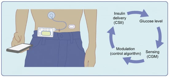
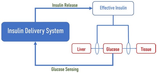
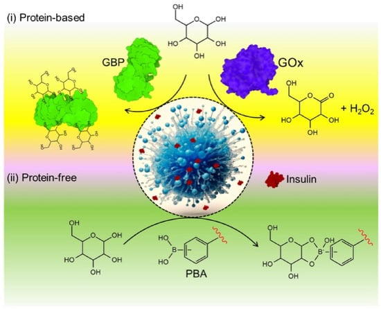
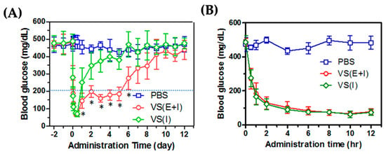
Glucose-Responsive Nanomedicine in Diabetes Therapy: Emerging Advances and Clinical Prospects
J. Funct. Biomater. 2026, 17(9), 424; https://doi.org/10.3390/jfb17090424 (registering DOI) - 22 Aug 2026
Abstract
Diabetes mellitus continues to impose a substantial global health burden, underscoring the need for therapeutic systems capable of achieving precise, adaptive, and patient-friendly glycemic control. Conventional diabetes treatments, including repeated insulin injections and oral hypoglycemic agents, are often constrained by non-physiological drug release,
[...] Read more.
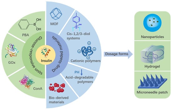
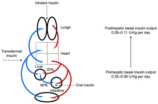
Diabetes mellitus continues to impose a substantial global health burden, underscoring the need for therapeutic systems capable of achieving precise, adaptive, and patient-friendly glycemic control. Conventional diabetes treatments, including repeated insulin injections and oral hypoglycemic agents, are often constrained by non-physiological drug release, poor adherence, systemic side effects, and the persistent risk of hypoglycemia. In this context, glucose-responsive nanomedicine has emerged as a promising platform for next-generation diabetes therapy by enabling self-regulated and glucose-triggered delivery of insulin and other antidiabetic agents. This review highlights recent advances in glucose-responsive nanomedicine, focusing on the principal sensing mechanisms, including glucose oxidase-based, phenylboronic acid-based, and lectin-mediated systems, as well as the nanoscale carriers engineered to support them, such as polymeric nanoparticles, nanogels, micelles, liposomes, and hybrid nanostructures. These smart platforms offer significant potential to improve drug stability, enhance targeting efficiency, reduce dosing frequency, and more closely mimic endogenous insulin secretion. The review further examines their emerging role in precision diabetes care, particularly in combination with continuous glucose monitoring technologies, wearable devices, and closed-loop therapeutic systems. Despite notable progress at the preclinical level, important barriers to clinical translation remain, including challenges related to biocompatibility, long-term safety, reproducibility, scalable manufacturing, and regulatory approval. Collectively, glucose-responsive nanomedicine represents a rapidly advancing and clinically relevant field with the potential to redefine diabetes management through intelligent and personalized therapeutic strategies. This review provides a focused overview of current developments, key translational challenges, and future directions toward clinical implementation.
Full article
(This article belongs to the Special Issue Applications of Nanomaterials in Drug Delivery Systems)
►
Show Figures
Open AccessArticle
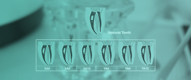
Modulating the Optical Properties of Initial Caries Lesions Through Low-Viscosity Resin Infiltration: A Spectrophotometric Evaluation
by
Paweł Maksymiuk, Maja Ptasiewicz, Joanna Zubrzycka, Ilona Wójcik-Chęcińska, Katarzyna Sarna-Boś, Piotr Stachurski and Renata Chałas
J. Funct. Biomater. 2026, 17(8), 423; https://doi.org/10.3390/jfb17080423 - 21 Aug 2026
Abstract
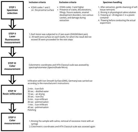
Objectives: Initial caries lesions (ICLs) are the earliest manifestation of dental caries, characterized by subsurface porosity that affects enamel’s optical properties while maintaining its surface integrity. Resin infiltration is a minimally invasive approach to managing these lesions by penetrating and sealing pores with
[...] Read more.
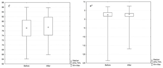
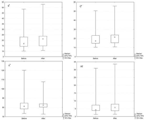
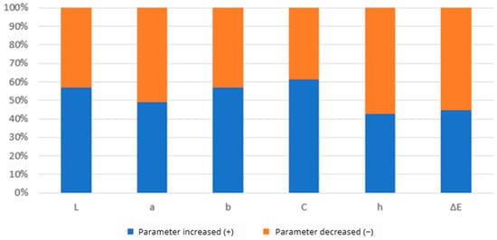
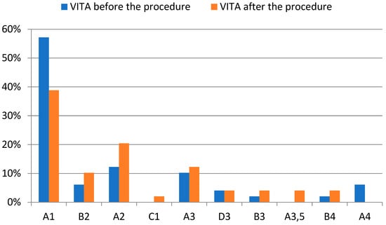
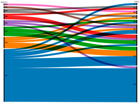
Objectives: Initial caries lesions (ICLs) are the earliest manifestation of dental caries, characterized by subsurface porosity that affects enamel’s optical properties while maintaining its surface integrity. Resin infiltration is a minimally invasive approach to managing these lesions by penetrating and sealing pores with low-viscosity resin, potentially modifying their visual appearance. In order to provide a quantifiable, objective assessment of this optical behavior, this study aimed to evaluate the modulation of color coordinates and VITA Classical shade transitions within the initial lesions immediately following infiltration with Icon Smooth Surface (DMG, Hamburg, Germany). Materials and Methods: Forty-nine extracted human premolars with naturally occurring proximal ICLs (subsurface demineralization) were selected. Lesions were assessed for fluorescence level using 655 nm laser fluorescence (DIAGNOdent pen). Reflectance spectrophotometry (SpectroShade Micro) was employed to record color coordinates in the CIELab and CIELCh spaces, alongside VITA Classical shades, before and after resin infiltration. Results: The infiltration procedure resulted in a statistically significant increase in b* (yellowness) and subsequent C* (chroma) parameters (p < 0.05). VITA Classical shade guide analysis showed an insignificant shift in color distribution according to value (p > 0.05). Conclusions: The present exploratory study provides quantitative insights into the immediate color-modifying effects of a resin infiltration procedure on natural initial proximal caries lesions, complementing the existing literature with objective CIELab/CIELCh and VITA Classical measurements. The p-values should be interpreted as descriptive indicators of potential trends rather than definitive statistical proof. Further research with larger cohorts and long-term follow-up is needed to validate the durability and clinical relevance of these esthetic outcomes.
Full article
(This article belongs to the Special Issue Advances in Biomaterials for Oral Health: From Dental Restoration to Regenerative Therapies)
►▼
Show Figures

Figure 1
Open AccessReview
Polyphosphate in Bone Tissue Engineering: From Molecular Mechanisms to Material Design
by
Zhangling Nie, Bingqiang Lu, Valentina K. Krut’ko, Anatoly I. Kulak and Feng Chen
J. Funct. Biomater. 2026, 17(8), 422; https://doi.org/10.3390/jfb17080422 - 21 Aug 2026
Abstract
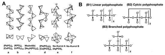
Polyphosphate (PolyP) is an inorganic polymer composed of orthophosphate units linked by high-energy phosphate anhydride bonds, widely found in various organisms from bacteria to mammals. In recent years, PolyP has attracted widespread attention in the field of bone tissue engineering due to its
[...] Read more.
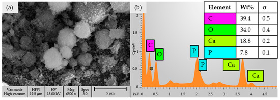
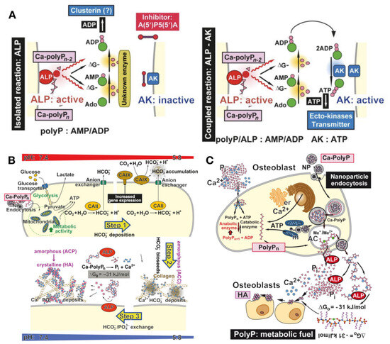
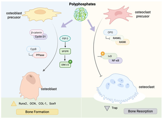
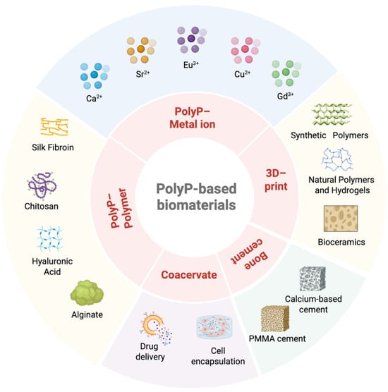
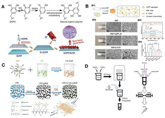
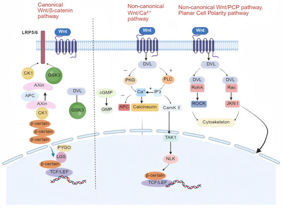
Polyphosphate (PolyP) is an inorganic polymer composed of orthophosphate units linked by high-energy phosphate anhydride bonds, widely found in various organisms from bacteria to mammals. In recent years, PolyP has attracted widespread attention in the field of bone tissue engineering due to its unique biological characteristics, possessing both osteoinductive activity and metabolic energy supply functions. This article systematically reviews the molecular structure, physicochemical properties, and multiple mechanisms by which PolyP promotes osteogenic differentiation, as well as biomaterial design strategies based on PolyP. PolyP can synergistically promote osteogenic differentiation through multiple mechanisms, including by acting as a phosphate donor, providing metabolic energy, regulating signaling pathways such as Wnt/β-catenin, and modulating the osteoprotegerin/receptor activator of nuclear factor κB ligand (OPG/RANKL) balance. In terms of material design, PolyP can form nano/microparticles with metal ions such as Ca2+, Sr2+, and Mg2+ and can also be compounded with polymers to construct various forms such as hydrogels, bone cement, and three-dimensional (3D)-printed scaffolds. Preclinical studies have shown that PolyP-incorporated materials exhibit excellent osteogenic performance and biocompatibility in bone defect repair, and preliminary clinical studies have also confirmed its feasibility. This article aims to provide a comprehensive overview of the current applications of PolyP-incorporated materials and delineate future directions, challenges, and necessary pathways for their clinical translation.
Full article
(This article belongs to the Special Issue Molecular Mechanisms and Biological Procedures of Biomaterials in Medical Applications (2nd Edition))
►▼
Show Figures

Figure 1

Open AccessArticle
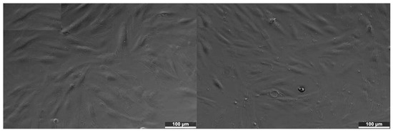
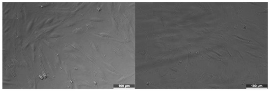
In Vitro Cytotoxicity of Three Dimethacrylate-Based Photopolymer Resins for 3D-Printed Dental Restorations: A Qualitative Morphological Screening on Human Fibroblasts
by
Teodora-Alina Tincea, Alexia-Ecaterina Cârstea, Vlad-Gabriel Vasilescu, Andreia Cucuruz, Adela Banciu, Adrian-Ionuț Nicoară, Claudia-Gabriela Mateiaș, Ana-Maria Cristina Țâncu, Silviu-Mirel Pițuru, Marina Imre and Lucian-Toma Ciocan
J. Funct. Biomater. 2026, 17(8), 421; https://doi.org/10.3390/jfb17080421 - 20 Aug 2026
Abstract
The rapid adoption of vat photopolymerization (3D printing) in restorative and prosthetic dentistry has outpaced the independent biological characterization of the dimethacrylate resins on which it relies, even though incompletely converted networks can release residual monomers that are cytotoxic to the adjacent oral
[...] Read more.
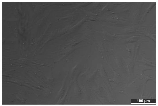
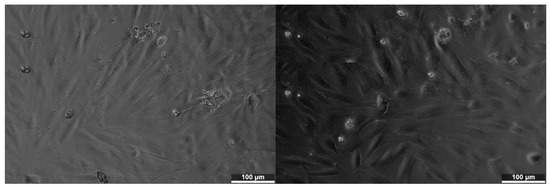
The rapid adoption of vat photopolymerization (3D printing) in restorative and prosthetic dentistry has outpaced the independent biological characterization of the dimethacrylate resins on which it relies, even though incompletely converted networks can release residual monomers that are cytotoxic to the adjacent oral tissues. This study reports a qualitative in vitro cytotoxicity screening of three commercial photopolymer resins optimized for additive manufacturing: a general-purpose resin (ANYCUBIC) and two resins with a declared dental indication (TEMP PRINT and V-PRINT) performed according to the morphological evaluation described in ISO 10993-5. Aqueous extracts were prepared following ISO 10993-12 and applied to normal human fibroblasts (BJ line, ATCC CRL-2522) at 100% and 50% concentration for 24 h; morphological reactivity was graded on the standardized 0–4 scale by inverted light microscopy against an unexposed control, and supported by a semi-quantitative visual estimate of the proportion of affected cells. All three materials produced a concentration-dependent response. TEMP PRINT retained the best cellular compatibility (grade 1 at 50%, grade 2 at 100%), V-PRINT was intermediate, and ANYCUBIC showed the most pronounced reactivity (approaching grade 3 at 100%), giving an estimated cytotoxicity order of ANYCUBIC > V-PRINT > TEMP PRINT. As a qualitative morphological screening, the findings indicate that the dental-grade resins elicited a milder fibroblast response than the general-purpose photopolymer and support the recommendation that general-purpose resins should not be substituted for certified materials in intraoral use. Confirmation by quantitative viability assays with independent replicates is required before firm conclusions are drawn. In line with the qualitative nature of ISO 10993-5 morphological screening, the reported proportions of morphologically affected cells represent visual scoring rather than direct measurements of cell viability, and the study is therefore presented as an initial biological screening.
Full article
(This article belongs to the Special Issue Advanced Dental Biomaterials for Prosthodontic Applications: Functional and Biological Perspectives)
►▼
Show Figures

Figure 1
Open AccessArticle
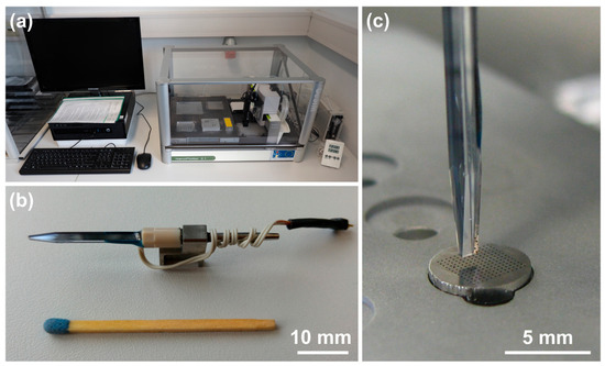
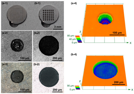
Inkjet Printing of Drugs into Surface-Embedded Micro-Reservoirs for Drug-Releasing Implants: Influence of Solvent Properties on Deposition Behavior
by
Robert Mau, Georg Schnell, Paul Oldorf and Hermann Seitz
J. Funct. Biomater. 2026, 17(8), 420; https://doi.org/10.3390/jfb17080420 - 20 Aug 2026
Abstract
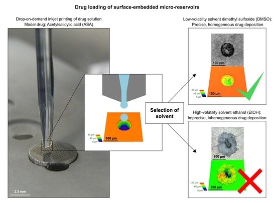
Background: Micro-reservoirs in implant surfaces represent a promising drug carrier concept for drug delivery systems. For drug loading, inkjet printing enables highly precise droplet positioning. However, droplet drying influences drug crystallization from printed drug solution. This study investigates how evaporation-driven phenomena affect the
[...] Read more.
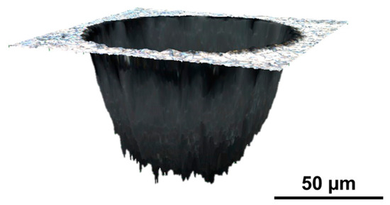
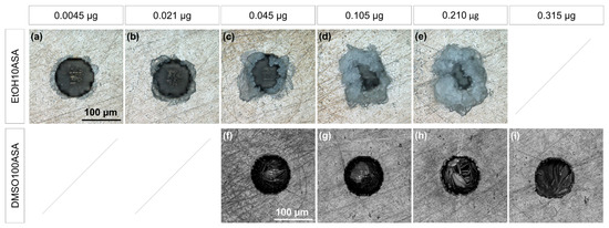
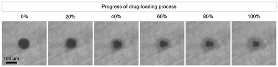
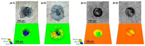
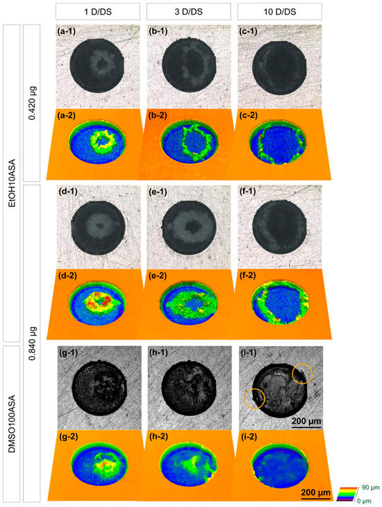
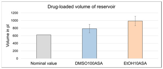
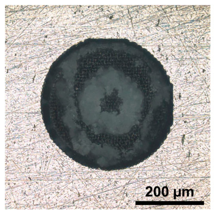
Background: Micro-reservoirs in implant surfaces represent a promising drug carrier concept for drug delivery systems. For drug loading, inkjet printing enables highly precise droplet positioning. However, droplet drying influences drug crystallization from printed drug solution. This study investigates how evaporation-driven phenomena affect the precision and homogeneity of inkjet-based deposition of a crystallizing drug into exemplary micro-reservoirs. The aim is to guide the selection of suitable solvents and inkjet process parameters. Methods: Laser-drilled micro-reservoirs were fabricated as blind holes with entrance diameters of 100 µm and 400 µm in the surface of specimens of EN 1.4404 (equivalent to AISI 316L) stainless steel, a commonly used biomaterial. The reservoirs were loaded with two different drug solutions using piezoelectric drop-on-demand inkjet printing. Acetylsalicylic acid (ASA) was applied as a model drug representing crystallizing small-molecule drugs. Solvents with markedly different evaporation rates, ethanol (EtOH) as a representative high-volatility solvent and dimethyl sulfoxide (DMSO) as a representative low-volatility solvent, were selected. The number of jetted droplets per dispensing step was varied. Precision and homogeneity of the drug deposition were investigated using light and laser scanning microscopy. Results: Over the course of droplet drying, two phenomena, the coffee-ring effect and creeping, can impair drug deposition quality. The coffee-ring effect leads to inhomogeneous, ring-shaped drug deposits. Creeping is the evaporation-driven spreading of crystalline structures and reduces the precision of drug deposition. The EtOH-based ASA solution (c = 10 g/L) was intensely affected by both phenomena. Inhomogeneities could be partially compensated via tailoring the droplet count per dispensing step. The DMSO-based solution (c = 100 g/L) exhibited a more compact crystallization of ASA (requiring ~20% less volume in an exemplary experiment), no coffee-ring effect, and only minor creeping. Conclusions: The DMSO-based ASA solution enabled a more precise and homogeneous drug deposition than the EtOH-based solution under the investigated printing and crystallization conditions. EtOH-related limitations could be counteracted by controlling the number of jetted droplets per dispensing step.
Full article
(This article belongs to the Special Issue Drug- and Ion-Releasing Implants)
►▼
Show Figures

Graphical abstract
Open AccessArticle
A Computational Framework for the Design and Mechanical Assessment of Biodegradable Airway Stents: Interaction with Rabbit Tracheal Tissue and Preliminary In Vivo Observations
by
Ada Ayechu-Abendaño, Letizia Cella, Carmen Sánchez-González, Carmen Sánchez-Matás, José Luis López-Villalobos, Cristina Díaz-Jiménez, Rocío Fernández-Parra and Mauro Malvè
J. Funct. Biomater. 2026, 17(8), 419; https://doi.org/10.3390/jfb17080419 - 20 Aug 2026
Abstract
Current airway stents, including silicone and metallic devices, remain associated with important complications such as migration, restenosis, mucus retention and the need for repeated interventions. Biodegradable stents offer a promising alternative by providing temporary mechanical support while avoiding the long-term presence of a
[...] Read more.
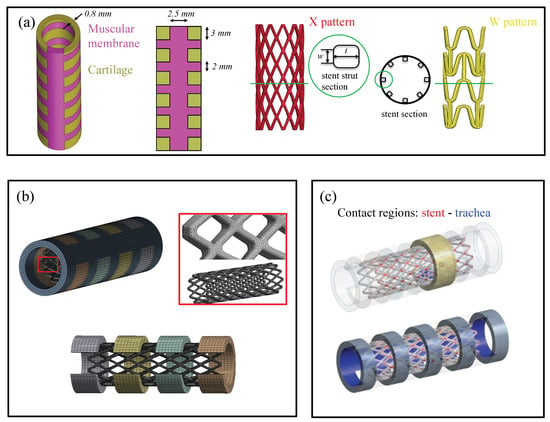
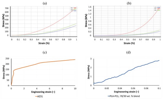
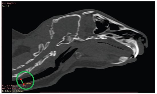
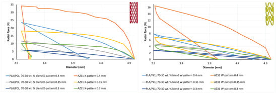
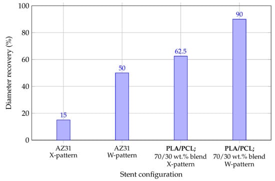
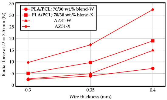
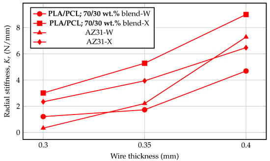
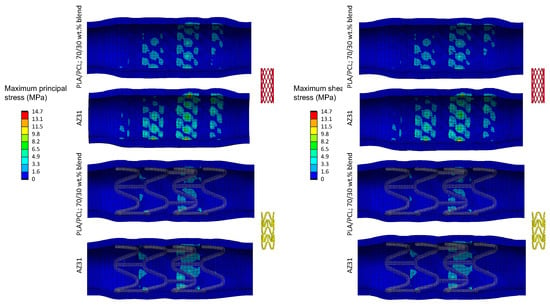
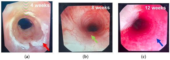
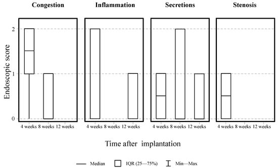
Current airway stents, including silicone and metallic devices, remain associated with important complications such as migration, restenosis, mucus retention and the need for repeated interventions. Biodegradable stents offer a promising alternative by providing temporary mechanical support while avoiding the long-term presence of a permanent implant. However, the influence of stent geometry and material properties on their mechanical performance and interaction with airway tissue is still not fully understood. This study presents a computational framework integrating computer-aided design and finite element analysis to investigate the mechanical behaviour of biodegradable tracheobronchial stents. Two stent architectures (X-pattern and W-pattern) were analysed over a range of wire thicknesses using two biodegradable materials: a PLA/PCL; 70/30 wt.% blend and AZ31 magnesium alloy. Radial compression, diameter recovery after radial compression and stent–tissue interaction simulations were performed to evaluate the influence of geometry, material selection and design parameters on device performance. The results suggested that both stent geometry and material properties strongly influence the mechanical behaviour of biodegradable airway stents, although they affect different aspects of the stent–tissue interaction. The X-pattern consistently exhibited greater resistance to radial compression, lower elastic diameter recovery after radial compression and improved maintenance of the expanded lumen compared with the W-pattern. Material properties primarily affected the magnitude of the mechanical response, as further confirmed by the quantitative contact-pressure analysis, with AZ31 providing greater radial support, while the spatial distributions of stress and strain within the tracheal wall were mainly governed by the stent architecture. Based on the computational analyses, X-pattern stents manufactured from the PLA/PCL; 70/30 wt.% blend were selected for in vivo evaluation in a rabbit model. Endoscopic observations revealed tissue features that were qualitatively consistent with the mechanical patterns predicted by the numerical simulations, although no direct causal relationship can be established from the available observations. These findings support the ability of the proposed framework to represent the principal aspects of stent–tissue interaction. The proposed computational framework provides a practical tool for the rational design and mechanical assessment of biodegradable airway stents and may facilitate the future development of customised airway prostheses.
Full article
(This article belongs to the Section Biomaterials for Tissue Engineering and Regenerative Medicine)
►▼
Show Figures

Figure 1
Open AccessArticle
Comparative Short-Term Electrochemical and Surface Characterization of Biodegradable Mg–Ca–Sr Alloys with Different Strontium Contents
by
Gabriela Leață, Dorin Ioan Cocoș, Ramona Feier, Corneliu Munteanu, Fabian Cezar Lupu, Ion Ciucă and Kamel Earar
J. Funct. Biomater. 2026, 17(8), 418; https://doi.org/10.3390/jfb17080418 - 20 Aug 2026
Abstract
Biodegradable Mg–Ca–Sr alloys are promising candidates for temporary implant applications, but their degradation behavior is strongly influenced by alloy composition and electrolyte chemistry. This comparative short-term study investigated how increasing the Sr content from 0.5 to 1.5 wt.% influences the electrochemical response and
[...] Read more.
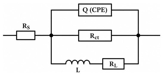
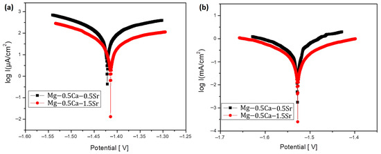
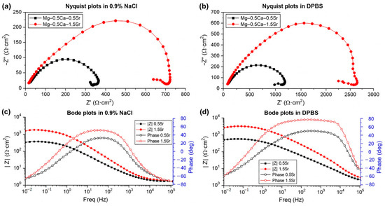
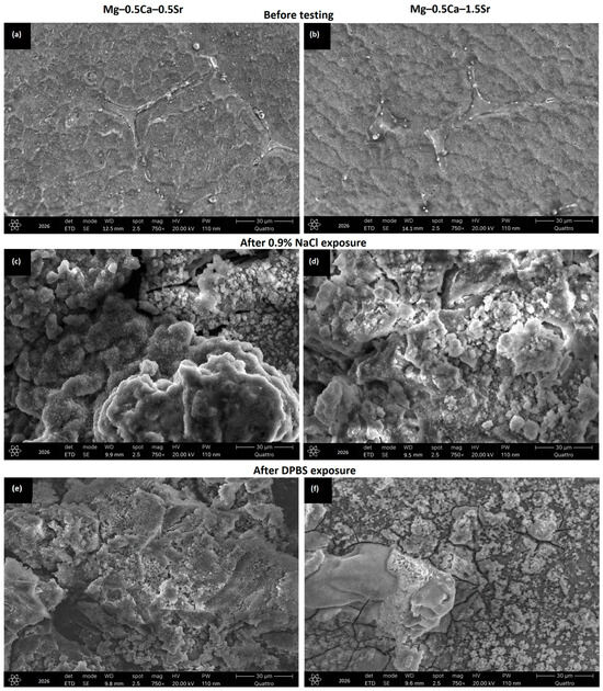
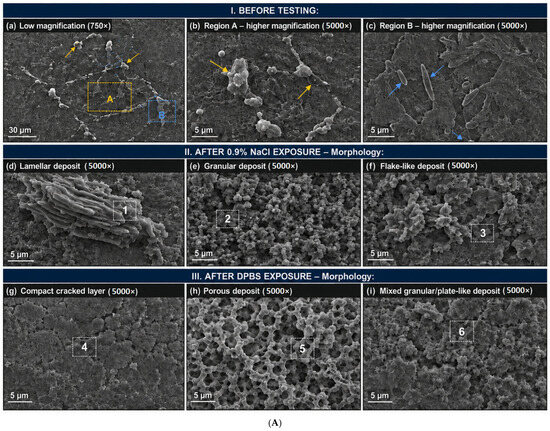
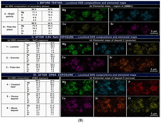
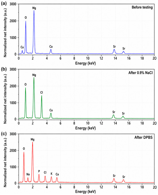
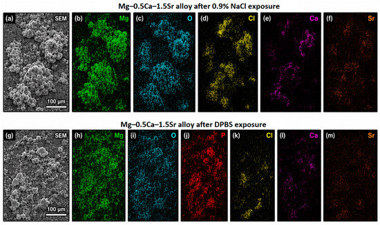
Biodegradable Mg–Ca–Sr alloys are promising candidates for temporary implant applications, but their degradation behavior is strongly influenced by alloy composition and electrolyte chemistry. This comparative short-term study investigated how increasing the Sr content from 0.5 to 1.5 wt.% influences the electrochemical response and surface characteristics of cast Mg–0.5Ca–xSr alloys in 0.9% NaCl and calcium- and magnesium-free Dulbecco’s phosphate-buffered saline (DPBS). Potentiodynamic polarization, electrochemical impedance spectroscopy, scanning electron microscopy, quantitative image analysis, and energy-dispersive X-ray spectroscopy were used. Increasing the Sr content reduced the corrosion-current density from 0.0110 to 0.0039 mA/cm2 in NaCl and from 0.297 to 0.169 mA/cm2 in DPBS, while the corresponding calculated corrosion rates decreased from 2.51 to 0.886 mm/year and from 6.66 to 3.79 mm/year, respectively. Replicated EIS measurements of both alloys showed that Mg–0.5Ca–1.5Sr exhibited higher charge-transfer resistance than Mg–0.5Ca–0.5Sr in both NaCl (253 ± 15 versus 184 ± 14 Ω·cm2) and DPBS (853 ± 48 versus 615 ± 42 Ω·cm2). NaCl-exposed surfaces showed more porous and discontinuous deposits and a greater number of visible pit-like defects, whereas DPBS produced comparatively continuous P-containing deposits. Overall, increasing the Sr content to 1.5 wt.% was associated with lower polarization-derived global corrosion kinetics and a more resistive interfacial response, whereas electrolyte composition strongly influenced both interfacial impedance and surface-deposit morphology. These findings represent comparative short-term electrochemical and surface-characterization data obtained at 23 ± 1 °C and should not be interpreted as measures of long-term physiological degradation or in vivo performance.
Full article
(This article belongs to the Special Issue Medical Application of Functional Biomaterials (3rd Edition))
►▼
Show Figures

Figure 1
Open AccessReview
Sustainable Valorization of Biogenic Waste for Bone Repair and Regeneration: A Comprehensive Review of Eggshell and Aquatic Biomaterials
by
Shazah Waqar, Tamer A. E. Ahmed and Maxwell T. Hincke
J. Funct. Biomater. 2026, 17(8), 417; https://doi.org/10.3390/jfb17080417 - 19 Aug 2026
Abstract
Bone loss represents a significant clinical burden that has driven the development of improved orthopedic graft substitutes. Although current grafting options, including autografts, allografts, and xenografts, have demonstrated considerable therapeutic potential, their widespread application is constrained by limitations such as donor scarcity, limited
[...] Read more.
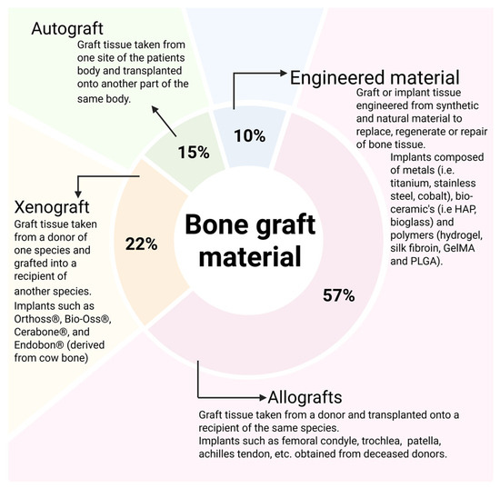
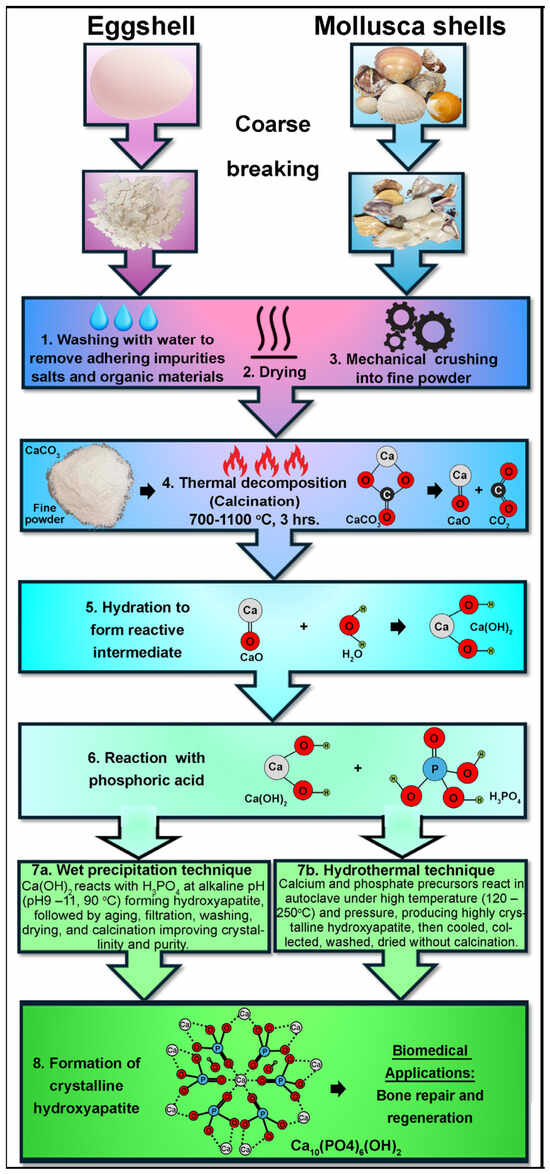
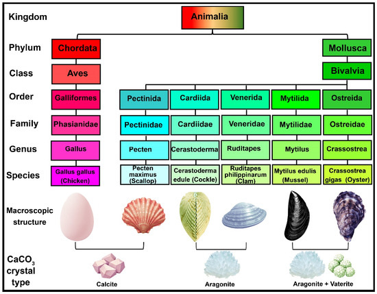
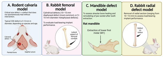
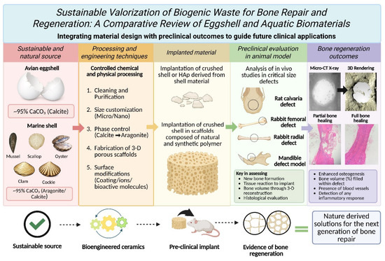
Bone loss represents a significant clinical burden that has driven the development of improved orthopedic graft substitutes. Although current grafting options, including autografts, allografts, and xenografts, have demonstrated considerable therapeutic potential, their widespread application is constrained by limitations such as donor scarcity, limited availability, and associated clinical risks. Consequently, biologically derived materials, including avian eggshells and marine bivalve shells, have emerged as promising alternative sources to produce bone precursor materials and next-generation bone graft substitutes. This review summarizes recent advances in avian eggshell- and marine shell-derived calcium carbonate (CaCO3) materials for bone regeneration and examines their preclinical evaluation in diverse animal models, including critical-size defects in calvarial, femoral, radial and mandibular bone. Relevant studies published over the past ten years were systematically analyzed, focusing on natural calcium carbonate systems derived from avian eggshell and marine shells, including oyster, mussel, clam, scallop, cockle, and sea urchins. A structured literature search was conducted using PubMed, Scopus, and Google Scholar to identify studies published between 2015 and 2025 investigating eggshell- and aquatic-derived biomaterials for bone repair and regeneration. Eligible studies were screened, and data were comparatively analyzed with respect to biomaterial source, scaffold fabrication, physicochemical characteristics, mechanical performance, biocompatibility, osteogenic potential, and the use of preclinical animal studies. Eggshell-derived biomaterials currently show the strongest translational evidence, while aquatic shell-derived biomaterials remain promising but underexplored for bone regeneration. Furthermore, this review critically examines the scientific, manufacturing, and regulatory challenges that must be addressed before clinical implementation.
Full article
(This article belongs to the Special Issue Functional Scaffolds for Hard Tissue Engineering and Surgery)
►▼
Show Figures

Figure 1

Open AccessArticle
A Functional TGF-β/Smad Assay for Targeted Profiling of Demineralized Bone Matrix-Derived Allograft Bioactivity
by
Adrian Lendvai, Tobias Weichhart, Hans Peter Weitzenböck, Christoph Wiesner, Rita Seeboeck, Narges Zamani, Michael Matzner, Monika Pichler, Bettina Steiner, Andrea De Luna, Stefan Nehrer and Harald Hundsberger
J. Funct. Biomater. 2026, 17(8), 416; https://doi.org/10.3390/jfb17080416 - 19 Aug 2026
Abstract
Demineralized bone matrix (DBM)-derived allografts retain extracellular matrix (ECM)-associated factors involved in bone repair, but biochemical protein recovery alone may not predict functional pathway activation. We evaluated the HEK-Blue™ TGF-β Reporter Assay as a targeted method for assessing Smad-dependent reporter activity in DBM-derived
[...] Read more.
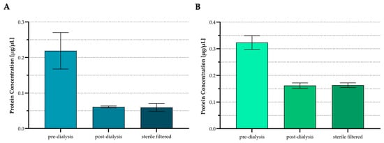
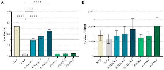
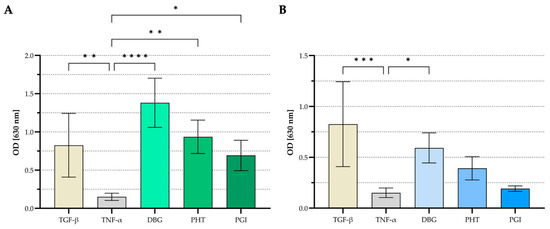
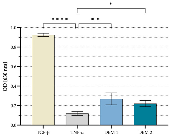
Demineralized bone matrix (DBM)-derived allografts retain extracellular matrix (ECM)-associated factors involved in bone repair, but biochemical protein recovery alone may not predict functional pathway activation. We evaluated the HEK-Blue™ TGF-β Reporter Assay as a targeted method for assessing Smad-dependent reporter activity in DBM-derived materials. ECM proteins were extracted from cortical demineralized bone granules (DBG) using guanidine hydrochloride (GuHCl) or urea and quantified after extraction, dialysis, and sterile filtration. Reporter cells were stimulated with extracted ECM proteins or directly with processed cortical and cancellous products, including DBG, wet heat-treated DBG formulated as Putty (PHT), and gamma-irradiated PHT (PGI). Non-pooled DBM sponge samples were also tested. Secreted embryonic alkaline phosphatase (SEAP) activity served as the functional reporter readout. Material-only no-cell controls assessed material-derived background. PrestoBlue™ readouts served as exploratory quality controls. The urea-derived extract yielded a higher apparent BCA-detectable protein concentration than the GuHCl-derived extract. Only the GuHCl-derived extract induced increasing SEAP activity at matched protein input. Direct stimulation showed reporter activation above the TNF-α pathway-negative cytokine control for cortical DBG, PHT, PGI, and cancellous DBG. DBM sponges also induced detectable SEAP activity. These findings support targeted functional screening of Smad-dependent TGF-β reporter activation in DBM-derived materials, but not total osteoinductivity assessment.
Full article
(This article belongs to the Section Bone Biomaterials)
►▼
Show Figures

Figure 1
Open AccessReview
Applications of DNA Hydrogels in Osteoporotic Bone Defects
by
Jiaqi Chen, Huiyu Jia, Xinyue Zhang, Da Liu, Xushuang Jia, Xintong Gu, Hongjuan Wen and Ye Jin
J. Funct. Biomater. 2026, 17(8), 415; https://doi.org/10.3390/jfb17080415 - 18 Aug 2026
Abstract
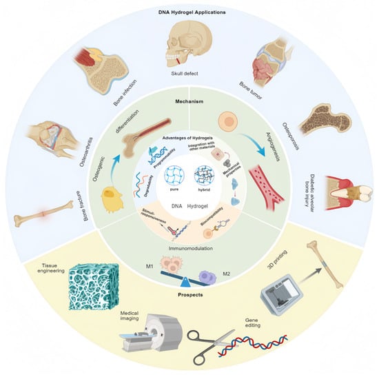
DNA hydrogels are an emerging class of biomaterials with programmability, biodegradability, biocompatibility, and dynamic responsiveness, enabling precise regulation of osteoblast and mesenchymal stem cell (MSC) proliferation and differentiation, activation of key signaling pathways, and promotion of angiogenesis and bone matrix mineralization. In contrast,
[...] Read more.
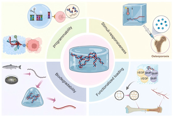
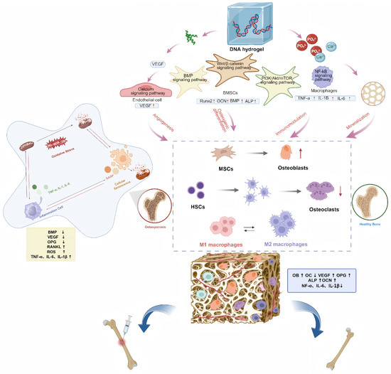
DNA hydrogels are an emerging class of biomaterials with programmability, biodegradability, biocompatibility, and dynamic responsiveness, enabling precise regulation of osteoblast and mesenchymal stem cell (MSC) proliferation and differentiation, activation of key signaling pathways, and promotion of angiogenesis and bone matrix mineralization. In contrast, conventional bone repair materials exhibit limitations including poor mechanical strength, uncontrollable degradation, and inadequate matching with native bone properties, restricting their application in osteoporotic defect repair. Current osteoporotic defect therapies, mainly anti-resorptive and anabolic agents, remain insufficient for many patients. Here, we propose pure and hybrid DNA hydrogels as novel therapeutic platforms to restore the dynamic balance between bone resorption and formation, thereby enhancing osteogenesis and facilitating bone regeneration and remodeling under osteoporotic conditions. Although challenges such as high production cost and long-term safety persist, integration with advanced technologies (e.g., 3D printing and gene editing) may provide theoretical support for further investigation of personalized and intelligent therapeutic strategies at the pre-clinical research stage, offering new insights into osteoporotic bone defects and bone tissue regeneration.
Full article
(This article belongs to the Section Bone Biomaterials)
►▼
Show Figures

Figure 1

Open AccessArticle
Effects of Intraoperative Ozone Application on Early Implant Stability: A Randomized Split-Mouth Clinical Trial
by
Zeliha Başak Çakır Erdil, Hüseyin Akıllı, Şahin Altuğ and Metin Çalışır
J. Funct. Biomater. 2026, 17(8), 414; https://doi.org/10.3390/jfb17080414 - 18 Aug 2026
Abstract
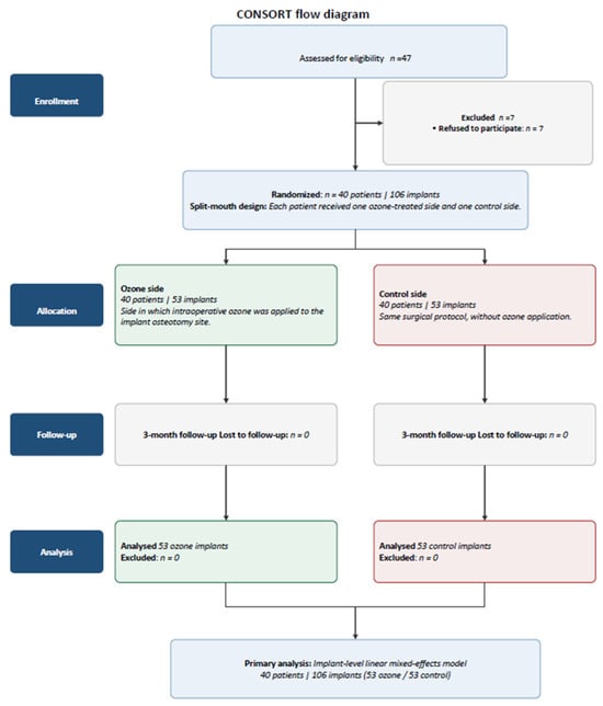
Background/Objectives: The aim of this study was to evaluate the effect of intraoperative gaseous ozone applied to the implant osteotomy immediately before implant placement on early implant stability using a split-mouth design and resonance frequency analysis (RFA). Methods: This prospective, randomized controlled split-mouth
[...] Read more.
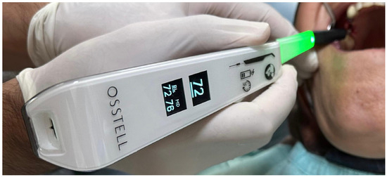
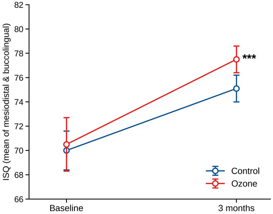
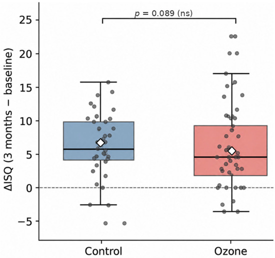
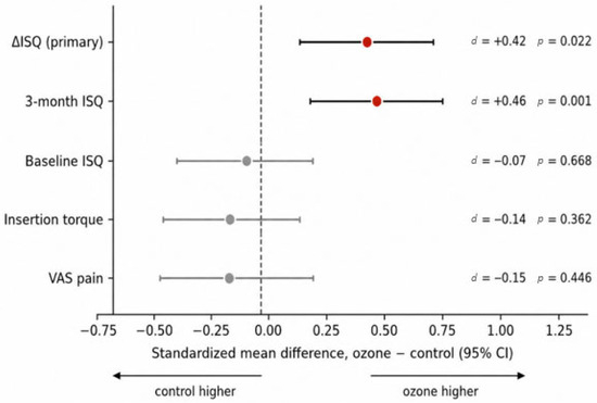
Background/Objectives: The aim of this study was to evaluate the effect of intraoperative gaseous ozone applied to the implant osteotomy immediately before implant placement on early implant stability using a split-mouth design and resonance frequency analysis (RFA). Methods: This prospective, randomized controlled split-mouth study included 40 patients receiving 106 implants in bilaterally symmetrical edentulous sites. Patients and outcome assessors were blinded. In the ozone group, ozone gas was applied to the implant site for 60 s immediately before implant placement; the control group underwent the same surgical protocol without ozone application. The primary outcome was the change in implant stability quotient from baseline to three months (ΔISQ). Secondary outcomes included baseline and three-month ISQ values, insertion torque, and postoperative pain assessed using a visual analog scale. Results: All 106 implants were analyzed. No significant between-group differences were observed in baseline ISQ (β = −0.49; 95% CI: −2.73 to 1.75; p = 0.668) or insertion torque (β = −0.62 Ncm; 95% CI: −1.96 to 0.72; p = 0.362). At three months, ISQ was higher in the ozone group (β = 1.91; 95% CI: 0.75 to 3.06; p = 0.001). ΔISQ was also significantly greater in the ozone group (β = 2.40; 95% CI: 0.34 to 4.45; p = 0.022; Cohen’s d = 0.42). Implant stability increased significantly in both groups (both p < 0.001). Postoperative pain did not differ between groups (β = −0.23; 95% CI: −0.83 to 0.36; p = 0.446). Although the effect direction was consistent across sensitivity analyses, ΔISQ was not statistically significant in the patient-level paired analysis (p = 0.089). No adverse events were reported. Conclusions: Intraoperative ozone application was associated with a modest increase in early implant stability without a significant difference in postoperative pain. However, given the small effect size and sensitivity of the findings to the analytical method, further large-scale, multicenter randomized controlled trials are required. Clinically, intraoperative ozone may be considered a potential adjunct to standard implant placement protocols, but the current evidence is insufficient to support its routine use.
Full article
(This article belongs to the Section Dental Biomaterials)
►▼
Show Figures

Figure 1

Open AccessArticle
Multidimensional Characterization of Surface Properties and Microbial Biofilm Formation in Commercial Flowable Resin-Based Dental Composites
by
Lucian Floare, Otilia Cornelia Bolos, Ramona Dumitrescu, Marioara Nicoleta Caraba, Iasmina-Mădălina Petculescu, Carmen Opris, Octavia Balean, Vanessa Bolchis, Bianca Ioana Todor, Cristina Elena Savencu, Atena Galuscan and Daniela Jumanca
J. Funct. Biomater. 2026, 17(8), 413; https://doi.org/10.3390/jfb17080413 - 18 Aug 2026
Abstract
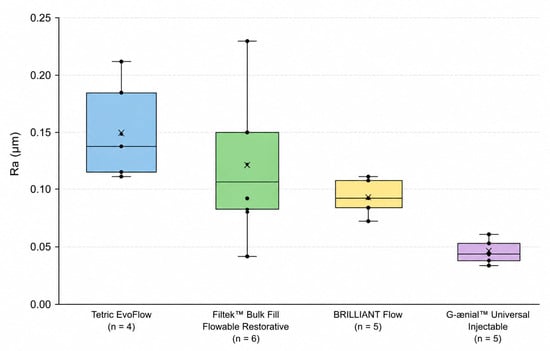
The interaction between restorative materials and microbial biofilms plays an important role in restoration longevity and the development of secondary caries. This study aimed to perform a multidimensional characterization of four commercially available flowable resin-based dental composites by evaluating their surface roughness, Vickers
[...] Read more.
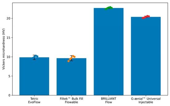
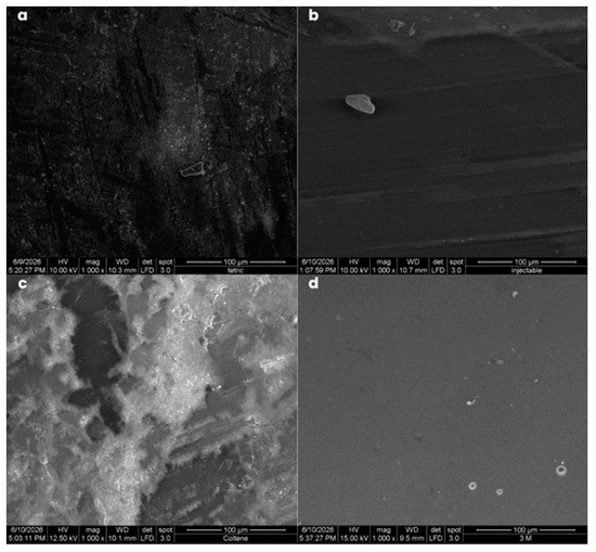
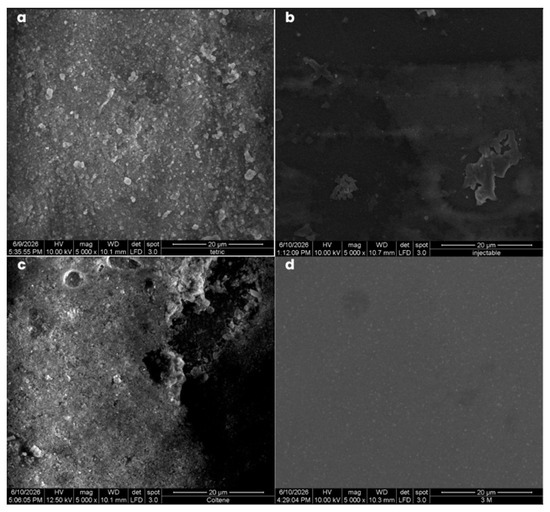
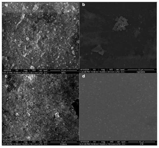
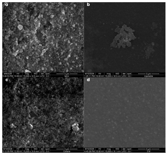
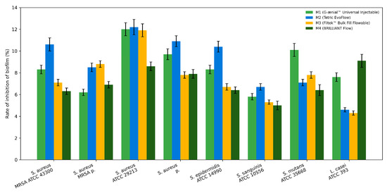
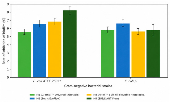
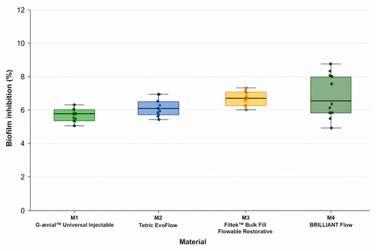
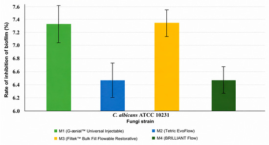
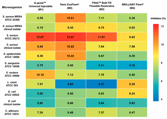
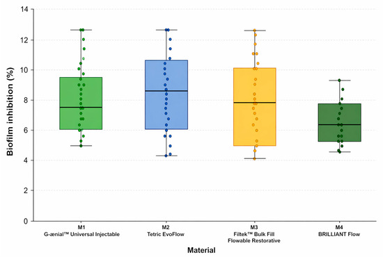
The interaction between restorative materials and microbial biofilms plays an important role in restoration longevity and the development of secondary caries. This study aimed to perform a multidimensional characterization of four commercially available flowable resin-based dental composites by evaluating their surface roughness, Vickers microhardness, surface morphology, and microbial biofilm formation under standardized in vitro conditions. Filtek™ Bulk Fill Flowable Restorative (3M), Tetric EvoFlow (Ivoclar Vivadent), BRILLIANT Flow (Coltene), and G-ænial™ Universal Injectable (GC) were investigated. Surface roughness was determined by contact profilometry, microhardness was assessed using the Vickers method, and microstructural characteristics were analyzed by scanning electron microscopy (SEM). Microbial biofilm biomass formed by Gram-positive bacteria, Gram-negative bacteria, and Candida albicans, including both reference strains and clinical isolates, was quantified using a crystal violet assay to compare material-dependent microbial colonization. G-ænial™ Universal Injectable exhibited the lowest roughness values, whereas BRILLIANT Flow showed the highest microhardness. SEM analysis revealed a more homogeneous surface morphology for Filtek™ Bulk Fill Flowable Restorative and G-ænial™ Universal Injectable, while Tetric EvoFlow and BRILLIANT Flow displayed increased topographical heterogeneity. Although modest differences in biofilm biomass accumulation were observed among the investigated composites (4.37–12.21% relative biomass reduction compared with the control), no material demonstrated a distinct advantage under the experimental conditions. The integrated evaluation of surface roughness, microhardness, surface morphology, and microbial biofilm formation provides a comparative characterization of commercially available flowable resin-based composites and contributes to a better understanding of material–biofilm interactions. These findings may support the future development of multifunctional restorative biomaterials with improved biofilm-modulating properties.
Full article
(This article belongs to the Section Dental Biomaterials)
►▼
Show Figures

Figure 1
Open AccessEditorial
State-of-the-Art Dental Adhesives and Restorative Composites
by
Jirun Sun
J. Funct. Biomater. 2026, 17(8), 412; https://doi.org/10.3390/jfb17080412 - 18 Aug 2026
Abstract
Since the pioneering work of Dr [...]
Full article
(This article belongs to the Special Issue State-of-the-Art Dental Adhesives and Restorative Composites)
Open AccessArticle
Core Buildup Composite Resins Bond Strength to Dentin and Microhardness Using Universal Adhesives
by
Iana Schmitt, Jorge Perdigão and Guilherme Carpena Lopes
J. Funct. Biomater. 2026, 17(8), 411; https://doi.org/10.3390/jfb17080411 - 18 Aug 2026
Abstract
The peer-reviewed literature on the use of universal adhesives (UAs) with core build-up composite resins (CBCRs) is limited. UAs may enhance the polymerization of dual-cured composite resins, leading to increased microhardness due to the “touch-cure” effect. This study compared the dentin bond strengths
[...] Read more.
The peer-reviewed literature on the use of universal adhesives (UAs) with core build-up composite resins (CBCRs) is limited. UAs may enhance the polymerization of dual-cured composite resins, leading to increased microhardness due to the “touch-cure” effect. This study compared the dentin bond strengths and microhardness of CBCRs bonded with UAs. A total of 480 bovine incisors were randomly allocated into 20 groups (n = 24). Two dual-cured CBCRs, ParaCore (PrCore) and Gradia Core (GrCore), one light-cured CBCR, Clearfil Photo Core (PhotoCore), and Filtek Z250 (Z250) were evaluated. For the dual-cured CBCRs, the UAs from the respective manufacturers were mixed with a dual-cure activator (Act): One Coat 7 Universal (OC7+Act) and G-Premio Bond (GrPB+Act). Three additional UAs were evaluated: Tokuyama Universal Bond II (TUB), Scotchbond Universal Plus Adhesive (SBUP), and Scotchbond Universal Adhesive with Dual Cure Activator (SBU+Act). Shear bond strength was evaluated after 24 h. Additional specimens were prepared for Vickers microhardness (VHN) evaluation, with or without UA application to the bottom surface. Bond strengths ranged from 7.4 ± 4.0 to 37.0 ± 7.5 MPa. GrCore and PrCore showed higher bond strengths with SBU+Act than with their manufacturer-recommended adhesives. Dual-cured CBCRs exhibited lower dentin bond strengths and lower VHN than the light-cured composite resins, and the touch-cure effect was more effective for GrCore and PhotoCore.
Full article
(This article belongs to the Special Issue Biomaterials in Restorative Dentistry and Endodontics (2nd Edition))
Open AccessArticle
Effect of n-Butyl Cyanoacrylate Coating on the Microhardness of MM-MTA After Exposure to Moisture and Human Blood
by
Jurica Matijević, Anja Baraba, Adna Vlahovljak Ferušić, Matea Vidov, Zoran Karlović and Ana Ivanišević
J. Funct. Biomater. 2026, 17(8), 410; https://doi.org/10.3390/jfb17080410 - 17 Aug 2026
Abstract
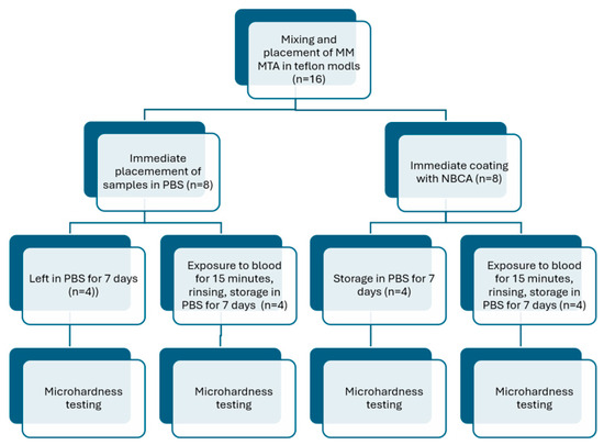
The aim of this study was to evaluate the influence of n-butyl cyanoacrylate (NBCA) coating of freshly mixed mineral trioxide aggregate (MTA) on its microhardness after setting under conditions of moisture and human blood exposure. MicroMega MM-MTA samples were prepared using Teflon molds
[...] Read more.
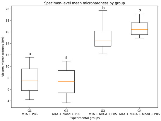
The aim of this study was to evaluate the influence of n-butyl cyanoacrylate (NBCA) coating of freshly mixed mineral trioxide aggregate (MTA) on its microhardness after setting under conditions of moisture and human blood exposure. MicroMega MM-MTA samples were prepared using Teflon molds (6 mm × 4 mm). Four experimental groups (n = 4) were established: control (MTA + phosphate-buffered saline (PBS)), MTA coated with NBCA (PeriAcryl) + PBS, MTA + blood + PBS, and MTA coated with NBCA + blood + PBS. Samples assigned to the blood-exposure groups were exposed to human blood for 15 min, rinsed, and subsequently stored in PBS. Prior to microhardness testing, all samples were incubated in PBS for seven days. Microhardness was measured using a Vickers microhardness tester, with five indentations performed on each specimen (20 measurements per group). Data were analyzed using the Shapiro–Wilk test for normality, Levene’s test, and two-way ANOVA, followed by Tukey’s post hoc test. The analysis revealed significant differences among the experimental groups (p < 0.001). Post hoc analysis demonstrated statistically significant differences between tissue glue-coated and uncoated samples (p < 0.05). Within the limitations of this in vitro study, coating MTA with NBCA tissue glue during the setting period significantly increased its microhardness following exposure to human blood and PBS.
Full article
(This article belongs to the Special Issue Property, Evaluation and Development of Dentin Materials)
►▼
Show Figures

Figure 1

Open AccessArticle
Electrosprayed Chitosan–Calcium Phosphate Microshell Composite Coatings on Silanized Titanium Substrates
by
Andrew Blass Watson, Matthew J. Atwill, Tomoko Fujiwara, Ranganathan Gopalakrishnan, Jessica Amber Jennings and Joel D. Bumgardner
J. Funct. Biomater. 2026, 17(8), 409; https://doi.org/10.3390/jfb17080409 - 17 Aug 2026
Abstract
Chitosan and calcium phosphate (CaP) are attractive bioactive coating materials for titanium (Ti) orthopedic implants, but coating approaches must provide uniform deposition, adequate adhesion, and cytocompatibility. This proof-of-concept study evaluated whether CaP microshells could be incorporated into electrosprayed chitosan coatings bonded to silanized
[...] Read more.
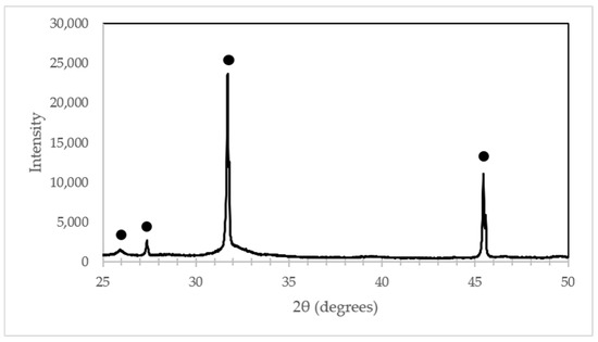
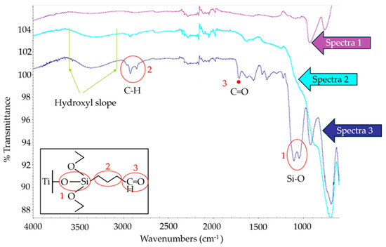
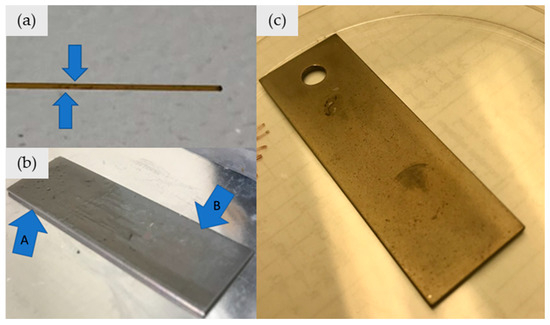
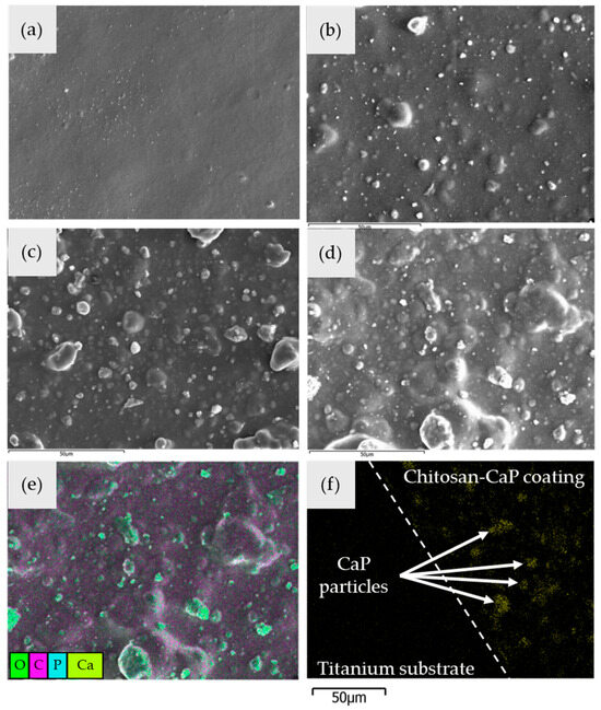
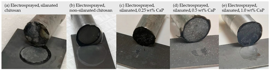
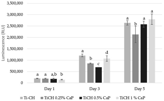
Chitosan and calcium phosphate (CaP) are attractive bioactive coating materials for titanium (Ti) orthopedic implants, but coating approaches must provide uniform deposition, adequate adhesion, and cytocompatibility. This proof-of-concept study evaluated whether CaP microshells could be incorporated into electrosprayed chitosan coatings bonded to silanized Ti substrates without compromising coating properties. CaP microshells were synthesized using carbon microsphere templates and added to chitosan electrospray solutions at 0.25, 0.5, and 1.0 wt% relative to chitosan. Electrospray parameters were adjusted, and coatings were characterized by scanning electron microscopy, energy dispersive X-ray spectroscopy, Fourier transform infrared spectroscopy, tensile adhesion testing, water contact angle measurements, and W-20-17 bone marrow stromal cell culture. Increasing capillary diameter and reducing pressure enabled stable deposition of uniform composite coatings containing up to 1.0 wt% CaP. CaP microshells were distributed across the coating surfaces and throughout the coating thickness. Silanization significantly increased coating adhesion compared with non-silanized Ti, while CaP incorporation up to 1.0 wt% did not significantly reduce bond strength. All coatings were hydrophilic and supported viable cell attachment and growth over five days. These findings support the feasibility of electrosprayed chitosan–CaP microshell coatings as adhesive, cytocompatible bioactive coating platforms for Ti implant materials.
Full article
(This article belongs to the Special Issue Drug- and Ion-Releasing Implants)
►▼
Show Figures

Figure 1
Open AccessArticle
Evaluation of Polytetrafluoroethylene (PTFE) Tape for Gingival Displacement Using CAD/CAM Analysis and Scanning Electron Microscopy (SEM)
by
Laura Monica Rusu, Antonio Pop, Stanca Cuc, Mircea Aurelian Rusu, Sorina Sava, Anca Labunet, Cristina Iosif and Adriana Objelean
J. Funct. Biomater. 2026, 17(8), 408; https://doi.org/10.3390/jfb17080408 - 17 Aug 2026
Abstract
Background: This in vitro study compared the effectiveness of polytetrafluoroethylene (PTFE) tape and a conventional cotton retraction cord for gingival displacement and evaluated the mechanical and microstructural characteristics of PTFE tape. Methods: Eight standardized prepared teeth with artificial gingiva were evaluated under three
[...] Read more.
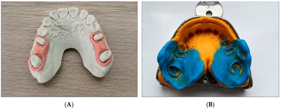
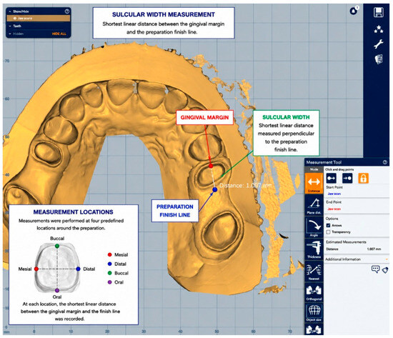
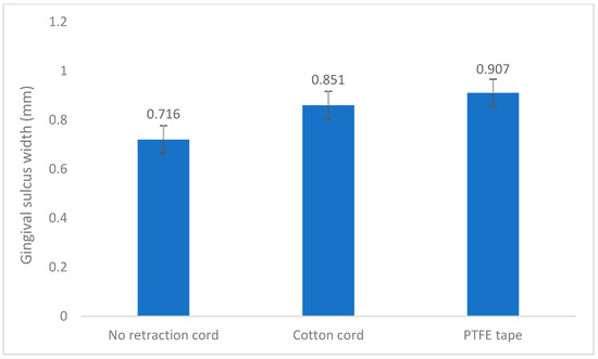
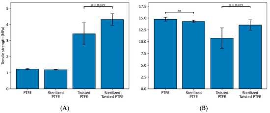
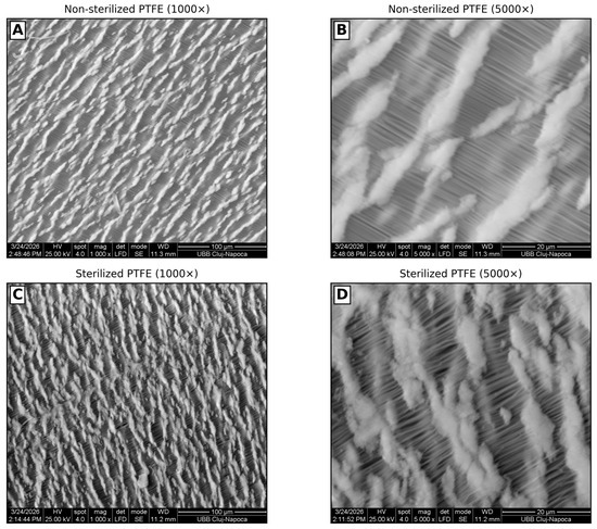
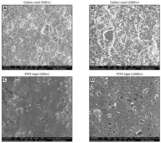
Background: This in vitro study compared the effectiveness of polytetrafluoroethylene (PTFE) tape and a conventional cotton retraction cord for gingival displacement and evaluated the mechanical and microstructural characteristics of PTFE tape. Methods: Eight standardized prepared teeth with artificial gingiva were evaluated under three conditions: control, cotton retraction cord, and PTFE tape. After each condition, impressions were digitized using a CAD/CAM system. Sulcular width was measured at four predefined locations and averaged for statistical analysis. PTFE tape was also evaluated by tensile testing before and after sterilization and by scanning electron microscopy (SEM). Results: Mean sulcular widths were 0.716 ± 0.118 mm (control), 0.851 ± 0.140 mm (cotton cord), and 0.907 ± 0.099 mm (PTFE). Repeated-measures ANOVA showed a significant effect of the displacement method (p < 0.001), with PTFE producing significantly greater sulcular enlargement than cotton cord. Qualitative SEM analysis revealed a more homogeneous impression surface morphology following PTFE removal. Sterilization did not significantly affect the tensile properties or microstructural integrity of PTFE tape. Conclusions: Under standardized in vitro conditions, PTFE tape produced the highest gingival sulcular enlargement and maintained favorable mechanical and microstructural properties, supporting its potential as an alternative gingival displacement material. Further clinical studies are required to confirm these findings.
Full article
(This article belongs to the Section Dental Biomaterials)
►▼
Show Figures

Figure 1
Open AccessReview
Confined Catalysis of Enzymes: Enhancement Strategies, Materials, and Biosensor Applications
by
Lijiao Zhang, Junyi Lu, Chenjie Yan, Wenjing Wang and Dajing Chen
J. Funct. Biomater. 2026, 17(8), 407; https://doi.org/10.3390/jfb17080407 - 15 Aug 2026
Abstract
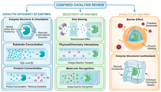
Confined catalysis enhances enzyme activity, stability, and selectivity by encapsulating enzymes in well-defined micro- or nanoscale environments. Unlike conventional immobilization strategies, which primarily stabilize enzymes, this approach actively regulates enzyme behavior, substrate transport, and product removal, addressing challenges in real-world biosensing. In this
[...] Read more.
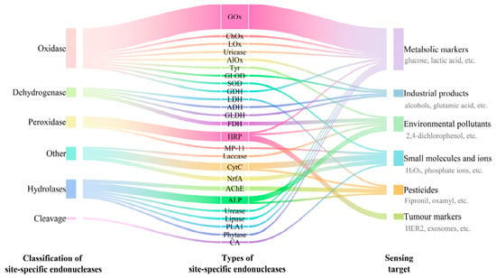
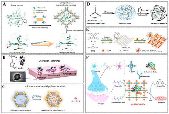
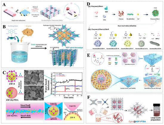
Confined catalysis enhances enzyme activity, stability, and selectivity by encapsulating enzymes in well-defined micro- or nanoscale environments. Unlike conventional immobilization strategies, which primarily stabilize enzymes, this approach actively regulates enzyme behavior, substrate transport, and product removal, addressing challenges in real-world biosensing. In this work, we connect confined catalysis principles with biosensor design requirements, emphasizing tailored microenvironments, hierarchical pore architectures, and cascade reaction optimization to improve sensitivity, selectivity, and operational stability. The review also highlights advanced strategies such as molecular imprinting, aptamer recognition, and DNA scaffold-guided co-localization, which enable precise substrate discrimination and efficient signal amplification. By integrating these confinement strategies with emerging materials, including biological macromolecular materials, amorphous porous materials, and crystalline framework materials, this work outlines pathways to next-generation biosensors with enhanced performance and practical applicability, addressing limitations overlooked in previous studies.
Full article
(This article belongs to the Section Biomaterials and Devices for Healthcare Applications)
►▼
Show Figures

Figure 1
Open AccessArticle
Influence of Mastication on Orthodontic Wire Sliding in Nickel–Titanium Brackets with Superelastic Wings
by
Jasmin Wichelhaus, Uwe Baumert, Corinna Seidel and Hisham Sabbagh
J. Funct. Biomater. 2026, 17(8), 406; https://doi.org/10.3390/jfb17080406 - 15 Aug 2026
Abstract
(1) Background: Orthodontic tooth movement (OTM) during sliding mechanics is influenced by a multitude of interactions. Studies suggest that vibration induced by mastication or usage of electric tooth brushes might impact resistance to sliding ( ) in conventional brackets. In superelastic
[...] Read more.
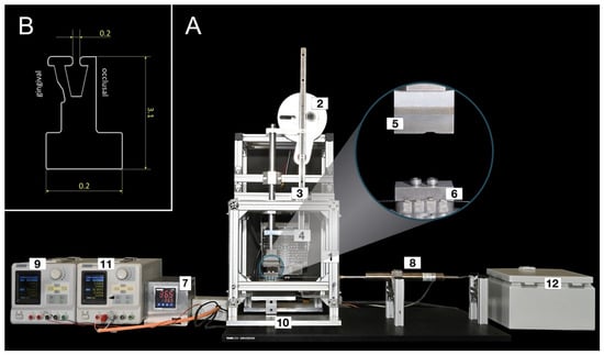
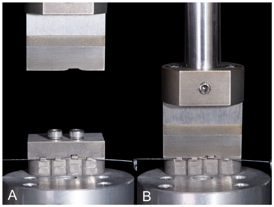
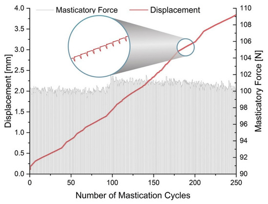
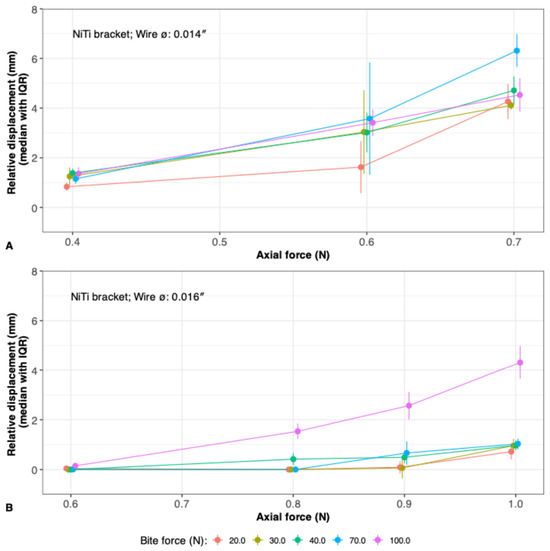
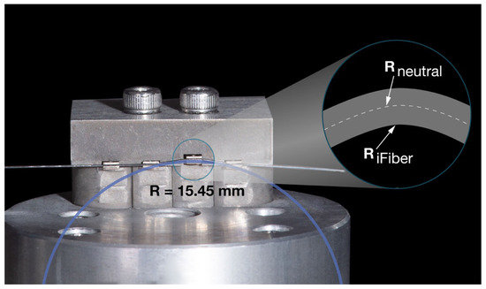
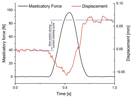
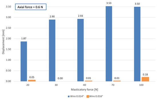
(1) Background: Orthodontic tooth movement (OTM) during sliding mechanics is influenced by a multitude of interactions. Studies suggest that vibration induced by mastication or usage of electric tooth brushes might impact resistance to sliding ( ) in conventional brackets. In superelastic brackets, archwire sliding might also be altered due to deformation of the bracket wings during mastication. Therefore, the aim of this study was to investigate how the superelastic properties of Nickel–Titanium (NiTi) brackets affect archwire sliding during mastication. (2) Methods: Orthodontic superelastic NiTi archwires with diameters of 0.014” and 0.016” were ligated into four superelastic NiTi brackets. One bracket was positioned with a vertical offset of 1 mm to simulate clinical vertical leveling. A weight force was applied to axially preload the archwire. The occlusal and axial forces affecting the movement of the archwires through the bracket were measured. (3) Results: Low chewing forces caused a measurable axial movement of the 0.014” wire. Increased chewing forces resulted in higher axial movements. The archwire diameter had an impact: 0.014” NiTi required lower masticatory forces to initiate wire motion as compared to the 0.016” NiTi. (4) Conclusions: The superelastic NiTi brackets examined showed that under simulated continuous dynamic masticatory loading, obstacles such as friction or binding could be overcome.
Full article
(This article belongs to the Special Issue Functional Dental Materials for Orthodontics and Implants)
►▼
Show Figures

Figure 1
Open AccessArticle
Development of an Electro-Responsive Sorafenib-Loaded Polypyrrole Coating-Modified Nickel–Titanium Alloy for Tumor Ablation Therapy
by
Lele Liu, Peng Gu, Dan Xia, Yonghao Wen, Baoe Li, Donghui Wang and Yaohong Wu
J. Funct. Biomater. 2026, 17(8), 405; https://doi.org/10.3390/jfb17080405 - 14 Aug 2026
Abstract
Nickel–titanium (NiTi) stents have been widely used for the palliative management of portal vein tumor thrombosis (PVTT) due to their excellent mechanical strength and biocompatibility. However, conventional NiTi implants are therapeutically passive and lack intrinsic antitumor activity, rendering them vulnerable to tumor ingrowth
[...] Read more.
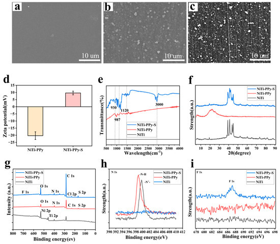
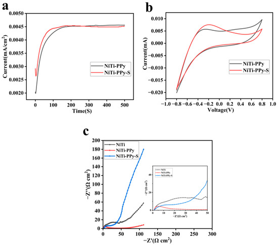
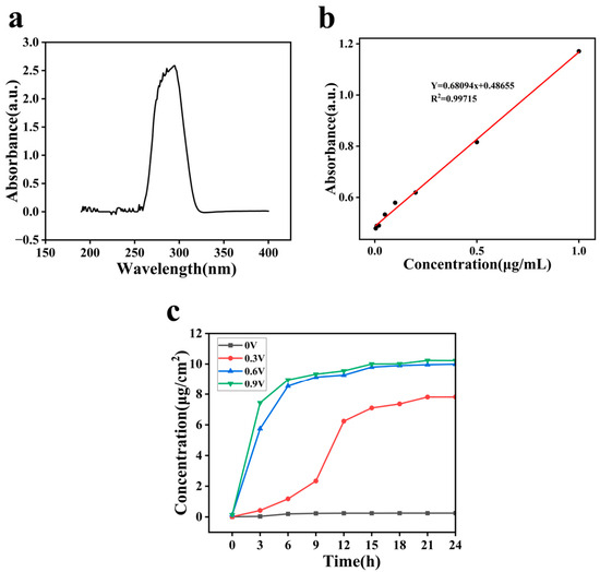
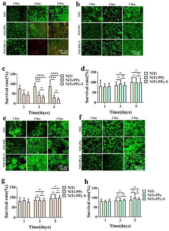
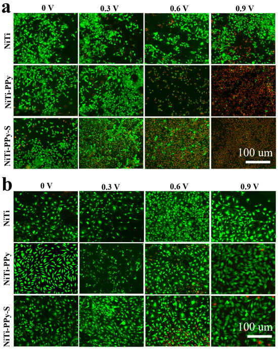
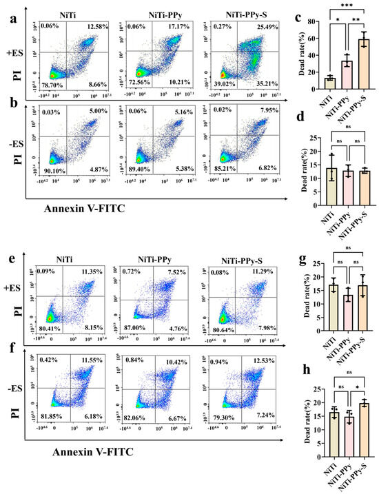
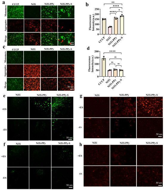
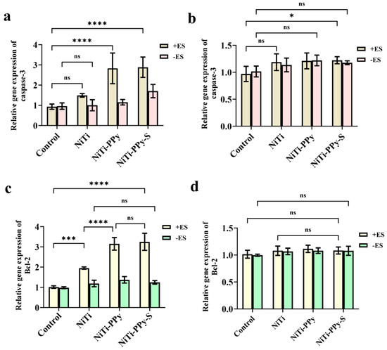
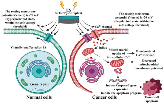
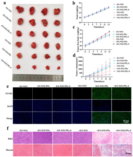
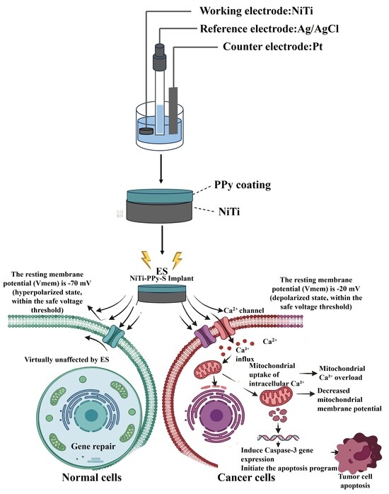
Nickel–titanium (NiTi) stents have been widely used for the palliative management of portal vein tumor thrombosis (PVTT) due to their excellent mechanical strength and biocompatibility. However, conventional NiTi implants are therapeutically passive and lack intrinsic antitumor activity, rendering them vulnerable to tumor ingrowth and subsequent restenosis. Electrical stimulation-mediated ablation offers a controllable physical strategy for local tumor clearance; however, the native TiO2 passivation layer on the NiTi surface restricts its interfacial electroactivity. Electrochemical impedance spectroscopy revealed that the PPy coating significantly reduced the interfacial impedance of the NiTi substrate. After loading with sorafenib, NiTi−PPy−S maintained good electrochemical responsiveness and enabled voltage-dependent drug release characteristics. Under an applied potential of 0.9 V for 24 h, approximately 10.522 ± 0.295 μg/cm2 of the loaded sorafenib was released mechanistically, whereas only 0.249 ± 0.171 μg/cm2 was released through passive diffusion over the same period. The antitumor effect is closely associated with ES-triggered Ca2+ influx, mitochondrial Ca2+ overload, loss of mitochondrial membrane potential, and caspase-3-dependent apoptosis. These findings demonstrate that electro-responsive PPy can transform passive NiTi implants into active antitumor therapeutic interfaces, offering a promising strategy for developing multifunctional implants for PVTT treatment.
Full article
(This article belongs to the Special Issue Biomaterials for Drug Delivery and Cancer Therapy)
►▼
Show Figures

Graphical abstract
Journal Menu
► ▼ Journal Menu-
- JFB Home
- Aims & Scope
- Editorial Board
- Topical Advisory Panel
- Early Career Editorial Board
- Instructions for Authors
- Special Issues
- Topics
- Sections & Collections
- Article Processing Charge
- Indexing & Archiving
- Editor’s Choice Articles
- Most Cited & Viewed
- Journal Statistics
- Journal History
- Journal Awards
- Conferences
- Editorial Office
Journal Browser
► ▼ Journal BrowserHighly Accessed Articles
Latest Books
E-Mail Alert
News
19 August 2026
The 2nd International Conference on Bioengineering: Bioengineering in an Era of AI (BIOENG 2026) Announces Distinguished Speakers Lineup and Late-Breaking Poster Submission Opportunity
Topics
Topic in
Applied Nano, Bioengineering, Coatings, JFB, Materials, Surfaces
Functional and Antiseptic Biomedical Surfaces and Interfaces
Topic Editors: Annett Dorner-Reisel, Aneta Zima, Nick Tucker, Tao WangDeadline: 31 August 2026
Topic in
IJMS, JFB, Macromol, Materials, J. Compos. Sci., Polymers
Recent Advances in Composite Biomaterials
Topic Editors: Diego Romano Perinelli, Florentina LupascuDeadline: 30 November 2026
Topic in
JFB, Materials, Prosthesis, IJMS, Oral, JCM
New Technological and Clinical Advances for Controlling Peri-Implantitis
Topic Editors: Javier Gil, Eugenio Velasco-Ortega, Aritza Brizuela-Velasco, Conrado AparicioDeadline: 31 December 2026
Topic in
Gels, JFB, Nanomaterials, Polymers, Polysaccharides
Advances in Bio-Based Functional Materials for Sustainable and Biomedical Applications
Topic Editors: Ricardo J. B. Pinto, Gabriela GuedesDeadline: 1 April 2027
Conferences
Special Issues
Special Issue in
JFB
Biopolymer-Based Materials for Biomedical Applications
Guest Editors: Kupnik Kaja, Mateja PrimožičDeadline: 28 August 2026
Special Issue in
JFB
Graphene Materials in Medical Applications
Guest Editors: Lílian Vanessa Rossa Beltrami, Daniele PerondiDeadline: 31 August 2026
Special Issue in
JFB
Emerging Trends of Nanomaterials in Biology
Guest Editors: Tapas K. Mandal, Nargish ParvinDeadline: 31 August 2026
Special Issue in
JFB
Advanced Nanomaterials in Medicine: Innovations in Diagnostics, Therapy, and Regeneration
Guest Editor: Rewati Raman UjjwalDeadline: 31 August 2026
Topical Collections
Topical Collection in
JFB
Feature Papers in Dental Biomaterials
Collection Editor: Gianrico Spagnuolo
Topical Collection in
JFB
Feature Papers in Biomaterials for Healthcare Applications
Collection Editor: Dimitrios Karamichos
Topical Collection in
JFB
Feature Papers in Antibacterial Biomaterials
Collection Editor: Masakazu Kawashita
Topical Collection in
JFB
Feature Papers in Biomaterials for Drug Delivery
Collection Editor: Junjie Li