


Abstract
Bakanae is one of the important diseases of rice in Bangladesh that causes substantial yield loss every year. We collected thirty isolates of Fusarium spp. from bakanae-infected rice plants from different agroecological zones of Bangladesh and investigated the variations in cultural and morphological characteristics and pathogenicity. Diversity was found in cultural characteristics, viz., colony features, phialide, chlamydospore formation, shape, and size of macro- and microconidia. Three variants of Fusarium species such as F. moniliforme, F. fujikuroi, and F. proliferatum were identified on PDA media based on their cultural and morphological characteristics. Isolate FM10 (F. moniliforme) exhibited the highest disease aggressiveness in developing elongated plants (26.50 cm), the highest number of chlorotic leaves (5.75), and a lower germination percentage. We evaluated different bioagents against the virulent isolate of F. moniliforme to develop a rice bakanae disease management approach. Four bioagents, viz., Trichoderma spp., Bacillus subtilis, Pseudomonas fluorescens, and Achromobacter spp., were evaluated for growth suppression of F. moniliforme. Among the bioagents, Achromobacter spp. and B. subtilis (BS21) showed 73.54% and 71.61% growth suppression, respectively. The investigation revealed that the application of Achromobacter spp. and B. subtilis (BS21) would be a potential candidate for effective and eco-friendly management of the bakanae disease of rice.
1. Introduction
Rice (Oryza sativa L.) is a prominent staple crop that serves as a significant food product for global populations, with a special emphasis on Asian societies. It provides sustenance for almost 60% of the global population [1]. Approximately 90% of global rice production and consumption exists in Asian regions [2]. Bangladesh is now the third largestrice-producing country in the world after China and India [3]. The total area of rice in Bangladesh is about 11.7 million hectares, with a production of 37.60 million metric tons [4]. The rice crop has a significantly diminished average yield as a consequence of the occurrence of specific illnesses. Rice is affected by a total of 50 diseases, encompassing 21 fungal, 6 bacterial, 12 viral, 4 nematode-related, and 7 other diseases and disorders [5]. These are the major issues responsible for the low yields in Bangladesh. Minor diseases such as false smut, bakanae, sheath rot, and grain discoloration, which were previously ignored, are now posing a severe danger to rice production [6].
Bakanae disease, caused by Fusarium moniliforme (anamorph of Gibberella fujikuroi Sawada),has recently emerged as a significantly important disease in Asia and other rice-growing countries of the world [7]. In Bangladesh, bakanae disease has been progressively increasing in prevalence, resulting in both qualitative and quantitative crop losses. Rice yield suffers a regular loss of 20–50% in Japan [8], 40% in Nepal [9], and 10–50% in Bangladesh [10] due to F. moniliforme. The first symptoms of bakanae disease are etiolation and aberrant elongation in affected plants through the production of gibberellic acid. Infected plants are taller than healthy plants and light yellowish in color. Roots form at the affected plants’ lower nodes. In the advanced stage, a white or light pinkish fungus develops on the lower nodes, and rotting of the foot region occurs, which leads to a few grains of poor quality or the complete death of infected plants [6,11]. Moreover, this fungus produces a diversity of mycotoxins comprised mainly of fumonisin B1 (FB1) during the progression of growth and incursion of rice grains [12]. FB1 is not only responsible for significant economic losses but has also been correlated with high occurrences of liver and esophageal cancer in numerous areas of the world [13].
The establishment of sustainable rice agricultural practices can be attained by the use of novel and enhanced cultivars, alongside the implementation of modern disease management strategies including resistant varieties, cultural practices, and biological and chemical control. All of these strategies have varying degrees of effectiveness in combating diseases. The only way to control bakanae diseases is to treat the seeds with chemical fungicides. However, this is expensive and harmful to both plants and the environment. Biological control is cost-effective and eco-friendly, and it is the most sustainable long-term solution [14]. Trichoderma spp., Pseudomonas spp., Achromobacter spp., and Bacillus spp. Have potential antagonist effects against phytopathogens [15]. Among them, P. fluorescens, B. cereus, and Trichoderma spp. isolates have an excellent potential to be used as biocontrol agents of F. fujikuroi in rice [16,17]. The capacity of these organisms to colonize roots and their beneficial interactions with plants can lead to an efficient defense against the disease, since they can trigger host defense responses against Fusarium spp. attack. In the present study, we evaluated the potential efficacy of bio-agents to suppress the rice bakanae disease causal organism F. moniliforme in in vitro conditions. The potential bioagents of the present investigation can be applied in the field for controlling bakanae disease.
2. Materials and Methods
2.1. Experimental Site
The experiments were carried out at the Laboratory of Bio-signaling, Bio-active compounds, and Bio-formulation of the Department of Plant Pathology and Professor Golam Ali Fakir Seed Pathology Centre (SPC), Bangladesh Agricultural University, Mymensingh-2202, during the period from January 2019 to December 2019. The laboratory experiment was laid out in a Completely Randomized Design (CRD) with three replications.
2.2. Isolation, Purification, and Identification of the Pathogen
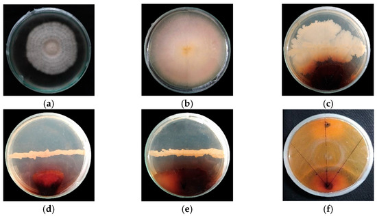
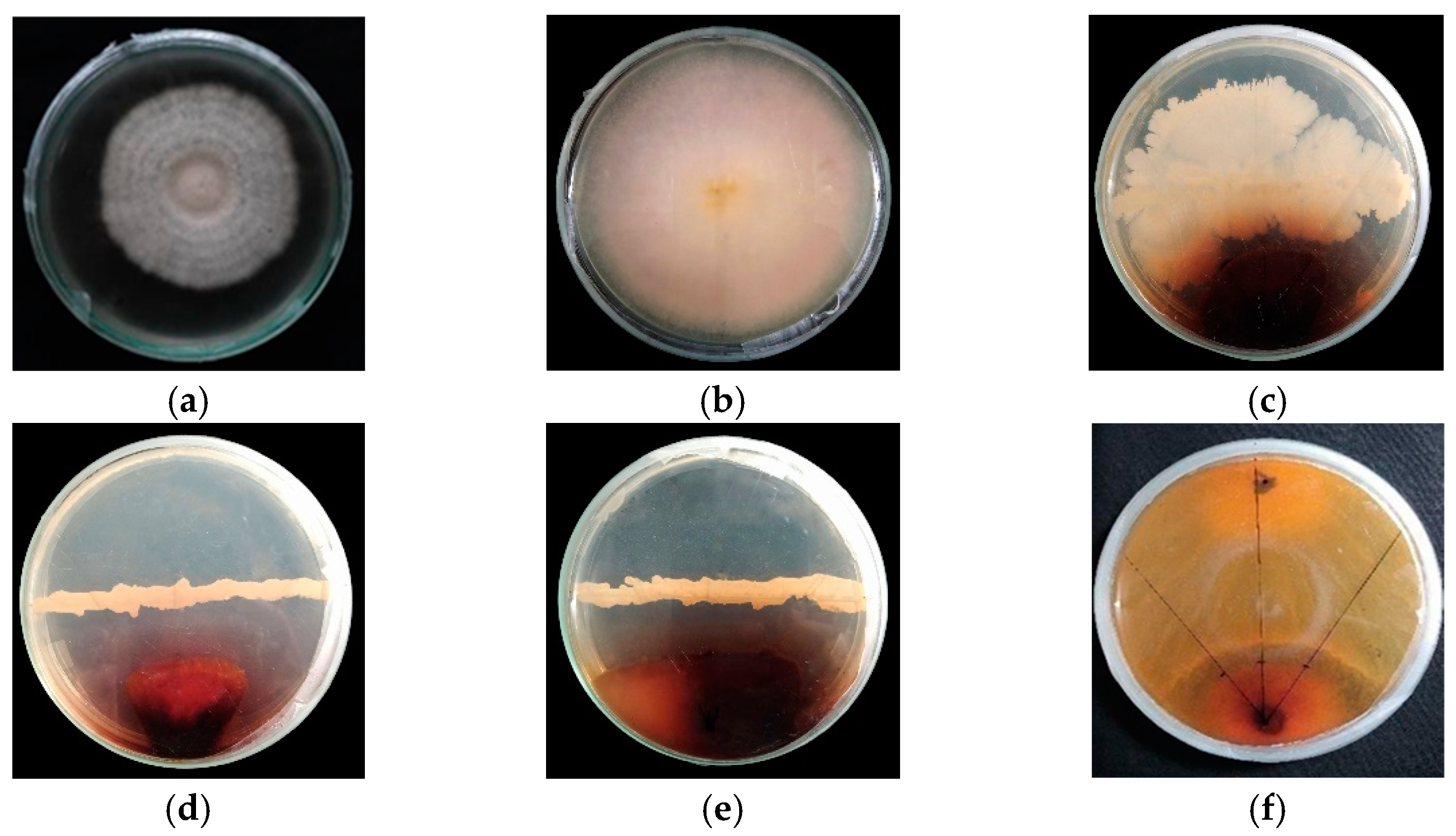
The diseased plant parts and soils around the infected root of bakanae disease were collected from different regions of Bangladesh (Table 1). Blotter incubation and soil dilution methods were followed to isolate Fusarium spp. from the infected plant parts of rice [18]. The infected roots of the plants were cut into small pieces of 5 mm length, with some healthy parts. Three layers of blotter papers (Whatman No. 1) were soaked in distilled water and placed at the bottom of plastic petri dishes. The roots were surface sterilized with 10% NaOCl solution for 30 s and subsequent washing with sterilized water three times to remove unwanted microorganisms from the surface of the infected parts. Then, 4–5 small pieces of infected plant roots were placed in petri dishes. Afterward, petri dishes were kept in an incubator at 25 ± 1 °C. After 7 days of incubation, the colony of Fusarium spp. was examined under a compound microscope, and pure culture was created by successive culture [7]. In the case of the soil dilution method, soil samples were air-dried and ground with mortar and pestle. One gram of soil sample was mixed into 9 mL distilled water, and then subsequent dilution series were prepared for isolating the fungus. One drop from each dilution was poured into separate 0.2% water agar media in petri dishes. After 7 days of incubation, the colony of fungi was examined under the microscope, and further pure culture was created [19,20,21] (Figure 1).
Table 1.
Isolates of Fusarium spp. collected from different agroecological zones of Bangladesh.
Figure 1.
Growth suppression ability of different bioagents against Fusarium moniliforme. (a) Pure culture of Fusarium moniliforme. (b) Control. (c) Bacillus subtilis. (d) Pseudomonas fluorescens. (e) Achromobacter spp. (f). Trichoderma spp.
2.3. Cultural and Morphologic
Mycelial discs (5 mm diameter) of 7-day-old cultures of Fusarium spp. of each isolate were transferred to the center of the PDA culture medium. The culture plates were incubated at 25 ± 1 °C for 7 days in an incubator. Colony color, substrate color, phialide, and chlamydospore formation were observed, which developed on the PDA medium from thirty isolates of Fusarium spp. Morphology in terms of shape, size, and septation of macro- and microconidia of thirty isolates of Fusarium spp. on PDA medium was measured and recorded. Ten-day-old cultures were considered for morphological variability. With the help of a compound microscope, observations of variation in conidial dimension were recorded [22].
2.4. Pathogenicity Test
A pathogenicity test was conducted by seed inoculation assay. The high-yielding rice cultivar BRRI dhan29 was used to test the pathogenicity. The surface of the seeds was disinfected by immersion in 70% ethanol for 1 min, then transferred to 1% sodium hypochlorite solution for 1 min and rinsed three times consecutively in sterile distilled water. Seeds were then left to dry inside the airflow cabinet. Suspension of fungal inoculum was prepared from a 15-day-old culture of F. moniliforme that was flooded with sterile water and scraped with a sterile spatula. The resulting suspensions were filtered through two layers of sterile cotton lint, and the concentrations of conidial suspensions were determined with a hemocytometer and adjusted to a concentration of 2 × 10⁶ spores/mL in sterile distilled water. Thirty rice seeds were soaked in 10 mL of inoculum suspension for 18 h at room temperature. Control seeds were soaked in sterile water only. Inoculated and control seeds were then sown in small plastic pots (three pots per isolate and ten seeds per pot) containing an autoclaved mixture of soil and sand at a ratio of 3:1. Fifteen days after inoculation, shoot length (cm), no. of chlorotic leaves, and germination percentage were assessed [22,23]. The seedlings were observed for symptoms of bakanae, as a number of slender and chlorotic, elongated leaves were observed, and a number of plants showed crown rot and produced roots on the lower nodes [24].
2.5. In Vitro Evaluation of Bioagents to Suppress the Growth of the Target Pathogen of Bakanae Disease of Rice
2.5.1. Multiplication of Bioagents
Bacillus subtilis, Pseudomonas fluorescens, Achromobacter spp., and Trichoderma spp. isolates used in the study were obtained from the laboratory of Bio-signaling, Bio-active compounds, and Bio-formulation of the Department of Plant Pathology, Bangladesh Agricultural University. Bacillus subtilis and Achromobacter spp. isolates were streaked on nutrient agar media, and Pseudomonas fluorescens isolates were streaked on King’s B media in glass petri dishes. Then, the petri dishes were kept in an incubator at 28 °C for 24 h. On the other hand, Trichoderma spp. was cultured on sterilized PDA plates. The plates were then incubated at 26 °C for 7 days. A 5 mm culture block from the plate of Trichoderma spp. was transferred to the new PDA plates to maintain pure culture and stored in a refrigerator at 4 °C for further studies.
2.5.2. In Vitro Growth-Suppressing Ability of Bioagents against Fusarium moniliforme
The growth suppression ability of B. subtilis, P. fluorescens, Achromobacter spp., and Trichoderma spp. isolates against F. moniliforme was examined by dual-culture technique [25] and incubation at 26 °C for 8–10 days. Three replications were maintained for each isolate. Observations on the width of the inhibition zone and radial mycelial growth at 9 and 12 days after incubation of the test pathogen were recorded, and percent inhibition of pathogen growth was calculated using the formula proposed by Vincent [26]:
where C = mycelial growth of the pathogen in control; T = mycelial growth of the pathogen in the dual-culture plate.
Percent inhibition (I) = (C − T/C) ×100
2.6. Disease Assessment
Disease assessment (Table 2) was done following the scale reported by Scherm et al. [27]
Table 2.
The disease intensity of bakanae is rated using a 0–4 scale. The scale spans over 5 classes [27].
2.7. Statistical Analyses
Statistical analyses were performed by R statistical software (http://www.R-project.org, accessed on 25 October 2023) to find out the significance of the differences among the bio-agents. ANOVA was performed by the Agricola R package [28], and mean differences were adjudged by Tukey’s HSD test.
3. Results
3.1. Cultural and Morphological Variation in Fusarium spp.
Different isolates of the pathogens associated with bakanae disease of rice were identified on PDA media as Fusarium moniliforme, F. fujikuroi, and F. proliferatum based on their cultural and morphological characteristics (Table 3 and Table 4). F. moniliforme is responsible for producing gibberellic acid, which causes abnormal elongation of plants, resulting in bakanae disease of rice. F. fujikuroi (FM3, FM11, FM13, FS16, FS19, FS20, FS21, FS26, FS34, FD35, FD47, FD48) isolates were identified based on characteristics like monophialide and simple polyphialide structure. Obovoid with flattened base, oval (0–1 septate), allantoid pyriform (rarely) microconidia (5.31–13.59 µm × 1.44–12.96 µm) were present. Macroconidia (57.35–118.51 µm × 11.17–23.05 μm) were falcate to almost straight and slender in shape (1–3 septation). F. moniliforme isolates (FM5, FM6, FM7, FM8, FM9, FM10, FM12, FS18, FS23, FD41) were characterized by simple and branch monophialide structures. The size of microconidia was 4.49–13.89 μm × 4.78–10.88 μm, and the shape was obovoid with a flattened base, oval to long oval, and elliptical. Macroconidia were falcate to straight, with three to five septate and a size of 54.68–177.33 μm × 11.29–18 μm. F. proliferatum isolates (FM4, FS22, FS24, FS25, FS29, FS31, FS32, FD45) have monophialide and polyphialide structures. Microconidia were obovoid with a flattened base, pyriform, and allantoid in shape, and sizes were 2.65–21.74 μm × 1.75–18.40 μm. Macroconidia were falcate to almost straight, slender, with one to four septate and a size of 57.07–254.98 μm × 15.81–33.10 μm. Both F. moniliforme and F. fujikuroi produce a large amount of toxins and metabolites that cause pigmentation in PDA media. The colors vary from light orange or pinkish to deep brown and sometimes grayish violet. On the other hand, F. proliferatum shows pinkish white to grayish black stripes.
Table 3.
Variation in colony characteristics of Fusarium spp. isolates on PDA media.
Table 4.
Variation in shape, size, and septation of microconidia and macroconidia of Fusarium spp. isolates on PDA media.
3.2. Evaluation of Pathogenic Variation in Different Isolates of Fusarium spp.
Thirty isolates of Fusarium spp. were subjected to a pathogenicity test to observe the elongated shoot length, number of chlorotic leaves, and germination percentage. The pathogenicity of different isolates was estimated as the degree of disease symptom expression 15 days after sowing (Table 5). The highest shoot length was observed in FM10 (26.50 cm), and the lowest length was recorded in FD35 (12.00 cm). The highest number of chlorotic leaves was observed in FM11 (5.75) isolates, followed by FM4, FM9, FM10, FS19, FS21, FS24, FS34, and FD45. At 15 DAS, the lowest germination percentage was observed in the FD35 isolate, which was statistically similar to the FM10 isolate. As such, the presence of a heavy pathogen dose of Fusarium spp. seed germination hampers plants drastically, and the FM10 (F. moniliforme) isolate was selected for further research experiments, as it showed the maximum aggressiveness in bakanae disease formation.
Table 5.
Assessing pathogenicity of different isolates of Fusarium moniliforme collected from different AEZs of Bangladesh by artificial inoculation in plants grown in pots (15DAS).
3.3. In Vitro Growth Suppression of Fusarium moniliforme by Bacillus subtilis
Eight Bacillus subtilis isolates were evaluated against F. moniliforme in in vitro conditions following a dual-culture plate technique on a PDA medium. The highest (54.64 mm) mycelial growth was observed on the control plate. The highest inhibition (71.61%) of mycelial growth over the control was observed in the BS21 isolate, and the lowest (18.69%) mycelial growth inhibition was found in the BS31 isolate (Table 6). Therefore, the BS21 isolate showed the most potent inhibition against F. moniliforme (Figure 1c).
Table 6.
In vitro evaluation of different isolates of Bacillus subtilis for suppressing the growth of Fusarium moniliforme.
3.4. In Vitro Growth Suppression of Fusarium moniliforme by Pseudomonas fluorescens
The application of eight P. fluorescens isolates was assessed against F. moniliforme in in vitro conditions. The highest (57.77 mm) mycelial growth was observed in the PF9 isolate, followed by the control plate (54.64 mm). The highest percent inhibition (58.64%) of mycelial growth over the control was observed in the case of isolate PF7. The lowest (4.52%) mycelial growth inhibition was found in isolate PF18 (Table 7). Among all the isolates of Pseudomonas fluorescens, PF7 showed the strongest inhibition against F. moniliforme (Figure 1d).
Table 7.
In vitro evaluation of different isolates of Pseudomonas fluorescens for suppressing the growth of Fusarium moniliforme.
3.5. In Vitro Growth Suppression of Fusarium moniliforme by Achromobacter spp.
Achromobacter spp. Was tested in vitro against F. moniliforme using a dual-culture plate technique on a PDA medium. Significant growth suppression (73.54%) of F. moniliforme by Achromobacter spp. was found (Table 8 and Figure 1e).
Table 8.
In vitro evaluation of Achromobacter spp. for suppressing the growth of Fusarium moniliforme.
3.6. In Vitro Growth Suppression of Fusarium moniliforme by Trichoderma spp.
Invitro evaluation of Trichoderma spp. against F. moniliforme was performed following a dual-culture plate technique on a PDA medium. A total of 69.84% radial mycelial growth inhibition of F. moniliforme by Trichoderma spp. was recorded (Table 9 and Figure 1f).
Table 9.
Invitro evaluation of Trichoderma spp. for suppressing the growth of Fusarium moniliforme.
4. Discussion
Rice bakanae disease is emerging as the most destructive disease for rice cultivation in Bangladesh. It causes greater yield losses across the rice-growing regions of the country. The pathogen primarily survives in seeds but is also known to survive in the soil [8]. Based on cultural, morphological, and pathogenicity tests, we identified the causal agent of bakanae disease of rice as F. moniliforme. Pandey et al. [7] also noted that F. moniliforme causes bakanae disease in rice in Nepal. These Fusarium species could differ with geographic as well as climatic variations [8]. Leslie et al. [29] identified Fusarium spp. using the morphological structure and different size and shape of the conidia. According to Nirenberg et al. [30], morphological features of the fungal isolates were assessed based on the size and the shape of micro- and macroconidia. Different isolates showed different colors of their colony in the PDA medium. The fungal isolates were identified on PDA media as F. moniliforme, F. fujikuroi, and F. proliferatum based on their colony features, phialide, and chlamydospore formation, as well as shape, septation, and size of macro- and microconidia. Among them, F. moniliforme is responsible for producing large quantities of gibberellic acid (GA3),which causes the seedling to have abnormal elongation and results in bakanae disease of rice. Sunder and Satyavir [31] reported that the isolates of F. moniliforme varied greatly in producing GA3 in liquid culture. There were several numbers of F. moniliforme, F. fujikuroi, and F. proliferatum isolated. Therefore, it was necessary to find the most pathogenic strain among the isolates. For this, pathogenicity tests of 30 isolates of Fusarium species were carried out using rice plants under a net house. Among them, the FM10 (F. moniliforme) isolate caused the highest shoot elongation in plants (26.50 cm), the highest number of chlorotic leaves (5.75), and the lowest germination percentage due to its maximum pathogenic virulence. Bashyal et al. [32] reported that Pusa Basmati 1121 rice was susceptible to bakanae disease and exhibited more abnormal elongation, rotting, and shrinkage of leaves. Therefore, effective strategies are essential to manage bakanae disease. This disease is controlled by chemical fungicides, but these are also severe environmental hazards [33]. To overcome this problem, bio-agents are used to suppress bakanae diseases and work through their production of various secondary metabolites [34]. In this study, we have used four bio-agents against F. moniliforme,viz., Trichoderma spp., Bacillus subtilis, Pseudomonas fluorescens, and Achromobacter spp., to control bakanae disease in plants of the next generation. Among the bio-agents, Achromobacter spp. and Bacillus subtilis (BS21) performed excellently and showed 73.54% and 71.61% inhibition, respectively. Sarwar et al. [34] reported that B. subtilis NH-100 and Bacillus sp. NH-217 and their surfactin exhibit remarkable antagonistic activity against bakanae disease in Super Basmati rice caused by F. moniliforme. Trichoderma spp. produced good inhibitory results under in vitro conditions, and it may represent an important biocontrol agent to control the bakanae disease of rice [6]. Similar observations were also made by Pal et al. [35] and Patkowska [15]. Gupta et al. [8] revealed that some strains of Trichoderma, Pseudomonas, and Bacillus have anti-fungal effects against bakanae disease. A similar finding was investigated by Jing and Suga [17]. Therefore, the introduction of biocontrol agents may be an innovative treatment capable of suppressing bakanae disease to be used instead of chemical treatment.
5. Conclusions
For sustainable and eco-friendly disease management, it is crucial to find an advanced and effective management technique superior to traditional practices. In modern agriculture, it is possible to use bio-agents that protect plants against phytopathogens. Trichoderma spp., Pseudomonas spp., Achromobacter spp., and Bacillus subtilis act as bio-agents that exhibit remarkable antagonistic activity against the bakanae disease of rice. The present study showed that Achromobacter spp. and Bacillus subtilis could be effective biocontrol agents against the bakanae disease in rice and should be incorporated into strategies for disease management. However, further research is necessary to replicate the experiment in field conditions in different AEZs for at least two years before a recommendation is made to farmers.
Author Contributions
A.A.A.: Experimental set up, treatment application, writing of original draft; M.H.A.: Analysis and interpretation of data; M.M.I.: Data collection and tabulation, development of graphs and tables; S.C. and M.H.K.: Review and editing of manuscript; M.A.R.K.: Design of experiment, finalization of manuscript, supervision of experiment. All authors have read and agreed to the published version of the manuscript.
Funding
This research was funded by the Ministry of Science and Technology (MoST) Project (Project no.: 2019/38/MoST) in Bangladesh.
Institutional Review Board Statement
Not applicable.
Informed Consent Statement
Not applicable.
Data Availability Statement
The manuscript contains all the necessary data.
Acknowledgments
This research work was a part of the MS research of the first author, financially supported by the National Science and Technology (NST), Ministry of Science and Technology, Govt. of the People’s Republic of Bangladesh.
Conflicts of Interest
The authors declare that there are no conflict of interest.
References
- Jiang, H.; Wu, N.; Jin, S.; Ahmed, T.; Wang, H.; Li, B.; Wu, X.; Bao, Y.; Liu, F.; Zhang, J.Z. Identification of Rice Seed-Derived Fusarium spp. and Development of LAMP Assay against Fusarium fujikuroi. Pathogens 2021, 10, 1. [Google Scholar] [CrossRef] [Scilit]
- Anjum, K.I.; Hossain, M.A. Nutritional and cooking properties of some rice varieties in Noakhali region of Bangladesh. Res. Agric. Livest. Fish. 2019, 6, 235–243. [Google Scholar] [CrossRef] [Scilit]
- Asmaul, H.; Asaduzzaman, M.M.; Nor, N.M.I.M. Rice bakanae disease: An emerging threat to rice production in Bangladesh. Asian J. Med. Biol. Res. 2020, 6, 608–610. [Google Scholar] [CrossRef] [Scilit]
- BBS. Yearbook of Agricultural Statistics-2020; Bangladesh Bureau of Statistics, Ministry of Planning, Government of the People’s Republic of Bangladesh: Dhaka, Bangladesh, 2021.
- Panda, K.K.; Mishra, M.K. Studies on physiological characteristics of Sarocladiumoryzae causing sheath rot of rice. Int. J. Curr. Microbiol. Appl. Sci. 2019, 8, 1767–1774. [Google Scholar] [CrossRef] [Scilit]
- Rawat, K.; Tripathi, S.B.; Kaushik, N.; Bashyal, B.M. Management of bakanae disease of rice using biocontrol agents and insights into their biocontrol mechanisms. Arch. Microbiol. 2022, 204, 401. [Google Scholar] [CrossRef] [Scilit] [PubMed]
- Pandey, P.; Timila, R.D.; Airee, S. Seeds infection of Fusarium moniliforme in different Rice varieties grown in mid-hills of Nepal. Arch. Agric. Environ. Sci. 2020, 5, 261–267. [Google Scholar] [CrossRef] [Scilit]
- Gupta, A.K.; Solanki, I.S.; Bashya, B.M.; Singh, Y.; Srivastava, K. Bakanae of Rice—An Emerging Disease in Asia. J. Anim. Plant Sci. 2015, 25, 1499–1514. [Google Scholar]
- Desjardins, A.E.; Plattner, R.D.; Nelson, P.E. Production of fumonisin B1 and moniliformin by Gibberella fujikuroi from rice from various geographic areas. Appl. Environ. Microbiol. 1997, 63, 1838–1842. [Google Scholar] [CrossRef] [Scilit]
- Hossain, K.S.; Miah, M.A.T.; Bashar, M.A. Preferred rice varieties, seed source, disease incidence and loss assessment in bakanae disease. J. Agrofor. Environ. 2011, 5, 125–128. [Google Scholar]
- Kumar, A.; Khilari, K.; Kumar, A.; Yadav, P.; Rathi, V.; Chaudhary, S. Effect of plant nutrient on incidence of bakanae disease of rice caused by Fusarium moniliforme in western Uttar Pradesh. J. Pharm. Innov. 2022, 11, 902–904. [Google Scholar]
- Lizárraga-Sánchez, G.J.; Leyva-Madrigal, K.Y.; Pánchez-Peña, S.; Quiroz-Figueroa, F.R.; Maldonado-Mendoza, I.E. Bacillus cereus sensulato strain B25 controls maize stalk and ear rot in Sinaloa, Mexico. Field Crops Res. 2015, 176, 11–21. [Google Scholar] [CrossRef] [Scilit]
- Chen, J.; Jia, J.; Song, Y.; Zhang, L. Fumonisins in China: Update on occurrence, epidemiology, exposure and regulation. Qual. Assur. Saf. Crops Foods 2015, 7, 63–72. [Google Scholar] [CrossRef] [Scilit]
- Bateman, I.J.; Carson, R.T.; Day, B.; Hanemann, M.; Hanley, N.; Hett, T.; Swanson, J. Economic Valuation with Stated Preference Techniques: A Manual; Department for Transport, Local Government and the Regions: London, UK, 2002; pp. 1–83.
- Patkowska, E. Bio-stimulants Managed Fungal Phytopathogens and Enhanced Activity of Beneficial Microorganisms in Rhizosphere of Scorzonera (Scorzonera hispanica L.). Agriculture 2021, 11, 347. [Google Scholar] [CrossRef] [Scilit]
- Kazempour, M.N.; Elahinia, S.A. Biological control of Fusarium fujikuroi the causal agent of Bakanae disease by rice associated antagonistic bacteria. Bulg. J. Agric. Sci. 2007, 13, 393. [Google Scholar]
- Jing, L.F.; Suga, H. Various methods for controlling the bakanae disease in rice. Rev. Agric. Sci. 2021, 9, 195–205. [Google Scholar] [CrossRef] [Scilit] [PubMed]
- Hossain, M.S.; Ali, M.A.; Khan, M.A.I.; Maniruzzaman, S. Prevalence and Transmission of Fusarium moniliforme: A Seed Borne Pathogen of Rice. Bangladesh Rice J. 2020, 24, 11–19. [Google Scholar] [CrossRef] [Scilit]
- Sharma, R.D.; Singh, R.S. A technique for selective isolation of Fusarium moniliforme from soil and plant tissues. J. Mycol. Plant Pathol. 1997, 3, 67–70. [Google Scholar]
- Saremi, H.; Farrokhi, F. Study on bakanae disease of rice and evaluation of cultivars in Gilan and Zanjan provinces, Iran. In Proceedings of the Fourth International Iran & Russia Conference, ShahreKord, Iran, 8–10 September 2004; pp. 358–364. [Google Scholar]
- Saremi, H.; Ammarellou, A.; Marefat, A.; Okhovvat, S.M. Binam a rice cultivar, resistant for root rot disease on rice caused by Fusarium moniliforme in Northwest, Iran. Int. J. Bot. 2008, 4, 383–389. [Google Scholar] [CrossRef] [Scilit]
- Raghu, S.; Yadav, M.K.; Prabhukarthikeyan, S.R.; Baite, M.S.; Lenka, S.; Jena, M. Occurrence, pathogenicity, characterization of Fusarium fujikuroi causing rice bakanae disease from Odisha and in vitro management. ORYZA-Int. J. Rice 2018, 55, 214–223. [Google Scholar] [CrossRef] [Scilit]
- Bashyal, B.M.; Aggarwal, R.; Rawat, K.; Sharma, S.; Gupta, A.K.; Choudhary, R.; Bhat, J.; Krishnan, S.G.; Singh, A.K. Genetic diversity and population structure of Fusarium fujikuroi causing Bakanae, an emerging disease of rice in India. Indian J. Exp. Biol. 2020, 58, 45–52. [Google Scholar]
- Wulff, E.G.; Sørensen, J.L.; Lübeck, M.; Nielsen, K.F.; Thrane, U.; Torp, J. Fusarium spp. associated with rice Bakanae: Ecology, genetic diversity, pathogenicity, and toxigenicity. Environ. Microbiol. 2010, 12, 649–657. [Google Scholar] [CrossRef] [Scilit]
- Ng, L.C.; Ngadin, A.; Azhari, M.; Zahari, N.A. Potential of Trichoderma spp. as biological control agents against bakanae pathogen (Fusarium fujikuroi) in rice. Asian J. Plant Pathol. 2015, 9, 46–58. [Google Scholar] [CrossRef] [Scilit]
- Vincent, J.M. Distortion of fungal hyphae in the presence of certain inhibitors. Nature 1947, 159, 850. [Google Scholar] [CrossRef] [Scilit]
- Scherm, B.; Balmas, V.; Infantino, A.; Aragona, M.; Valente, M.T.; Desiderio, F.; Marcello, A.; Phanthavong, S.; Burg, D. Clonality, spatial structure, and pathogenic variation in Fusarium fujikuroi from ess, L.W. and Rau rain-fed rice in southern Laos. PLoS ONE 2019, 14, e0226556. [Google Scholar] [CrossRef] [Scilit]
- R Core Team. R: A Language and Environment for Statistical Computing; R Foundation for Statistical Computing: Vienna, Austria, 2020; Available online: https://www.rproject.org/index.html (accessed on 25 October 2023).
- Leslie, J.F.; Summerell, B.A. The Fusarium Laboratory Manual; John Wiley & Sons: Hoboken, NJ, USA, 2008. [Google Scholar]
- Nirenberg, H.I.; O’Donnell, K. New Fusarium species and combinations within the Gibberella fujikuroi species complex. Mycologia 1998, 90, 434–458. [Google Scholar] [CrossRef] [Scilit]
- Sunder, S.; Satyavir, S. Vegetative compatibility, biosynthesis of GA3 and virulence of Fusarium moniliforme isolates from bakanae disease of rice. Plant Pathol. 1998, 47, 767–772. [Google Scholar] [CrossRef] [Scilit]
- Bashyal, B.M.; Aggarwal, R.; Sharma, S.; Gupta, S.; Singh, U.B. Single and combined effects of three Fusarium species associated with rice seeds on the severity of bakanae disease of rice. J. Plant Pathol. 2016, 98, 405–412. [Google Scholar]
- Chan, Z.L.; Ding, K.J.; Tan, G.J. Research progress of rice bakanae disease. J. Anhui Agric. Univ. 2002, 30, 880–883. [Google Scholar]
- Sarwar, A.; Hassan, M.N.; Imran, M.; Iqbal, M.; Majeed, S.; Brader, G.; Sessitsch, A.; Hafeez, F.Y. Biocontrol activity of surfactin A purified from Bacillus NH-100 and NH-217 against rice bakanae disease. Microbiol. Res. 2018, 209, 1–13. [Google Scholar] [CrossRef] [Scilit] [PubMed]
- Pal, S.; Khilari, K.; Jain, S.K.; Singh, J.; Kumar, A.; Kumar, A. Management of Bakanae Disease of Rice through Combination of Trichoderma spp. and Fungicides. Int. J. Curr. Microbiol. Appl. Sci. 2019, 8, 494–501. [Google Scholar] [CrossRef] [Scilit]

Disclaimer/Publisher’s Note: The statements, opinions and data contained in all publications are solely those of the individual author(s) and contributor(s) and not of MDPI and/or the editor(s). MDPI and/or the editor(s) disclaim responsibility for any injury to people or property resulting from any ideas, methods, instructions or products referred to in the content. |
© 2024 by the authors. Licensee MDPI, Basel, Switzerland. This article is an open access article distributed under the terms and conditions of the Creative Commons Attribution (CC BY) license (https://creativecommons.org/licenses/by/4.0/).