Article

In Vivo Analysis of Intraoral Scanner Precision Using Open-Source 3D Software

by
Roberto Lo Giudice
1,*,
Cosimo Galletti
2,
João Paulo Mendes Tribst
3,
Laia Perez Melenchón
2,
Marco Matarese
4,5,*,
Alessandra Miniello
6,
Filippo Cucinotta
7 and
Fabio Salmeri
7
1
Department of Clinical and Experimental Medicine, Messina University, 98123 Messina, Italy
2
Integrated Adult Dentistry, Faculty of Medicine and Health Sciences, University of Barcelona, 08007 Barcelona, Spain
3
Department of Dental Materials, Academic Centre for Dentistry Amsterdam (ACTA), University of Amsterdam and Vrije Universiteit Amsterdam, 1081 Amsterdam, The Netherlands
4
Department of Biomedical and Dental Sciences, Morphological and Functional Images, School of Dentistry, University of Messina, Policlinico G. Martino, Via Consolare Valeria, 98100 Messina, Italy
5
IRCCS Centro Neurolesi “Bonino-Pulejo”, 98100 Messina, Italy
6
Department of Neuroscience, Reproductive Sciences and Dentistry, University of Naples Federico II, Via S. Pansini 5, 80131 Napoli, Italy
7
Department of Engineering, Messina University, Contrada Di Dio (S. Agata), 98166 Messina, Italy
*
Authors to whom correspondence should be addressed.
Prosthesis 2022, 4(4), 554-563; https://doi.org/10.3390/prosthesis4040045
Submission received: 24 August 2022 / Revised: 23 September 2022 / Accepted: 28 September 2022 / Published: 9 October 2022
(This article belongs to the Special Issue Digital Technologies, Materials and Telemedicine in Dentistry)

Abstract

:
Intraoral three-dimensional scanning techniques could be used to improve dental practice, leading to an improved overall quality of the prosthetic devices and improved comfort for the patient. An accurate and precise intraoral scanner allows proper diagnosis, follow-up evaluation, and prosthesis application. The aim of this research is to evaluate the precision of an intraoral scanners (Medit i500, Medit Corp., Seoul, Korea), using open-source software in the digital workflow. The precision was compared through repetitions of the scanning process of the upper dental arch, following superimpositions in the whole 3D arch area. It was possible to display colorimetric maps for qualitative comparison, and the deviations of the values were classified as clinically acceptable. Within the limitation of this study, the clinically acceptable in vivo frequency of points’ deviation, or the precision, was obtained in 98.8% ± 1.4%; therefore, the use of open-source software can be a viable option in the digital workflow, improving patient follow ups with the 3D model superimposition.

1. Introduction

Intraoral scanners (IOSs) play a fundamental role in dentistry as the starting point of a digital workflow for prosthetic planning and fabrication. This workflow could also include the computer-aided design/computer-assisted manufacturing (CAD/CAM) phase to create precise prosthetic parts [1,2,3].
When compared to analog impressions, the digital ones are considered faster, easier to store and distribute, have improved patient acceptance, and allow a real-time three-dimensional previsualization of the scanning surfaces [2,4,5]. Nevertheless, the presence of saliva, crevicular fluid, or blood over the tooth surfaces and gingiva, as well as the patients’ movements, can lead to scanning errors [3,6,7].
Intraoral scanners are based on many noncontact optical technologies such as confocal microscopy, optical coherence tomography, active and passive stereovision and triangulation, interferometry, and phase shift principles; moreover, these technologies are usually combined for use in the intraoral cavity [8].
The information collected by these cameras are software processed, which accurately reconstructs the 3D model of the desired structures. This technology is the same as that used by industrial scanners [9].
In all IOSs, the light projection is recorded as individual images or video and compiled by a software after recognition of the POI (points of interest). Moreover, the surface reconstruction can be obtained from a compilation of images, a video, that comprises several images per second in a continuous data flow, or per wave analysis [10].
Thus, despite the advantages obtained through the use of IOSs, more in vivo evaluations of the accuracy of different scanners should be performed, in order to establish digital impressions as a suitable alternative to conventional impressions, which are still considered the gold standard in fixed prosthodontics [3,11].
Accuracy is represented by the combination of trueness and precision. Trueness, defined as the ability to capture the real entity of a measure [3,12,13,14], can be evaluated by comparing digital impressions to conventional impressions made with highly accurate impression materials such as polyvinyl siloxane or polyether [3].
On the other hand, precision is defined as the ability to capture the same measures (set of values) with subsequent digital impressions [12,13,14]. The smaller the dispersion of the data set, the higher the precision obtained [15].
During the intraoral scanning process, multiple images are obtained and superimposed to create the final 3D surface data (stereolithography). Errors mainly occur during the process of integration of that scan data, as the larger an object to be scanned is, the bigger is the number of images to be processed, giving, as a result, more dimensional inaccuracies [16].
The precision of full-arch scans using IOSs has been widely evaluated with in vitro studies using dental casts as the reference object to be scanned, thereby not evaluating the associated in vivo scan errors due to optical reflections of natural teeth, intraoral environment humidity, or patient movement [7]. However, the number of studies that have evaluated the precision of full-arch scans directly from a patient’s oral cavity is limited [16,17]. Kwon M. et al. reported an absolute mean precision value of 56.6 ± 52.4 μm for different intraoral scanners (i500, CS3600, Trios 3, iTero, and CEREC Omnicam), showing that it is possible to perform this kind of evaluation with an in vivo subject [16].
Medit i500 is an intraoral scanner that uses imaging processing based on a 3D-in-motion video technology. The manufacturer declares an in vitro precision of 3.2 µm ± 0.49 for a single tooth, 6.3 µm ± 0.63 for quadrants, and 22.6 µm ± 7.55 for full-arch models. In addition, lower precision was reported when the company performed in vivo measurement (precision values of 10 µm, 25 µm, and 50 µm, respectively).
The software available to perform a 3D superimposition comparison are not always available free of cost, making the open-source approach an interesting option.
Open-source software are developed with the aim of being applied as freely available solutions that can be used and modified [18]. However, the dental literature reported that open-source tools in dentistry were classified as scarce, incomplete, and methodologically presenting low-quality information [18].
Therefore, the aim of the present clinical study is to evaluate, in vivo, the precision of the Medit i500 intraoral scanner (IOS) in subsequent full-arch scans using open-source software. The null hypotheses were as follows:
(1)
There are no differences in the measures (set of values) caught in the successive digital impressions.
(2)
No significant differences in each scan expressed as clinically acceptable when considering a threshold of <0.3 mm [19].

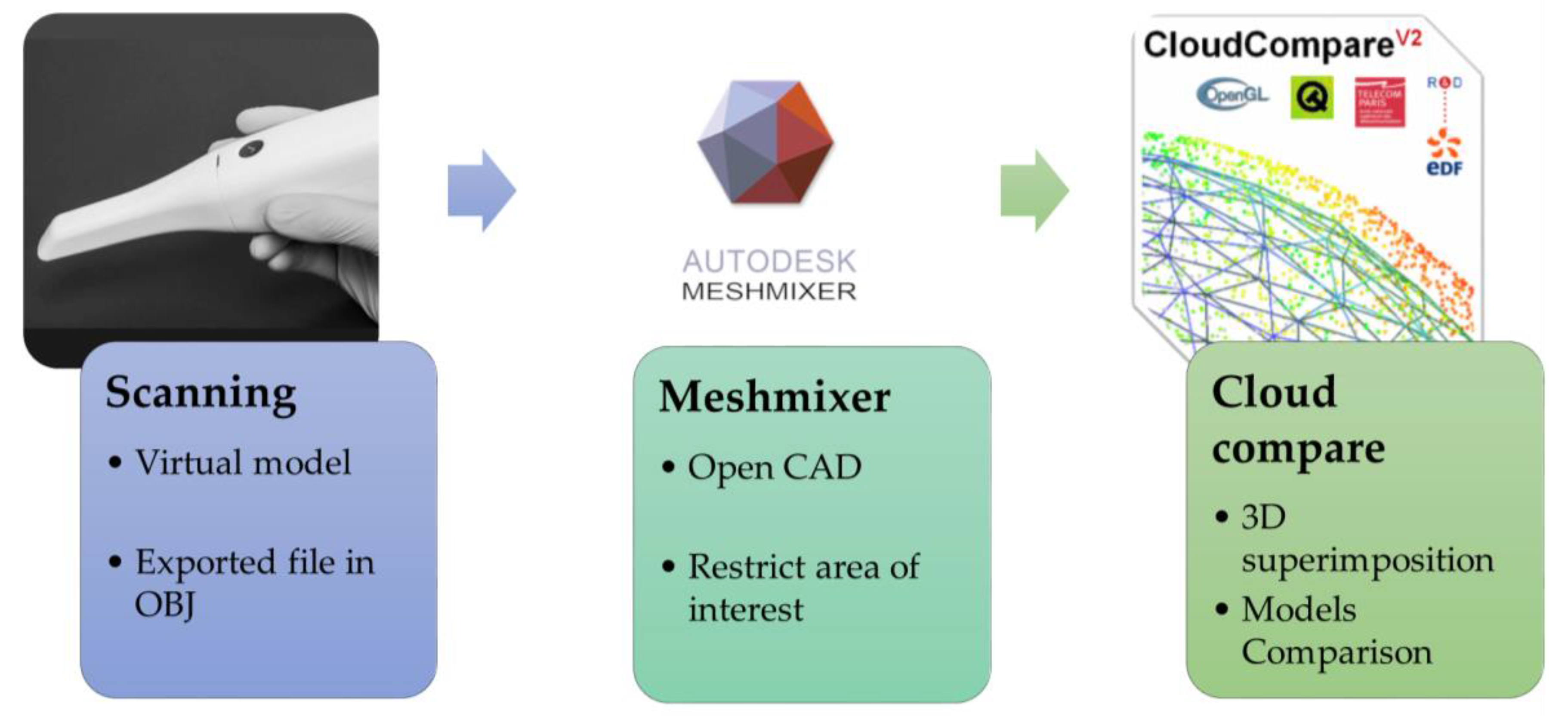
2. Materials and Methods

The present study included 5 intraoral scans of 1 voluntary participant. The decayed, missing, filled teeth index (DMFT) of the patient, considered was 0 considering the maxillary arch and excluding all the third molars. The study protocol was performed in accordance with the institutional research committee and the 1964 Helsinki declaration and its later amendments.
The volunteer’s maxilla was scanned 5 times in sequence, after a ten-minute rest-period, by the same skilled operator, with a conventional intraoral scanner Medit i500 (Medit Corp., Seoul, Korea) using the company’s software (Medit Link version 2.5.2 build 376 Revision 27 520).
The operator is defined as skilled as they graduated in dentistry more than 10 years ago and use an IOS on a daily basis.
This scanner device does not require powder opacification of the teeth to be scanned, so the teeth were only air dried. Prior to each scan, the IOS was calibrated following the manufacturer’s instruction. Then, the lip retractor (Mirahold, Hager, Germany) was placed, and teeth were dried with compressed air.
The scans were performed following the manufacturer’s instruction for the scan strategy; beginning from the occlusal surface of the second right molar, proceeding toward the second left molar, going back scanning the buccal side in the left to right direction and the palatal side in the right to left direction. In each scanning process, careful attention was given to the scan reliability map showing the most precise color scale value (green).
The scanner software, during the manual movements, expresses the approximate reliability using a colorimetric scale from red to green (less reliable to more reliable), allowing the operator to complete the scan with the best-fit 3D data.
The 3D files were exported from the scanner software in a standard OBJ format (Figure 1).
The exported file also contains the gingival surface, which is a source of error for a deviation in measurement. To overcome this inconvenience, the elimination of the soft tissue was performed before proceeding with the 3D superimposition analysis (Figure 2). This step is optional and can be applied for a better qualitative view of the interest region. For that, an open-source software (Autodesk Meshmixer version 3.5.4) was used.
This software is classified as a prototype design tool based on high-resolution dynamic triangle meshes, allowing the use of a filter based on the crease angle, which makes it possible to eliminate the soft tissue and keep only the teeth in a user-friendly process.
Using another open-source software (Cloud compare version 2.12), the 3D files from the maxillary arch were aligned. The alignment was first manually performed, identifying 6 pairs of coincident points and then using the Iterative Closest Point (ICP) algorithm to perform the 3D superimposition. Subsequently, the alignment allows the evaluation of the difference in terms of deviations.
The aligned 3D files were then converted into point clouds with approximately five million points for each virtual model. Individual deviation analysis was performed for each obtained model using a color map, in order to test the precision of the measurements. This process simulates the follow up performed in different visits to the dental office, allowing the comparison between different periods of a patient’s life.
Statistical analysis was performed using SPSS for Windows, version 22.0 (IBM, New York, NY, USA), considering the deviation distribution expressed in percentage, mean values, and standard deviation.

3. Results

Each impression was categorized as a crescent number, and for each comparison between two 3D files (number vs. number), the deviation data were exported and expressed in millimeters (Figure 3 and Figure 4). It is possible to observe that there is a similar trend between the comparisons regardless of the model that was used.
The use of a colorimetric map shows the data using a colorimetric scale in the scanned models’ comparison. A representative map is summarized in Figure 5 showing the regions with highest divergence in terms of precision.
The cumulative frequency of points with deviation below various thresholds for each pair of models was expressed in percentage with the mean value and standard deviation (Table 1).
The analysis of the data for the clinically acceptable threshold of <0.3 mm precision shows an overall cumulative frequency of point deviation precision of 98.8% ± 1.4%, and for <0.2 mm a precision of 95.9 ± 3%.

4. Discussion

To date, the reliability and accuracy of intraoral digital impressions using IOSs can be guaranteed for those prosthetic preparations with visible margins scanned in the absence of blood and/or saliva flow. Besides, scientific evidence has proven digital impressions with IOSs to be a suitable alternative, or even superior, to conventional impressions, performed with highly accurate impression materials such as polyvinyl siloxane or polyether, in cases of a single tooth or short spans [20,21,22,23,24,25].
On the other hand, in the current literature, there is no concordance about the dimensional accuracy of full-arch digital impression when compared to the accuracy of conventional impressions. Literature reports that one of the weak points of IOSs is full-arch scans where multiple restorations on teeth and/or implants are involved [16,26,27].
The main reason for this dimensional inaccuracy is the cumulative and systematic errors in image or video superimposition while the scan of the arch is being executed. [26] This explains why when larger intraoral spans are scanned, errors and, therefore, inaccuracy may increase; a larger number of raw images or videos that need to be processed and superimposed leads to errors in the arch dimension and deformation of the arch shape. This aspect is reported as particularly evident in the molar region [16,26,27,28].
In order to reduce this issue, the need for scanning protocols or precise sequences becomes evident. These impression techniques, usually described and recommended by the manufacturer, allow a reduction in the final amount of stitching (superimpositions), thus decreasing the final cumulative error obtained [29,30,31,32,33].
In the case of the IOS i500 (Medit), the impression strategy for the upper jaw, recommended by the manufacturer and followed in this study, starts scanning the occlusal surfaces from the left/right maxillary second molar to the right/left maxillary second molar, followed by the buccal surfaces and ending with the palatal ones.
However, an in vitro study compared the three-dimensional (3D) distortion of complete-arch scans scanned with six IOSs (i500, Medit; TRIOS2; 3Shape A/S, TRIOS3; 3Shape A/S, CS3500; Carestream Dental, CS3600; Carestream Dental and Primescan; Dentsply Sirona) using two different scan strategies and reported that a modified scan strategy based on first scan from the left maxillary second molar to the right maxillary canine and then from the left maxillary canine to the right maxillary second molar improved the accuracy of the IOSs in complete-arch scans [34].
The precision can be considered in two ways: repeatability, as in the precision when the operator measures the same object several times in the same environment; and reproducibility, which evaluates whether the results are consistent when different operators perform the scan or when the scanning environment has changed [35,36,37].
The precision of full-arch scans using IOSs has been widely evaluated in in vitro studies using dental casts as the reference object to be scanned, avoiding then, associated in vivo scan errors such as natural teeth optical reflections linked to the humid intraoral environment, as well as patient movement, which can increase full-arch scan inaccuracies [6,38,39,40,41,42].
The aim of this paper was to evaluate whether repeated in vivo scans show dimensional deviation. One strong point of our research is related to the in vivo analysis setting which resembles a real clinical use of the device and thus gives clinically reliable data.
Considering the IOS software’s stitching process, our analysis provides information on the reliability of this process to understand whether some factors could modify the overall scans’ precision and, thus, the precision of the prosthesis that will be manufactured.
The analysis of the data for the clinically acceptable threshold (<0.3 mm) shows an overall cumulative frequency of point deviation precision of 98.8% ± 1.4%; therefore, the null hypotheses were rejected.
The colormap analysis showed no clinically relevant discrepancy when evaluating and comparing the different scanned areas.
Further, previous authors [42] investigated the impact of soft tissue on the precision obtained in full-arch scans with different partial edentulous spans, by using two intraoral scanners (Medit i500 and Carestream CS3600). Regardless of the type of intraoral scanners used, a similar trend was shown. Surface deviations were primarily located on the side that included the partially edentulous area; moreover, the results exhibit that the precision was significantly lower when scanning full dental arches with five or more missing teeth.
Another strong point of this research was the evaluation of the in vivo precision of IOS full-arch scans, without the soft tissue. The main source of deviation in similar analyses could be related to the soft tissue scans’ difficulty, which is linked to increased deviation data; therefore, to obtain clinically reliable data we decided to evaluate only the teeth area.
As previously mentioned, the main limitation of an actual IOS is represented by the process of stitching together a larger number of raw images or videos. Moreover, it has been reported that objects that have a complex geometric shape, such as the occlusal surface of teeth, produce more reliable reconstructions [37]. Contrarily, in those scanned items with flat and smooth surfaces, for instance, edentulous areas, errors in the alignment of scan data may be greater. This fact justifies the reduction in the repeatability of full-arch scan data in cases of large edentulous sites [37,38].
A similar conclusion was reached in a posterior in vitro study, wherein, after measuring the virtual files in a linear manner instead of superimposing 3D data, authors reported significant differences in precision between edentulous versus dentate full-arch scans (precision values of <441 μm versus <311 μm, respectively), as well as, in partial edentulous spans (precision values of <97 μm for edentulous and <54 μm for dentate scans). The A.A. concluded that full-arch scans with IOSs, especially, the edentulous ones, are less reliable in comparison with the sectorial ones and that, with respect to sectorial scans, all IOSs have better accuracy in spans of approximately 16 to 22 mm in relation to the complete-arch scan [37].
Ender A. et al. [31] also evaluated the accuracy of numerous IOSs, such as the i500 Medit, for complete and partial dental impressions. They evidence, as in previous studies, suggested that digital intraoral impressions have become a suitable alternative to conventional ones in cases of partial-arch impressions showing the highest accuracy in posterior areas, with deviations below 50 μm for all test groups, in comparison to anterior ones. Besides, it was concluded that the accuracy obtained in full-arch impressions remains challenging.
Although previous authors [31,38] described in their studies precision values from 52.3 to 66.3 μm for in vitro full-arch scans with the IOS i500 Medit, Michelinakis et al. [43] reported lower precision values, but in any case, they exceeded an average accuracy of 100 μm.
In an in vivo study, Kwon M. et al. reported an absolute mean precision and a trueness value of 56.6 ± 52.4 μm and 76.6 ± 79.3, respectively for all intraoral scanners evaluated (i500, CS3600, Trios 3, iTero, and CEREC Omnicam) [16].
An important aspect that needs to be considered when evaluating the data retrieved from the literature is in regard to the operator’s experience, which has been shown have a direct impact on outcome.
The greater the experience an operator has, the lesser time/number of images required to complete the scan [44]. Although there was no consistent decline in the study by Róth, the variation in the scanning time and the number of images needed to have a complete reconstruction sheds light on the importance of the operator’s experience [45].
In our study, we decided to perform the intraoral scans using an experienced operator to have less bias related to this aspect.
If we consider a threshold value of <150 μm of accuracy for a fixed dental prosthesis [2,37,44], and a clinically acceptable range between 50 and 90 μm for implant-supported multiunit restorations [46,47,48,49], the results on accuracy with IOSs reported in the previous studies indicate that digital impressions direct from IOSs are a suitable alternative to conventional impressions in cases of partial spans and that this becomes controversial in the case of complete-arch scans.
The limitations of this study include a reduced sample of one participant, as well as, that the accuracy of the complete-arch scan it is only assessed in terms of precision. Further in vivo studies with a larger number of participants are needed in order to evaluate in more realistic practice the accuracy obtained in full-arch scans with IOSs under clinical conditions in terms of trueness and precision.

5. Conclusions

Within the limitations of this study, the clinically acceptable in vivo frequency of point deviation precision (<0.3 mm) was obtained as 98.8% ± 1.4%. Moreover, the use of open-source software appears to be a reliable option to assess the precision of the Medit i500 scanner.

Author Contributions

Conceptualization R.L.G. and C.G.; methodology, F.C.; software, F.S.; validation, M.M.; formal analysis, J.P.M.T. and L.P.M.; investigation, R.L.G.; data curation, F.S.; writing—original draft preparation, R.L.G. and A.M.; writing—review and editing, M.M.; supervision, J.P.M.T.; project administration, R.L.G. All authors have read and agreed to the published version of the manuscript.

Funding

This research received no external funding.

Institutional Review Board Statement

Not applicable.

Informed Consent Statement

Not applicable.

Data Availability Statement

Not applicable.

Conflicts of Interest

The authors declare no conflict of interest.

References

  1. Mörmann, W.H. The evolution of the CEREC system. J. Am. Dent. Assoc. 2006, 137, 7S–13S. [Google Scholar] [CrossRef] [PubMed]
  2. Pradíes, G.; Zarauz, C.; Valverde, A.; Ferreiroa, A.; Martínez-Rus, F. Clinical evaluation comparing the fit of all-ceramic crowns obtained from silicone and digital intraoral impressions based on wavefront sampling technology. J. Dent. 2015, 43, 201–208. [Google Scholar] [CrossRef] [Green Version]
  3. Giachetti, L.; Sarti, C.; Cinelli, F.; Russo, D.S. Accuracy of digital impressions in fixed prosthodontics: A systematic review of clinical studies. Int. J. Prosthodont. 2020, 33, 192–201. [Google Scholar] [CrossRef] [PubMed]
  4. Lee, S.J.; Gallucci, G.O. Digital vs. conventional implant impressions: Efficiency outcomes. Clin. Oral Implant. Res. 2013, 24, 111–115. [Google Scholar] [CrossRef]
  5. Ting-Shu, S.; Jian, S. Intraoral digital impression technique: A review: Intraoral digital impression review. J. Prosthodont. 2015, 24, 313–321. [Google Scholar] [CrossRef] [PubMed]
  6. Flügge, T.V.; Schlager, S.; Nelson, K.; Nahles, S.; Metzger, M.C. Precision of intraoral digital dental impressions with iTero and extraoral digitization with the iTero and a model scanner. Am. J. Orthod. Dentofac. Orthop. 2013, 144, 471–478. [Google Scholar] [CrossRef] [PubMed]
  7. Oh, H.; Lim, Y.; Kim, B.; Kim, M.; Kwon, H.; Baek, Y. Effect of scanning-aid agents on the scanning accuracy in specially designed metallic models: A laboratory study. PLoS ONE 2022, 17, e0267742. [Google Scholar] [CrossRef]
  8. Logozzo, S.; Zanetti, E.M.; Franceschini, G.; Kilpelä, A.; Mäkynen, A. Recent advances in dental optics—Part I: 3D intraoral scanners for restorative dentistry. Opt. Lasers Eng. 2014, 54, 203–221. [Google Scholar] [CrossRef]
  9. Cucinotta, F.; Raffaele, M.; Salmeri, F. A Topology Optimization of a Motorsport Safety Device. Lect. Notes Mech. Eng. 2020, 400–409. [Google Scholar]
  10. Taneva, E.; Kusnoto, B.; Evans, C.A. 3D Scanning, Imaging, and Printing in Orthodontics; Chapter 9 Issues in Contemporary Orthodontics; IntechOpen: London, UK, 2015; pp. 147–188. [Google Scholar]
  11. Tsirogiannis, P.; Reissmann, D.R.; Heydecke, G. Evaluation of the marginal fit of single-unit, complete-coverage ceramic restorations fabricated after digital and conventional impressions: A systematic review and meta-analysis. J. Prosthet. Dent. 2016, 116, 328–335.e2. [Google Scholar] [CrossRef]
  12. Ender, A.; Mehl, A. Accuracy of complete-arch dental impressions: A new method of measuring trueness and precision. J. Prosthet. Dent. 2013, 109, 121–128. [Google Scholar] [CrossRef]
  13. Imburgia, M.; Logozzo, S.; Hauschild, U.; Veronesi, G.; Mangano, C.; Mangano, F.G. Accuracy of four intraoral scanners in oral implantology: A comparative in vitro study. BMC Oral Health 2017, 17, 1–13. [Google Scholar] [CrossRef]
  14. Mangano, F.; Gandolfi, A.; Luongo, G.; Logozzo, S. Intraoral scanners in dentistry: A review of the current literature. BMC Oral Health 2017, 17, 149. [Google Scholar] [CrossRef] [Green Version]
  15. Richert, R.; Goujat, A.; Venet, L.; Viguie, G.; Viennot, S.; Robinson, P.; Farges, J.C.; Fages, M.; Ducret, M. Intraoral Scanner Tech-nologies: A Review to Make a Successful Impression. J. Healthc. Eng. 2017, 8427595, 1–9. [Google Scholar] [CrossRef]
  16. Kwon, M.; Cho, Y.; Kim, D.-W.; Kim, M.; Kim, Y.-J.; Chang, M. Full-arch accuracy of five intraoral scanners: In vivo analysis of trueness and precision. Korean J. Orthod. 2021, 51, 95–104. [Google Scholar] [CrossRef]
  17. Goracci, C.; Franchi, L.; Vichi, A.; Ferrari, M. Accuracy, reliability, and efficiency of intraoral scanners for full-arch impres-sions: A systematic review of the clinical evidence. Eur. J. Orthod. 2016, 38, 422–428. [Google Scholar] [CrossRef] [Green Version]
  18. Chruściel-Nogalska, M.; Smektała, T.; Tutak, M.; Sporniak-Tutak, K.; Olszewski, R. Open-source software in dentistry: A systematic review. Int. J. Technol. Assess. Health Care 2017, 33, 487–493. [Google Scholar] [CrossRef]
  19. Osnes, C.A.; Wu, J.H.; Venezia, P.; Ferrari, M.; Keeling, A.J. Full arch precision of six intraoral scanners in vitro. J. Prosthodont. Res. 2020, 64, 6–11. [Google Scholar] [CrossRef] [PubMed]
  20. Güth, J.-F.; Keul, C.; Stimmelmayr, M.; Beuer, F.; Edelhoff, D. Accuracy of digital models obtained by direct and indirect data capturing. Clin. Oral Investig. 2013, 17, 1201–1208. [Google Scholar] [CrossRef]
  21. Boeddinghaus, M.; Breloer, E.S.; Rehmann, P.; Wöstmann, B. Accuracy of single-tooth restorations based on intraoral digital and conventional impressions in patients. Clin. Oral Investig. 2015, 19, 2027–2034. [Google Scholar] [CrossRef] [PubMed]
  22. de Andrade, G.S.; Pinto, A.B.A.; Tribst, J.P.M.; Chun, E.P.; Borges, A.L.S.; de Siqueira Ferreira Anzaloni Saavedra, G. Does overlay preparation design affect polymerization shrinkage stress distribution? A 3D FEA study. Comput. Methods Biomech. Biomed. Eng. 2021, 24, 1026–1034. [Google Scholar] [CrossRef]
  23. Zarauz, C.; Valverde, A.; Martinez-Rus, F.; Hassan, B.; Pradies, G. Clinical evaluation comparing the fit of all-ceramic crowns obtained from silicone and digital introral impressions. Clin. Oral Investig. 2016, 20, 799–806. [Google Scholar] [CrossRef] [PubMed]
  24. Vecsei, B.; Joós-Kovács, G.; Borbély, J.; Hermann, P. Comparison of the accuracy of direct and indirect three-dimensional digitizing processes for CAD/CAM systemsean in vitro study. J. Prosthodont. Res. 2017, 61, 177–184. [Google Scholar] [CrossRef]
  25. Arcas, L.P.B.; Tribst, J.P.M.; Baroudi, K.; Amaral, M.; da Silva-Concílio, L.R.; Vitti, R.P. Dimensional accuracy comparison of physical models generated by digital impression/3D-printing or analog impression/plaster methods. Int. J. Odontostomatol. 2021, 15, 562–568. [Google Scholar] [CrossRef]
  26. Moreno, A.; Gimenez, B.; Ozcan, M.; Pradies, G. A clinical protocol for intraoral digital impressions of screw-retained CAD/CAM framework on multipleimplants based on wavefront sampling technology. Implant. Dent. 2013, 22, 320–325. [Google Scholar] [CrossRef] [PubMed] [Green Version]
  27. Gimenez, B.; Ozcan, M.; Martinez-Rus, F.; Pradies, G. Accuracy of a digital impression system based on parallel confocal la-ser technology for implants with consideration of operator experience and implant angulation ande depth. Int J. Oral Maxillofac. Implant. 2014, 853–862. [Google Scholar] [CrossRef] [PubMed]
  28. Ferrari, M.; Keeling, A.; Mandelli, F.; Lo Giudice, G.; Garcia-Godoy, F.; Joda, T. The ability of marginal detection using different intraoral scanning systems: A pilot randomized controlled trial. Am. J. Dent. 2018, 31, 272–276. [Google Scholar] [PubMed]
  29. Lee, J.H.; Yun, J.H.; Han, J.S.; Yeo, I.S.L.; Yoon, H.I. Repeatability of intraoral scanners for complete arch scan of partially eden-tulous dentitions: An in vitro study. J. Clin. Med. 2019, 8, 1187. [Google Scholar] [CrossRef] [PubMed] [Green Version]
  30. Müller, P.; Ender, A.; Joda, T.; Katsoulis, J. Impact of digital intraoral scan strategies on the impression accuracy using the TRIOS Pod scanner. Quintessence Int. 2016, 47, 343–349. [Google Scholar]
  31. Ender, A.; Zimmermann, M.; Mehl, A. Accuracy of complete- and partial-arch impressions of actual intraoral scanning systems in vitro. Int. J. Comput. Dent. 2019, 22, 11–19. [Google Scholar]
  32. Medina-Sotomayor, P.; Agustín Pascual, M.; Isabel Camps, A. Accuracy of four digital scanners according to scanning strategy in complete-arch impressions. PLoS ONE 2018, 13, e0202916. [Google Scholar] [CrossRef] [PubMed] [Green Version]
  33. Passos, L.; Meiga, S.; Brigagão, V.; Street, A. Impact of different scanning strategies on the accuracy of two current intraoral scanning systems in complete-arch impressions: An in vitro study. Int. J. Comput. Dent. 2019, 22, 307–319. [Google Scholar] [PubMed]
  34. Son, K.; Jin, M.-U.; Lee, K.-B. Feasibility of using an intraoral scanner for a complete-arch digital scan, part 2: A comparison of scan strategies. J. Prosthet. Dent. 2021, in press. [Google Scholar] [CrossRef] [PubMed]
  35. Schmidt, A.; Klussmann, L.; Wöstmann, B.; Schlenz, M.A. Accuracy of digital and conventional full-arch impressions in patients: An update. J. Clin. Med. 2020, 9, 688. [Google Scholar] [CrossRef] [Green Version]
  36. Sanda, K.; Yasunami, N.; Okada, M.; Furuhashi, A.; Ayukawa, Y. Accuracy of the intra- and extra-oral scanning technique for transferring the intaglio surface of a pontic of provisional restorations to definitive restorations. Materials 2021, 14, 6489. [Google Scholar] [CrossRef]
  37. Braian, M.; Wennerberg, A. Trueness and precision of 5 intraoral scanners for scanning edentulous and dentate complete-arch mandibular casts: A comparative in vitro study. J. Prosthet. Dent. 2019, 122, 129–136.e2. [Google Scholar] [CrossRef] [Green Version]
  38. Lee, K.C.; Park, S.-J. Digital intraoral scanners and alginate impressions in reproducing full dental arches: A comparative 3D assessment. Appl. Sci. 2020, 10, 7637. [Google Scholar] [CrossRef]
  39. Li, H.; Lyu, P.; Wang, Y.; Sun, Y. Influence of object translucency on the scanning accuracy of a powder-free intraoral scanner: A laboratory study. J. Prosthet. Dent. 2017, 117, 93–101. [Google Scholar] [CrossRef]
  40. Mangano, F.; Lerner, H.; Margiani, B.; Solop, I.; Latuta, N.; Admakin, O. Congruence between meshes and library files of implant scanbodies: An in vitro study comparing five intraoral scanners. J. Clin. Med. 2020, 9, 2174. [Google Scholar] [CrossRef]
  41. Troiano, A.; Lo Giudice, G.; de Luca, R.; Lo Giudice, F.; D’Amato, S.; Tartaro, G.; Colella, G. Salvage of dental implant located in mandibular odontogenic cyst. A conservative surgical treatment proposal. Dent. J. 2020, 8, 49. [Google Scholar] [CrossRef]
  42. Lee, S.J.; Kim, S.-W.; Lee, J.J.; Cheong, C.W. Comparison of intraoral and extraoral digital scanners: Evaluation of surface topography and precision. Dent. J. 2020, 8, 52. [Google Scholar] [CrossRef] [PubMed]
  43. Michelinakis, G.; Apostolakis, D.; Tsagarakis, A.; Kourakis, G.; Pavlakis, E. A comparison of accuracy of 3 intraoral scanners: A single-blinded in vitro study. J. Prosthet. Dent. 2020, 124, 581–588. [Google Scholar] [CrossRef] [PubMed]
  44. Revell, G.; Simon, B.; Mennito, A.; Evans, Z.P.; Renne, W.; Ludlow, M.; Vág, J. Evaluation of complete-arch implant scanning with 5 different intraoral scanners in terms of trueness and operator experience. J. Prosthet. Dent. 2021. Available online: https://pubmed.ncbi.nlm.nih.gov/33832761/ (accessed on 23 September 2022). [CrossRef] [PubMed]
  45. Róth, I.; Czigola, A.; Joós-Kovács, G.L.; Dalos, M.; Hermann, P.; Borbély, J. Learning curve of digital intraoral scanning—An in vivo study. BMC Oral Health 2020, 20, 1–7. [Google Scholar] [CrossRef] [PubMed]
  46. ISO I. 5725-1: 1994; Accuracy (Trueness and Precision) of Measurement Methods and Results e Part 1: General Principles and Definitions. International Organization for Standardization: Geneva, Switzerland, 1994.
  47. Braian, M.; De Bruyn, H.; Fransson, H.; Christersson, C.; Wennerberg, A. Tolerance measurements on internal- and external-hexagon implants. Int. J. Oral Maxillofac. Implant. 2014, 29, 846–852. [Google Scholar] [CrossRef] [Green Version]
  48. Tribst, J.P.M.; Dal Piva, A.M.d.O.; Lo Giudice, R.; Borges, A.L.S.; Bottino, M.A.; Epifania, E.; Ausiello, P. The Influence of Custom-Milled Framework Design for an Implant-Supported Full-Arch Fixed Dental Prosthesis: 3D-FEA Study. Int. J. Environ. Res. Public Health 2020, 17, 4040. [Google Scholar] [CrossRef]
  49. Giudice, R.L.; Famà, F. Health care and health service digital revolution. Int. J. Environ. Res. Public Health 2020, 17, 4913. [Google Scholar] [CrossRef]
Figure 1. Flowchart summarizing the software sequence and the objective of each step.
Prosthesis 04 00045 g001
Figure 2. Representative scheme of the removed area (soft tissue) and the area of interest for measurement (teeth).
Prosthesis 04 00045 g002
Figure 3. Deviation distribution as cumulative frequency: interscan comparison (scan number vs. scan number).
Prosthesis 04 00045 g003
Figure 4. Deviation distribution as frequency: interscan comparison (scan number vs. scan number).
Prosthesis 04 00045 g004
Figure 5. Colorimetric map generated by the 3D models’ superimposition.
Prosthesis 04 00045 g005
Table 1. Percentage of points below determined deviation thresholds.
Pairs of ScansDeviation < 0.01 mmDeviation < 0.05 mmDeviation < 0.1 mmDeviation < 0.2 mmDeviation < 0.3 mmDeviation < 0.4 mm
1 vs. 22.9%48.5%77.9%96.2%99.8%100.0%
1 vs. 32.4%49.2%82.1%94.8%98.4%99.4%
1 vs. 43.5%79.5%97.5%99.6%99.9%100.0%
1 vs. 52.3%46.0%75.6%97.2%100.0%100.0%
2 vs. 32.3%36.2%67.2%92.3%97.0%99.1%
2 vs. 42.5%59.4%82.9%97.0%99.5%99.7%
2 vs. 56.6%78.7%96.1%99.6%99.9%100.0%
3 vs. 41.9%42.0%77.7%93.4%97.3%98.9%
3 vs. 51.2%29.4%59.9%90.1%95.9%98.4%
4 vs. 52.8%55.6%83.0%98.6%100.0%100.0%
Mean2.8%52.4%80.0%95.9%98.8%99.6%
Standard dev.1.4%15.6%10.9%3.0%1.4%0.5%
Publisher’s Note: MDPI stays neutral with regard to jurisdictional claims in published maps and institutional affiliations.

