Review

CCR6–CCL20-Mediated Immunologic Pathways in Inflammatory Bowel Disease

School of Health Sciences, College of Health and Medicine, University of Tasmania, Launceston, TAS 7250, Australia
*
Author to whom correspondence should be addressed.
Gastrointest. Disord. 2019, 1(1), 15-29; https://doi.org/10.3390/gidisord1010003
Submission received: 17 July 2018 / Revised: 19 August 2018 / Accepted: 19 August 2018 / Published: 21 August 2018

Abstract

:
Inflammatory bowel disease (IBD) has evoked significant interest in human immunobiology given its tactical immune evasion methodologies resulting in acute immune destabilization. IBD comprising Crohn’s disease and Ulcerative colitis manifests as chronic inflammation in the gut mucosa, leading to complexities involving immune dysregulation in the T helper lymphocyte arm, effecting disease pathogenicity. The mucosa of the alimentary canal is constantly exposed to a myriad of food antigens and luminal microorganisms for which a consistent host-protective mechanism is operative in healthy people. Lowered mucosal immune expression which allows penetration of the epithelial barrier by infective pathogenic microbes elicits both innate and adaptive immune responses in the gut, culminating in aberrant intestinal inflammation. Interestingly, the IBD leukocyte repertoire is significantly entwined with chemokine-assisted chemotactic navigation into the sites of inflammation, which is also thought to generate favorable immune-suppressive responses. The functions of the cognate chemokine receptor, CCR6, which binds with its unique ligand CCL20, are expected to tilt the balance between upregulation of homeostatic tolerance and inflammatory pathophysiology. This review aims to critically examine the CCR6-driven immune pathways: TH1/TH2, TH1/TH17, TH17/Treg, IL-23/IL-17, Akt/ERK-1/2, ILC3, and TH9/TH2 for systematic investigation of its underlying mechanisms in the future and to underpin its importance in resolving IBD pathology. Thus, CCR6 occupies an exclusive position in gut immunology which renders it an invaluable therapeutic tool for the production of novel medicaments to treat IBD.

1. Introduction

Immune-compromised diseases of the gastrointestinal tract have been a topic which has attracted active interest for decades and is one that continues to test the robustness of our immune system. Among the many disease-related models of human immune inactivation, IBD has been a pertinent system within the gut mucosal biology. Importantly, a new immune concept involving the chemokine receptor 6 is gaining momentum with chemokine-activated immune mechanisms coming to the fore.
Chemokines are a superfamily of immune-modulatory small protein molecules, which regulate leukocyte migration to inflammatory sites through their chemoattractant properties. Among a plethora of functions, chemotactic navigation of effector T helper cell cohorts to the gut mucosa from lymphatics appears to be primarily a CCR6-driven mechanism, which synchronizes immune homeostasis during IBD pathophysiology. The CCR6–CCL20 axis is a prominent immune modulator in both innate and adaptive immune responses of a wide range of inflammatory diseases. It is associated with tissue damage and injury, human immunodeficiency virus (HIV), ophthalmic disorders, lung and kidney disorders, autoimmune diseases, brain disease, arthrosclerosis, obesity, diabetes, and cancer [1].

2. Chemokines

Chemokines are a specific group of cytokines that are small molecular proteins known to perform several different functions in the human immune system. Salient functions include T helper lymphocyte differentiation and cell chemotaxis aiding cell migration towards inflammatory locations. Other functions of chemokines are angiogenesis, development of embryos, B cell maturation and differentiation, lymphatic organogenesis, wound healing, inflammation, and cancer metastasis. Chemokines preferentially aid leukocyte homeostasis by directing cell movement towards sites of injury and have been referred to as a specific cell navigational mechanism positioned within the immune system. The total chemokine repertoire consists of 50 chemokines and they belong to receptor and ligand cohorts. Chemokines consists of four subgroups named XC, CX3C, CXC, and CC, in which the naming is based on their receptors. A receptor is denoted by R, and therefore, CC chemokines bind with CCR chemokine receptors. Chemokines display a common molecular structure of having three beta-pleated sheets with an alpha helix at the carbon terminal in which the cysteine motifs are joined by disulfide bonds. The entire chemokine is composed of 67–127 amino acid residues. Chemokine receptors are exhibited on the cell surface and they are described as seven transmembrane domains joined to guanine nucleotide-coupled G1 class protein receptors. One chemokine receptor can bind with many ligands, several ligands can bind with one receptor, or simply, one receptor would bind with one sole ligand. In the case of chemokine receptor 6, it has only one monogamous partner, the chemokine ligand CCL20. Chemokines play a prominent role in inflammation and the spreading of cancer, and interestingly, chemokine inhibition has yielded anti-inflammatory attributes in inflammatory disorders [1].

Chemokine Receptor 6 (CCR6) and Chemokine Ligand CCL20

CCR6 is denoted by a string of different molecular identities, such as: CD196, CKRL3, GPR29, CKR-L3, CMKBR6, GPRCY4, STRL22, BN-1, DCR2, DRY6, CCR-6, and CC-CKR-6 or C-C CKR-6. CCR6 is said to transduce signals that mobilize intracellular calcium ion flux upon binding to its ligand, CCL20. Apart from CCL20, human beta defensins (HBD), which are a group of microbicidal peptides, have been identified as additional epitopes, although this needs thorough validation. Yet, there is increasing evidence that the counterpart of the CCL20 ligand, human beta defensin-2 (HBD2), concomitantly contributes to epithelial cell migration in maintaining intestinal epithelial barrier integrity, and as such, acts as a frontline defense against microbial invasion. Vongsa et al. in 2009 [2] reported equipotent functionality of HBD2 to CCL20, demonstrating that it stimulates active migration of the human intestinal cell lines Caco2, T84, and non-transformed IEC6 ex vivo. It further confirmed that HBD2 is capable of stimulating intracellular calcium influx, which innervates phosphoinositide 3-kinase signal transduction via mobilizing the guanosine triphosphatase (GTPase), RhoA, and phosphorylated myosin light chain, leading to F-actin accumulation, thus shedding light on a possible canonical wound healing mechanism. Therefore, HBD is thought to augment immune cell patrolling, microbial defense, and intestinal barrier restitution. These results significantly underpin the notion that HBD2 parallels the functions of CCL20 in its role as an additional partnering ligand of CCR6.
CCL20 was recognized as the principal ligand through calcium ion flux experiments in a K562 cell line devoid of other chemokine receptors but transfected with CCR6. CCL20 has other synonyms, such as LARC (liver and activation-regulated chemokine), MIP-3α (macrophage inflammatory protein), and Exodus-1. CCR6 is predominantly expressed in the appendix, pancreas, lymph nodes, and spleen, with lesser expression in the fetal liver, testis, colon, small intestine, and thymus. Expression of CCL20 is upregulated by intestinal enterocytes responsive to bacteria displaying flagellar movement as well as antigen-presenting dendritic and macrophage subsets, Langerhans cells in the skin, endothelial cells, natural killer (NK cells), neutrophils, B cells, and TH17 cells. CCR6 is signatory on the T lymphocyte subdivisions of TH17 and Treg cells, innate lymphoid cells (ILC)-3, neutrophils, NK T cells, B cells, and immature dendritic cells. Distinctively, CCR6 is thought to trigger the PI3 kinase (phosphoinositide 3 kinase) signal transduction cascade, which leads to phosphorylation that provides energy for cell chemotaxis [1,2].

3. Importance of IBD

IBD consists of an immune-compromised disease complex that includes two sub phenotypes: Crohn’s disease (CD) and Ulcerative colitis (UC), characterized by chronic inflammation within the intestine. These are multifactorial, heritable diseases, which have more than 200 single nucleotide polymorphisms (SNP), including NOD2, ATR16GL, XBPI, IL23R, STAT3, JAK2 and CCR6, that sometimes manifest in genetically predisposed individuals [3]. Recently, heritability was endowed with only 25% probability for being recognized as the cardinal factor causing disease because a multitude of environmental factors are also now held accountable [4]. Fifty percent concordance of CD in monozygotic twins, rising incidence of disease in the past 60 years without changes in the genetic makeup, lower rates of IBD in underdeveloped countries, and development of IBD in immigrants to countries of high prevalence have all emphasized the importance of multiple environmental influences on the inception of the disease [5]. Rather than blaming it all on genetic predisposition and the microbiome, the trend now is to focus on new signaling pathways: one of these is the ER-stress-induced apoptosis due to the unfolded protein response, resulting in the autophagy of Paneth, goblet, and intraepithelial cells [6].
Population-based studies reveal IBD to be a global disease of the 21st century, with highest prevalence in Europe and North America. A recent USA-based study demonstrated a 1.3% increase in prevalence and showed that disease was more common in people who are unemployed, living in poverty, and lacking a high school education [7]. Increasing rates of IBD have been shown in a number of newly industrialized countries in Asia, Africa, South America, and the Middle East since the 1990s [8]. A recent population survey identified Australia to have the highest prevalence of IBD alongside Canada, Denmark, and New Zealand, with CD being more common than UC in the Australian patients [9]. Data also show that IBD is becoming more severe and complex in Australia, with a projected estimate of 100,000 patients in 2022 [10]. Among the Australians surveyed, factors such as smoking, childhood immunological events such as exposure to tonsillectomy or chickenpox, and frequent intake of fast food are factors associated with increased risk of developing IBD, while frequent drinking of caffeine and owning pets provided protection against developing the disease. Studies also show that IBD most commonly presents in young adults aged between 15 and 29 years of age [11]. Another peak age group is those aged between 60 and 70 years of age [11].
Although the etiology of IBD is not yet completely understood, it is commonly attributed to gastrointestinal (GI) tract stimulation by excessive and abnormal adaptive immune responses via induction of proinflammatory cytokines. Such immune activation is produced against the 100 trillion or so luminal microbial flora inhabiting the intestine, which spans about 200–400 m2 in extent. Dysregulated innate and adaptive immunity followed by microbial dysbiosis due to disruption of the mucosal barrier which exposes persons to numerous luminal antigens plays a significant role in disease development [12]. Decreased rates of IBD in rural environments and increased rates of IBD in overly clean environments have been reported, while enteric infections may trigger onset in settings such as refugee camps. Multiple causative factors, newly defined as the “exposome”, consisting of all the environmental contributory agents are now implicated in IBD and include the gut microbiota (both commensal and pathogenic), excessive usage of antibiotics, nutrition, smoking, industrialization with more exposure to pollutants, a Westernized lifestyle, poor access to hygienic toileting, sleep disorders, anxiety and depression, appendectomy, exposure to bright sunlight, and increased vitamin D [4]. Consumption of milk protein, animal protein, polyunsaturated fats, and high-sugar foods (as included in the so-called Western diet) is known to increase the risk of IBD, with inhalation of tobacco posing as another risk factor in CD [13]. Passive inhalation of smoke either during pregnancy or childhood is also thought to increase incidence of CD [4].
CD and UC can be differentiated by the location in the gastrointestinal tract, severity of inflammation appearing in the intestinal wall, and peculiarities in pathophysiology. CD is a chronic, transmural, segmental inflammatory disease that involves any part of the gut from mouth to anus, but mostly affects the terminal ileum. While CD is characterized by the finding of mucosal granuloma, it can be complicated by the development of fistulas and strictures. UC is also a chronic, inflammatory disease which affects only the mucosa of the colon and the rectum.
Both CD and UC feature a relapsing–remitting disease course. Symptoms of both can include rectal bleeding, abdominal pain, tenesmus, urgency to evacuate, prolonged intermittent diarrhea, anorexia, fatigue, and weight loss [14]. Extraintestinal manifestations include arthritis, particular skin rashes, and involvement of the eyes, liver, and kidneys. The severity of the symptoms varies from mild to severe. The most common complication of CD is the blockage of the intestine due to swelling, which results in the thickening of the bowel wall. Afflicted persons often encounter problems related to malnutrition, triggered by poor nutrient absorption [13].
Although various biomarkers can be present in IBD, no single blood test is diagnostic of IBD. Abnormalities include anemia, elevated inflammatory markers, electrolyte abnormalities due to diarrhea, vitamin deficiencies as seen in CD, and low albumin indicative of both inflammation and poor absorption of nutrients [13]. Individuals with severe or complicated disease despite medical therapies may require surgical intervention. While colectomy in individuals with UC essentially cures the disease, those with CD requiring surgery commonly have postoperative recurrence [15]. Long-standing CD and UC is also associated with increased risk of colitis-associated carcinoma after 8–10 years of active disease [16]. IBD patients also become highly susceptible to chronic immune disorders such as HIV, psoriasis, primary sclerosing cholangitis, and ankylosing spondylitis. IBD-associated mortality has been reported by some studies, while persons suffering from CD displaying comorbidities with cardiovascular and respiratory disease are also documented [17].

4. Immune Mechanisms of CCR6–CCL20

The principal mechanisms of adaptive immunity encompass CD4+ T cells and its subsets in enforcing immune activation in the gut. Naïve TH0 helper cells are transformed into effector T helpers upon antigen sampling by dendritic cells and or macrophages in the mesenteric lymph nodes. Effector T helpers consist of four congenial immune subtypes: TH1, TH2, TH17, and regulatory Treg cells, although recent interest has been rigorously focused on the two subgroups of TH17 and Treg cells in the immune induction of IBD. These two subgroups have demonstrated distinctively opposing functional roles primarily attributable to their alienated cytokine profiles, represented by inflammation-triggering IL-17 and inflammation-dampening IL-10, respectively. In a nutshell, TH17 induces disease activation by releasing inflammatory cytokines leading to tissue damage, while Treg cells promote immune tolerance induced by inflammation-suppressive cytokines aiding tissue restitution [18,19]. The underlying mechanisms favoring these two opposing roles are not yet clear, but the expression of the chemokine receptor CCR6 is considered the cardinal determinant of their selective proliferation in maintaining immune homeostasis. The CC-motif of chemokine receptor 6 initiates chemoattractant migration of leukocytes towards the intestinal epithelium, which bind with its partnering chemokine, CCL20, produced by intestinal epithelial cells when provoked by microbial stimulation [20]. This underpins the concordant, yet enigmatic role the CCR6–CCL20 axis performs in primarily resolving the IBD immune mechanism by promoting immune tolerance [21]. The discovery of factors which skew the selection of TH17 and Treg polarization in the gut mucosa would be a novel breakthrough in IBD therapy [22].
Multiple cytokines and transcription factors serve to upregulate CCR6 expression on TH17 cells. Transforming growth factor-beta sulphate (TGF-β), IL-6, IL-17, IL-21 and IL-23, as well as the lineage-selective master transcription factors, retinoic-acid-receptor-related orphan nuclear receptor gamma (RORγt) and retinoic-acid-receptor-related orphan nuclear receptor alpha (RORα), are important regulators in upholding the immune induction of CCR6, which is the hallmark of the TH17 cell cohort, and they are strongly associated with autoimmune disease [23]. IBD invariably falls under the direct influence of the proinflammatory apparatus driven by CCR6, including TH17 cell differentiation and proliferation. Well-documented studies report that neutralizing IL-17 as well as TH17 lacking CCR6 receptors serve to markedly inhibit several autoimmune disorders [24]. Intriguingly, CCR6 represents a double-edged sword by its ability to mobilize the immune-suppressive T cell population: the natural regulatory Treg cells induced by the transcription factor FoxP3. Treg cells are highly effective in modulating autoimmune disease progression by actively suppressing proinflammatory T cell proliferation.
Concomitantly, CCR6 drives chemotactic recruitment of TH17 and Treg cell subsets to sites of infection in a self-sustained feedback loop, because TH17 cells also have the ability to express CCL20, induced by the cytokines TGF-β and IL-6 and the transcription factor STAT3. Given the dubious role played by CCR6 in disease amelioration, there obviously remains hitherto unidentified factors such as cytokines, adhesion, and costimulatory molecules and a whole repertoire of innate immune induction factors associated with CCR6 which contribute towards the selective mobilization of TH17 cells versus Treg distribution at the sites of inflammation. The most recently introduced immunological concepts about IBD links the deregulated TH17 and Treg axis as the pivotal point which decides the fate of IBD resolution [21,25].

Immune Mechanisms of CCR6–CCL20 Specific to IBD

The small intestine is the primary site of inflammation as it is exposed to a load of bacterial antigens and nutrients and is covered by a single layer of epithelium, containing epithelial cells, Paneth cells, entero-endocrine cells, microfold (M) cells, and mucous-secreting goblet cells. Most of the immune action takes plays in Peyer’s patches, where the follicle-associated epithelium (FAE) is crossed by antigen samplers which stimulate naive T and B cells to develop into effectors. Effector T and B lymphocytes are always found patrolling the mucosal surfaces in the gut. Inflammation is part and parcel of the intestinal mucosa to help maintain a healthy protective immune response to the numerous colonies of symbiotic microbes present within the gut. Gut tolerance mainly occurs in Peyer’s patches and isolated lymphoid follicles. Antigen uptake in the gut-associated lymphoid tissue (GALT) mostly occurs via M cells in the FAE in Peyer’s patches by dendritic cells. CCR6-bearing dendritic cells are attracted towards CCL20-producing epithelial cells and thus migrate towards the mucosa-associated connective tissue, named the lamina propria (LP), to activate naive T and B lymphocytes. Primed effector cells drain out of afferent lymphatics to the mesenteric lymph nodes, from where they enter the thoracic duct and then join the bloodstream and return through the network of vessels back to the intestinal mucosa [26]. Activated T helper cells stimulate naive B cells into antibody-producing plasma cells. Antigen-primed B lymphocytes are transformed into class-switching B plasma cells that secrete IgA in the gut mucosa upon antigen presentation and activation by dendritic cells [27].
CCR6-deficient models display an overall change in the architecture of the intestinal epithelium, with smaller Peyer’s patches, a lesser number of subepithelial domes, absence of isolated lymphoid follicles, few intestinal M cells, and high resistance to bacteria which enter through M cell conduits. Other significant IBD-relevant responses included marked elevation in the number of TH17 cells in the spleen and lymph nodes; less preference for migration to inflamed sites and less suppressive capabilities of Treg cells; DSS- and TNBS-induced colitis, producing moderate and severe disease, respectively; and reconstitution of Rag2−/− SCID mice with naïve T cells from healthy mice resulting in acute inflammation [21,28,29,30,31,32].

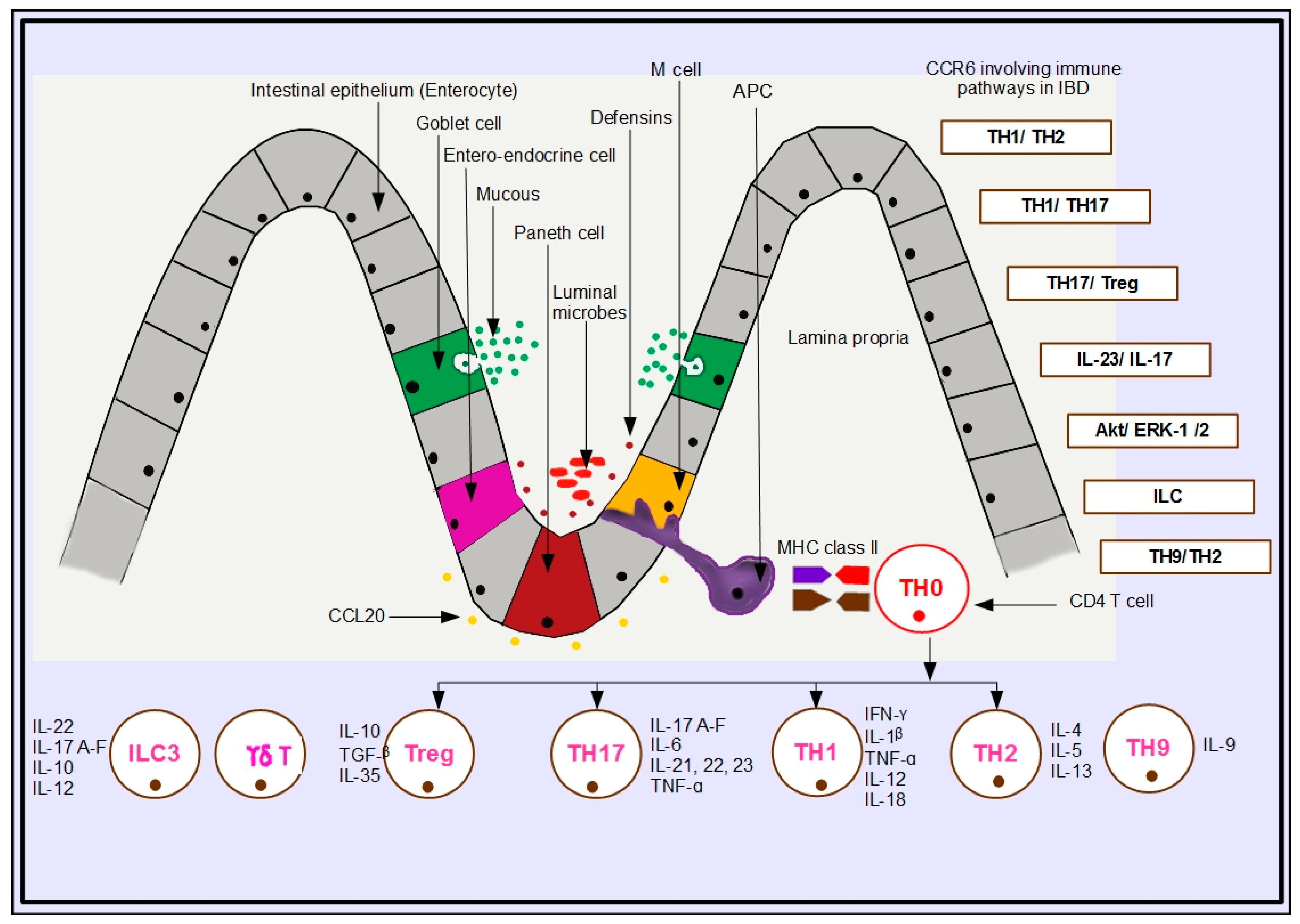
5. Immunologic Pathways in IBD Involving CCR6

An interesting feature of immune-regulated diseases highlighted by GWAS is gene loci similarity across a wide range of such disorders that indicates shared etiology. Meta-analysis of shared single nucleotide polymorphisms (SNP) referred to as the “Immunochip”, consisting of a microarray of SNPs, has been designed to reveal deep replication and fine mapping among immune-controlled pathogeneses. Among the large number of disorders surveyed, many displayed co-occurrence and familial incidence, suggesting that a person afflicted with one immune-mediated affliction runs a high risk of contracting another of the same type. This overlapping etiology is attributed to common clinical and immunological parameters concomitantly shared by those disorders, which led us to a systematized examination of the various immune-activation pathways involving the chemokine receptor 6, as shown in Figure 1 as well as Table 1, that culminate in immune-mediated disease pathogenicity in IBD [32].

5.1. TH1/TH2 Pathway

The immunological background of CD and UC in the past had been ascribed to the traditional dichotomy of TH1 (IFN-γ, IL-1β, IL-18, TNF-α, GM-CSF) and TH2 (IL-4, IL-5, IL-13, IL-22) cytokine pathways, in which disease susceptibility to CD was promoted by TH1-associated cytokines, whereas UC was modulated by TH2 cytokines. This old version has been challenged recently by a more modernized concept of the TH17 versus Treg imbalance paradigm, playing a greater, more convincing role in determining IBD pathogenesis [33]. In certain autoimmune diseases, including IBD, TH1 cells are known to upregulate the receptor CCR6, which could be fairly assumed to play a pathogenic role given the proinflammatory cytokines secreted by this cell subtype [1].

5.2. TH1/TH17 Pathway

IL-17 is considered as a major role player in IBD given its release from TH17 cells, a prominent cellular marker of IBD pathogenesis. Blockade of TH17 cells has strongly demonstrated decreased inflammation in the gut leading to lowered severity of acute colitis. IL-17 is well documented as a proinflammatory cytokine which has several isoforms, IL-17A to IL-17F, whose mRNA levels are persistently high in both CD and UC patients. IL-17 is proactive at the innate immunity level by stabilizing tight junctions in the intestinal epithelium. There are reports of IL-17 acting to recruit T cells into the lamina propria (LP) during an inflammatory episode, demonstrating adaptive immunity. The proinflammatory TH17 cells are characterized by the transcription factor RORγt and surface markers IL23R and CCR6, and upregulate the cytokines IL-17A, IL-17F, IL-21, IL-22 and IL-26 and the chemokine ligand CCL20. TGF-β, which is essential for the development of both murine and human TH17 cells, is also known to reciprocally regulate both TH17 and regulatory Treg development. GWAS has confirmed that TH17 differentiation is under the influence of the genes coded by IL-23R, IL-12B, JAK2, STAT3, CCR6 and TNFSF15, which are linked to increased risk of developing CD and, partly, UC. Collectively, inflammation in CD is selectively mediated through the polarization of TH1 and TH17 pathways, as evidenced by immune therapy utilizing anti-IL-12/IL-23p40 antibodies, which produced reasonable efficacy in treating CD [32,33,34,35].
Contradicting responses have been recorded for IL-17A, as its inhibition produced attenuated inflammation as well as stimulation of experimental colitis. TH17 is an important immunological milepost in IBD pathogenesis as it drives the TH17/Treg imbalance paradigm, which is the modernized version of the TH1/TH17 pathway central to delineating the IBD progression [36]. The STAT3-innervated IL-17 pathway is notably relevant to colitis as it is a transcription factor specific to TH17 cells. Overexpression of STAT3 induces the differentiation and proliferation of TH17 cells, whereas the absence of STAT3 decisively depresses the differentiation of TH17 cells from naïve T cells [37].
IL-17A–F are potent cytokines, which preferentially induce granulocyte recruitment, tissue damage, and production of IL-21 and IL-22. Ex-vivo cell cultures taken from the inflamed mucosa of individuals with IBD produced higher levels of IL-17A than samples from healthy control subjects [33,37,38]. IL-21 is said to drive the TH1/TH17 mechanisms in the gut. This is evidenced by two facts: blockade of IL-21 resulted in the inhibition of LP mononuclear cells from producing IFN-γ and IL-17A, and secondly, IL-21-deficient mice became resistant to TH1/TH17-driven colitis [39,40]. The interferon regulatory factor proteins IRF5 and IRF8, which are also transcription factor proteins, have been strongly linked with CD and UC in directing transcription of IL-23A, IL-12A and IL-12B as well as suppressing IL-10, which leads to a powerful TH1/TH17 response. IRF5 induces the M1 antimicrobial phenotype in macrophages, while IRF8 is vital for the development of dendritic cells and monocytes. Mutation of the IRF8 gene is associated with primary immunodeficiency [33].

5.3. TH17/Treg

Treg cells are a loosely defined large cohort of immune-suppressive reactors comprising several sub phenotypes such as T helper 3 cells, CD8+ suppressor cells, NK-like cells, Tr1 cells, and some γδ T cell populations. However, the central players bearing the Treg identity are classified into two common classical subtypes, consisting of the FoXP3+ CD25+ CD4+ T cells, named the natural Treg, which develop within the thymus; and the inducible or adaptive Treg, which can develop from naïve T cells in the peripheral lymphoid organs. These key Treg cells function in two ways. In the first type, the cytokines IL-10, TGF-β, and IL-35, expressed by Treg collectively, suppress local effector T cells irrespective of antigen specificity. However, in the second type, the original clone persists beyond its lifespan and can be adoptively transferred between animals, and tends to generate potent interest in clinical transplantation and cell therapy against autoimmunity [40,41].
There are four major suppressive mechanisms by which Treg cells activate immunity: secretion of cytokines, surface molecule signaling, cytolysis, and metabolic control. Loss of function mutations of FoxP3 are associated with decreased numbers of functional Tregs and exacerbation of autoimmune disease [41]. Interestingly, the CCR6–CCL20 axis plays a critical role in selecting the upregulation of Tregs in the periphery following inflammation [22]. In IBD, proliferation of Treg cells as opposed to the proinflammatory TH17 is a remarkable factor which determines induction of tolerance and, therefore, anti-inflammatory behavior [21]. The number of CD4+ CD8 CD25+ Tregs was markedly increased in inflamed and noninflamed tissue of IBD patients compared to healthy controls, and Tregs were localized in the LP and the muscularis mucosa. Tregs isolated from the mucosa of patients with IBD show potent suppressor activity in vitro [41].

5.4. IL-23/IL-17 Pathway

Advanced clinical trials of agents that interfere with IL-23 have shown promise for successful treatment of colitis. IL-23 is the master cytokine which drives not only the differentiation of TH17 cell repertoires, but also is responsible for evoking remarkable antimicrobial responses in the gut. The IL-23/IL-17 immune mechanistic pathway denotes a central tool which operates to heighten IBD-related pathology and is also validated by the results of GWASs. This pathway is known to involve many susceptibility loci of different genes, of which the SNP of the IL23R gene, encoding the large subunit of the IL-23 receptor, has been a common defect in a large number of individuals with IBD. STAT3 and JAK2 are two other gene variants which produce chronic intestinal inflammation and are also linked to IL-23 signal transduction in the IL-23/IL-17 axis. The remaining high-risk variants of this pathway are IL12b, which initiates development of the common subunit of the IL-12 receptor, and CCR6, which encodes the chemokine receptor that is preferentially expressed by TH17 cells [27,41]. Collectively, all of these genetic aberrations provide ample evidence that they act as sentinels which contribute towards IBD immunogenicity. It is of interest to note that in a murine arthritic model, IL-23 induced IL-17 release in CD4+ T cells via the activation of STAT3, JAK 2, PI3K/Akt, and NF-κB, which also disclosed a link between IL-1, IL-17, and IL-23 as the arthritic mice were an IL-1R antagonist-deficient population [42].

5.5. Akt/ERK-1/2 Pathway

The extracellular signal-regulated protein kinases 1 and 2, which belong to the mitogen-activated protein kinase superfamily, are known to mediate cell proliferation. The Ras–Raf–MEK–ERK signal transduction pathway leads to the translocation of ERK 1/2 to the nucleus to produce mitogenic responses and is a pathway which has much therapeutic potential. The mRNA expression of CCL20 was quantified in intestinal epithelial cells by a study group, and this data was correlated with similar data obtained from the inflamed lesions in the colon of CD patients and those with colorectal cancer (CRC). They also measured increased IL-8 protein levels and CCL20-activated Akt/ERK-1/2 and SAPK/JNK MAP kinases, emphasizing evidence of CCL20 mediation in these pathways. CCL20 activation produced a 2.6-fold increase in cell migration of both IEC and CRC cells, along with significantly enhanced cell proliferation. They concluded that CCR6 mediates a critical aspect of intestinal homeostasis and intestinal inflammation by inducing chemotaxis of IEC and CRC cells in the gut [42,43].

5.6. Innate Lymphoid Cells (ILC)

A recent cutting-edge breakthrough in the study of innate immunity has been the discovery of innate lymphoid cells (ILC), which could provide an important mechanism to understand the causes of IBD. These are a specific set of immune cells exhibiting some interesting characteristics, such as (i) a relationship to lymphoid tissue inducer cells in development and function; (ii) morphological similarity to lymphoid cells, but lacking antigen-specific receptors; (iii) absence of phenotypic markers usually present on immune cells; and, importantly; (iv) production of cytokines relevant to IBD which offer protective immunity. Earlier, these were thought to be related to natural killer (NK) cells, but are now recognized as a group of novel cells which are useful in delineating IBD immune-mediated pathways. Three subdivisions of ILC have been recognized, named ILC1, ILC2, and ILC3, based on their similarity of origin due to the sharing of transcription factors T-bet, GATA-3, and RORγt, respectively. The prominent factor which highlights the closeness of ILC with T effector lymphocytes is that both these groups produce the same cytokines in IBD. The ILC3 group displays the CCR6 receptor and falls into two cell lines named natural cytotoxicity receptor positive (NCR+) and negative (NCR), and each of these categories produce different cytokines; the former secreting IL-22, while the latter, being similar to TH17 cells, secretes both IL-17 and IL-23. ILC3 cells, which share similarities with TH17, stimulate neutrophils via IL-17 to produce the enzyme elastase and oxygen free radicals, which cause apoptosis in the intestinal epithelium.
Mononuclear phagocytes are said to activate ILC in the gut. There is intercommunication between the ILC subgroups; NCR+ ILC3 cells tend to release IL-12, which in turn activates the IFN-γ-producing ILC1 subtype. By producing IL-23, ILC1sB activate NCR+ ILC3 cells to express major histocompatibility complex class II epitopes, which could initiate the transformation of CD4+ T cells into effectors. IL-22-dependent CCR6-mediated ILC3 trafficking to the Peyer’s patches in the intestinal epithelium is known to exist, and intriguingly, ILC3 are also able to produce IL-10, retinoic acid, and TGF-β to aid the differentiation of Treg populations. A decrease in IL-22 manifests as a loss of immune tolerance, and as a consequence of poor or no immune tolerance, IBD clinical pathologies are shown to develop [26,44,45,46].

5.7. TH9/TH2 Pathway

Another recently discovered CCR6 signaling immune mechanism involves the intriguing TH9 cells, which act in concert with the TH2 subset in promoting a distinctive TH9/TH2 inflammatory pathway. TH9 cells, which characteristically upregulate IL-9, are induced by the cytokines TGF-β and IL-4 and reportedly express three chemokine receptors (CCR3, CCR6, and CXCR3) important in cell migration to inflammatory microenvironments during inflammatory disorders. The exact migratory and homing prowess of TH9 is not well studied yet, but the fact that it utilizes more than one chemokine receptor in a given inflammatory situation is demonstrated by TH9 utilizing both CCR6 and CCR3 in allergic inflammation and the use of CXCR3 and CCR6 by TH9 in experimental autoimmune encephalitis (EAE). TH9 was so named because it lacked the expression of the lineage-specific nuclear receptors T-bet, GATA-3, RORγt, and FoxP3, but produced IL-9, which is normally associated with antiparasitic or allergic immunity, eliciting a TH2-type immune response. The concordance of TH9/TH2 immune responsiveness is further supported by IL-9 production in T cells of Balb/c mice infected with the intracellular parasite Leishmania major, which strongly evokes TH2-type immunity [47]. However, TH9 is believed to be involved in both positive and negative inflammatory regulation because it is distinctively linked to an antitumor immune response, as reported in a mouse model of pulmonary melanoma, in which TH9/IL-9 promoted dendritic cell recruitment to tumor tissues in a CCR6–CCL20-dependent pathway [47,48].

5.8. CD4+/CD8α+ Cells (DP8-Alpha Cells)

A gut-derived colonic Treg cell subset, which is identified as CD4+/CD8α+, reportedly expresses CCR6 and proliferates in response to the fecal microbe Faecalibacterium prausnitzii, a bacterium of the firmicute phylum belonging to the Clostridium IV group, known to produce the anti-inflammatory cytokine IL-10. In IBD patients, the presence of F. prausnitzii is markedly low, although it is found in the healthy fecal microbiota of non-IBD subjects. These cells, identified as CCR6+ CXCR6+ DP8-alpha (double-positive CD8 alpha) cells, have been found to produce IL-10 and inhibit T cell proliferation by promoting CD39 activity. The proportions of this cell subset were quantified within a population of CD3+ T cells using peripheral blood samples collected from patients having IBD, infectious colitis, and healthy controls, in which the DP8-alpha numbers were remarkably low. The study suggested that these cells need to be evaluated further regarding their potency as a biomarker to be used in diagnosing IBD [49].

6. Future Directions

The CCR6–CCL20 axis has not yet been investigated fully with respect to its role in the pathogenesis of IBD. Although many reported experiments have delineated aspects of IBD pathogenesis, little work has focused on advancing our understanding of the critical roles that the chemokine receptor 6 plays in orchestrating adaptive immune responses during gut inflammation. The development and use of specific spontaneous colitis models in preference to models featuring chemically-induced colitis (such as DSS and TNBS) would further enhance the investigation of these immune mechanisms. Preclinical models such as CCR6/CCL20 double knockouts as well as humanized murine clones and randomized clinical trials utilizing antibodies, novel CCR6 inhibitors, and deeper investigation of TH22 cells associated with inflammatory response are suggested as future initiatives which might bring us closer to elucidating the immunogenic contributions of the chemokine receptor 6. Studies utilizing proteomics and metabolomics methodologies, combined with chemokine receptor 6 biology, may also be useful in identifying the multiple immune pathways that are dysregulated in IBD. A comparison of immune mechanisms with those delineated so far in other autoimmune disorders would also shed some light given shared genetic and molecular etiologic factors.

Author Contributions

Concept, writing and illustration by R.R. Editing and guidance provided by R.E.

Funding

This research received no external funding.

Acknowledgments

The first author is funded by a research training program (RTP) scholarship given by the Australian Government.

Conflicts of Interest

The authors declare no conflict of interest.

Abbreviations

Aktprotein kinase B
Bbursa-derived (lymphocyte)
β defensinbeta defensin
CDCrohn’s disease
CD4+ Tcluster of differentiation 4 positive thymocyte
CcR6CC chemokine receptor 6
CCL20CC chemokine ligand 20
CCr6gene for CCR6
CCR6−/−CCR6-deficient
CRCcolorectal carcinoma
DCdendritic cell
DNAdeoxyribonucleic acid
DSSdextran sodium sulphate
ERendoplasmic reticulum
ERKextracellular signal-regulated kinases
FAEfollicle-associated epithelium
FoxP3forkhead box P3
GALTgut-associated lymphoid tissue
GATAtranscription factor
GI tractgastrointestinal tract
GM-CSFgranulocyte macrophage colony-stimulating factor
GWASgenome-wide association studies
γδ T cellgamma delta T cell
HIVhuman immunodeficiency virus
IBDinflammatory bowel disease
IECintestinal epithelial cell
IFN-γgamma interferon
ILinterleukin
IL-1βinterleukin one beta
ILC3innate lymphoid cell 3
IRFinterferon regulatory factor protein
JNKJun kinase
KOknockout
LPlamina propria
LARCliver and activation-regulated chemokine
M cellmicrofold cell
MAPKmitogen-activated protein kinase
MEKdual threonine and tyrosine recognition kinase
MIP-3αmacrophage inflammatory protein-3 alpha
NCRnatural cytotoxicity receptor
NF-kBnuclear factor kappa B
NKnatural killer
NODnucleotide-binding and oligomerization domain
pprotein
PBMCperipheral blood mononuclear cells
PI3Kphosphoinositide-3-kinase
Rreceptor
Ras/Rafguanine nucleotide exchange factor
RNAribonucleic acid
RORγtretinoic-acid-receptor-related orphan nuclear receptor gamma
SAPKstress-activated protein kinase
SCIDsevere combined immune deficiency
SNPsingle nucleotide polymorphism
STAT3signal transducer and activator of transcription 3
Tthymus-derived lymphocyte/thymocyte
T-betT-box transcription factor
Tgtransgenic
TH1T helper 1
TH2T helper 2
TH17thymocyte helper 17
TGF-βtransforming growth factor-beta sulphate
TNBStrinitro benzene sulfonic acid
TNF-αtumour necrosis factor-alpha
Tregregulatory thymocyte cell
UCulcerative colitis
WTwild type

References

  1. Ranasinghe, R.; Eri, R. Pleiotropic immune functions of chemokine receptor 6 in health and disease. Medicines 2018, 5, 69. [Google Scholar] [CrossRef] [PubMed]
  2. Vongsa, R.A.; Zimmerman, N.P.; Dwinell, M.B. CCR6 regulation of the actin cytoskeleton orchestrates human beta defensin-2-and CCL20-mediated restitution of colonic epithelial cells. J. Biol. Chem. 2009, 284, 10034–10045. [Google Scholar] [CrossRef] [PubMed]
  3. Graham, D.B.; Xavier, R.J. From genetics of inflammatory bowel disease towards mechanistic insights. Trends Immunol. 2013, 34, 371–378. [Google Scholar] [CrossRef] [PubMed] [Green Version]
  4. Shouval, D.S.; Rufo, P.A. The role of environmental factors in the pathogenesis of inflammatory bowel diseases: A review. JAMA Pediatr. 2017, 171, 999–1005. [Google Scholar] [CrossRef] [PubMed]
  5. Rosen, M.J.; Dhawan, A.; Saeed, S.A. Inflammatory bowel disease in children and adolescents. JAMA Pediatr. 2015, 169, 1053–1060. [Google Scholar] [CrossRef] [PubMed]
  6. Kaser, A.; Zeissig, S.; Blumberg, R.S. Inflammatory Bowel Disease. Annu. Rev. Immunol. 2010, 28, 573–621. [Google Scholar] [CrossRef] [PubMed] [Green Version]
  7. Dahlhamer, J.M.; Zammitti, E.P.; Ward, B.W.; Wheaton, A.G.; Croft, J.B. Prevalence of Inflammatory Bowel Disease among Adults Aged ≥18 Years—United States, 2015. MMWR Morb. Mortal. Wkly. Rep. 2016, 65, 1166–1169. [Google Scholar] [CrossRef] [PubMed]
  8. Friedrich, M.J. Inflammatory bowel disease goes global. JAMA 2018, 319, 648. [Google Scholar] [CrossRef] [PubMed]
  9. Niewiadomski, O.; Studd, C.; Hair, C.; Wilson, J.; Ding, N.S.; Heerasing, N.; Ting, A.; McNeill, J.; Knight, R.; Santamaria, J.; et al. Prospective population-based cohort of inflammatory bowel disease in the biologics era: Disease course and predictors of severity. J. Gastroenterol. Hepatol. 2015, 30, 1346–1353. [Google Scholar] [CrossRef] [PubMed]
  10. Crohn’s and Colitis Foundation Australia Website. 2013. Available online: http://www.crohnsandcolitis.com.au.
  11. Niewiadomski, O.; Studd, C.; Wilson, J.; Williams, J.; Hair, C.; Knight, R.; Prewett, E.; Dabkowski, P.; Alexander, S.; Allen, B.; et al. Influence of food and lifestyle on the risk of developing inflammatory bowel disease. J. Intern. Med. 2016, 46, 669–676. [Google Scholar] [CrossRef] [PubMed]
  12. Cader, M.Z.; Kaser, A. Recent advances in inflammatory bowel disease: Mucosal immune cells in intestinal inflammation. Gut 2013, 62, 1653–1664. [Google Scholar] [CrossRef] [PubMed]
  13. Fakhoury, M.; Negruli, R.; Mooranian, A.; Al-salam, H. Inflammatory bowel disease: Clinical aspects and treatments. J. Inflamm. Res. 2014, 7, 113–120. [Google Scholar] [CrossRef] [PubMed]
  14. Jin, J. Inflammatory bowel disease. JAMA 2014, 311, 2034. [Google Scholar] [CrossRef] [PubMed]
  15. Blumberg, R.S. Crohn Disease. JAMA 2008, 300, 439–440. [Google Scholar] [PubMed]
  16. Cima, R.R.; Pemberton, J.H. Medical and surgical management of chronic ulcerative colitis. Arch. Surg. 2005, 140, 300–310. [Google Scholar] [CrossRef] [PubMed]
  17. Luo, C.; Zhang, H. The role of proinflammatory pathways in the pathogenesis of colitis-associated colorectal cancer. Mediat. Inflamm. 2017, 2017, 5126048. [Google Scholar] [CrossRef] [PubMed]
  18. Hanauer, S.B. Inflammatory bowel disease: Epidemiology, pathogenesis and therapeutic opportunities. Inflamm. Bowel Dis. 2006, 12, S3–S9. [Google Scholar] [CrossRef] [PubMed]
  19. Geremia, A.; Biancheri, P.; Allan, P.; Corazza, G.R.; Di Sabatino, A. Innate and adaptive immunity in inflammatory bowel disease. Autoimmun. Rev. 2014, 13, 3–10. [Google Scholar] [CrossRef] [PubMed]
  20. Kmiec, Z.; Cyman, M.; Slebioda, T.J. Cells of the innate and adaptive immunity and their interactions in inflammatory bowel disease. Adv. Med. Sci. 2017, 62, 1–16. [Google Scholar] [CrossRef] [PubMed]
  21. Skovdhal, H.K.; Granlund, A.V.; Ostvik, A.E.; Bruland, T.; Bakke, I.; Torp, S.H.; Damas, J.K.; Sandvik, A.K. Expression of CCL20 and its corresponding receptor CCR6 is enhanced in active inflammatory bowel disease and TLR3 mediates CCL20 expression in colonic epithelial cells. PLoS ONE 2015, 10, e0141710. [Google Scholar] [CrossRef] [PubMed] [Green Version]
  22. Lee, A.Y.S.; Eri, R.; Lyons, A.B.; Grimm, M.C.; Korner, H. CC chemokine ligand CCL20 and its cognate receptor CCR6 in mucosal T cell immunology and inflammatory bowel disease: Odd couple or axis of evil? Front. Immunol. 2013, 4, 194–206. [Google Scholar] [CrossRef] [PubMed]
  23. Proudfoot, A.E. Chemokine receptors: Multifaceted therapeutic targets. Nat. Rev. Immunol. 2002, 2, 106–115. [Google Scholar] [CrossRef] [PubMed]
  24. Yamazaki, T.; Yang, X.O.; Chung, Y.; Fukunaga, A.; Nurieva, R.; Pappu, B.; Martin-Orozco, N.; Kang, H.S.; Ma, L.; Panopoulos, A.D.; et al. CCR6 regulates the migration of inflammatory and regulatory T cells. J. Immunol. 2008, 181, 8391–8401. [Google Scholar] [CrossRef] [PubMed]
  25. Moldoveanu, A.C.; Diculescu, M.; Braticevici, C.F. Cytokines in inflammatory bowel disease. Rom. J. Intern. Med. 2015, 53, 118–127. [Google Scholar] [CrossRef] [PubMed] [Green Version]
  26. Murphy, K.M. Janeway’s Immunobiology, 8th ed.; Garland Publishing Inc.: San Francisco, CA, USA, 2012. [Google Scholar]
  27. Ohman, L.; Astrom, R.; Hornquist, E.H. Impaired B cell responses to orally administered antigens in lamina propria but not peyer’s patches of Gαi2 deficient mice. Immunology 2005, 115, 271–278. [Google Scholar] [CrossRef] [PubMed]
  28. Basheer, W.; Kunde, D.; Eri, R. Role of chemokine ligand CCL20 and its receptor CCR6 in intestinal inflammation. Immunol. Infect. Dis. 2013, 1, 30–37. [Google Scholar]
  29. Varona, R.; Cadenas, V.; Flores, J.; Martinez-A, C.; Marquez, G. CCR6 has a non-redundant role in the development of inflammatory bowel disease. Eur. J. Immunol. 2003, 33, 2937–2946. [Google Scholar] [CrossRef] [PubMed] [Green Version]
  30. Varona, R.; Villares, R.; Carramolino, L.; Goya, I.; Zaballos, A.; Gutierrez, J.; Torres, M.; Martinez-A, C.; Marquez, G. CCR6 deficient mice have impaired leukocyte homeostasis and altered contact hypersensitivity and delayed type hypersensitivity responses. J. Clin. Investig. 2001, 107, R37–R45. [Google Scholar] [CrossRef] [PubMed]
  31. Lee, A.Y.S.; Phan, T.K.; Hulett, M.D.; Korner, H. The relationship between CCR6 and its binding partners: Does the CCR6-CCL20 axis have to be extended? Cytokine 2015, 72, 97–101. [Google Scholar] [CrossRef] [PubMed]
  32. Parkes, M.; Cortes, A.; van Heel, D.A.; Brown, M.A. Genetic insights into common pathways and complex relationships among immune-mediated diseases. Nat. Rev. Genet. 2013, 14, 661–673. [Google Scholar] [CrossRef] [PubMed]
  33. Brand, S. Crohn’s disease: Th1, Th17 or both? The change of a paradigm: New immunological and genetic insights implicate Th17 cells in the pathogenesis of Crohn’s disease. Gut 2009, 58, 1152–1167. [Google Scholar] [CrossRef] [PubMed] [Green Version]
  34. Ueno, A.; Ghosh, A.; Hung, D.; Li, J.; Jijon, H. Th17 plasticity and its changes associated with inflammatory bowel disease. World J. Gastroenterol. 2015, 21, 12283–12295. [Google Scholar] [CrossRef] [PubMed]
  35. Korn, T.; Bettelli, E.; Oukka, M.; Kuchroo, V.K. IL-17 and Th17 cells. Annu. Rev. Immunol. 2009, 27, 485–517. [Google Scholar] [CrossRef] [PubMed]
  36. Lee, S.H.; Kwon, J.; Chao, M.L. Immunological pathogenesis of inflammatory bowel disease. Intestig. Res. 2018, 16, 26–42. [Google Scholar] [CrossRef] [PubMed] [Green Version]
  37. Beurel, E.; Lowell, J.A. Th17 cells in depression. Brain Behav. Immun. 2018, 69, 28–34. [Google Scholar] [CrossRef] [PubMed]
  38. Nadeema, A.; Ahmada, S.F.; Al-Harbia, N.O.; Fardana, A.S.; El-Sherbeenyb, A.M.; Ibrahimc, K.E.; Attiaa, S.M. IL-17A causes depression-like symptoms via NFκB and p38MAPK signalling pathways in mice: Implications for psoriasis associated depression. Cytokine 2017, 97, 14–24. [Google Scholar] [CrossRef] [PubMed]
  39. Chao, M.L.; Kang, J.W.; Moon, Y.M.; Nam, H.J.; Jhun, J.Y.; Heo, S.B.; Jin, H.T.; Min, S.Y.; Ju, J.H.; Park, K.S.; et al. STAT3 and NF-_B signal pathway is required for IL-23-mediated IL-17 production in spontaneous arthritis animal model IL-1 receptor antagonist-deficient mice. J. Immunol. 2006, 176, 5652–5661, Copyright © 2006 by The American Association of Immunologists, Inc.. [Google Scholar] [CrossRef]
  40. Griffin, M.D.; Ritter, T.; Mahon, B.P. Immunological Aspects of Allogeneic Mesenchymal Stem Cell Therapies. Hum. Gene Ther. 2010, 21, 1641–1655. [Google Scholar] [CrossRef] [PubMed] [Green Version]
  41. Boden, E.K.; Snapper, S.B. Regulatory T cells in inflammatory bowel disease. Cur. Opin. Gastroenterol. 2008, 24, 733–741. [Google Scholar] [CrossRef]
  42. Mebratu, Y.; Tesfaigzi, Y. How ERK1/2 Activation Controls Cell Proliferation and Cell Death Is Subcellular Localization the Answer? Cell Cycle 2009, 8, 1168–1175. [Google Scholar] [CrossRef] [PubMed]
  43. Brand, S.; Olszak, T.; Beigel, F.; Diebold, J.; Otte, J.M.; Eichhorst, S.T.; Goke, B.; Dambacher, J. Cell differentiation dependent expressed CCR6 mediates ERK-1/2, SAPK/JNK, and Akt signalling resulting in proliferation and migration of colorectal cancer cells. J. Cell. Biochem. 2006, 97, 709–723. [Google Scholar] [CrossRef] [PubMed]
  44. Geremia, A.; Arancibia-Carcamo, C.V. Innate lymphoid cells in intestinal inflammation. Front. Immunol. 2017, 8, 1296. [Google Scholar] [CrossRef] [PubMed]
  45. Geremia, A.; Arancibia-Carcamo, C.V.; Fleming, M.P.; Rust, N.; Singh, B.; Mortensen, N.J.; Travis, S.P.; Powrie, F. IL-23-responsive innate lymphoid cells are increased in inflammatory bowel disease. J. Exp. Med. 2011, 208, 1127–1133. [Google Scholar] [CrossRef] [PubMed] [Green Version]
  46. Buonocore, S.; Ahern, P.P.; Uhlig, H.H.; Ivanov, I.I.; Littman, D.R.; Maloy, K.J.; Powrie, F. Innate immune lymphoid cells drive IL-23 dependent innate intestinal pathology. Nature 2010, 464, 1371–1375. [Google Scholar] [CrossRef] [PubMed]
  47. Kara, E.E.; Comerford, I.; Bastow, C.R.; Fenix, K.A.; Litchfield, W.; Handel, T.M.; McColl, S.R. Distinct chemokine receptor axes regulate T helper 9 cell trafficking to allergic and autoimmune inflammatory sites. J. Immunol. 2013, 191, 1110–1117. [Google Scholar] [CrossRef] [PubMed]
  48. Lu, Y.; Qian, J.; Yi, Q. Th9 cells promote antitumor immune responses in vivo. J. Clin. Investig. 2012, 122, 4160–4171. [Google Scholar] [CrossRef] [PubMed] [Green Version]
  49. Godefroy, E.; Alameddine, J.; Montassier, E.; Mathé, J.; Desfrançois-Noël, J.; Marec, N.; Bossard, C.; Jarry, A.; Bridonneau, C.; Le Roy, A.; et al. Expression of CCR6 and CXCR6 by gut-derived CD4+/CD8alpha+ T-regulatory cells which are decreased in blood samples from patients with inflammatory bowel diseases. Gastroenterology 2018. [Google Scholar] [CrossRef] [PubMed]
Figure 1. Schematic representation of the CCR6-involving immune pathways in IBD in the gut mucosa and priming of naïve CD4 T cells into effectors in mesenteric lymph nodes with their associated cytokines. Legend: APC—Antigen Presenting Cell; TH—T helper lymphocyte; M—Microfold Cell; Treg—Regulatory Treg Cell; ILC3—Innate Lymphoid Cell 3; MHC Class II—Major Histocompatibility Complex Class II; γδ T—Gamma delta T Cell.
Gastrointestdisord 01 00003 g001
Table 1. Different CCR6-driven immune pathways, their mechanisms, and disease outcomes.
PathwayLink to CCR6MechanismOutcomeReference
TH1/TH2CCR6+ TH1 cellsMigration of CCR6+ TH1 cells to the intestine, attracted by CCL20 produced by the IEC, given their release of proinflammatory cytokines induces inflammation in the intestine.Inflammation[33]
TH1/TH17CCR6+ TH17 cellsInduced by IL-23, RORγt, and TGF-β, TH17 cells, upon differentiation, release IL-17A–F, which drives CCR6+ TH17 cell recruitment towards the intestinal epithelium, attracted by CCL20 produced by the IEC.Inflammation[32,33,34,35,36,37,38,39,40]
TH17/TregCCR6+ Treg cellsInduced by TGF-β and FoxP3, regulatory Treg cells, after differentiation, release IL-10, which drives CCR6+ Treg cells towards the intestinal epithelium, attracted by CCL20 produced by the IEC.Resolution[40,41,42]
IL-23/IL-17CCR6+ TH17IL-12, STAT3, and IL-23-induced CCR6+ TH17 cells migrate towards the intestinal epithelium, stimulated by IL-17A–F and attracted by CCL20 produced by the IEC.Inflammation[27,41,42]
Akt/ERK-1/2CCR6+ TH17/TregCCL20 activates Akt/ERK-1/2 and SAPK/JNK MAP kinases to increase cell proliferation and cell mobilization in the intestinal epithelium.Homeostasis[42,43]
ILCCCR6+ NCR+ ILC3Induced by IL-22, CCR6+ NCR+ ILC3 release IL-12 and activate IFN-γ-producing ILC1 cells.Inflammation[44,45,46]
IL-23-producing ILC1 activate NCR+ ILC3 to express MHC class II epitopes to initiate transformation of naïve T helper cells into effectors.Inflammation/Resolution
Induced by IL-22, CCR6+ ILC3 migrate to Peyer’s patches, attracted by CCL20 produced by the IEC. Decrease in IL-22 results in loss of immune tolerance.
TH9/TH2CCR6+ TH9 cellsInduced by retinoic acid, TGF-β and IL-10 released by ILC3 effect differentiation of Treg cells in the intestine.Inflammation[47,48]
Induction by IL-9 elicits antiparasitic or allergic immunity, evoking a TH2-type immune response.

