

The Onset and Early Stages of Dynamic Wetting of Superspreading and Non-Superspreading Trisiloxane Surfactant Solutions on Hydrophobic Surfaces

Laboratory of Technical Physics, University of Liverpool, Brownlow Hill, Liverpool L69 3GH, UK
Colloids Interfaces 2024, 8(1), 5; https://doi.org/10.3390/colloids8010005
Submission received: 15 September 2023
/
Revised: 5 December 2023
/
Accepted: 21 December 2023
/
Published: 2 January 2024
Abstract
:The onset and early stages of dynamic wetting on different hydrophobic surfaces is investigated experimentally for aqueous solutions of two commercial trisiloxane surfacants of similar chemical structure, one of which exhibits superspreading behaviour, in order to investigate the spreading dynamics independently of the surface activity. Superspreading, or the ability of a surfactant solution to spread on a surface beyond the state determined by thermodynamic equilibrium, has been investigated for more than 30 years however its physical mechanism remains poorly understood to date despite its important applications in the formulation of agrochemicals. Surfactant solutions were prepared by dissolving S233 and S240 surfactants (Evonik Industries AG, Essen, Germany) into de-ionised water at a weight concentration of 0.1%. Drops of surfactant solutions and pure water were deposited on three horizontal substrates with different wettability (equilibrium contact angle of water ranging between and ), and observed from below with a high-frame rate camera to visualise the advancing contact line. The spreading ratio of drops as a function of time was extracted from high-speed videos by digital image processing. Results reveal that the superspreading solution exhibits an intermittent spreading rate, as well as peculiar features of the contact line, which are not observed for the non-superspreading solution, and confirm the superspreading effect becomes less significant when the surface energy of the substrate is decreased.
1. Introduction
In surface sciences, the term superspreading refers to the ability of a drop of a surfactant solution to spread on a hydrophobic surface to an area greater than ≈80 times the area covered by a drop of pure water on the same surface [1]. This enhanced spreading ability makes superspreading surfactants highly valuable in practical applications such as the distribution of agrochemicals, where they maximise crop protection while reducing chemical waste and soil pollution, the formulation of paints and coatings, where they ensure even coverage and prevent defects like streaking or uneven drying, the formulation of drugs with improved solubility and bioavailability, and environmental remediation, where they facilitate the removal of oil spills from water surfaces.
Remarkably, superspreading is observed only for certain trisiloxane surfactants that have the ability to form bilayer aggregates instead of micelles when dissolved into aqueous solution above a critical concentration. This phenomenon seems to be at odds with Thermodynamics because such large spreading ratios often exceed those observed for fluids with lower surface tension, which should exhibit higher spreading ratios according to the Young-Laplace equation. Since its was first observed more than 30 years ago [2], this phenomenon has been extensively investigated, both experimentally and numerically, but remains poorly understood to date.
The huge volume of frequently controversial literature on this subject has been patiently catalogued and discussed critically in some excellent reviews [3,4,5,6,7]. Despite a certain confusion and several misleading results, often due to either the use of surfactants with lower surface activity than trisiloxanes as comparison [8,9], or too low surfactant concentrations [10,11], or excessively hydrophobic substrates [12,13], there seems to be consensus on a number of points. First, the surfactant concentration in the solution must be sufficiently high to allow the formation of surfactant aggregates, which are thought to act as a reservoir of molecules during the spreading of the droplets [6]. There is multiple evidence such aggregates consist of flat bilayers in superspreading trisiloxane surfactants, as opposed to non-superspreading surfactants which tend to aggregate into an isotropic micellar phase [14,15].
Second, the adsorption kinetics of trisiloxane surfactants is very fast on both the liquid-air and the liquid-substrate interfaces [2,14,16,17]. This affects the spreading coefficient, , where is the surface tension of the solid, is the interfacial tension between solid and liquid, and is the surface tension of the liquid. Since the adsorption of surfactant molecules reduces the interfacial energies and (see e.g., [18]), this may result into a positive spreading coefficient, which is a condition for complete wetting [7]. In addition, there are indications that the adsorption layer of superspreading trisiloxanes at the interface between the liquid and the substrate consists of strongly anisotropic structures and/or structures with a preferred directional alignment [19,20], while non-superspreading trisiloxanes form spherical micelles (e.g., [14]), i.e., strongly curved aggregates.
Finally, another condition required for the occurrence of superspreading is humidity of air, since no superspreading can be observed in a dry air atmosphere [4]. This suggests the interplay of evaporation and condensation near the contact line [21] may play a role in superspreading by affecting the local surfactant concentration as well as the adsorption kinetics at the liquid-air and liquid-substrate interfaces.
Over the years, various mechanisms have been proposed to explain why superspreading occurs and how [4,7,22,23], however none of them can be validated either by direct measurements or by molecular dynamics simulations of the surfactant dynamics with the current state-of-the-art of technology. In particular, it is not possible to observe experimentally or simulate numerically the fast and continuous transport of surfactant molecules from the bulk fluid to the interfaces throughout the duration of the spreading process.
The present work investigates the onset and the early stages of drop spreading on substrates of different wettability by high-speed imaging, comparing the behaviour of a superspreading surfactant solutions with that of a non-superspreading one. Although superspreading is typically observed on relatively long time scales (of the order of s), the aim is to verify whether the advancing contact line of a superspreading solution drop exhibits any peculiar features since the very beginning. Previous investigations of the fast dynamics during the initial stages of spreading [24] rely on side view images of spreading drops, therefore do not allow either a precise quantification of the spreading ratio or the observation of the contact line morphology. Results show the contact line dynamics exhibits peculiar features for the superspreading solution, which are not observed for the non-superspreading solution, in addition, they confirm the superspreading effect becomes less significant when the surface energy of the substrate is decreased.
2. Materials and Methods
Surfactant solutions with a weight concentration of 0.1% were obtained by dissolving surfactants Break-Thru S233 (a non-ionic trisiloxane) and Break-Thru S240 (a polyether trisiloxane), both supplied as a liquid formulation by Evonik Industries AG; the resulting equilibrium surface tension of the solutions, measured with a Wilhelmy tensiometer (Kruss Easydyne, Hamburg, Germany) was 23 mN/m and 22 mN/m, respectively. Due to the low concentration, the viscosity of both solutions is almost identical to that of pure water. The two surfactants used have similar chemical structure, but only one, S240, is a superspreader, whereas S233 does not show superspreading; this allows one to investigate superspreading independently of the chemical structure of the surfactant.
A schematic layout of the experimental set-up is displayed in Figure 1. Liquid drops were created at the tip of a blunt hypodermic needle (gauge 21—i.d. 0.495 mm) using a screw-driven syringe pump. The equilibrium drop diameters, calculated from drop weight measurements (, where m is the drop mass and is the fluid density), were mm for water, mm for the S233 solution, and mm for the S240 solution, respectively. The needle tip was placed at a distance mm above the substrate to prevent contact between the drop and the surface before detachment, ensuring uniform drop size, and to deposit drops minimising the inertial spreading.
Three horizontal substrates of different wettability were used: clear polycarbonate, acrylic glass, and Parafilm™. Several samples of the same material were obtained from a single sheet. The equilibrium contact angles of water, measured from side view images of sessile drops using an image processing method [25], were on Parafilm, on the acrylic glass, and on polycarbonate, respectively. All substrates were rinsed several times with de-ionised water and dried with compressed air prior to experiments, and used only for a single experiment. The surface inspection with a digital microscope showed the three substrates used have a similar, negligible roughness.
The early stages of drop spreading, as well as the contact line details, were recorded using a high-speed CMOS camera (Vision Research Phantom v9.1) mounted vertically below the substrate. Transmitted light illumination was provided by an optic fiber halogen illuminator (ThorLabs, Newton, NJ, USA). To visualise the contact line of spreading drops, the camera was equipped with a Sony TV Zoom Lens (magnification range of 1×–2.5×), and images with a size of 480 × 480 pixels and resolution of 34.5 m/pixel were captured at a rate of 1000 fps. To visualise the microscopic contact line details, the camera was equipped with a Keyence VH-100ZR zoom lens (magnification range of 100×–1000×), and images with a size of 480 × 480 pixels and resolution of 5.6 m/pixel were captured at a rate of 5000 fps. For each set of experimental parameters, the experiment was repeated three times to verify repeatibility, and to calculate the mean value as well as the standard deviation.
A digital image processing algorithm running in Matlab environment was used to extract quantitative information from high-speed videos of spreading droplets [26]. First, videos were pre-processed to enhance contrast by equalizing the intensity histogram, and to remove the background by subtracting pixel-by-pixel a reference image of the substrate from each frame of the captured videos:
where is the pixel intensity of the processed image, the pixel intensity of the background image, and the pixel intensity of the raw image. The contact line of spreading drops was identified by smoothing images with a Gaussian filter with a variance of of the maximum intensity, and calculating the maximum intensity gradient. The contact line length, L, was obtained by placing the drop centroid in the point with coordinates equal to the arithmetic mean of the contour line coordinates [27]. Finally, the equivalent diameter of the spreading drop was calculated as .
3. Results and Discussion
3.1. Onset of Spreading
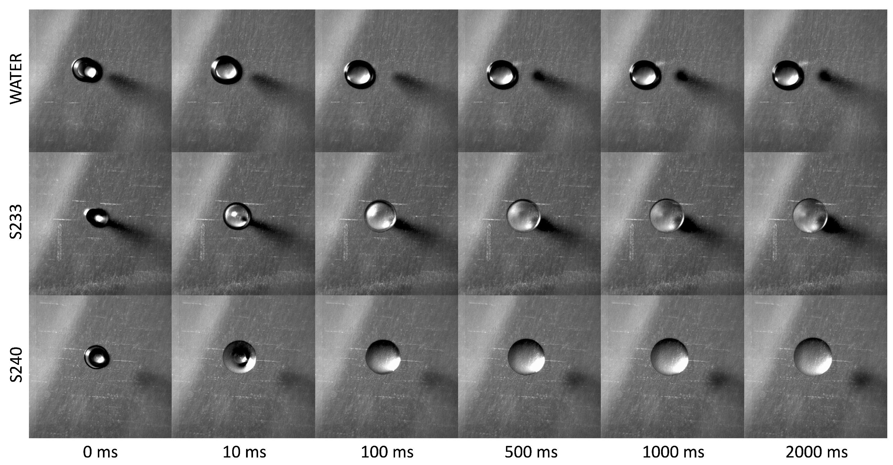
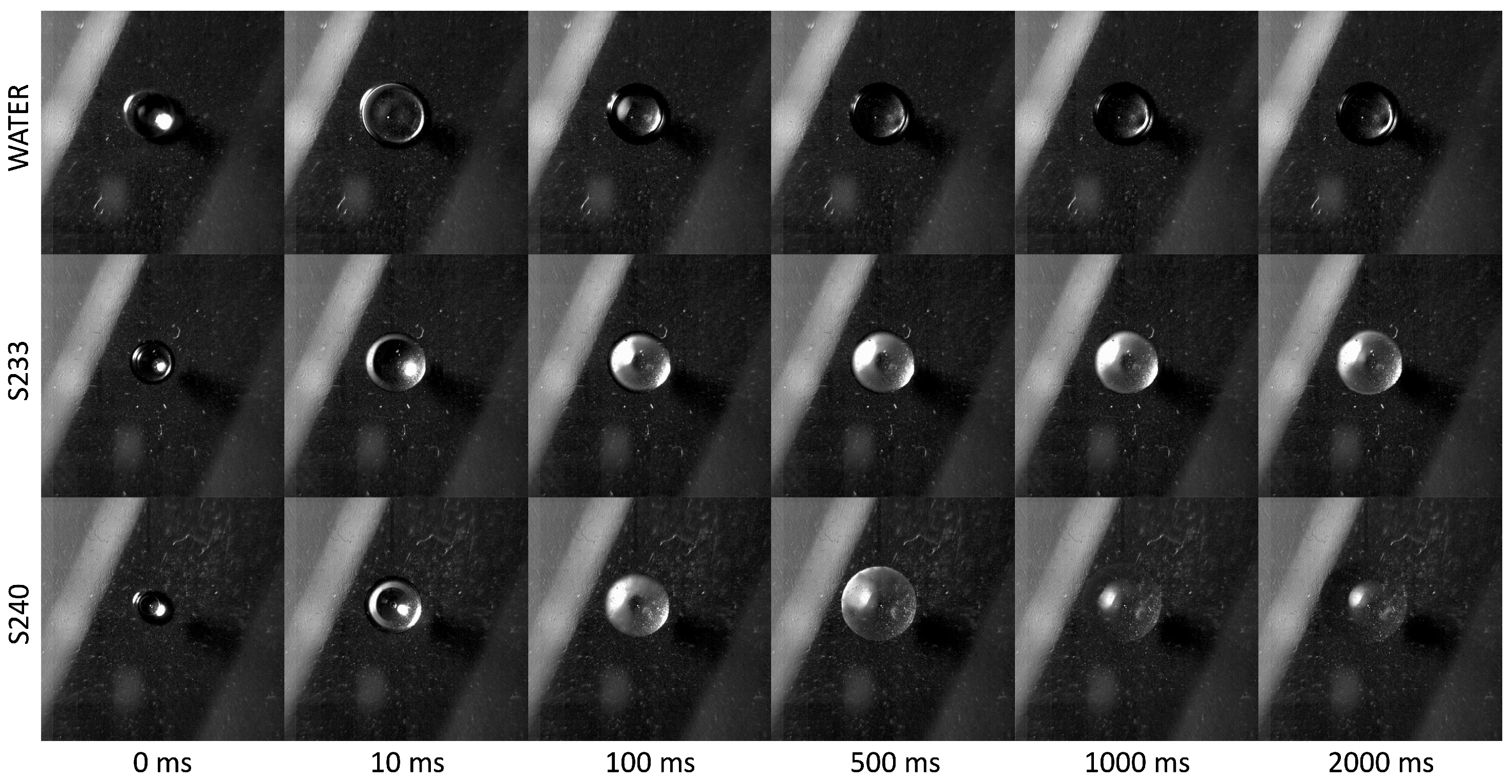
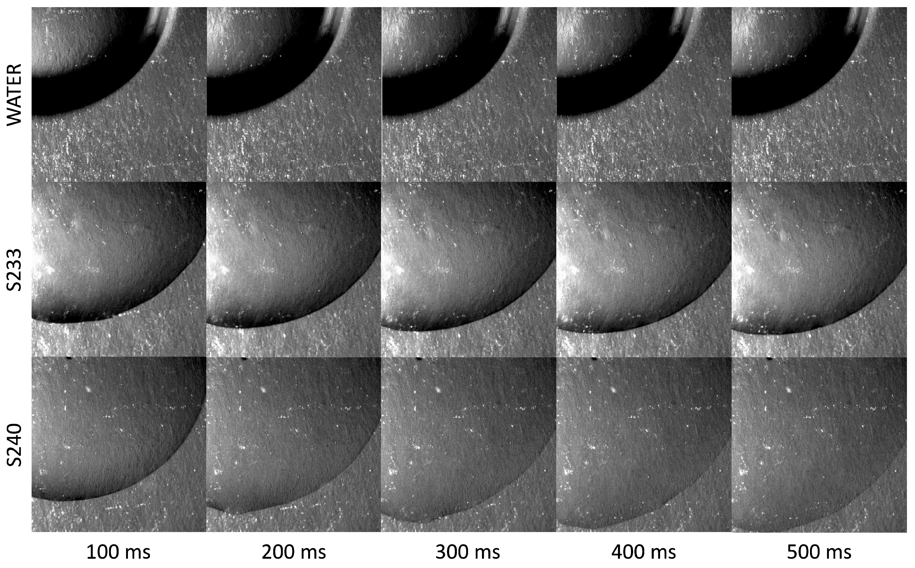
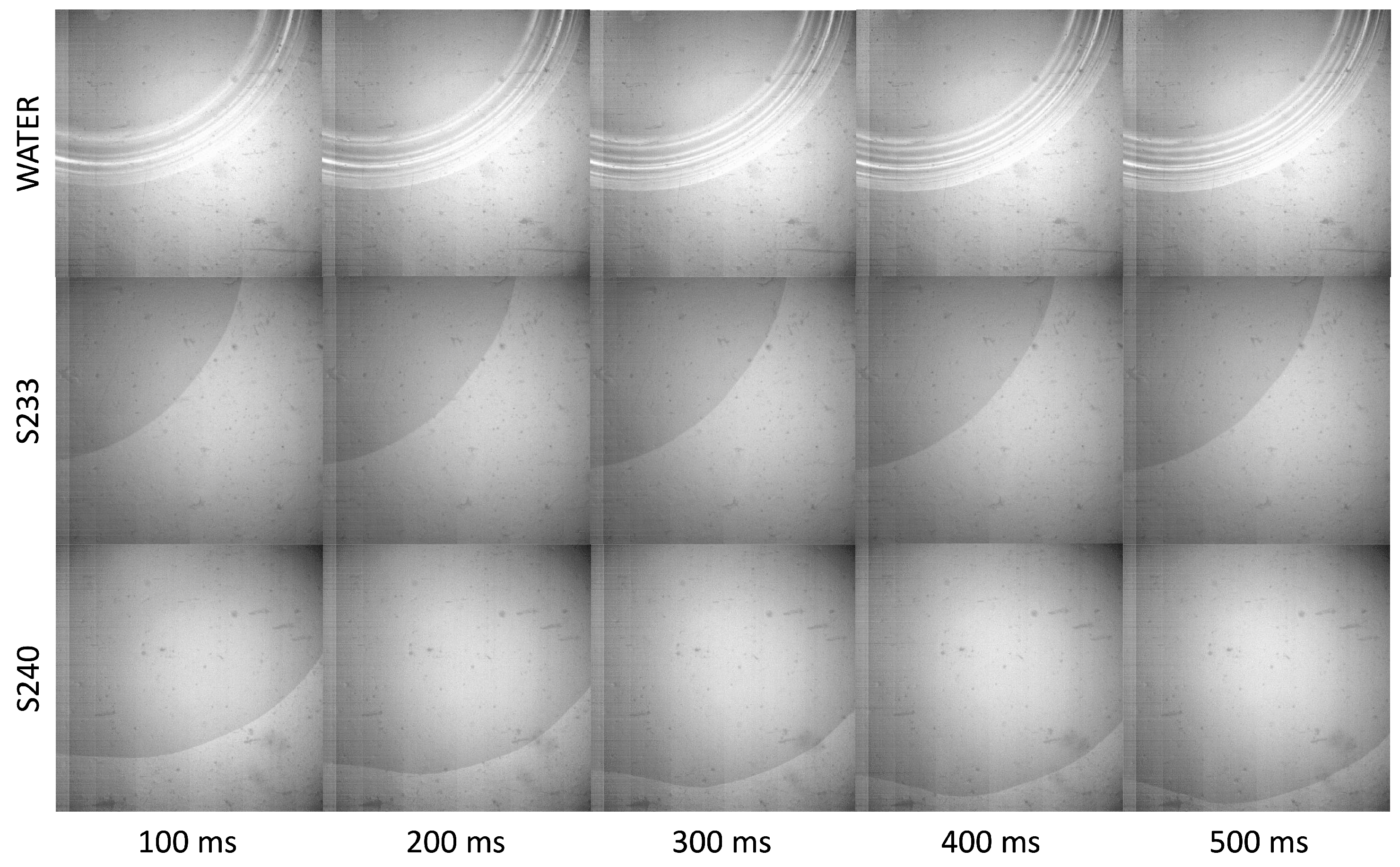
The onset of drop spreading for the three fluids considered during the first two seconds after drop deposition is displayed in Figure 2, Figure 3 and Figure 4, respectively on the Parafilm™ (Figure 2), the acrylic glass (Figure 3), and the polycarbonate (Figure 4) substrates. In all cases, the enhanced spreading of the surfactant solutions is noticeable already 10 ms after deposition. However, its magnitude is strongly dependent on the surface wettability.
While on the Parafilm™ and the acrylic glass surfaces the contact line remains perfectly circular for all fluids, on the polycarbonate substrate, which has the highest wettability (), the contact line of surfactant solution drops departs from the initial circular shape almost immediately upon deposition and becomes quickly highly irregular, and only the contact line of water drops remains circular. Such contact line instability is likely due to the physicochemical interaction between the surfactant molecules and the surface, and may depend on the manufacturing process of the substrate.
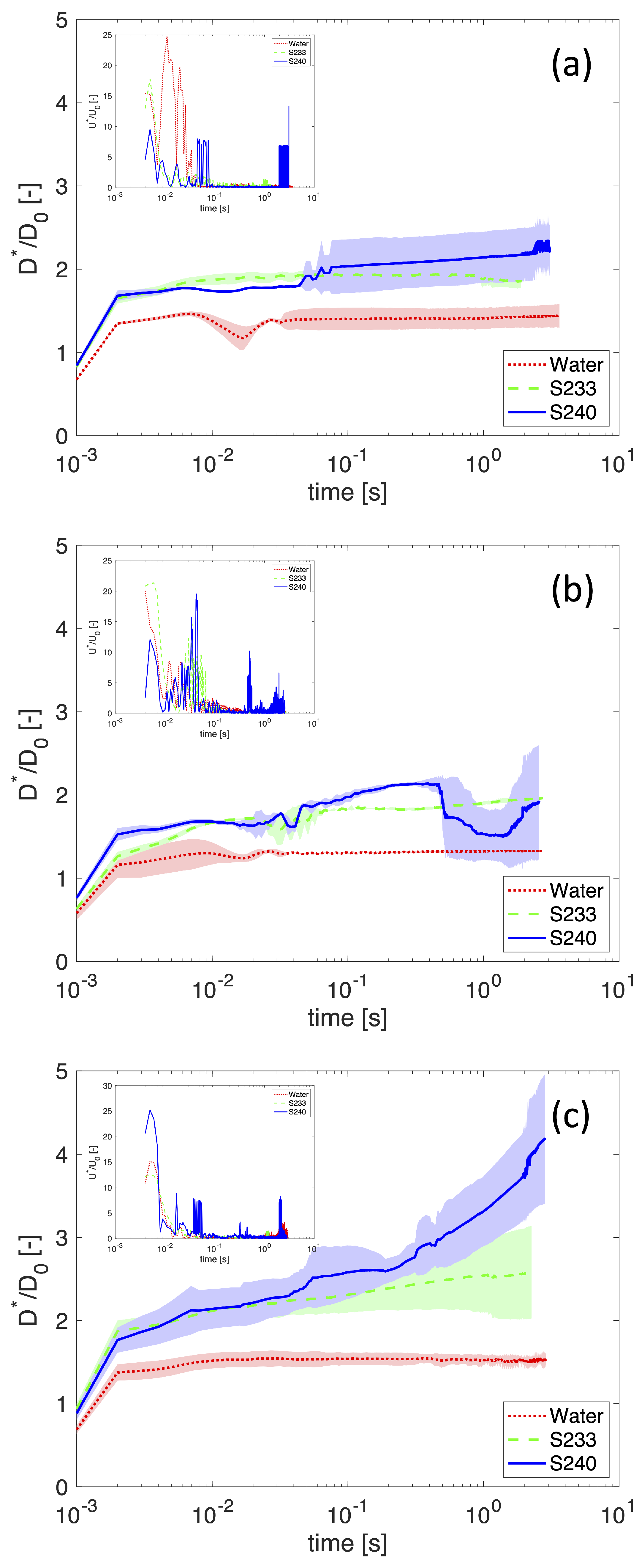
A more quantitative analysis can be obtained from the spreading ratio, , where L is the length of the contour line, which is shown in Figure 5 respectively for the Parafilm™ (Figure 5a), the acrylic glass (Figure 5b), and the polycarbonate (Figure 5c) substrates. The insets in Figure 5 display the magnitude of the dimensionless spreading velocity, , where is the growth rate of the equivalent base diameter, and is the impact velocity.
During deposition, the spreading ratio increases rapidly under the drop own weight for about 2 ms. At the end of this stage, the spreading ratio of surfactant solution drops is already bigger in comparison with water drops, however there is no significant difference between the non-superspreader and the superspreader surfactant solutions. Thus, the initial difference between water and the two surfactant solutions can be solely attributed to the different values of the surface tension.
After deposition, spreading continues at a much slower rate, driven by capillary forces. Irrespective of the substrate material, water drops attain equilibrium in approximately 50 ms, when their spreading ratio becomes constant, indicating there is only partial wetting with a defined contact angle. The two surfactant solution drops spread at a similar rate to each other until ms after deposition. Then, the non-superspreader surfactant solution drop continues to spread at the same slow rate, while the superspreader surfactant solution drop starts to spread at a faster rate. The difference between the spreading rates of the two surfactant solutions is strongly dependent on the surface wettability, increasing for the substrates with higher surface energy. In addition, the spreading rate of the superspreader surfactant solution is not constant, but periods of faster spreading rate alternate with periods of slower spreading rate.
On all surfaces, the divergence of the spreading ratio of the superspreader solution from that of the non-superspreader solution begins with a period of faster spreading ( ms). On the least wettable Parafilm™ surface (Figure 5a), a second period of faster spreading can be observed around ms, while on the most wettable polycarbonate surface (Figure 5c) there are two subsequent periods of faster spreading at ms and ms, respectively.
On the surface with intermediate wettability (Figure 5b), the spreading ratio exhibits a sudden drop at ms, which is clearly an artefact of the image processing algorithm caused by an unexpected temporary change of the illumination conditions (see Figure 3, bottom row), however one can still observe short periods of faster spreading alternating with longer periods of slower spreading. The irregular alternating pattern of the spreading rate in the superspreader surfactant solution appears clearly in the plots displaying the dimensionless spreading velocity (see the insets in Figure 5). Such peculiar dynamic behaviour of the superspreader surfactant solution cannot be observed either for pure water or for the non-superspreader surfactant solution.
These results seem to indicate that (i) superspreading does not take place simultaneously with the formation of a contact line, but only after a short delay of ms, and (ii) superspreading is not a smooth and continuous process, but alternates faster and slower stages. In addition, the peculiar dynamics observed for the superspreader solution occurs also on more hydrophobic surfaces, although the growth rate of the spreading ratio is much smaller so that it is not possible to attain large spreading ratios before the liquid evaporation becomes significant. Remarkably, these features would be very difficult if not impossible to observe if the spreading ratio was calculated based on the base diameter measured from side view images of spreading drops at a slower sampling rate, because they would be hidden by the measurement uncertainty.
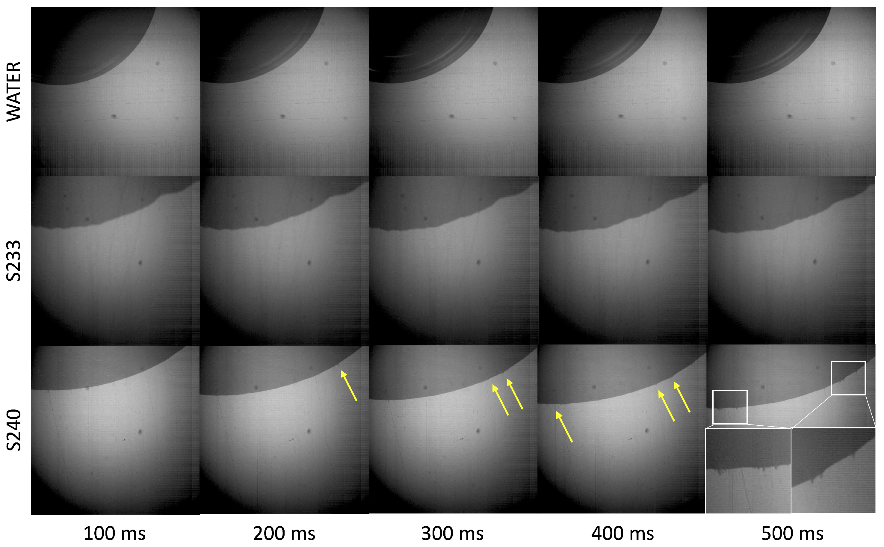
3.2. Microscopic Contact Line Morphology
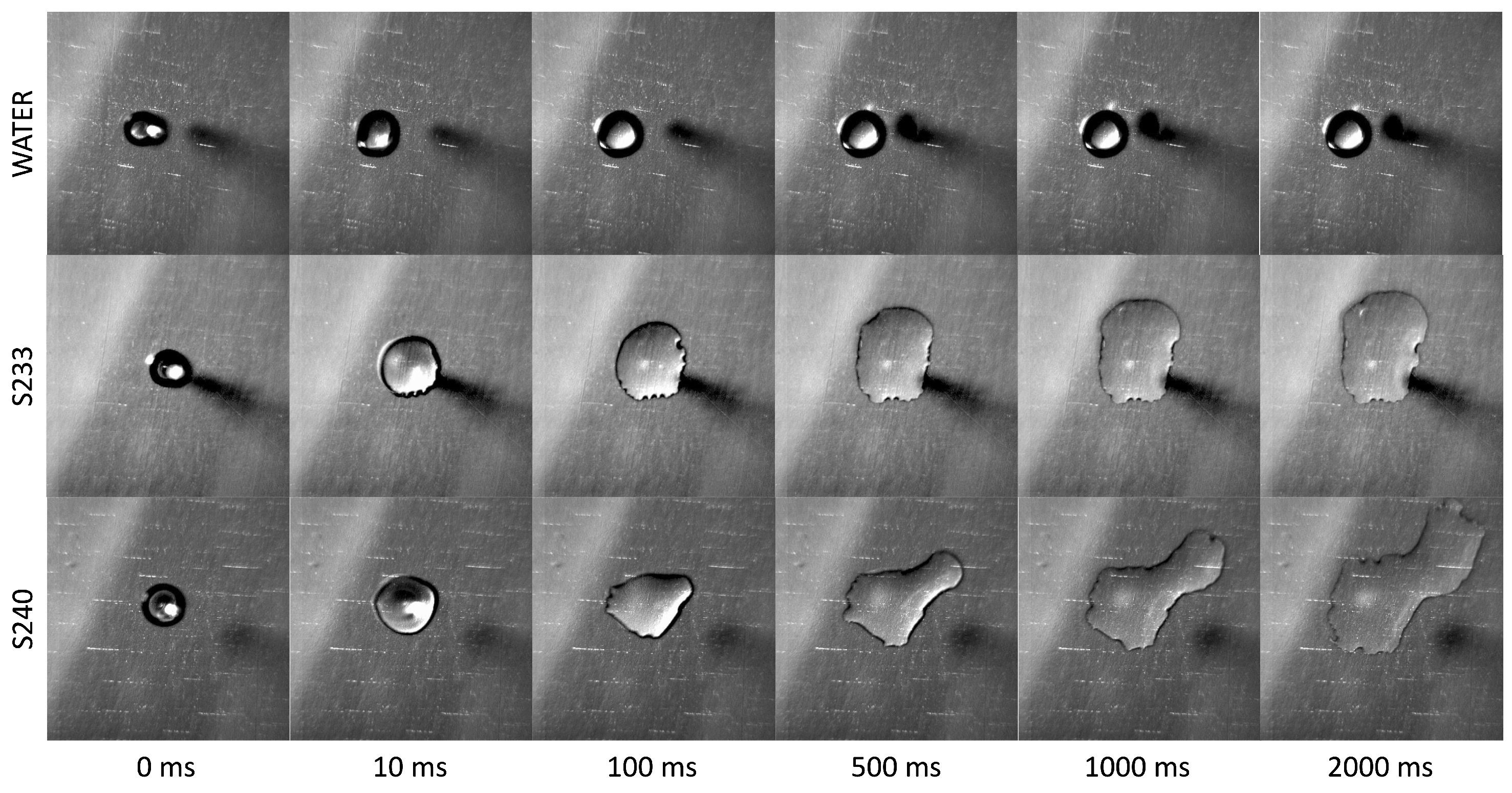
The contact line details of the three fluids considered during the first 500 ms after drop deposition are displayed in Figure 6, Figure 7 and Figure 8, respectively on the Parafilm™ (Figure 6), the acrylic glass (Figure 7), and the polycarbonate (Figure 8) substrates. On all surfaces, the edge of pure water drops appears surrounded by concentric rings which indicate the existence of a precursor film; the rings are very clearly displayed in Figure 7, top row, but can also be observed in the other two Figure 6 and Figure 8. On the contrary, images relative to surfactant solutions, whether superspreading or not, do not display similar fringes, which confirm the absence of a precursor film, as reported previously [7].
While the contact line of water drops appears perfectly smooth and circular, the contact line of surfactant solutions appears more irregular. In the case of pure fluids, this would be attributed to a non-homogeneous surface (e.g., not cleaned prior to experiments), however this can be excluded in the present experiments since drops of pure water do not show similar behaviour. Thus, one can attribute the irregular contour of the contact line of surfactant solutions to some between the surfactant and the surface.
Remarkably, when the advancing contact line of a superspreader surfactant solution drop is observed at the microscale on the least hydrophobic surface (Figure 8, bottom row), one can clearly visualise the formation of microscopic fingers at certain preferred positions along the contact line, which should not be mistakenly interpreted as background dark spots. A previous study shows that in the longer term (several seconds after the drop deposition), these fingers rapidly develop into semi-dendritic structures, which pull the liquid in the droplet beyond the contact line and therefore enhance spreading [28]. This peculiar morphology cannot be observed in the case of non-superspreader trisiloxane surfactant solutions, which exhibit a generally smooth contact line.
4. Conclusions
The early-stage dynamics of drop spreading on hydrophobic surfaces reveals peculiar features of superspreading surfactant solutions as compared with non-superspreading solutions. Irrespective of the surface wettability, the spreading ratio of superspreader solution drops starts to increase more than that of non-superspreader solution drops about 50 ms after deposition. In addition, one can observe that the spreading of superspreader solutions does not evolve as a somooth and continuous process in time, but consists of a sequence of alternating faster and slower stages. If confirmed for different superpreader surfactants and a wider range of experimental conditions, this finding will lead to reconsider the superspreading mechanisms proposed so far.
The superspreading process maintains its qualitative features on all the surfaces considered, however its magnitude reduces significantly as the surface hydrophobicity is increased, so that it is not possible to attain large spreading ratios before the liquid evaporation becomes significant.
The microscopic analysis of the contact line during spreading seems to confirm the absence of a precursor film during the spreading of surfactant solution drops, as opposed to pure fluids. In addition, the contact line of surfactant solution drops aappears irregular as compared with the smooth and circular contact line of drops of pure fluids. During the spreading on a moderately hydrophobic surface (), the contact line of the superspreader surfactant solution exhibits microscopic fingering, which deserves further investigation.
Funding
This research received no external funding.
Data Availability Statement
Raw data available upon request.
Acknowledgments
Samples of surfactants Break-Thru S233 and Break-Thru S240 (Evonik Industries AG) were kindly donated by Joachim Venzmer.
Conflicts of Interest
The author declares no conflict of interest.
References
- Humble, G.; Simpelkamp, J.; Kennedy, M. Use of Non-Spreading Silicone Surfactants in Agrochemical Compositions. U.S. Patent No. AU 2002301504 B2, 11 May 2004. [Google Scholar]
- Ananthapadmanabhan, K.; Goddard, E.; Chandar, P. A study of the solution, interfacial and wetting properties of silicone surfactants. Colloids Surf. 1990, 44, 281–297. [Google Scholar] [CrossRef]
- Hill, R.M. Superspreading. Curr. Opin. Colloid Interface Sci. 1998, 3, 247–254. [Google Scholar] [CrossRef]
- Venzmer, J. Superspreading—20 years of physicochemical research. Curr. Opin. Colloid Interface Sci. 2011, 16, 335–343. [Google Scholar] [CrossRef]
- Venzmer, J. Chapter 6—Superspreading. In Droplet Wetting and Evaporation; Brutin, D., Ed.; Academic Press: Oxford, UK, 2015; pp. 71–84. [Google Scholar] [CrossRef]
- Sankaran, A.; Karakashev, S.I.; Sett, S.; Grozev, N.; Yarin, A.L. On the nature of the superspreaders. Adv. Colloid Interface Sci. 2019, 263, 1–18. [Google Scholar] [CrossRef]
- Venzmer, J. Superspreading—Has the mystery been unraveled? Adv. Colloid Interface Sci. 2021, 288, 102343. [Google Scholar] [CrossRef] [PubMed]
- Ivanova, N.; Zhantenova, Z.; Starov, V. Wetting dynamics of polyoxyethylene alkyl ethers and trisiloxanes in respect of polyoxyethylene chains and properties of substrates. Colloids Surf. A Physicochem. Eng. Asp. 2012, 413, 307–313. [Google Scholar] [CrossRef]
- Kovalchuk, N.M.; Matar, O.K.; Craster, R.V.; Miller, R.; Starov, V.M. The effect of adsorption kinetics on the rate of surfactant-enhanced spreading. Soft Matter 2016, 12, 1009–1013. [Google Scholar] [CrossRef]
- Chengara, A.; Nikolov, A.D.; Wasan, D.T. Spreading of a Water Drop Triggered by the Surface Tension Gradient Created by the Localized Addition of a Surfactant. Ind. Eng. Chem. Res. 2007, 46, 2987–2995. [Google Scholar] [CrossRef]
- Ivanova, N.A.; Kovalchuk, N.M.; Sobolev, V.D.; Starov, V.M. Wetting films of aqueous solutions of Silwet L-77 on a hydrophobic surface. Soft Matter 2016, 12, 26–30. [Google Scholar] [CrossRef]
- Ivanova, N.; Starov, V.; Johnson, D.; Hilal, N.; Rubio, R. Spreading of Aqueous Solutions of Trisiloxanes and Conventional Surfactants over PTFE AF Coated Silicone Wafers. Langmuir 2009, 25, 3564–3570. [Google Scholar] [CrossRef]
- Starov, V.; Ivanova, N.; Rubio, R.G. Why do aqueous surfactant solutions spread over hydrophobic substrates? Adv. Colloid Interface Sci. 2010, 161, 153–162. [Google Scholar] [CrossRef]
- He, M.; Hill, R.M.; Lin, Z.; Scriven, L.E.; Davis, H.T. Phase behavior and microstructure of polyoxyethylene trisiloxane surfactants in aqueous solution. J. Phys. Chem. 1993, 97, 8820–8834. [Google Scholar] [CrossRef]
- Wagner, R.; Wu, Y.; Czichocki, G.; Berlepsch, H.; Rexin, F.; Perepelittchenko, L. Silicon-modified surfactants and wetting: II. Temperature-dependent spreading behaviour of oligoethylene glycol derivatives of heptamethyltrisiloxane. Appl. Organomet. Chem. 1999, 13, 201–208. [Google Scholar] [CrossRef]
- Kumar, N.; Couzis, A.; Maldarelli, C. Measurement of the kinetic rate constants for the adsorption of superspreading trisiloxanes to an air/aqueous interface and the relevance of these measurements to the mechanism of superspreading. J. Colloid Interface Sci. 2003, 267, 272–285. [Google Scholar] [CrossRef]
- Ritacco, H.A.; Ortega, F.; Rubio, R.G.; Ivanova, N.; Starov, V.M. Equilibrium and dynamic surface properties of trisiloxane aqueous solutions: Part 1. Experimental results. Colloids Surf. A Physicochem. Eng. Asp. 2010, 365, 199–203. [Google Scholar] [CrossRef]
- Israelachvili, J. Intramolecular and Surface Forces, 3rd ed.; Academic Press: Cambridge, MA, USA, 2011. [Google Scholar]
- Kumar, N.; Maldarelli, C.; Couzis, A. An infrared spectroscopy study of the hydrogen bonding and water restructuring as a trisiloxane superspreading surfactant adsorbs onto an aqueous–hydrophobic surface. Colloids Surf. A Physicochem. Eng. Asp. 2006, 277, 98–106. [Google Scholar] [CrossRef]
- Williams, K.; Bertola, V.; Martin, D. Experimental investigation of a flowing superspreader solution using Reflection Anisotropy Spectroscopy. Results Phys. 2016, 6, 277–279. [Google Scholar] [CrossRef]
- Papakonstantinou, C.; Chen, H.; Bertola, V.; Amirfazli, A. Effect of condensation on surface contact angle. Colloids Surf. A Physicochem. Eng. Asp. 2022, 632, 127739. [Google Scholar] [CrossRef]
- Nikolov, A.; Wasan, D. Superspreading mechanisms: An overview. Eur. Phys. J. Spec. Top. 2011, 197, 325. [Google Scholar] [CrossRef]
- Ruckenstein, E. Superspreading: A possible mechanism. Colloids Surf. A Physicochem. Eng. Asp. 2012, 412, 36–37. [Google Scholar] [CrossRef]
- Wang, X.; Venzmer, J.; Bonaccurso, E. Surfactant-Enhanced Spreading of Sessile Water Drops on Polypropylene Surfaces. Langmuir 2016, 32, 8322–8328. [Google Scholar] [CrossRef] [PubMed]
- Biolè, D.; Bertola, V. A goniometric mask to measure contact angles from digital images of liquid drops. Colloids Surf. A Physicochem. Eng. Asp. 2015, 467, 149–156. [Google Scholar] [CrossRef]
- Biolè, D.; Bertola, V. Measuring fluid interfaces, corners, and angles from high-speed digital images of impacting drops. J. Flow Vis. Image Process. 2021, 28, 1–19. [Google Scholar] [CrossRef]
- Thoroddsen, S.; Sakakibara, J. Evolution of the Fingering Pattern of an Impacting Drop. Phys. Fluids 1998, 10, 1359. [Google Scholar] [CrossRef]
- Biolè, D.; Bertola, V. The role of the microscale contact line dynamics in the wetting behaviour of complex fluids. Arch. Mech. 2015, 67, 401–414. [Google Scholar]
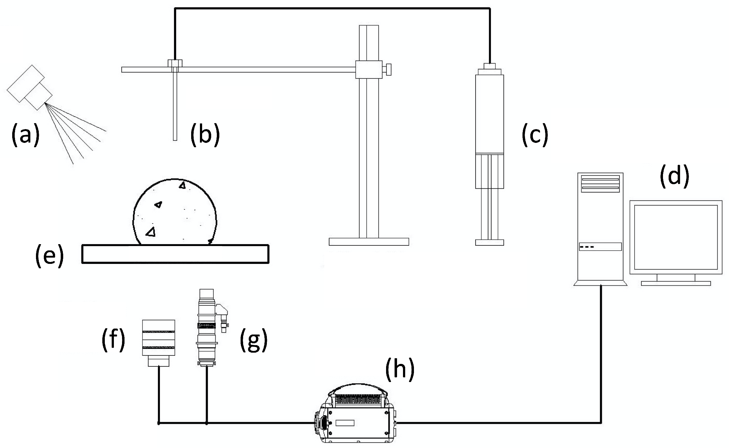
Figure 1.
Schematic of the experimental set-up: (a) Halogen illumination. (b) Dispensing needle. (c) Syringe pump. (d) Data acquisition system. (e) Substrate. (f) Low-magnification lens. (g) High-magnification lens. (h) High-speed camera.
Figure 1.
Schematic of the experimental set-up: (a) Halogen illumination. (b) Dispensing needle. (c) Syringe pump. (d) Data acquisition system. (e) Substrate. (f) Low-magnification lens. (g) High-magnification lens. (h) High-speed camera.

Figure 2.
Initial stages of spreading of drops deposited on a Parafilm™ substrate (). (Top row): water. (Middle row): S233 solution. (Bottom row): S240 solution. Frame size: 16.5 mm.
Figure 2.
Initial stages of spreading of drops deposited on a Parafilm™ substrate (). (Top row): water. (Middle row): S233 solution. (Bottom row): S240 solution. Frame size: 16.5 mm.

Figure 3.
Initial stages of spreading of drops deposited on an acrylic glass substrate (). (Top row): water. (Middle row): S233 solution. (Bottom row): S240 solution. Frame size: 16.5 mm.
Figure 3.
Initial stages of spreading of drops deposited on an acrylic glass substrate (). (Top row): water. (Middle row): S233 solution. (Bottom row): S240 solution. Frame size: 16.5 mm.

Figure 4.
Initial stages of spreading of drops deposited on a clear polycarbonate substrate (). (Top row): water. (Middle row): S233 solution. (Bottom row): S240 solution. Frame size: 16.5 mm.
Figure 4.
Initial stages of spreading of drops deposited on a clear polycarbonate substrate (). (Top row): water. (Middle row): S233 solution. (Bottom row): S240 solution. Frame size: 16.5 mm.

Figure 5.
Spreading ratio, , of water and surfactant solution drops on substrates of different wettability. (a): Parafilm™ (). (b): Acrylic glass (). (c): polycarbonate (). Insets display the magnitude of the dimensionless spreading velocity, .
Figure 5.
Spreading ratio, , of water and surfactant solution drops on substrates of different wettability. (a): Parafilm™ (). (b): Acrylic glass (). (c): polycarbonate (). Insets display the magnitude of the dimensionless spreading velocity, .

Figure 6.
Contact line detail of drops deposited on a Parafilm™ substrate () during the early stages of spreading. (Top row): water. (Middle row): S233 solution. (Bottom row): S240 solution. Frame size: 2.7 mm.
Figure 6.
Contact line detail of drops deposited on a Parafilm™ substrate () during the early stages of spreading. (Top row): water. (Middle row): S233 solution. (Bottom row): S240 solution. Frame size: 2.7 mm.

Figure 7.
Contact line detail of drops deposited on an acrylic glass substrate () during the early stages of spreading. (Top row): water. (Middle row): S233 solution. (Bottom row): S240 solution. Frame size: 2.7 mm.
Figure 7.
Contact line detail of drops deposited on an acrylic glass substrate () during the early stages of spreading. (Top row): water. (Middle row): S233 solution. (Bottom row): S240 solution. Frame size: 2.7 mm.

Figure 8.
Contact line detail of drops deposited on a clear polycarbonate substrate () during the early stages of spreading. (Top row): water. (Middle row): S233 solution. (Bottom row): S240 solution. Frame size: 2.7 mm. The insets show microscopic protrusions emerging from the contact line.
Figure 8.
Contact line detail of drops deposited on a clear polycarbonate substrate () during the early stages of spreading. (Top row): water. (Middle row): S233 solution. (Bottom row): S240 solution. Frame size: 2.7 mm. The insets show microscopic protrusions emerging from the contact line.

Disclaimer/Publisher’s Note: The statements, opinions and data contained in all publications are solely those of the individual author(s) and contributor(s) and not of MDPI and/or the editor(s). MDPI and/or the editor(s) disclaim responsibility for any injury to people or property resulting from any ideas, methods, instructions or products referred to in the content. |
© 2024 by the author. Licensee MDPI, Basel, Switzerland. This article is an open access article distributed under the terms and conditions of the Creative Commons Attribution (CC BY) license (https://creativecommons.org/licenses/by/4.0/).
Share and Cite
MDPI and ACS Style
Bertola, V. The Onset and Early Stages of Dynamic Wetting of Superspreading and Non-Superspreading Trisiloxane Surfactant Solutions on Hydrophobic Surfaces. Colloids Interfaces 2024, 8, 5. https://doi.org/10.3390/colloids8010005
AMA Style
Bertola V. The Onset and Early Stages of Dynamic Wetting of Superspreading and Non-Superspreading Trisiloxane Surfactant Solutions on Hydrophobic Surfaces. Colloids and Interfaces. 2024; 8(1):5. https://doi.org/10.3390/colloids8010005
Chicago/Turabian StyleBertola, Volfango. 2024. "The Onset and Early Stages of Dynamic Wetting of Superspreading and Non-Superspreading Trisiloxane Surfactant Solutions on Hydrophobic Surfaces" Colloids and Interfaces 8, no. 1: 5. https://doi.org/10.3390/colloids8010005