Article

Ultrasonic-Assisted Electrodeposition of Mn-Doped NiCo2O4 for Enhanced Photodegradation of Methyl Red, Hydrogen Production, and Supercapacitor Applications

by
Kuan-Ching Lee
1,
Timm Joyce Tiong
1,*,
Guan-Ting Pan
2,
Thomas Chung-Kuang Yang
3,
Kasimayan Uma
4,
Zong-Liang Tseng
4,
Aleksandar N. Nikoloski
2,* and
Chao-Ming Huang
5,*
1
Department of Chemical and Environmental Engineering, University of Nottingham, Broga Road, Semenyih 43500, Selangor Darul Ehsan, Malaysia
2
College of Science, Health, Engineering & Education, Murdoch University, 90 South Street, Murdoch, WA 6150, Australia
3
Department of Chemical Engineering and Biotechnology, National Taipei University of Technology, Taipei 10608, Taiwan
4
Organic Electronics Research Center, Ming Chi University of Technology, New Taipei City 24301, Taiwan
5
Green Energy Technology Research Center, Department of Advanced Applied Materials Engineering, Kun Shan University, No. 195, Kunda Rd., Yongkang Dist., Tainan 710303, Taiwan
*
Authors to whom correspondence should be addressed.
J. Compos. Sci. 2024, 8(5), 164; https://doi.org/10.3390/jcs8050164
Submission received: 27 February 2024 / Revised: 17 April 2024 / Accepted: 23 April 2024 / Published: 29 April 2024
(This article belongs to the Special Issue Nanocomposites for Supercapacitor Application)

Abstract

:
This paper presents a novel ultrasonic-assisted electrodeposition process of Mn-doped NiCo2O4 onto a commercial nickel foam in a neutral electroplating bath (pH = 7.0) under an ultrasonic power of 1.2 V and 100 W. Different sample properties were studied based on their crystallinity through X-ray diffraction (XRD), morphology was studied through scanning electron microscopy (SEM), and photodegradation was studied through ultraviolet–visible (UV–Vis) spectrophotometry. Based on the XRD results, the dominant crystallite phase obtained was shown to be a pure single NiCo2O4 phase. The optical properties of the photocatalytic film showed a range of energy band gaps between 1.72 and 1.73 eV from the absorption spectrum. The surface hydroxyl groups on the catalytic surface of the Mn-doped NiCo2O4 thin films showed significant improvements in removing methyl red via photodegradation, achieving 88% degradation in 60 min, which was approximately 1.6 times higher than that of pure NiCo2O4 thin films. The maximum hydrogen rate of the composite films under 100 mW/cm2 illumination was 38 μmol/cm2 with a +3.5 V external potential. The electrochemical performance test also showed a high capacity retention rate (96% after 5000 charge–discharge cycles), high capacity (260 Fg−1), and low intrinsic resistance (0.8 Ω). This work concludes that the Mn-doped NiCo2O4 hybrid with oxygen-poor conditions (oxygen vacancies) is a promising composite electrode candidate for methyl red removal, hydrogen evolution, and high-performance hybrid supercapacitor applications.

Graphical Abstract

1. Introduction

Semiconductor photocatalysts have been widely studied and used in the treatment of environmental pollution owing to their ability to produce hydrogen and oxygen from water. Titanium dioxide (TiO2) is among the most used semiconductor photocatalysts due to its high efficiency of 13% [1,2]. However, titanium dioxide has a large 3.2 eV band gap energy, reducing its efficiency under visible light. Therefore, many studies have attempted to improve TiO2 photocatalyst activity under visible light, including through oxygen deficiency generation [3,4] and metal doping [5,6]. As a result of these efforts, despite the light absorption of TiO2 having been increased to the visible light region [7], its thermal instability or carrier recombination centers increasingly affect these processes [8,9,10,11]. However, improvements are still required if TiO2 is to be used as a photocatalyst, causing researchers to venture into other materials for use within the visible light region.
Recently, Cui et al. [12] found transition metal oxide NiCo2O4 nanoplatelets to be novel and promising photocatalysts for methyl blue degradation due to their small particle size (80–150 nm) and excellent optical properties (band gap between 2.06 and 3.63 eV). Wan et al. [13] performed an investigation on NiCo2O4 catalyst preparation for the degradation of methyl red under visible light irradiation, and they found that the methyl red photodegradation efficiency of mesoporous NiCo2O4 can reach 95.1% under visible light irradiation for 2 h. Additionally, the demonstrated effectiveness of NiCo2O4 in photodegradation within the visible region has enhanced the popularity of this mixed oxide compound, mainly due to its multiple valence states, hypotonicity, ultrastability, and containable structure [12]. To further enhance the properties of NiCo2O4, one typical method that has gained popularity over recent years is performing transition metal doping onto a semiconductor film [14,15]. Han et al. [16] synthesized Cu-doped NiCo2O4 nanosheets using a thermal method for energy storage applications. The electrochemical properties of the Cu–NiCo2O4 electrode can be improved due to its high conductivity and effective mass transfer. Additionally, it is worth noting that the electrochemical properties of NiCo2O4 can also be enhanced by Al doping due to the transportation of electrons/ions and the large specific surface area for redox reactions in the reaction system [14]. In the context of Mn doped onto NiCo2O4, there have been several recent reports on the enhancement that Mn provides in their respective domains. Nguyen et al. [17] successfully doped Mn onto NiCo2O4 using hydrothermal methods followed by a subsequent calcination route. They showed significant increments in the power density of their supercapacitor. Similar results were obtained by Pradeepa et al. [18], in which Mn-doped NiCo2O4 improved the specific capacitance, electrode stability, and transfer speed of charge carriers. Liang et al. [19] found that spinel Mn–NiCo2O4 can effectively shorten the transmission path of Li ions, thereby improving the overall conductivity of Li-ion batteries.
Research on preparing binder-free composite electrodes using nickel as the base material has aroused great interest in recent years [7,20,21,22,23]. Binder-free electrodes enhance the contact between the active electrode material and the current collector, significantly reducing the risk of catalyst shedding during cycling and improving cycle stability [7]. Among the synthesis methods for binder-free electrodes, electrodeposition synthesis offers a wide range of benefits due to its improved catalyst uniformity, providing consistent grain sizes across the catalytic surface [24]. One common approach to enhancing the electrodeposition technique is to employ ultrasound during the synthesis process to improve the anti-corrosivity [25], magnetitic intensity [26], degree of wear [27], and hardness [28] properties of composite films. Researchers have reported several ultrasonic-assisted synthesis methods. For example, Costa et al. [29] found that using ultrasound for catalyst preparation improved their catalyst’s surface chemical group properties. Additionally, Shi et al. [30] reported that the introduction of ultrasonic-assisted electrodeposition can improve the particle’s surface distribution and impede catalyst aggregation on the substrate. The ultrasonic-assisted electrodeposition approach can provide a benefit in that hydrogen bubbles and film oxides are removed, leading to porosity in thin films and improvements in their electrode efficiency in systems [31]. Despite the emerging popularity of NiCo2O4 and the increased use of Mn as a dopant to improve overall catalytic activity, there are still no records on electrodeposition synthesis using ultrasound for ternary metal oxide catalyst preparation from a precursor mixture.
In this work, we report a facile combined ultrasonic-assisted electrodeposition method for preparing Mn-doped NiCo2O4 composite electrodes to investigate the effects of different molar ratios of Ni/Co/Mn on the characteristics of catalysts, such as their morphology, crystallinity, and functional groups. The resulting Mn-doped NiCo2O4 composite electrodes show high potential for environmental protection through methyl red removal, energy utility in hydrogen evolution, and energy storage.

2. Materials and Methods
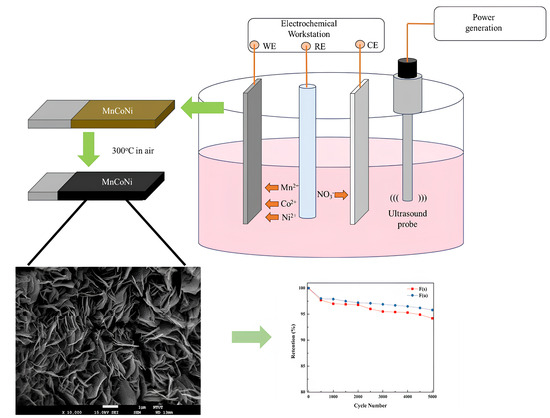
2.1. Materials

The precursors of Ni2+, Co2+, and Mn2+ ions for the reaction of Mn/Ni/Co/O thin films were analytical-grade nickel(II) nitrate hexahydrate (Ni(NO3)2•6H2O), cobalt(II) nitrate hexahydrate (Co(NO3)2•6H2O), and manganese(II) nitrate hydrate (Mn(NO3)2•H2O) with purities of >99%, all of which were purchased from Merck. The commercial nickel foam substrates (2 × 3 cm) were acquired from Sigma-Aldrich Co (Taipei City, Taiwan). Before the deposition process, the commercial nickel foam substrates were cleaned in different baths of acetone, deionized (DI) water, and ethyl alcohol using an ultrasonic cleaning machine for 30 min in each bath. The nickel foam substrates were then blown dry with nitrogen gas.

2.2. Preparation of Mn-Doped NiCo2O4 Electrodes

Mn-doped NiCo2O4 thin films were prepared on the commercial nickel foam substrates via ultrasonic-assisted electrochemical deposition. The solution (250 mL) of electrochemical deposition was made of aqua solutions of Mn(NO3)2•H2O (0.04M), Co(NO3)2•6H2O (0.04 M), Na2SO4 (0.04 M), and Ni(NO3)2•6H2O (0.04 M). All of the samples were prepared with varying molar ratios of Ni/Co/Mn, as summarized in Table 1. All thin films were deposited using a galvanostat (Autolab Model PGSTAT 30, Herisau, Switzerland) with a standard three-electrode system (the commercial nickel foam, a 4 × 10 cm2 platinum sheet, and a saturated calomel electrode were respectively used as a working electrode, a counter electrode, and reference electrodes) connected to an electrochemical Pyrex glass cell. Figure 1 shows the setup of the ultrasonic-assisted electrochemical deposition of the Mn-doped NiCo2O4 electrodes. Deposition voltage and time were set at 1.2 V and 15 min for all samples. During the electrodeposition process, an additional ultrasonic power of 100 W (Qsonica, Q700, 20 kHz, Newtown, CT, USA) was employed for the ultrasonicated samples. After deposition, dark brown films were deposited on the nickel foam substrates (working electrodes) in the three-electrode electrodeposition system. After this, all of the samples were annealed in a furnace at 500 °C for 60 min following a stable ramping rate (5 °C/min) from 30 °C. The temperature was then gradually cooled down to an ambient temperature (25 °C) in an air atmosphere.

2.3. Characterization of the Mn-Doped NiCo2O4 Electrodes

Crystalline patterns of all of the samples were obtained using X-ray diffraction (Rigaku RINT 2000, TX, USA) with Cu Kα radiation (λ = 1.5405 Å) in a 2θ scan range of 10° to 80°. The spectral absorbance of all of the samples was measured using ultraviolet-visible (UV–Vis) spectrophotometry (PerkinElmer Lambda 900, Shelton, CT, USA) with a wavelength range from 500 to 1100 nm at ambient temperature. The thin film morphology in this study was analyzed using a field emission scanning electron microscope (JEOL JSM 6700F, Peabody, MA, USA) with energy-dispersive X-ray analysis (OXFORD X-MaxN, accelerating voltage, 15.0 kV, High Wycombe, UK). The textural properties of the samples were obtained via the Barrett–Emmett–Teller approach using a Micromeritics Instrument Corporation device (TriStar II Plus, Norcross, GA, USA).

2.4. Photocatalytic Performance Evaluation of the Mn-Doped NiCo2O4 Electrodes

Photocatalytic performance was evaluated via photodegradation of the methyl red solution under visible irradiation using the Mn-doped NiCo2O4 electrodes. Before the visible radiation evaluation, the methyl red aqueous solution (60 mL, 10 ppm) was stirred using a magnetic stirrer (CORNING, 200 rpm, New York, NY, USA) for adsorption analysis under dark surroundings for 30 min. Visible irradiations were acquired using a 500 W xenon lamp equipped with cut-off filters to eliminate wavelengths of light lower than 420 nm. The methyl red concentration in the solution was analyzed using ultraviolet–visible spectrophotometry at 30 min intervals for 150 min.

2.5. Electrochemical Measurements

Half-cell electrochemical measurements of different molar ratios of Ni/Co/Mn for the Mn-doped NiCo2O4 electrodes were performed in a 6 M KOH electrolyte. For the half-cell measurements, active electrode material/Ni foam was used as the working electrode, saturated calomel electrode (SCE) as the reference electrode, and platinum plate as the counter electrode. Cyclic voltammetry (CV) analyses were recorded from 100 to 5 mVs1. Galvanostatic charge and discharge (GCD) analyses were recorded from 10 to 1 Ag1. Electrochemical impedance spectroscopy (EIS) analyses were recorded at AC frequencies from 10 kHz to 0.1 Hz.

3. Results and Discussion

3.1. XRD Analysis

Figure 2 shows the XRD patterns of the Mn-doped NiCo2O4 electrodes (a) without and (b) with ultrasonic-assisted samples at different molar ratios in the order of increasing Mn load (moving from samples A to F). Generally, standard NiCo2O4 diffraction peaks (JCPDS No. 20-0781) could be observed for samples with and without ultrasonic assistance. A pure cubic NiCo2O4 phase can be seen in Figure 2a, indicating that diffraction peaks of Mn alloys and other compounds were not in the samples. Two deviations from the XRD diffraction patterns were observed for those NiCo2O4 samples doped with Mn, due to the different atomic radii of Ni (0.124 nm) and Mn (0.140 nm). The main diffraction peak of the Mn-doped NiCo2O4 electrode shifted significantly, and the crystallinities of the samples decreased as the manganese concentration in the reaction solution increased [17]. Sun et al. [30] reported that Mn atoms occupy Ni seats in NiCo2O4 structures. According to the results of the XRD pattern, the major diffraction peaks of the NiCo2O4 electrodes experienced a light shift with an increase in manganese concentration in the reaction solution, suggesting that the nickel sites were occupied by manganese atoms, with the Mn3+ ions preferring to replace the Ni3+ ions within NiCo2O4. This substitution improved the electronic structure and enhanced the ability for charge transfer, thereby improving the conductivity and efficiency of electron movement [28,31,32]. After doping with Mn, the ratio of Ni and Co atoms changed slightly, indicating that Mn atoms replaced the positions of Ni and Co. As Ni, Co, and Mn have different atomic radii (0.124, 0.167, and 0.140 nm, respectively), it is evident that Mn atoms replacing the positions of Ni and Co in a typical NiCo2O4 electrode will result in slight deviations in the XRD diffraction of NiCo2O4. The structural representation of a typical NiCo2O4 electrode demonstrates that Ni (or Mn) ions occupy octahedral sites, while Co ions occupy octahedral and tetragonal sites [17]. Interestingly, the electrical conductivity of electrodes can be improved to enhance electron transport, owing to the shift phenomenon of XRD, which can be attributed to the differences in the atomic radii of nickel and manganese.
A noticeable decrease in the crystallinities of the samples treated with ultrasound can be seen in Figure 2b. The intensity of the XRD diffraction patterns decreased due to the smaller grain size in the ultrasonic-assisted samples [33]. The crystalline forms resulted in no changes in the samples. Based on the XRD results, the peak intensities of the samples that underwent ultrasonic-assisted synthesis were reduced by approximately 0.2 times compared to the original samples. Recently, numerous studies have reported that ultrasonic techniques can generate microturbulence, microjets, and shock waves and disrupt van der Waals forces, thereby enhancing the particle deagglomeration in solution systems and leading to more uniform particle size distribution. The grain size refinement of thin films can also be improved [34]. In addition, no impurities were found in the X-ray diffraction patterns. Based on the Scherrer formula, the crystallite sizes of NiCo2O4 and Mn-doped NiCo2O4 are listed in Table 2. The grain size of NiCo2O4 was 16.92 nm for A(u) and 15.23 for A(s). The crystallite size of the Mn-doped NiCo2O4 samples decreased as the concentration of the Mn doping increased with/without ultrasonic-assisted electrodeposition [35].
Figure 3 shows Raman spectra of the Mn-doped NiCo2O4 electrodes. Raman peaks at 455, 505, and 648 align with the Raman peaks of NiCo2O4. No other phases or impurities were observed. Compared to Figure 3a,b, the peaks in Figure 3b had a negative shift, which means that the sample with ultrasonic-assisted synthesis had more oxygen vacancies [28]. The reduced Raman spectra intensities of the NiCo2O4 phase were accompanied by an increased Mn concentration in the reaction.

3.2. Morphology and XPS Analyses

Figure 4 shows the microstructure of the A(u) to F(s) samples at 10,000× magnification. The microstructures of all of the samples changed from highly dense to porous due to various ultrasonic-assisted electrochemical deposition parameters in the electrodeposition reaction. The SEM results show that the morphology of the samples changed from a sphere to a tube to a sheet according to different Mn doping amounts. The ultrasound-treated sample showed smaller particles than the non-ultrasound-treated sample. The resulting morphologies are similar to those of Min et al. [36]. The microstructure could have become agglomerated and cracked as a result of an increased oxygen vacancy concentration in the samples [37]. Notably, the particle size was smaller than that of the samples without ultrasonic treatment, owing to the fact that aggregated cluster particles can be largely decreased by the ultrasonic conditions during electrodeposition [34]. The atomic ratios of the A(u) to F(s) samples are shown in Table 3. The atomic ratio values for Ni/Co/O/Mn were found to be 1.00:3.88/4.21:5.55/5.10:0/0.015. While the atomic ratios of Mn increased in the samples, the ratios of Co/Ni increased gradually. Additionally, all of the samples were prepared under oxygen-poor conditions with an increased concentration of Mn in the electrodeposition reaction, meaning that the oxygen vacancy defects were caused by doping of the Mn element [36]. Replacement of the Ni element by Mn caused oxygen vacancy defects, indicated in the EDS results by the ratio of Mn increasing while the ratio of O decreased. To provide evidence of oxygen deficiency, XPS analysis was conducted. Figure 5 shows the XPS spectra of F(u) and F(s) obtained with and without ultrasonic-assisted electrodeposition to confirm oxygen deficiencies and the replacement of Ni by Mn. An increase in the peak area for O 1s (531.63 eV) was observed for F(s), indicating that the ultrasonic-assisted synthesis successfully introduced abundant oxygen vacancies [38]. Figure 6 shows the XPS spectra of A(u) and F(u), both before and after Mn doping for Mn 2p, Co 2p, Ni 2p, and O 1s. When Mn was doped, the XPS strength of Co and Ni in F(s) significantly decreased due to the decrease in their content. On the contrary, the XPS strength of O 1s increased due to the rise in oxygen vacancies present in F(s). These findings are consistent with the trends observed in the XRD analysis [38].

3.3. UV–Vis Diffuse Reflectance Spectroscopy Analysis

Figure 7a,b show the UV diffuse reflectance spectroscopy of the A(u) to F(s) samples. All of the samples demonstrated absorption of the visible light range. The value of the absorption edges of the samples was around 920 nm (1.35 eV). The Mn-doped NiCo2O4 samples that underwent ultrasonic-assisted treatment had the same band gap, corroborating the value of 1.35 V found by Wan et al. [13].

3.4. Porosity and Surface Area Characterization

The nitrogen adsorption and desorption isotherms of the A(u) to F(s) samples are shown in Figure 8a,b. The hysteresis loop curves of all of the samples were IV isotherms, indicating that the Mn-doped NiCo2O4 electrodes were a classic mesoporous type. The relative pressure at 0.9 among all of the samples experienced sharp growth, showing an increase in the adsorbed volume and capillary condensation, which was carried out in the nitrogen adsorption isotherm system. Xing et al. [39] reported that the H2-type hysteresis loop of a steep triangular desorption branch can provide highly interconnected ink-bottle-like pores. Table 4 shows the textural properties of the Mn-doped NiCo2O4. The increased surface area was due to the microstructure transfer from a highly dense structure to a porous structure with gradually increased doping of manganese elements in the electrodeposition reaction. As shown in Table 4, it can be concluded that the order of the specific surface area is F(u) > E(u) > D(u) > C(u) > B(u) > A(u). These results indicate that the surface area of those samples without ultrasonic assistance could be improved through doping with manganese. Additionally, the surface area of those samples with ultrasonic-assisted synthesis in the electrodeposition reaction was higher than those samples without ultrasonic-assisted synthesis, owing to the mass transfer at the particle surface, which can be improved during the mixing of pre-cursors for electrodeposition reaction systems [40]. The textural properties of the Mn-doped NiCo2O4 were determined using the BET equation and the Barrett–Joyner–Halenda (BJH) model, based on the nitrogen adsorption and desorption isotherms among all of the samples. The pore volume, pore size, and surface area of the Mn-doped NiCo2O4 samples without ultrasonic-assisted treatment were 0.49–0.82 cm³/g, 120–170 Å, and 65.11–126.54 m²/g, respectively, indicating that the textural properties belong to hierarchical porous structures. Figure 9 shows the pore size distributions of all of the samples obtained via calculation using the BJH approach. Interestingly, the mesoporous structure of narrower pores (3 to 100 nm) could be detected among those electrodes with ultrasonic-assisted preparation. In contrast, the central pore size distribution of those electrodes without ultrasonic-assisted preparation was around 48 nm, which increased to the macropore range. Hao et al. [26] reported ideal materials that have hierarchical macroporous structures to transport electrons and ions efficiently for excellent photoactivity. The textural properties of the samples agreed with the morphology results. The surface area of the Mn-doped NiCo2O4 samples with ultrasonic-assisted treatment was higher than that of the A(u) to F(u)) samples, largely because ultrasonic treatment provided an effective method to improve ion deposition compatibility through the leveling effect [22]. Costa et al. [28] reported that the particle deagglomeration in the electrodeposition process combined with the ultrasound technique could be promoted due to microturbulence, microjets, shock waves, and van der Waals forces causing physical and chemical property changes. However, large aggregates of particles in the electrodes in the plating solution could be observed and incorporated inside the thin films [37].

3.5. Photocatalytic Activity Evaluation

Figure 10 shows the photodegradation of the methyl red solution of all of the samples with a 1.35 eV band gap. As seen in Figure 10, the F(s) sample showed the highest photocatalytic degradation, amounting to approximately 90% of methyl red degradation among all of the samples. Table 4 shows the first-order rate constant of all of the samples using the Langmuir–Hinshelwood kinetic model. The value of the first-order rate constant for those samples without ultrasonic-assisted synthesis increased in the order F(u) > E(u) > D(u) > C(u) > B(u) > A(u) when comparing the value of the first-order rate constant for those samples without ultrasonic-assisted synthesis, which is in agreement with the order of the textural property results. The first-order rate constant was improved from 0.0055 to 0.0063 min−1 when the preparation of the electrode was accompanied by ultrasound. As seen in Table 5, it is noteworthy that the first-order rate constant, with an increase in the oxygen vacancy concentration and a high surface area, was in the order of F(s) > E(s) > D(s) > C(s) > B(s) > A(s), owing to the large surface area and nano-size particles of the thin films. This situation may be attributed to the dispersion of the metal catalyst upon ultrasonic treatment, improving the overall surface area and hence improving photocatalytic performance, including the crystalline structures, microstructures, and defect levels in the nanomaterials, which can be controlled by means of ultrasonic assistance in the electrodeposition reaction. Additionally, the F(s) sample exhibited the best photocatalytic performance in degrading methyl red among all of the samples, indicating that a special mesoporous structure and high surface area improve the abilities of light harvesting and methyl red adsorption. The enhancement in the photodegradation performance of the Mn-doped NiCo2O4 samples prepared via ultrasonic-assisted electrodeposition [39] might be due to their high surface area and defect properties.
The hydroxyl groups of the Mn-doped NiCo2O4 samples played a significant role in the photodegradation of the methyl red solution. To further explain the hydroxyl groups in the photocatalysts, the photocatalysts were put in the DRIFT and heated from room temperature to 250 °C to remove the adsorbed water from the photocatalyst’s surface. A broad and strong band from 3000 to 3700 cm−1 among the samples can be observed in Figure 11. The F(s) sample had the strongest hydroxyl group band among all of the samples, meaning that the Brønsted acidity properties and chemical adsorption on the photocatalytic surface can be improved by employing ultrasonic-assisted synthesis. Salam [41] reported that a tunable pore architecture with richer OH groups can be obtained via ultrasound to enhance the interaction capacity of the atoms, ions, and molecules in photocatalysts. The F(s) sample had the highest photocatalytic performance among all of the samples, with an excellent crystalline and microporous structure, the highest surface area, and the strongest hydroxyl groups band. The hydrogen evolution of all of the samples is shown in Figure 12. During the hydrogen evolution, the F(s) sample had the maximum value (38 μmol/cm2) among all of the samples based on 300 W Xe lamp irradiation. It was around 1.9 times higher than that of the F(u) sample. The highest hydrogen production efficiency was mainly attributed to the improved electrical conductance, meaning that the Ni atoms were substituted with Mn atoms to form donor-type defects. The number of Mn sites in the crystal lattice under ultrasonic-assisted synthesis was higher than that in the sample without ultrasonic-assisted synthesis.

3.6. Electrochemical Performance of the Mn-Doped NiCo2O4 Electrodes

A three-electrode setup was used to analyze the electrochemical behavior of the synthesized Mn-doped NiCo2O4 electrodes. The results of the galvanostatic charge and discharge (GCD), cyclic voltammetry (CV), and electrochemical impedance spectroscopy (EIS) analyses are shown in Figure 13. The average specific capacitance of the samples can be calculated from the CV curves by integrating the area under the current–potential curve:
C = 1 m v ( V c V a ) V a V c I V d V
where C is the specific capacitance (F g−1), m is the mass of the active material in the electrode, n is the potential scan rate (mVs−1), Vc − Va is the potential window, and I(V) is the response current (mA) [42].
The mechanism of redox conversions at Mn-doped NiCo2O4 electrodes in KOH electrolyte could be represented as follows:
MnNiCo + H2O + OH → CoOOH + MnOOH + NiOOH + e
CoOOH + OH ↔ CoO2 + H2O + e
MnOOH + OH ↔ MnO2 + H2O + e
NiOOH + OH ↔ NiO2 + H2O + e
The potentials of all these redox reactions are very close, so all the redox peaks merge into one. Therefore, only one peak can be observed in the CV curve. The redox peaks are mainly attributed to redox reactions associated with N-O/N-O-OH (where N stands for Co, Ni, or Mn). Samples synthesized with ultrasound assistance showed stronger redox reactions than those without. The excellent performance of the ultrasonic-assisted synthesized sample is mainly attributed to its higher specific surface area, which reduces its intrinsic resistance and thus leads to better electrochemical performance [43,44].
Table 6 presents the specific capacitance values and intrinsic resistance values for electrodes from the F(u) (192 F g−1,1.3 Ω) and F(s) (260 F g−1, 0.8 Ω) samples; the ultrasonic-assisted synthesis results showed superior capacitance and lower impedance. In Figure 13f, capacitance retention was tested under a current of 1 Ag−1 for 5000 cycles; the retention of the F(u) and F(s) samples was 94% and 96%, respectively, showing good asymmetric supercapacitor electrode potential.

4. Conclusions

This study demonstrated the hydrogen evolution, photodegradation, and supercapacitor electrochemical properties of Mn-doped NiCo2O4 electrodes prepared via ultrasonic-assisted electrodeposition. This study has two distinctive features: (1) it introduced a rapid and efficient method for modifying/introducing oxygen vacancies in electrodes, and (2) it encompassed a comprehensive set of characterizations for hydrogen evolution, photodegradation, and supercapacitor performance. The preparation parameters were varied to achieve different degrees of oxygen vacancies, and comparisons were made with and without the use of ultrasound assistance. Increasing oxygen vacancies altered the microstructure of the electrodes, forming layered 3D flower-like porous nanoplates and enhancing the specific surface area and carrier density. Applying ultrasonic-assisted technology resulted in the lowest AC resistance and highest charge transfer efficiency. Consequently, the Mn-doped NiCo2O4 electrodes with the richest oxygen vacancies prepared by means of ultrasonic-assisted electrodeposition exhibited the highest hydrogen evolution, photodegradation efficiency, and electrochemical performance.

Author Contributions

K.-C.L.: methodology, writing—original draft. T.J.T.: conceptualization. G.-T.P.: validation, formal analysis. T.C.-K.Y.: conceptualization. K.U.: data curation. Z.-L.T.: data curation, Resources. A.N.N.: validation. C.-M.H.: writing—review and editing. All authors have read and agreed to the published version of the manuscript.

Funding

This work was financially supported by the National Science and Technology Council (NSTC) of Taiwan under project NSTC 112-2637-E-168-007. The authors gratefully acknowledge the use of code XRD005100 and use of the machine equipment belonging to the Core Facility Center of National Cheng Kung University.

Data Availability Statement

Data are contained within the article.

Conflicts of Interest

We certify that the submission is original work and is not under review at any other publication. Hence, we declare no conflict of interest.

References

  1. El Sharkawy, H.M.; Shawky, A.M.; Elshypany, R.; Selim, H. Efficient photocatalytic degradation of organic pollutants over TiO2 nanoparticles modified with nitrogen and MoS2 under visible light irradiation. Sci. Rep. 2023, 13, 8845. [Google Scholar] [CrossRef]
  2. Kale, G.; Arbuj, S.; Chothe, U.; Khore, S.; Nikam, L.; Kale, B. Highly Crystalline Ordered Cu-dopedTiO2Nanostructure by Paper Templated Method: Hydrogen Production and Dye Degradation under Natural Sunlight. J. Compos. Sci. 2020, 4, 48. [Google Scholar] [CrossRef]
  3. Pan, X.; Yang, M.-Q.; Fu, X.; Zhang, N.; Xu, Y.-J. Defective TiO2 with oxygen vacancies: Synthesis, properties and photocatalytic applications. Nanoscale 2013, 5, 3601–3614. [Google Scholar] [CrossRef]
  4. Bi, X.; Du, G.; Kalam, A.; Sun, D.; Yu, Y.; Su, Q.; Xu, B.; Al-Sehemi, A.G. Tuning oxygen vacancy content in TiO2 nanoparticles to enhance the photocatalytic performance. Chem. Eng. Sci. 2021, 234, 116440. [Google Scholar] [CrossRef]
  5. Dubey, R.S.; Jadkar, S.R.; Bhorde, A.B. Synthesis and characterization of various doped TiO2 nanocrystals for dye-sensitized solar cells. ACS Omega 2021, 6, 3470–3482. [Google Scholar] [CrossRef]
  6. Basavarajappa, P.S.; Patil, S.B.; Ganganagappa, N.; Reddy, K.R.; Raghu, A.V.; Reddy, C.V. Recent progress in metal-doped TiO2, non-metal doped/codoped TiO2 and TiO2 nanostructured hybrids for enhanced photocatalysis. Int. J. Hydrog. Energy 2020, 45, 7764–7778. [Google Scholar] [CrossRef]
  7. Erusappan, E.; Pan, G.-T.; Chung, H.-Y.; Chong, S.; Thiripuranthagan, S.; Yang, T.C.-K.; Huang, C.-M. Hierarchical nickel–cobalt oxide and glucose-based carbon electrodes for asymmetric supercapacitor with high energy density. J. Taiwan Inst. Chem. Eng. 2020, 112, 330–336. [Google Scholar] [CrossRef]
  8. Herrmann, J.-M.; Tahiri, H.; Ait-Ichou, Y.; Lassaletta, G.; Gonzalez-Elipe, A.; Fernandez, A. Characterization and photocatalytic activity in aqueous medium of TiO2 and Ag-TiO2 coatings on quartz. Appl. Catal. B Environ. 1997, 13, 219–228. [Google Scholar] [CrossRef]
  9. Moon, J.; Takagi, H.; Fujishiro, Y.; Awano, M. Preparation and characterization of the Sb-doped TiO2 photocatalysts. J. Mater. Sci. 2001, 36, 949–955. [Google Scholar] [CrossRef]
  10. Anpo, M.; Takeuchi, M. The design and development of highly reactive titanium oxide photocatalysts operating under visible light irradiation. J. Catal. 2003, 216, 505–516. [Google Scholar] [CrossRef]
  11. Umebayashi, T.; Yamaki, T.; Yamamoto, S.; Miyashita, A.; Tanaka, S.; Sumita, T.; Asai, K. Sulfur-doping of rutile-titanium dioxide by ion implantation: Photocurrent spectroscopy and first-principles band calculation studies. J. Appl. Phys. 2003, 93, 5156–5160. [Google Scholar] [CrossRef]
  12. Cui, B.; Lin, H.; Liu, Y.Z.; Li, J.B.; Sun, P.; Zhao, X.C.; Liu, C.J. Photophysical and photocatalytic properties of core-ring structured NiCo2O4 nanoplatelets. J. Phys. Chem. C 2009, 113, 14083–14087. [Google Scholar] [CrossRef]
  13. Wan, Y.; Chen, J.; Zhan, J.; Ma, Y. Facile synthesis of mesoporous NiCo2O4 fibers with enhanced photocatalytic performance for the degradation of methyl red under visible light irradiation. J. Environ. Chem. Eng. 2018, 6, 6079–6087. [Google Scholar] [CrossRef]
  14. Gaim, Y.T.; Yimanuh, S.M.; Kidanu, Z.G. Enhanced photocatalytic degradation of amoxicillin with MN-doped Cu2O under sunlight irradiation. J. Compos. Sci. 2022, 6, 317. [Google Scholar] [CrossRef]
  15. Omarova, A.Z.; Ayazbaev, T.; Yesdauletova, Z.S.; Aldabergen, S.A.; Kozlovskiy, A.L.; Moldabayeva, G.Z. Evaluation of the Applicability of Modifying CdSe Thin Films by the Addition of Cobalt and Nickel to Enhance the Efficiency of Photocatalytic Decomposition of Organic Dyes. J. Compos. Sci. 2023, 7, 460. [Google Scholar] [CrossRef]
  16. Han, X.; Song, L.; Ding, J.; Hu, L.; Xu, C.; Wang, Y. Design and preparation of Cu-doped NiCo2O4 nanosheets with intrinsic porosities for symmetric supercapacitors. Mater. Lett. 2020, 278, 128400. [Google Scholar] [CrossRef]
  17. Van Nguyen, T.; Van Thuy, V.; Thao, V.D.; Hatsukano, M.; Higashimine, K.; Maenosono, S.; Chun, S.E.; Thu, T.V. Facile synthesis of Mn-doped NiCo2O4 nanoparticles with enhanced electrochemical performance for a battery-type supercapacitor electrode. Dalton Trans. 2020, 49, 6718–6729. [Google Scholar] [CrossRef]
  18. Pradeepa, S.; Rajkumar, P.; Diwakar, K.; Sutharthani, K.; Subadevi, R.; Sivakumar, M. A Facile One-Pot Hydrothermal Synthesis of Zn, Mn Co-Doped NiCo2O4 as an Efficient Electrode for Supercapacitor Applications. Chem. Sel. 2021, 6, 6851–6862. [Google Scholar] [CrossRef]
  19. Liang, Z.; Tu, H.; Kong, Z.; Yao, X.; Xu, D.; Liu, S.; Shao, Y.; Wu, Y.; Hao, X. Urchin like inverse spinel manganese doped NiCo2O4 microspheres as high performances anode for lithium-ion batteries. J. Colloid Interface Sci. 2022, 616, 509–519. [Google Scholar] [CrossRef]
  20. Hong, Z.-Y.; Chen, L.-C.; Li, Y.-C.M.; Hsu, H.-L.; Huang, C.-M. Response Surface Methodology Optimization in High-Performance Solid-State Supercapattery Cells Using NiCo2S4–Graphene Hybrids. Molecules 2022, 27, 6867. [Google Scholar] [CrossRef]
  21. Hsieh, M.-C.; Chen, B.-H.; Hong, Z.-Y.; Liu, J.-K.; Huang, P.-C.; Huang, C.-M. Fabrication of 5 V High-Performance Solid-State Asymmetric Supercapacitor Device Based on MnO2/Graphene/Ni Electrodes. Catalysts 2022, 12, 572. [Google Scholar] [CrossRef]
  22. Ismail, M.M.; Hong, Z.-Y.; Arivanandhan, M.; Yang, T.C.-K.; Pan, G.-T.; Huang, C.-M. In Situ binder-free and hydrothermal growth of nanostructured NiCo2S4/Ni electrodes for solid-state hybrid supercapacitors. Energies 2021, 14, 7114. [Google Scholar] [CrossRef]
  23. MacDonald, M.; Zhitomirsky, I. Capacitive Properties of Ferrimagnetic NiFe2O4-Conductive Polypyrrole Nanocomposites. J. Compos. Sci. 2024, 8, 51. [Google Scholar] [CrossRef]
  24. Devendra, B.K.; Praveen, B.; Tripathi, V.; Nagaraju, G.; Prasanna, B.; Shashank, M. Development of rhodium coatings by electrodeposition for photocatalytic dye degradation. Vacuum 2022, 205, 111460. [Google Scholar] [CrossRef]
  25. Pandiyarajan, S.; Ganesan, M.; Liao, A.-H.; Manickaraj, S.S.M.; Huang, S.-T.; Chuang, H.-C. Ultrasonic-assisted supercritical-CO2 electrodeposition of Zn-Co film for high-performance corrosion inhibition: A greener approach. Ultrason. Sonochem. 2021, 72, 105463. [Google Scholar] [CrossRef]
  26. Hajnorouzi, A.; Modaresi, N. Direct sono electrochemical method for synthesizing Fe3O4 nanoparticles. J. Magn. Magn. Mater. 2020, 505, 166732. [Google Scholar] [CrossRef]
  27. Zhang, H.; Wang, J.; Chen, S.; Wang, H.; He, Y.; Ma, C. Ni–SiC composite coatings with improved wear and corrosion resistance synthesized via ultrasonic electrodeposition. Ceram. Int. 2021, 47, 9437–9446. [Google Scholar] [CrossRef]
  28. Mladenović, I.; Lamovec, J.; Radović, D.V.; Radojević, V.; Nikolić, N.D. Influence of intensity of ultrasound on morphology and hardness of copper coatings obtained by electrodeposition. J. Electrochem. Sci. Eng. 2022, 12, 603–615. [Google Scholar] [CrossRef]
  29. Costa, J.M.; de Almeida Neto, A.F. Ultrasound-assisted electrodeposition and synthesis of alloys and composite materials: A review. Ultrason. Sonochem. 2020, 68, 105193. [Google Scholar] [CrossRef]
  30. Duan, B.; Zhu, Z.; Sun, C.; Zhou, J.; Walsh, A. Preparing copper catalyst by ultrasound-assisted chemical precipitation method. Ultrason. Sonochem. 2020, 64, 105013. [Google Scholar]
  31. Bengoa, L.N.; Ispas, A.; Bengoa, J.F.; Bund, A.; Egli, W.A. Ultrasound assisted electrodeposition of Cu-SiO2 composite coatings: Effect of particle surface chemistry. J. Electrochem. Soc. 2019, 166, D244. [Google Scholar] [CrossRef]
  32. Zhang, H.; Wang, J.; Duan, H.; Ren, J.; Zhao, H.; Zhou, C.; Qi, J. Mn3+ partially substituting the Ni3+ of NiCo2O4 enhance the charge transfer kinetics and reaction activity for hybrid supercapacitor. Appl. Surf. Sci. 2022, 597, 153617. [Google Scholar] [CrossRef]
  33. Ádám, A.A.; Szabados, M.; Varga, G.; Papp, B.; Musza, K.; Kónya, Z.; Kukovecz, A.; Sipos, P.; Pálinkó, I. Ultrasound-assisted hydrazine reduction method for the preparation of nickel nanoparticles, physicochemical characterization and catalytic application in Suzuki-Miyaura cross-coupling reaction. Nanomaterials 2020, 10, 632. [Google Scholar] [CrossRef] [PubMed]
  34. Li, B.; Mei, T.; Chu, H.; Wang, J.; Du, S.; Miao, Y.; Zhang, W. Ultrasonic-assisted electrodeposition of Ni/diamond composite coatings and its structure and electrochemical properties. Ultrason. Sonochem. 2021, 73, 105475. [Google Scholar] [CrossRef] [PubMed]
  35. Xu, S.; Wu, Q.; Wu, J.; Kou, H.; Zhu, Y.; Ning, C.; Dai, K. Ultrasound-assisted synthesis of nanocrystallized silicocarnotite biomaterial with improved sinterability and osteogenic activity. J. Mater. Chem. B 2020, 8, 3092–3103. [Google Scholar] [CrossRef] [PubMed]
  36. Min, J.W.; Yim, C.J.; Im, W.B. Preparation and electrochemical characterization of flower-like Li1.2Ni0.17Co0.17Mn0.5O2 microstructure cathode by electrospinning. Ceram. Int. 2014, 40, 2029–2034. [Google Scholar] [CrossRef]
  37. Ding, Q.; Dou, Y.; Liao, Y.; Huang, S.; Wang, R.; Min, W.; Chen, X.; Wu, C.; Yuan, D.; Liu, H.K.; et al. Oxygen Vacancy-Rich Ultrathin Co3O4 Nanosheets as Nanofillers in Solid-Polymer Electrolyte for High-Performance Lithium Metal Batteries. Catalysts 2023, 13, 711. [Google Scholar] [CrossRef]
  38. Liu, G.; Liu, Y.; Luo, J.; Wang, Y.; He, J.; Shi, X.; Nie, Z.; Lu, X. Surface Corrosion Engineering Endows Mn-Doped NiCo2O4 Nanosheets with High Capacity and Long Life for Alkaline Zn-Based Batteries. ACS Appl. Energy Mater. 2024, 7, 2234–2240. [Google Scholar] [CrossRef]
  39. Xing, Z.; Ju, Z.; Zhao, Y.; Wan, J.; Zhu, Y.; Qiang, Y.; Qian, Y. One-pot hydrothermal synthesis of Nitrogen-doped graphene as high-performance anode materials for lithium ion batteries. Sci. Rep. 2016, 6, 26146. [Google Scholar] [CrossRef]
  40. Dillinger, B.; Suchicital, C.; Clark, D. The effects of ultrasonic cavitation on the dissolution of lithium disilicate glass. Sci. Rep. 2022, 12, 20398. [Google Scholar] [CrossRef]
  41. Salam, M.A.; Imdadulhaq, E.S.; Al-Romaizan, A.N.; Saleh, T.S.; Mostafa, M.M.M. Ultrasound-Assisted 1, 3-Dipolar Cycloadditions Reaction Utilizing Ni-Mg-Fe LDH: A Green and Sustainable Perspective. Catalysts 2023, 13, 650. [Google Scholar] [CrossRef]
  42. Tsai, Y.-C.; Yang, W.-D.; Lee, K.-C.; Huang, C.-M. An effective electrodeposition mode for porous MnO2/Ni foam composite for asymmetric supercapacitors. Materials 2016, 9, 246. [Google Scholar] [CrossRef] [PubMed]
  43. Vazhayil, A.; Thomas, J.; Thomas, T.; Hasan, I.; Thomas, N. Mn-substituted NiCo2O4/rGO composite electrode for supercapacitors and their electrochemical performance boost by redox additive in alkaline electrolyte. J. Energy Storage 2024, 84, 110789. [Google Scholar]
  44. Raman, V.; Mohan, N.V.; Balakrishnan, B.; Rajmohan, R.; Rajangam, V.; Selvaraj, A.; Kim, H.J. Porous shiitake mushroom carbon composite with NiCo2O4 nanorod electrochemical characteristics for efficient supercapacitor applications. Ionics 2020, 26, 345–354. [Google Scholar] [CrossRef]
Figure 1. Deposition method of Mn-doped NiCo2O4 electrodes.
Jcs 08 00164 g001
Figure 2. XRD patterns of Mn-doped NiCo2O4 electrodes (a) without ultrasonic-assisted synthesis and (b) with ultrasonic-assisted synthesis. (c) Main peak without ultrasonic-assistance. (d) Main peak with ultrasonic-assistance.
Jcs 08 00164 g002
Figure 3. Raman spectra of Mn-doped NiCo2O4 samples (a) without ultrasonic-assisted synthesis and (b) with ultrasound assisted–synthesis.
Jcs 08 00164 g003
Figure 4. SEM images of samples A(u)–F(u) and for A(s)–F(s). at a magnification of 10,000×.
Jcs 08 00164 g004aJcs 08 00164 g004b
Figure 5. XPS survey patterns of F(u) and F(s): (a) Mn 2p, (b) Co 2p, (c) Ni 2p, (d) O 1s.
Jcs 08 00164 g005
Figure 6. Mn 2p, Co 2p, Ni 2p, and O 1s XPS spectra of A(u) and F(u): (a) Mn 2p, (b) Co 2p, (c) Ni 2p, (d) O 1s.
Jcs 08 00164 g006
Figure 7. UV diffuse reflectance spectroscopy of Mn-doped NiCo2O4 electrodes: (a) samples without ultrasonic-assisted synthesis; (b) samples with ultrasonic-assisted synthesis.
Jcs 08 00164 g007
Figure 8. The hysteresis loop for electrodes between nitrogen adsorption and desorption isotherms: (a) samples without ultrasonic-assisted synthesis; (b) samples with ultrasonic-assisted synthesis.
Jcs 08 00164 g008
Figure 9. Pore size distribution for (a) samples without ultrasonic—assisted synthesis and (b) samples with ultrasonic—assisted synthesis.
Jcs 08 00164 g009
Figure 10. Photodegradation of methyl red solution by Mn-doped NiCo2O4 samples (a) without ultrasonic-assisted synthesis and (b) with ultrasound assisted-synthesis.
Jcs 08 00164 g010
Figure 11. FTIR spectra of Mn-doped NiCo2O4 samples (a) without ultrasonic-assisted synthesis and (b) with ultrasound assisted-synthesis.
Jcs 08 00164 g011
Figure 12. The hydrogen evolution performance of Mn-doped NiCo2O4 samples (a) without ultrasonic-assisted synthesis and (b) with ultrasonic-assisted synthesis.
Jcs 08 00164 g012
Figure 13. Electrochemical properties of Mn-doped NiCo2O4 electrodes: (a) CV curves of sample F(u); (b) GCD of sample F(u); (c) CV curves of sample F(s); (d) GCD of sample F(s); (e) EIS values of F(s) and F(u); (f) capacity retention rate of sample F(s) and F(u).
Jcs 08 00164 g013aJcs 08 00164 g013b
Table 1. Summary of synthesis parameters in the reaction.
SamplesMole Ratios of Ni:Co:MnpH ValueUltrasound Power (W)Annealed Temperature (°C)
A(u)1:1:0.07-500
B(u)1:1:0.27-500
C(u)1:1:0.47-500
D(u)1:1:0.67-500
E(u)1:1:0.87-500
F(u)1:1:1.07-500
A(s)1:1:0.07100500
B(s)1:1:0.27100500
C(s)1:1:0.47100500
D(s)1:1:0.67100500
E(s)1:1:0.87100500
F(s)1:1:1.07100500
(u) un-sonicated; (s) sonicated.
Table 2. Crystallite sizes of NiCo2O4 and Mn-doped NiCo2O4 samples.
SamplesParticle Size (nm)
A(u)16.92
B(u)16.11
C(u)15.85
D(u)15.21
E(u)14.57
F(u)14.13
A(s)15.23
B(s)14.50
C(s)14.31
D(s)13.62
E(s)13.11
F(s)12.75
(u) un-sonicated; (s) sonicated.
Table 3. Atomic ratios of Ni:Co:O:Mn from sample A(u) to sample F(s).
SampleMole Ratios of Ni:Co:MnAtomic Ratios of Ni:Co:O:Mn
A(u)1:1:0.01:3.88:5.55:0.000
B(u)1:1:0.21:3.91:5.45:0.005
C(u)1:1:0.41:3.96:5.31:0.007
D(u)1:1:0.61:4.05:5.21:0.009
E(u)1:1:0.81:4.11:5.12:0.012
F(u)1:1:1.01:4.21:5.10:0.015
A(s)1:1:0.01:3.90:5.53:0.000
B(s)1:1:0.21:3.93:5.49:0.005
C(s)1:1:0.41:4.01:5.35:0.008
D(s)1:1:0.61:4.07:5.21:0.010
E(s)1:1:0.81:4.13:5.15:0.013
F(s)1:1:1.01:4.19:5.09:0.015
(u) un-sonicated; (s) sonicated.
Table 4. Textural properties of Mn-doped NiCo2O4 electrodes.
SamplesSurface Area (m²/g)Pore Volume (cm³/g)Pore Size (Å)
A(u)65.110.49120
B(u)70.320.51135
C(u)80.560.62146
D(u)95.350.69150
E(u)102.360.75155
F(u)112.450.82157
A(s)84.120.56145
B(s)90.230.62153
C(s)98.540.73162
D(s)112.350.78166
E(s)126.540.82170
F(s)130.210.91173
(u) un-sonicated; (s) sonicated.
Table 5. The first-order rate constant of all samples as measured by the Langmuir–Hinshelwood kinetic model.
SamplesFirst-Order Rate Constant (min−1)
A(u)0.0016
B(u)0.0023
C(u)0.0030
D(u)0.0037
E(u)0.0048
F(u)0.0055
A(s)0.0018
B(s)0.0025
C(s)0.0035
D(s)0.0041
E(s)0.0054
F(s)0.0063
(u) un-sonicated; (s) sonicated.
Table 6. Specific capacitance values of Mn-doped NiCo2O4 electrodes obtained from cyclic voltammetry study and the intrinsic resistance of electrodes (Rs) from electrochemical impedance spectra.
SamplesScan Rate (V/s)Capacity (F/g)Intrinsic
Resistance of Electrode (Ω)
Retention
Rate after 5000 Cycles Test
F(u)0.051921.394%
F(s)0.052600.896%
(u) un-sonicated; (s) sonicated.
Disclaimer/Publisher’s Note: The statements, opinions and data contained in all publications are solely those of the individual author(s) and contributor(s) and not of MDPI and/or the editor(s). MDPI and/or the editor(s) disclaim responsibility for any injury to people or property resulting from any ideas, methods, instructions or products referred to in the content.

