The Effects of Apigenin and Curcumin on Autophagy Related Cell Death and Apoptosis †

Abstract
:1. Introduction
2. Methods
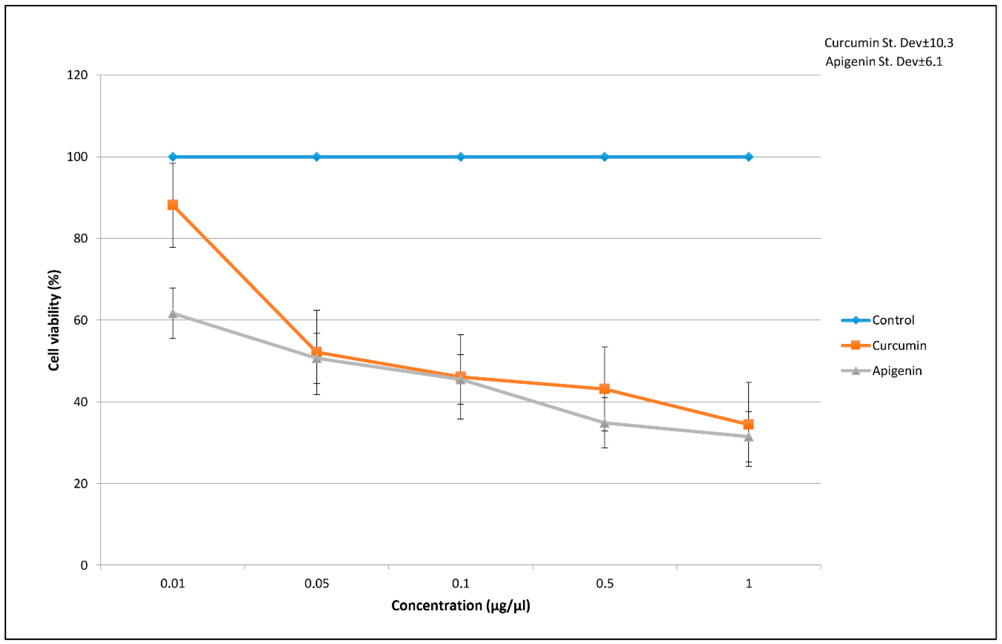
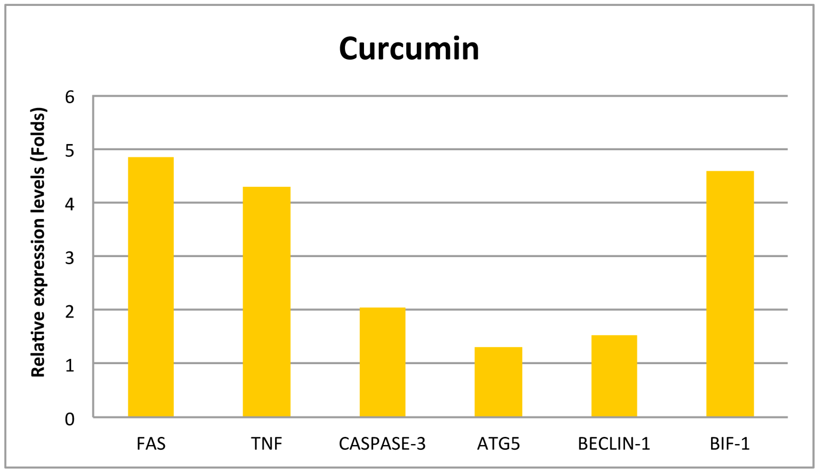
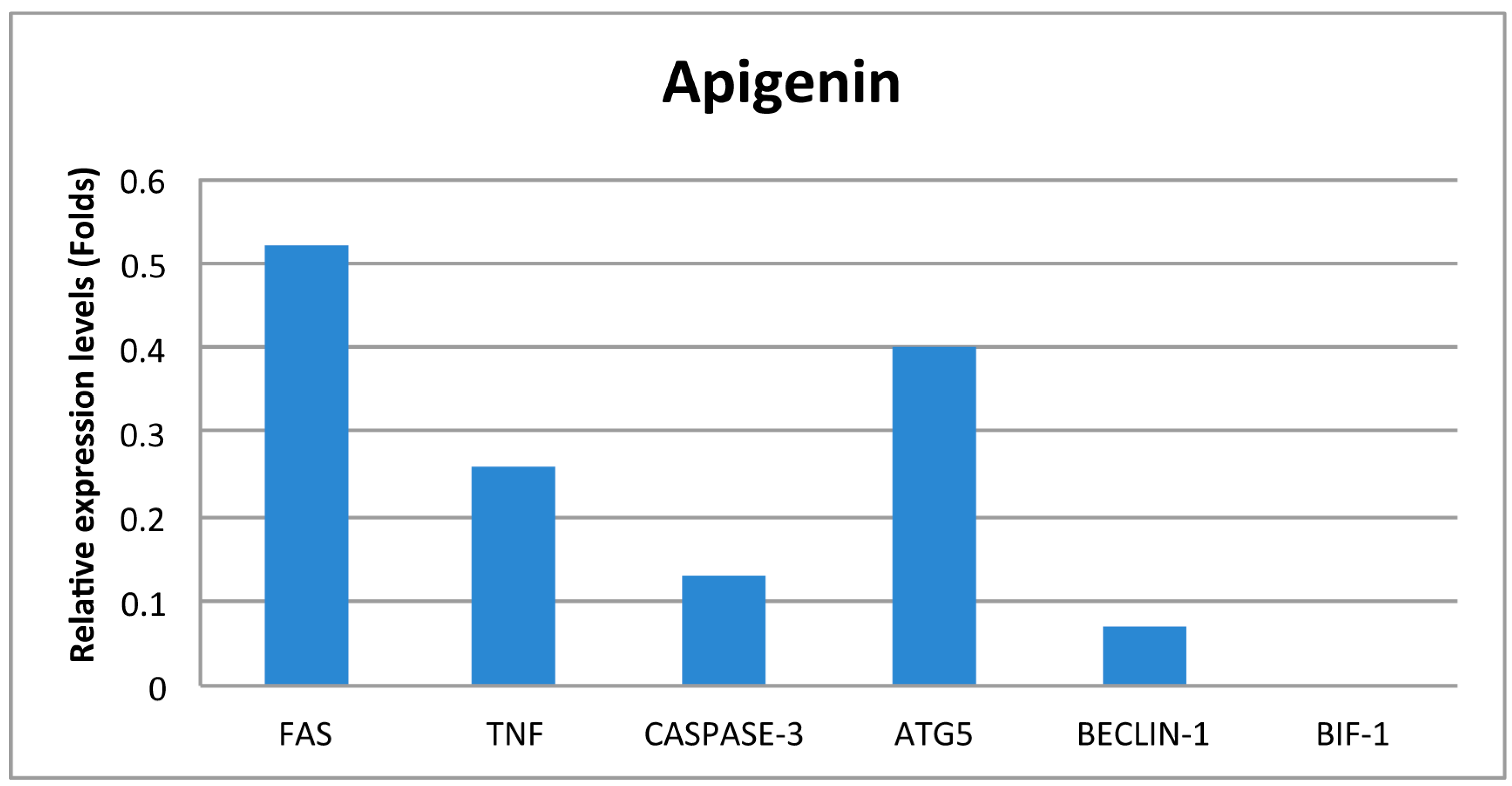
3. Results
4. Discussion
5. Conclusions
Author Contributions
Conflicts of Interest
References
- Salmani, J.M.M.; Zhang, X.P.; Jacob, J.A.; Chen, B.A. Apigenin’s anticancer properties and molecular mechanisms of action: Recent advances and future prospectives. Chin. J. Nat. Med. 2017, 5, 321–329. [Google Scholar] [CrossRef]
- Shehzad, A.; Lee, Y.S. Molecular mechanisms of curcumin action: Signal transduction. Biofactors 2013, 39, 27–36. [Google Scholar] [CrossRef]
- Galluzzi, L.; Vitale, I.; Abrams, J.M.; Alnemri, E.S.; Baehrecke, E.H.; Blagosklonny, M.V.; Dawson, T.M.; Dawson, V.L.; El-Deiry, W.S.; Fulda, S.; et al. Molecular definitions of cell death subroutines: Recommendations of the nomenclature committee on cell death 2012. Cell Death Differ. 2012, 19, 107–120. [Google Scholar] [CrossRef] [PubMed]
- Jain, V.M.; Paczulla, A.M.; Klonisch, T.; Dimgba, F.N.; Rao, S.B.; Roberg, K.; Schweizer, F.; Lengerke, C.; Davoodpour, P.; Palicharla, V.R.; et al. Interconnections between apoptotic, autophagic and necrotic pathways: Implications for cancer therapy development. J. Cell. Mol. Med. 2013, 17, 12–29. [Google Scholar] [CrossRef] [PubMed]
- Gordy, C.; He, Y.W. The crosstalk between autophagy and apoptosis: Where does this lead? Protein Cell 2012, 3, 17–27. [Google Scholar] [CrossRef] [PubMed]
- Kim, S.Y.; Song, X.; Zhang, L.; Bartlett, D.L.; Lee, Y.J. Role of Bcl-xL/Beclin-1 in interplay between apoptosis and autophagy in oxaliplatin and bortezomib-induced cell death. Biochem. Pharmacol. 2014, 88, 178–188. [Google Scholar] [CrossRef] [PubMed]
- Ozsoylemez, O.D.; Ozturk, M.; Sutlupinar, N.; Kayacan, S.; Tuncdemir, M.; Ozan, G. The effects of colchicum baytopiorum on regulatory genes of apoptotic and autophagic cell death in hela cells. Curr. Pharm. Biotech. 2016, 17, 1369–1376. [Google Scholar] [CrossRef] [PubMed]
- Takahashi, Y.; Karbowski, M.; Yamaguchi, H.; Kazi, A.; Wu, J.; Sebti, S.M.; Youle, R.J.; Wang, H.G. Loss of Bif-1 suppresses Bax/Bak conformational change and mitochondrial apoptosis. Mol. Cell. Biol. 2005, 25, 9369–9382. [Google Scholar] [CrossRef] [PubMed]


Publisher’s Note: MDPI stays neutral with regard to jurisdictional claims in published maps and institutional affiliations. |
© 2018 by the authors. Licensee MDPI, Basel, Switzerland. This article is an open access article distributed under the terms and conditions of the Creative Commons Attribution (CC BY) license (https://creativecommons.org/licenses/by/4.0/).
Share and Cite
Kayacan, S.; Yilancioglu, K.; Akdemir, A.S.; Dagistanli, F.K.; Melikoglu, G.; Ozturk, M. The Effects of Apigenin and Curcumin on Autophagy Related Cell Death and Apoptosis. Proceedings 2018, 2, 1586. https://doi.org/10.3390/proceedings2251586
Kayacan S, Yilancioglu K, Akdemir AS, Dagistanli FK, Melikoglu G, Ozturk M. The Effects of Apigenin and Curcumin on Autophagy Related Cell Death and Apoptosis. Proceedings. 2018; 2(25):1586. https://doi.org/10.3390/proceedings2251586
Chicago/Turabian StyleKayacan, Sera, Kaan Yilancioglu, Ayse Seda Akdemir, Fatma Kaya Dagistanli, Gulay Melikoglu, and Melek Ozturk. 2018. "The Effects of Apigenin and Curcumin on Autophagy Related Cell Death and Apoptosis" Proceedings 2, no. 25: 1586. https://doi.org/10.3390/proceedings2251586