Sensor and Embedded Control System for Liquid Crystal Implantable Eye Lens †


1
Department of Electrical Engineering, ESAT-MICAS, KU Leuven, 3001 Leuven, Belgium
2
Laboratory of Soft Matter and Biophysics, Department of Physics and Astronomy, KU Leuven, 3001 Leuven, Belgium
3
Department of Ophthalmology, University Hospitals Leuven, 3000 Leuven, Belgium
4
Eye and Refractive Center, 8900 Ieper, Belgium
*
Author to whom correspondence should be addressed.
†
Presented at the Eurosensors 2018 Conference, Graz, Austria, 9–12 September 2018.
Proceedings 2018, 2(13), 936; https://doi.org/10.3390/proceedings2130936
Published: 11 December 2018
(This article belongs to the Proceedings of EUROSENSORS 2018)
Abstract
:A miniature sensor and control system is developed to facilitate human eye ciliary muscle movement detection and to drive the corresponding liquid crystal based lens to create an autofocusing lens for cataract patients. The movement of the ciliary muscle is detected by a marker that detunes a Colpitts oscillator. The change in oscillation frequency is measured by the implantable circuit and sent to an external control unit. This external unit calculates the corresponding focal length and returns corresponding commands to the implantable system to change differential signal driving the lens. The system is built with state-of-the-art Commercial-Off-The-Shelf (C.O.T.S.) components around a miniature ultra-low power Filed Programmable Gate Array (F.P.G.A) and a hand full analog components. The system fits on a 10 mm outer diameter Printed Circuit Board (PCB), consumes less than 2.5 mW and is able to measure up to 1 mm ciliary muscle displacements.
1. Introduction
A well-known eye disease that blurs the human vision is cataract. It can be solved with minimal surgery, replacing the natural lens by e.g., PMMA/Silicone/Acrylic lens. Research about a miniature focal adjustable Liquid Crystal (LC) based lens [1] could lead to next generation active eye lens devices. This paper focusses on how to equip such an eye lens with minimal electronic circuit to detect ciliary muscle movement and to drive the LC based lens.
Therefore, a miniature sensor system and an embedded control system is developed for the next generation implantable eye lens, enabling natural autofocus behavior without physical contact between the lens and ciliary muscle/processes. This solution circumvents common problems with existing lens implants, like e.g., Z-syndrome [2], who use ciliary muscle lens implant contact to adjust focal distance in a mechanical way.
The prototype is built with state-of-the-art Commercial-Off-The-Shelf (C.O.T.S.) components around a miniature ultra-low power Field programmable Gate Array (FPGA). The system fits on a 10 mm diameter Printed Circuit Board (PCB) and consumes less than 2.5 mW. The system should be placed in the lens capsule as illustrated in Figure 1. The autofocus system is based on movement detection of a metallic marker placed on the ciliary muscle and is able to detect marker displacements of 1 mm. The developed system controls a first prototype liquid crystal lens to adapt the focal length.
2. Materials and Methods
2.1. Sensor Design and Simulation
In order to detect ciliary muscle movement a miniature magnetic-inductive, distance measurement Eddy current sensor is developed consisting in a miniature marker and Colpitts oscillator. The position of the marker will influence the magnetic environment of the miniature detection coil, changing the coils inductance and in turn the corresponding Colpitts oscillation frequency. Given limited space available in the human eye [3], marker sizes and materials were evaluated by a COMSOL Multiphysics® model (Figure 2). With a maximum marker size of 1.4 × 0.7 × 0.25 [mm] and a coil, with 2.5 mm outer diameter and 2 mm length, coil inductance and corresponding in oscillation frequency deviation where explored as shown in Figure 2b. The Colpitts frequency was tuned and set to 1 MHz oscillation frequency for the measurement experiment.
The marker’s shape was determined and designed to ease mounting on the ciliary processes of the ciliary muscle as shown in Figure 1. For a measurement experiment with Rhesus Monkey Eye, a set of markers were fabricated out of high Mu stainless steel from MuMETAL® by laser cutting, as illustrated in Figure 3a. Non-ferro markers will be used later for biocompatible reasons and will result in less but still measurable frequency shifts.
2.2. Embedded Control System
To control the LC based lens, a digital VHDL based system was developed and implemented in the Lattice ICE40UL1K16 Ultra Low (UL) Power smallest FPGA (1.48 mm × 1.48 mm µBGA). The system, contains an 8bit ATMEL AT90S2313 alike AX8 soft-core processor acting as a programmable state machine, a frequency counter to determine the Colpitts frequency, a bi-phase pulse width modulator to drive the LC lens. The system includes also a universal asynchronous serial communication unit to communicate with an external control unit. Only 8 internal work registers and 128 16bit wide program words are used to implement the full system functionality. The implantable system is controlled by the external unit via inductive communication and by calling one out of 6 direct or 5 indirect callable embedded functions. The FPGA system runs on a 1.2 V core voltage and 3.3 V I/O voltage and consumes less than 200 µW. The digital design uses 93% of the logical tiles and only 22% of internal FPGA block RAM which contains the embedded assembly code.
A limited number of analog components are used to implement the required powering, ASK demodulation, load modulation and provide low power system clock generation. Full block diagram is shown in Figure 4. Two LDO39130 Low Drop voltage regulators are used to provide 1.2 V FPGA core and 3.3 V system voltage. A SiT8021 based micro power MEMS oscillator chip provides the system clock. The Colpitts oscillator is built around a NESG2046M33 NPN SiGe transistor and the output is fed via a TS985 based Schmitt trigger into the FPGA. The entire analog circuitry only consumes around 2.2 mW.
The ASK demodulation circuit is implemented with classic Schottky diode RC circuit followed by dual MAX40100 op-amp high pass filter circuit and TS985 Schmitt trigger circuit extract and deliver the Rx signal to de FPGA. Load modulation, to transmit data to the external device is built with a simple logic level CSD13380 MOS transistor and load resistor.
3. Results and Discussions
The final semi-rigid PCB design is shown in Figure 5. The central FPGA is programmable via a disposable flex programing interface connector. The current design fits in a human eye, requires an amount of power in the mW range, and fulfils required functionality. Measurements have shown acceptable but less frequency deviation than expected. This could be attributed to the fact that the markers have less volume and surface in comparison with the simplified marker beam model used in COMSOL. Further miniaturization is possible by using e.g., state-of-the-art embedded PCB components if available form electronic manufactures.
4. Conclusions
In this paper an embedded control system for LC based eye lens implant was proposed. The system is built upon a hand full of state-of-the-art C.O.T.S. components. The final design is miniaturized to fit the confined space of a human eye, consuming less than 2.5 mW. This work is a step towards next generation active implantable eye lens system.
Future work will focus on possible circuit improvement, lens integration, packaging and further development of the required inductive powering and communication glasses.
Acknowledgments
Authors would like to express their gratitude towards KU Leuven master student Nick De Strycker and Thomas More Colleague Jurre De Weerdt for support in the Bionic Eye project. The research leading to these results has received funding from the European Research Council under the European Union’s Seventh Framework Program (FP7/2007–2013)/ERC grant agreement n° 340931.
Conflicts of Interest
The authors declare no conflict of interest.
References
- Doornaert, D.; De Gresem, H.; Glorieux, C.; Puers, B.; Spileers, W.; Blankaert, J. Intraocular Electro-Optic Lens with Ciliary Muscle Controlled Accommodation. In Proceedings of the 35th Annual International Conference of the IEEE Engineering in Medicine and Biology Society—EMBC, Osaka, Japan, 3–7 July 2013; pp. 3190–3193. [Google Scholar]
- Yuen, L.; Trattler, W.; Wachler, B.S.B. Two cases of Z syndrome with Crystalens after uneventful cataract surgery. J. Cataract. Refract. Surg. 2008, 34, 1986–1989. [Google Scholar] [CrossRef] [PubMed]
- Doornaert, D.; Glorieux, C.; Puers, R.; De Gresem, H.; Spileers, W.; Blankaert, J. Physiological constraints for an intraocular inductive distance sensor. In Proceedings of the 36th Annual International Conference of the IEEE Engineering in Medicine and Biology Society—EMBC, Chicago, IL, USA, 26–30 August 2014; pp. 646–649. [Google Scholar]
Figure 1.
An illustration, based on Heidelberg engineering Eye-Anterior-Description of the developed implantable eye lens system.
Figure 1.
An illustration, based on Heidelberg engineering Eye-Anterior-Description of the developed implantable eye lens system.
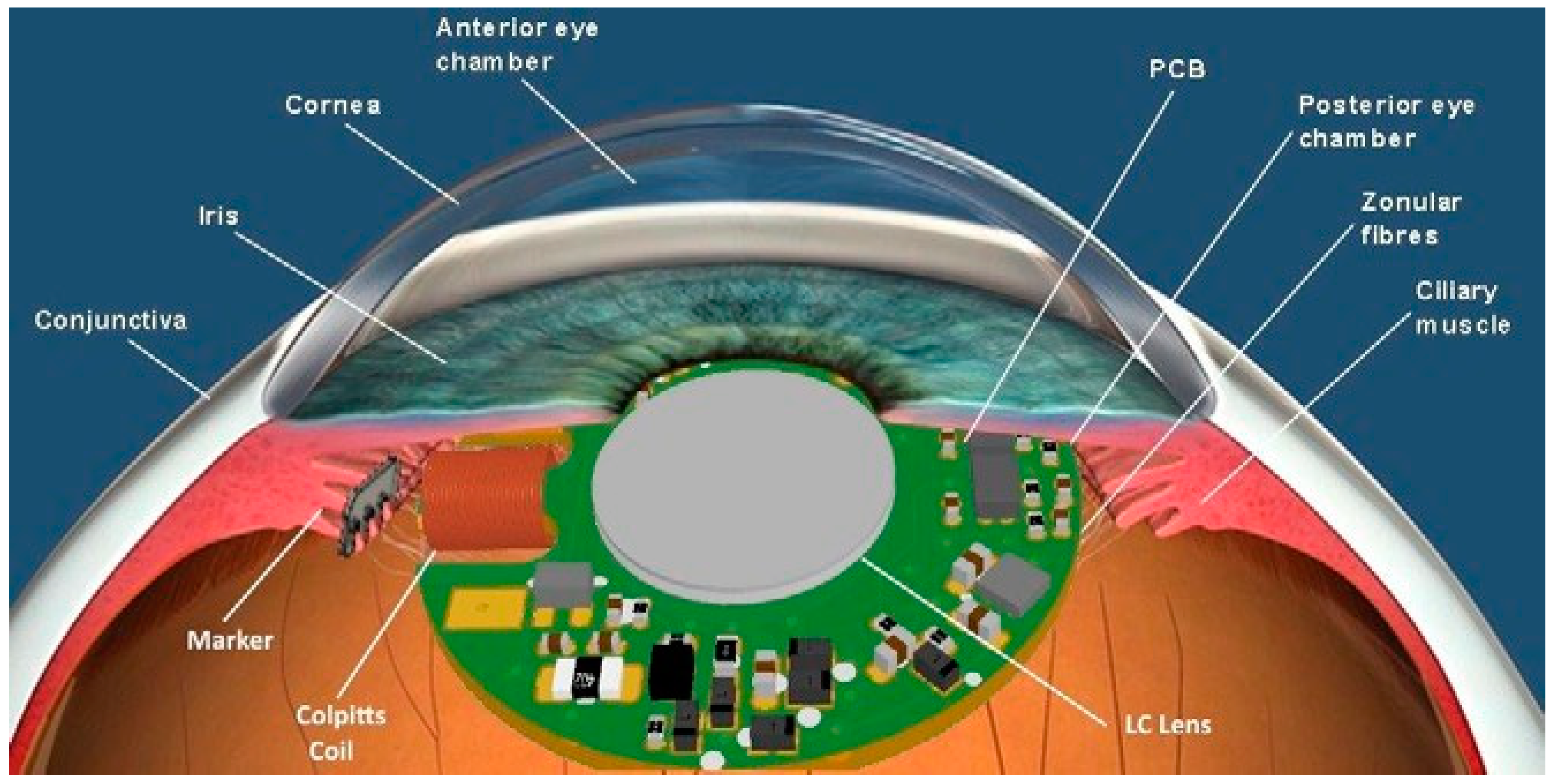
Figure 2.
(a) COMSOL coil-marker simulation to determine magnetic behavior and coil inductance change. With a. the ½ modeled coil and b. the marker; (b) Simulated Colpitts frequency deviation as a function of marker to coil distance.
Figure 2.
(a) COMSOL coil-marker simulation to determine magnetic behavior and coil inductance change. With a. the ½ modeled coil and b. the marker; (b) Simulated Colpitts frequency deviation as a function of marker to coil distance.
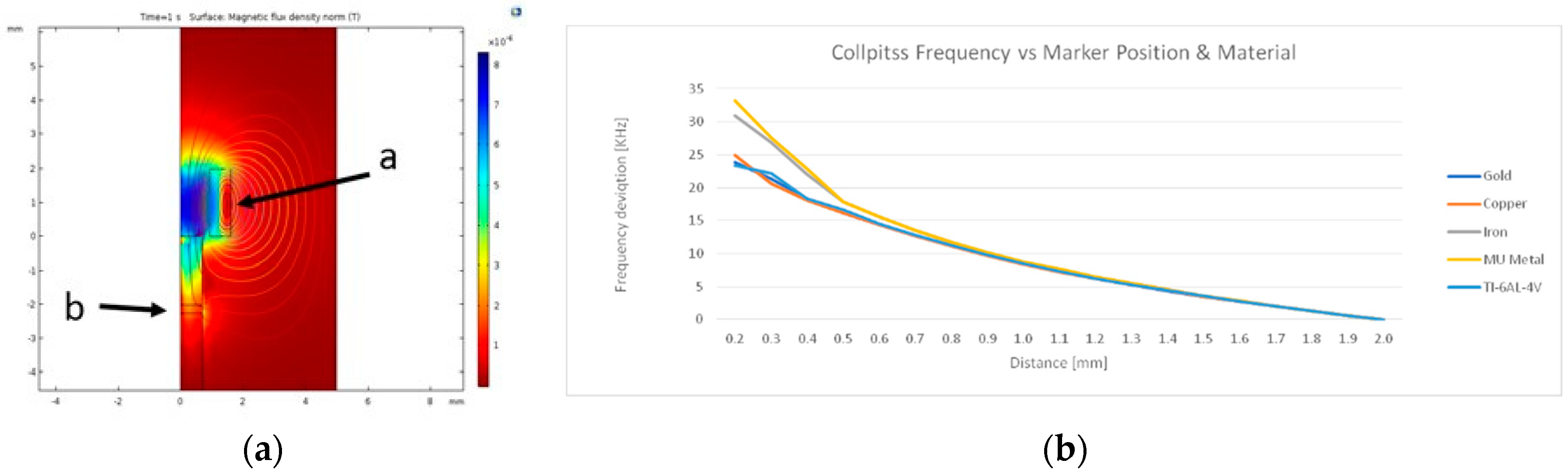
Figure 3.
(a) A set of different laser cut markers on top of a Eurocent coin to illustrate marker size; (b) Marker mounted on ciliary processes of a Rhesus monkey eye. With a. corpus ciliary; b. equator lens; c. anterior capsule and d. top view of the marker. (c) With a. NESG2046M33 based Colpitts oscillator and b. the wire wound detection coil.

Figure 4.
Block schematic of the full embedded control system.
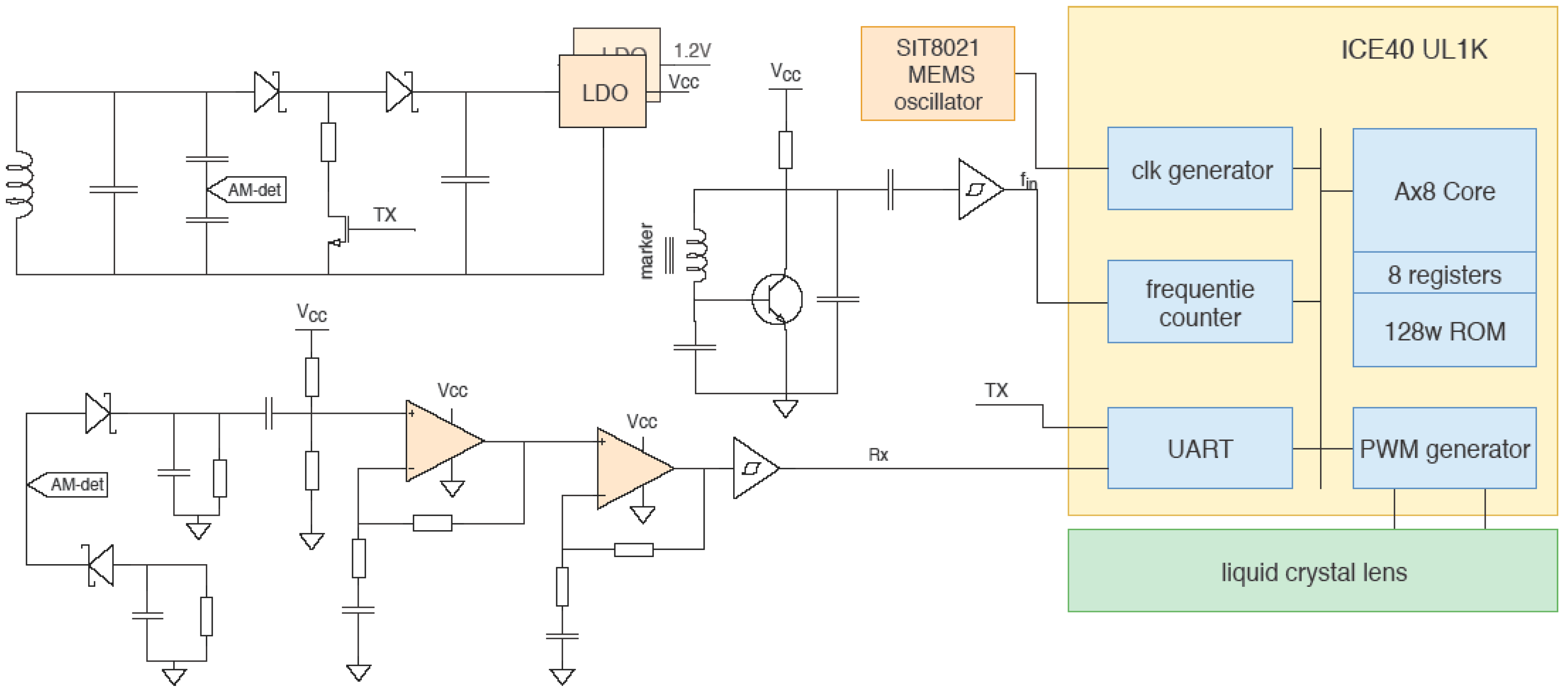
Figure 5.
Final Semi-Rigid PCBD design result with flex interface for FPGA programming.

Publisher’s Note: MDPI stays neutral with regard to jurisdictional claims in published maps and institutional affiliations. |
© 2018 by the authors. Licensee MDPI, Basel, Switzerland. This article is an open access article distributed under the terms and conditions of the Creative Commons Attribution (CC BY) license (https://creativecommons.org/licenses/by/4.0/).
Share and Cite
MDPI and ACS Style
Pelgrims, P.; Glorieux, C.; Blanckaert, J.; Puers, R. Sensor and Embedded Control System for Liquid Crystal Implantable Eye Lens. Proceedings 2018, 2, 936. https://doi.org/10.3390/proceedings2130936
AMA Style
Pelgrims P, Glorieux C, Blanckaert J, Puers R. Sensor and Embedded Control System for Liquid Crystal Implantable Eye Lens. Proceedings. 2018; 2(13):936. https://doi.org/10.3390/proceedings2130936
Chicago/Turabian StylePelgrims, Patrick, Christ Glorieux, Johan Blanckaert, and Robert Puers. 2018. "Sensor and Embedded Control System for Liquid Crystal Implantable Eye Lens" Proceedings 2, no. 13: 936. https://doi.org/10.3390/proceedings2130936