Article

Changes in the Serum Biochemical Indices and Intestinal Microbial Community of Rabbitfish (Siganus oramin) in Three Different Habitats

1
Key Laboratory of South China Sea Fishery Resources Exploitation & Utilization, Ministry of Agriculture and Rural Affairs, South China Sea Fisheries Research Institute, Chinese Academy of Fishery Sciences, Guangzhou 510300, China
2
National Agricultural Experimental Station for Fishery Resources and Environment Dapeng, Shenzhen Base of South China Sea Fisheries Research Institute, Chinese Academy of Fishery Sciences, Shenzhen 518121, China
*
Authors to whom correspondence should be addressed.
Fishes 2024, 9(3), 96; https://doi.org/10.3390/fishes9030096
Submission received: 18 January 2024 / Revised: 21 February 2024 / Accepted: 1 March 2024 / Published: 4 March 2024

Abstract

:
The rabbitfish Siganus oramin is an important naturally caught and aquaculture fish species. Intestinal microbiota can affect the metabolism and immunity of fish, which is closely related to the habitat of the host. In this study, we collected the wild fry S. oramin from a natural sea area, and cultured them in outdoor and indoor ponds, respectively, and investigated the changes in serum biochemical indexes and intestinal microbial community in three different habitats. The results showed that compared with the wild population, the serum total protein content of the outdoor culture population increased significantly. The indoor culture population had significantly higher triglyceride and total cholesterol contents than that of the outdoor culture population. Additionally, the intestinal microbial richness indexes ACE and Chao1 of the cultured population were higher than those of the wild population, especially the indoor culture, but Shannon and Simpson had no obvious changes. The relative abundances of Firmicutes, Spirochaetae and Bacteroidetes increased in the outdoor culture population, but decreased in the indoor culture population; those of Proteobacteria and Cyanobacteria were completely the opposite. Some putative beneficial bacteria (Lactobacillus, Clostridium sensu stricto 1, and Intestinibacter) and harmful bacteria (Vibrio, Photobacterium, Desulfovibrio, Streptococcus) all decreased in the intestines of the cultured population when compared with the wild population, while Akkermansia and Faecalibacterium were enriched in the outdoor culture population. These results reveal that a change in habitat environment, whether an outdoor or an indoor pond, positively influenced the intestinal microbiota of the rabbitfish, which is beneficial to the healthy culture of the fish from the perspective of microbial community.
Key Contribution: A change in the habitat environment affects the serum biochemical indexes and microbial community of the rabbitfish, which is beneficial to the healthy culture of the fish from the perspective of microbial community.

1. Introduction

The rabbitfish (Siganus oramin), also known as S. canaliculatus, is a small coastal economic fish found in tropical and subtropical sea areas. S. oramin is an important fish species, both caught and cultured, and it is widely distributed in the South China Sea, the southern East China Sea, and the Taiwan Strait in China [1]. S. oramin is a omnivorous fish, which has the advantages of a short feeding cycle and low breeding cost, giving it great market development potential. In recent years, with the continuous expansion of market demand, the culture methods of S. oramin have gradually diversified, ranging from mixed culture to single culture, and from cage culture to outdoor pond culture and indoor pond culture. Consequently, the farming output of S. oramin has been increasing year by year [2]. Therefore, exploring better farming techniques for rabbitfish can enhance its industrial development.
The intestines of fish harbor a large number of microbial communities that are closely related to their growth, development, and production [3]. Fish inhabit aquatic environments, and the composition of their intestinal microbiota is closely linked to their living environment, physiological state, and developmental stage [4]. A balanced intestinal microflora plays a crucial role in aiding fish in digesting and absorbing nutrients, regulating immune defense and metabolic function, and maintaining overall organismal health [5]. Conversely, an imbalanced intestinal microflora can lead to physiological disorders of the host and increased susceptibility to diseases [6]. It is shown that the intestinal microbiota of basket fish is related to its rearing water and different diets [7,8]. Exploring the characteristics of fish intestinal microbiota in different habitats holds significant importance for optimizing aquaculture techniques and preventing diseases.
Blood parameters have been widely utilized for assessing the health status of cultured fish, due to their ability to provide reliable information on metabolic disorders, deficiencies, and chronic stress conditions before clinical symptoms manifest [9]. Several studies on various fish species, including Schizothorax [10], Dicentrarchus labrax [11], and Misgurnus anguillicaudatus [12], have revealed that aquaculture practices have a significant impact on the physiological and biochemical aspects of fish blood. The differences in nutritional status, physiological condition, and overall health of fish in different environments contribute to variations in blood parameters, as blood is linked to the body’s metabolic processes. When fish undergo physiological changes due to external factors, these changes are reflected in the physiological indicators of their blood [13].
However, currently, there is still a lack of research on the physiological changes and differences in intestinal microbiota of S. oramin in different habitats. In this study, we collected wild S. oramin fry from marine habitats, and reared them for 100 days in both outdoor and indoor ponds, respectively. Then, using biochemical analysis and high-throughput sequencing of 16S rDNA, we investigated the changes in blood biochemical indicators and intestinal microbial communities of S. oramin in these three different habitats, namely the wild environment of natural sea area, outdoor pond culture, and indoor pond culture. Our findings contribute to the cultivation of S. oramin and the exploration of intestinal microbial resources.

2. Materials and Methods

2.1. Experimental Design and Feeding Trial

S. oramin fry with an average 1.28 ± 0.02 g of initial weight were collected from the natural sea area of Daya Bay in Shenzhen, China (latitude N 22°37′34″, longitude E 114°4′06″) in June 2020. These wild population (WP) fish were immediately transferred to an outdoor pond (OP) and indoor pond (IP) for cultivation. The indoor pond was an industrial recirculating aquaculture system (IRAS) with eight 30 m2 culture ponds, and the culture density was 70–80 fish per m2, while the outdoor pond was a 667 m2 high pond laying bottom membrane, with a culture density of 40–45 fish per m2. The fish were fed twice a day (at 8:00 and 18:00) with compound feed containing nutritional ingredients such as crude protein (44.1%), crude fat (9.4%), ash (12.8%), and moisture (8.0%). The seawater used for the culture ponds was directly sourced from the same sea area in Daya Bay and underwent sand filtration for the rearing water. Throughout the experiment, the water quality of both the indoor and outdoor ponds, as well as the sea area, was continuously monitored (Table S1). The culture experiment was conducted at the Shenzhen Base of South China Sea Fisheries Research Institute, Chinese Academy of Fishery Sciences (Shenzhen, China).

2.2. Sample Collection

After 100 days of fish culture, samples were collected for this study. Three groups were included: the wild population (WP) group, indoor pond (IP) group, and outdoor pond (OP) group. At the 100th day, the average body weight of the fish in the IP group was 82.96 ± 10.02 g, while that of the OP group was 73.08 ± 12.74 g. Remote sensing monitoring technology was used to determine the location of the initial wild population, which were collected during the same period, and served as the control (WP group), with an average body weight of 53.62 ± 3.48 g.
Healthy fish from each group were randomly selected for sampling, ensuring that their body surfaces showed no signs of injury or infection. Euthanasia was performed using 10 mg/L eugenol (Shanghai Medical Instruments Co., Ltd., Shanghai, China). Venous blood was collected from 9 fish using sterile syringes and combined to create one sample. The sample was allowed to stand at 4 °C for 1 h, followed by centrifugation (3000 rpm, 10 min, 4 °C) to collect the serum for biochemical index determination. The complete intestines of these nine fish were sampled and combined to create one microbial sample. Each group consisted of three serum samples and three microbial samples. All the serum and intestinal microbial samples were stored at −80 °C until further analysis. All the animal processes followed strictly the protocols for the Animal Care and Use Committee of the South China Sea Fisheries Research Institute, Chinese Academy of Fishery Sciences.

2.3. Biochemical Analysis

The serum samples were dissolved in ice, and then the biochemical indexes were determined with a Beckman automatic biochemical analyzer (HITACHI-7180), including total protein (TP), albumin (ALB), glucose (GLU), triglyceride (TG), total cholesterol (TC), high-density lipoprotein (HDL-C), low-density lipoprotein (LDL-C), and alkaline phosphatase (ALP), alanine aminotransferase (ALT), and aspartate aminotransferase (AST).

2.4. 16S rDNA Sequencing of Intestinal Microbiota

The genomic DNA of intestinal microbiota from each group was extracted using the Fast DNA SPIN kit (MPBIO, CA, USA). After assessing the quality and concentration, the purified DNA was used to amplify the V3–V4 fragment of the 16S rDNA gene of intestinal microbes using specific primers. PCR amplification was conducted using the Phusion High-Fidelity PCR Master Mix Kit (New England Biolabs, Ipswich, MA, USA). The qualified PCR products were pooled in equal proportions and purified using the Gene Jet Gel Extraction Kit (Thermo Scientific, Waltham, MA, USA). Subsequently, a sequencing library was constructed using the MetaVx™ Library Construction Kit (South Plainfield, NJ, USA), and the sequencing process was carried out on the Illumina MiSeq platform.

2.5. Bioinformatics Analysis of Intestinal Microbiota

The obtained reads were filtered using FLASH v1.2.7 software and assigned to each sample based on the unique barcode. Sequence analysis was performed using UPARSE v10 software, and operational taxonomic units (OTUs) were defined with a similarity threshold of ≥97% [14]. α-diversity was evaluated using Mothur v1.30.1 software with ACE, Chao1, Simpson, and Shannon indexes. β-diversity was assessed by performing PCA polt using the R software v3.4.4 package. The relative abundance of intestinal bacterial communities was analyzed at the phylum and genus levels. Differential bacterial taxa among the three groups were identified using linear discriminant analysis effect size (LEfSe) [15]. Additionally, based on the abundance of bacterial OTUs, the correlation and difference networks of the intestinal microbiota were constructed using Cytoscape software (http://www.cytoscape.org, accessed on 21 Feburary 2024). The metabolic function of the intestinal microbiota was predicted using the PICRUSt 2.0 software.

2.6. Statistical Analysis

Statistical analysis of the intestinal microbiota such as bacterial diversity, LEfSe, and PICRUSt were conducted using R package v3.4.4 software. PICRUSt analyses were performed based on the KEGG level 3 using the R RandomForest package. Biochemical index data were analyzed by one-way ANOVA using SPSS 17.0 software, and the data were presented as mean ± SE. When significant differences were observed, multiple comparisons between groups were performed using Dunnett’s test. Significance was set at p < 0.05.

3. Results

3.1. Changes in Serum Biochemical Indexes

The serum biochemical indexes of the fish in the three habitats are shown in Table 1. Compared with the WP group, the TP content was increased in the OP group (p < 0.05). The content of ALB was decreased in the IP group, while ALP and ALT were increased in the OP and IP groups. GLU, TG, TC, LDL-C, and AST were decreased in the OP group but increased in the IP group, while HDL-C was increased in the OP group but decreased in the IP group, although these changes were not significant (p > 0.05).

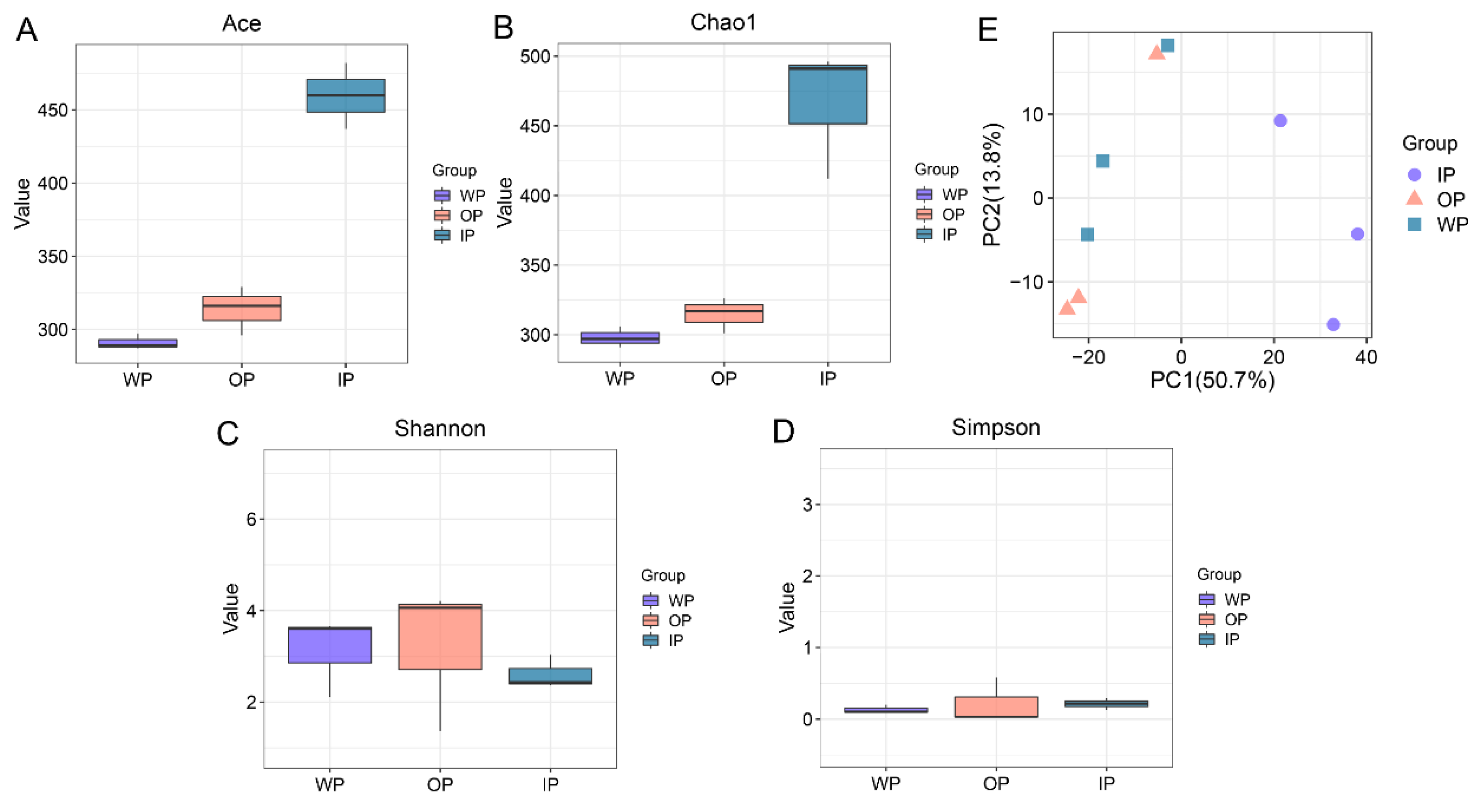
3.2. Changes in the Diversity of Intestinal Microbiota

After performing 16S rDNA sequencing, a total of 1,024,693 raw reads were obtained from the nine microbial samples of the three groups, and 700,163 clean reads were generated, with an average of 227,710 clean reads per sample (Table S2). After quality control, 51,612 clean reads per sample were used for bioinformatics analysis, and the sequencing depth was effective (Figure S1). A total of 657 OTUs were identified in this study, including 354 OTUs, 388 OTUs, and 527 OTUs in the WP, OP, and IP groups, respectively (Figure S2, Table S2). Among them, 245 OTUs were shared by the three groups, with the highest number of unique OTUs observed in the IP group, followed by the OP group and the WP group. The α-diversity indices of intestinal microbes, such as ACE and Chao1, were higher in the OP and IP groups compared to the WP group, and the highest level was in the IP group (Figure 1A,B). The Shannon index increased in the OP group but decreased in the IP group, while the Simpson index showed the opposite trend (Figure 1C,D). But these α-diversity data were not statistically significant (p > 0.05). The β-diversity based on the PCA plot revealed distinct patterns of intestinal microbiota among the three groups, with a closer relationship observed between the OP and WP groups (Figure 1E).

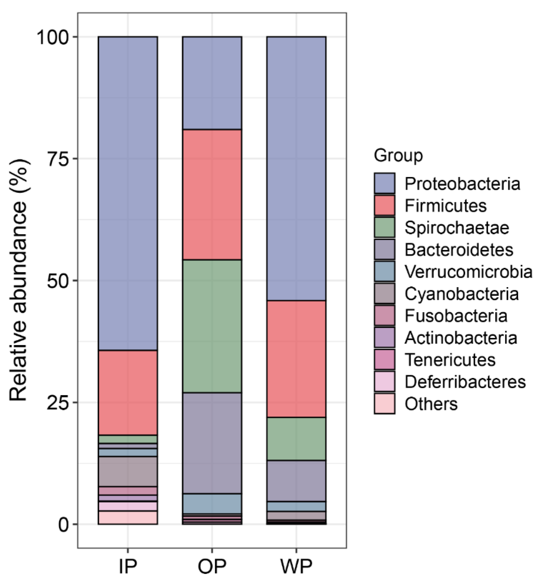
3.3. Changes in the Community Composition of Intestinal Microbiota

The abundance of dominant intestinal bacteria varied across different habitats. Twenty bacterial phyla were identified, with Proteobacteria, Firmicutes, Spirochaetae, and Bacteroidetes being the most dominant. At the phylum level, compared to the WP group, the relative abundances of Firmicutes, Spirochaetae, and Bacteroidetes were increased in the OP group but decreased in the IP group. Conversely, the relative abundances of Proteobacteria and Cyanobacteria were decreased in the OP group but increased in the IP group (Figure 2).
Furthermore, the relative abundances of several intestinal bacterial genera differed among the three groups, and the top 50 bacterial genera are listed in Figure S3. Among these bacteria, we mainly paid attention to several potentially beneficial or harmful bacteria. For example, in comparison to the WP group, Akkermansia and Faecalibacterium were increased in the OP group, but decreased in the IP group; Vibrio, Photobacterium, Streptococcus, Clostridium sensu stricto 1, Lactobacillus, Desulfovibrio, and Intestinibacter were decreased in the OP and IP groups (Figure 3).

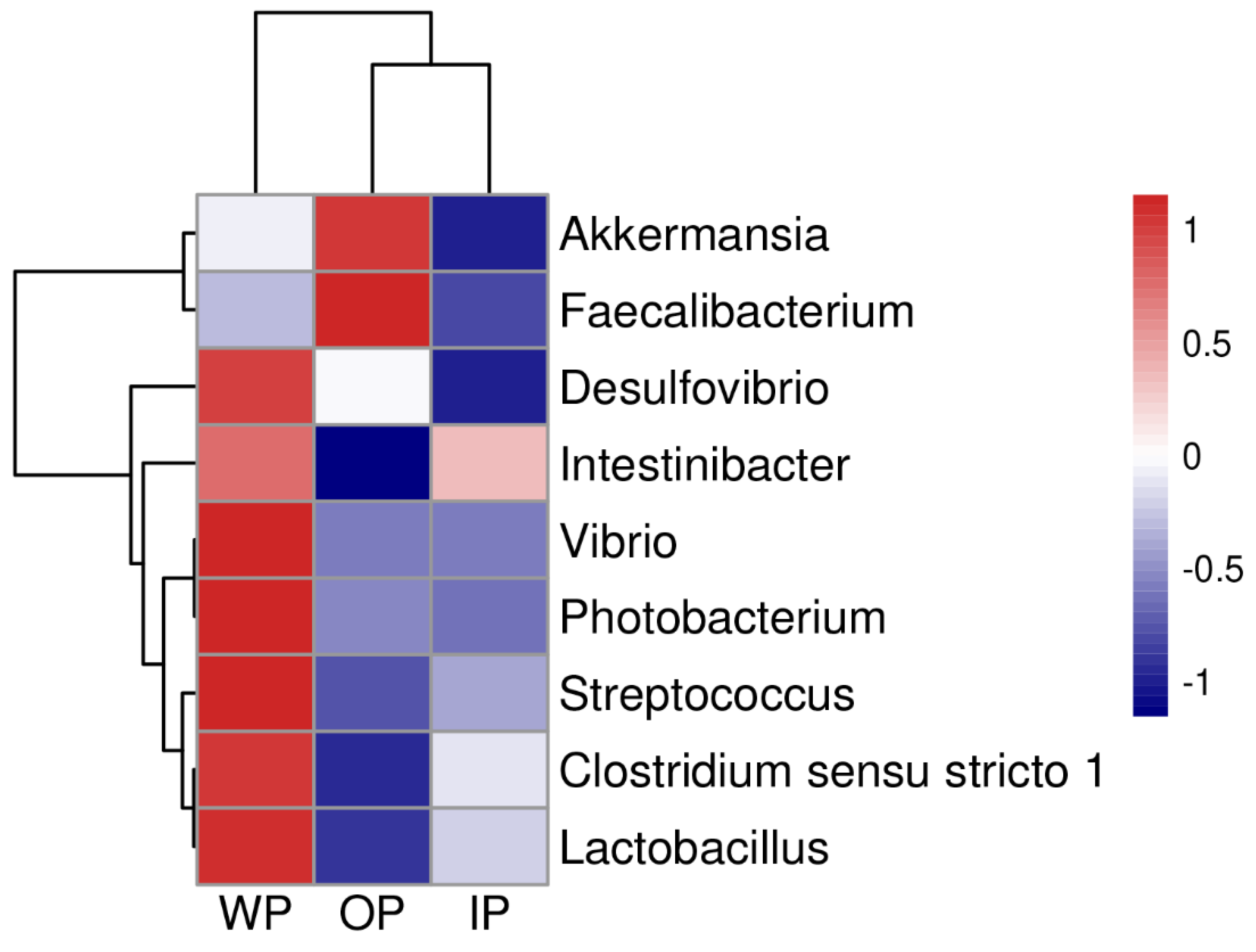
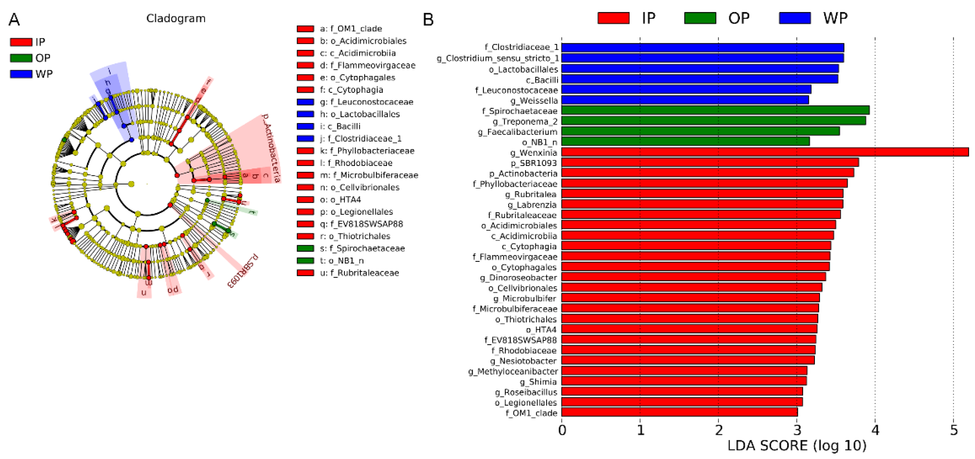
3.4. Differential Analysis of Intestinal Bacteria

LEfSe analysis further revealed the differences in intestinal bacterial composition among the different habitats. The families Clostridiaceae and Leuconostocaceae were enriched in the WP group, while the family Spirochaetaceae was enriched in the OP group. Additionally, the families Flammeovirgaceae, Microbulbiferaceae, Phyllobacteriaceae, and Rhodobiaceae were enriched in the IP group (Figure 4A). Based on an LDA score greater than 3.0, the genera Clostridium sensu stricto 1 and Weissella were enriched in the WP group, while Faecalibacterium and Treponema 2 were enriched in the OP group. Moreover, the genera Dinoroseobacter, Labrenzia, Methyloceanibacter, Microbulbifer, Nesiotobacter, Roseibacillus, Rubritalea, Shimia, and Wenxinia were enriched in the IP group (Figure 4B).

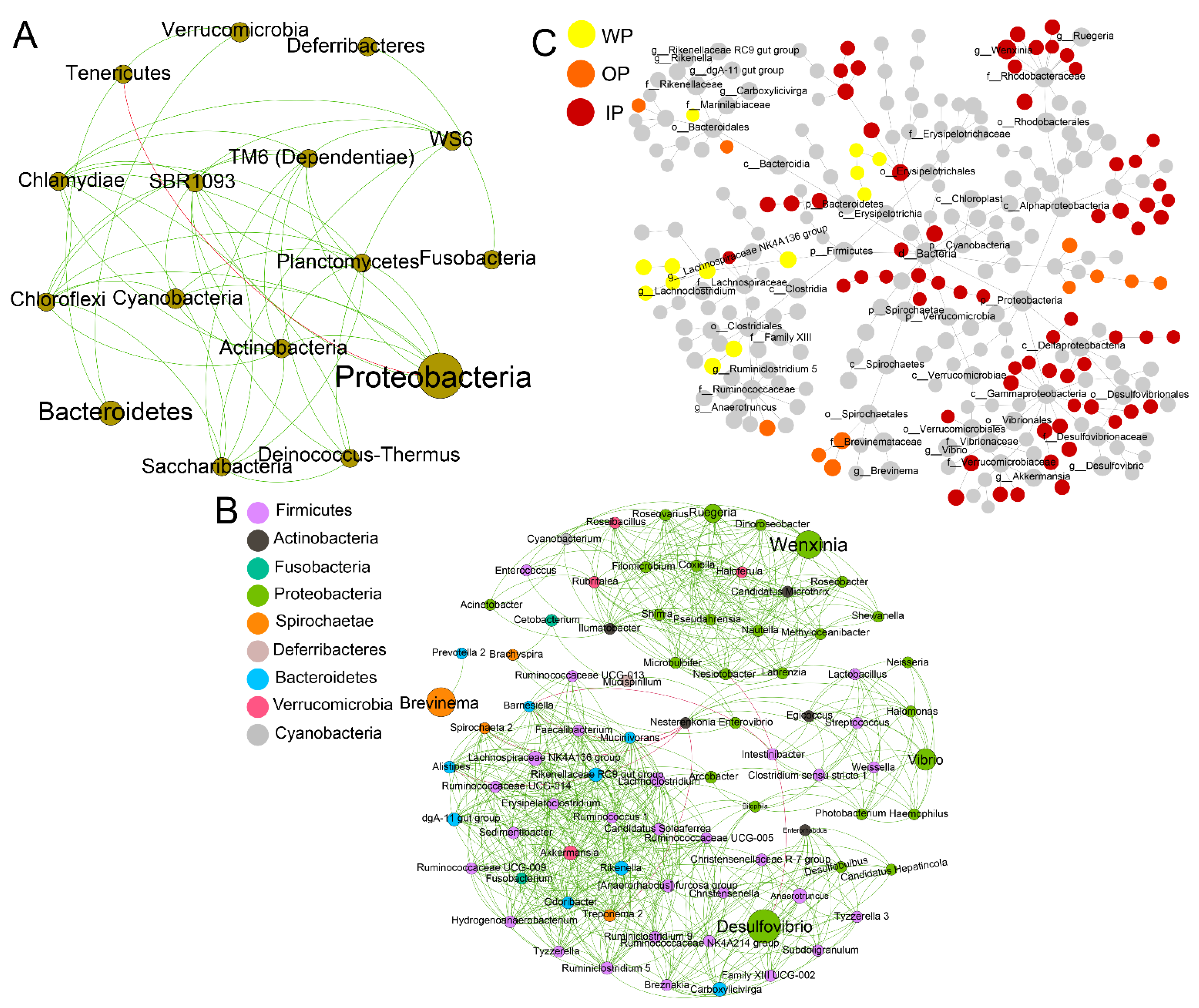
3.5. Network Relationships of Intestinal Microbial Community

The correlation and differential networks of the intestinal microbiota were further analyzed. At the phylum level, Tenericutes, Bacteroidetes, and Verrucomicrobia exhibited positive correlations with each other, while Tenericutes showed a negative correlation with Proteobacteria (Figure 5A). At the genus level, Streptococcus had a positive correlation with Lactobacillus, Clostridium sensu stricto 1, and Intestinibacter. Similarly, Photobacterium was positively correlated with Lactobacillus and Clostridium sensu stricto 1. Furthermore, Streptococcus exhibited a positive correlation with Photobacterium (Figure 5B). In the differential network, Wenxinia dominated in the IP group (Figure 5C).

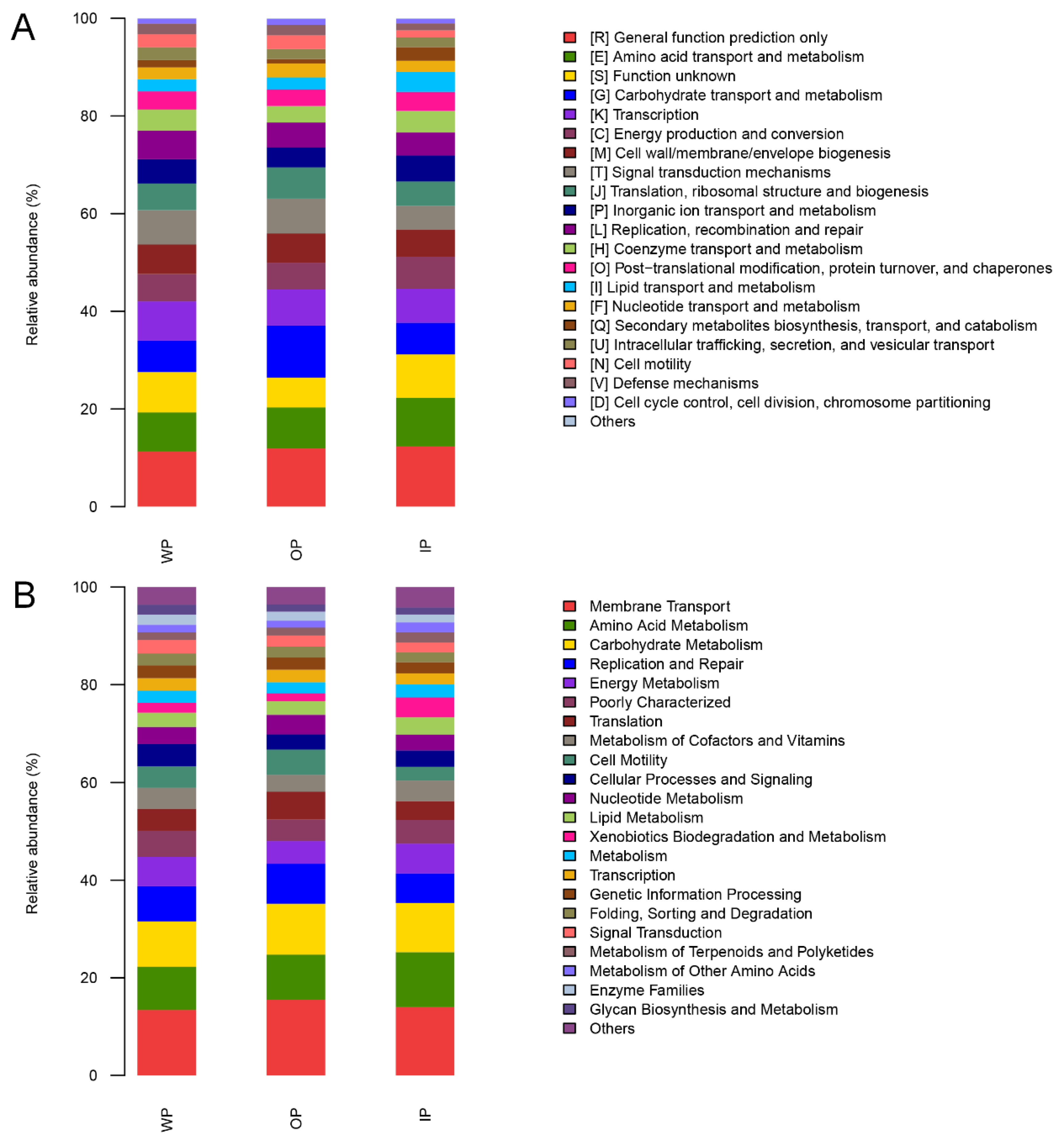
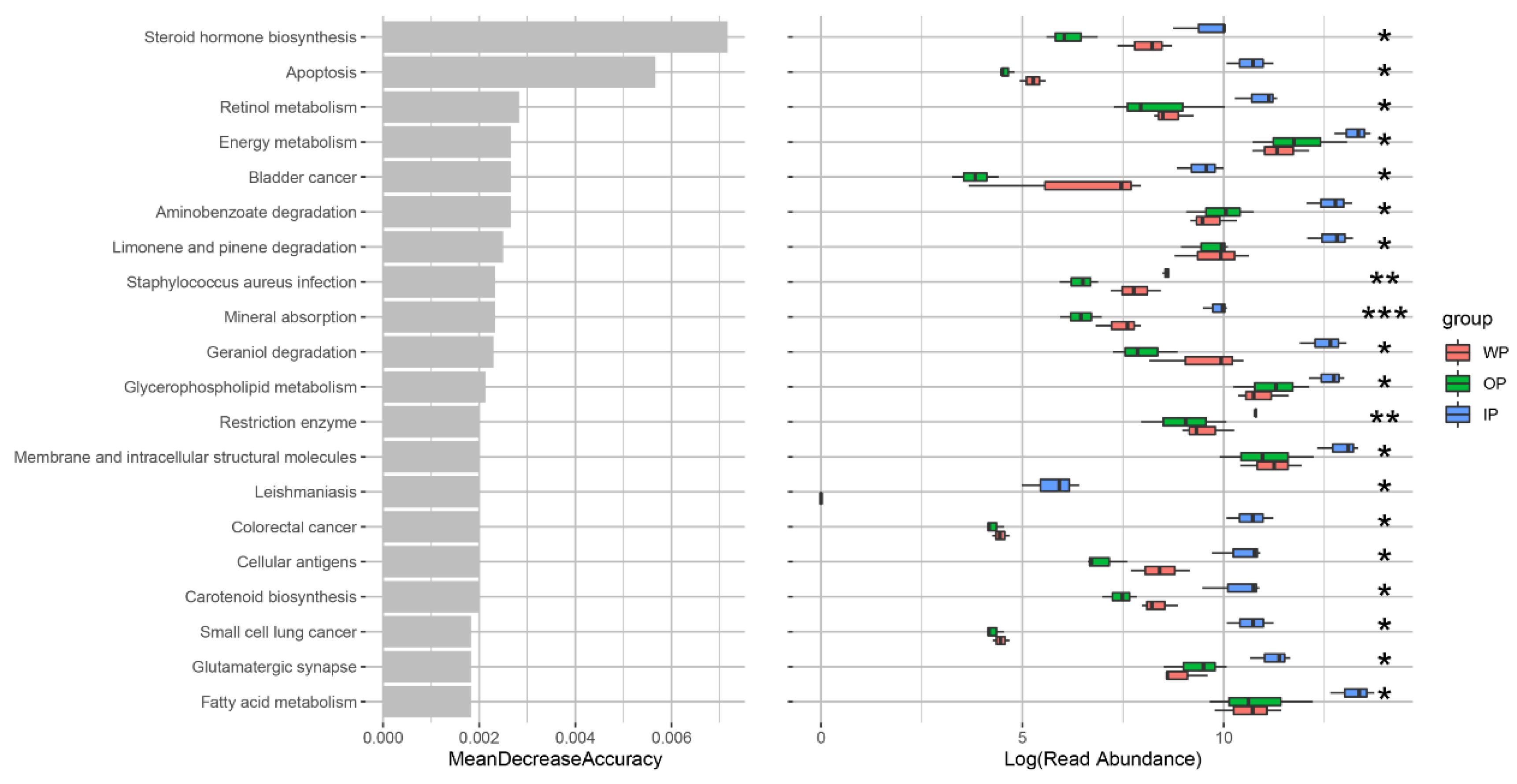
3.6. Changes in Intestinal Microbial Metabolic Function

Based on COG and KEGG annotations, the functions of the intestinal microbiota were primarily related to “membrane transport”, “amino acid metabolism”, “carbohydrate metabolism”, and “energy metabolism”, etc. (Figure 6). Compared with the WP group, the functions of “energy metabolism” and “glycerophospholipid metabolism” were increased in the OP and IP groups (p < 0.05). Furthermore, the functions of “mineral absorption”, “steroid hormone biosynthesis”, “carotenoid biosynthesis”, “retinol metabolism”, and “fatty acid metabolism” were increased in the IP group but decreased in the OP group (p < 0.05) (Figure 7).

4. Discussion

4.1. Response of Serum Biochemical Indicators to Three Different Habitats

Serum biochemical indicators are considered as important clinical diagnostic criteria, often used to estimate the health status of fish and assess the impact of natural stressors. Factors such as nutritional status (artificial feed versus natural prey) and stocking density directly affect the health of animals, leading to changes in blood and liver parameters [16,17]. In the wild, the fish face harsh survival conditions, including hunger, predation pressure, slow growth, and low condition factor. In the indoor pond culture, density, operational, and nutritional stress from a monotonous diet result in fast growth and a high condition factor. Outdoor pond culture, on the other hand, entails relatively less stress, where fish can feed on both natural algae and artificial feed, avoiding dietary restrictions and hunger stress, resulting in better overall health conditions. These ecological variations often lead to significant fluctuations in various blood indices.
TG and TC are commonly used as important indicators to evaluate the health of fish, with increased levels indicating lipid and lipoprotein metabolism disorders [12]. HDL-C, synthesized mainly in the liver, transports excess lipids and cholesterol from the bloodstream to the liver for breakdown, thus acting as a scavenger for blood waste [18]. LDL-C concentration is closely associated with the occurrence of cardiovascular diseases. Reducing LDL-C in fish can reduce the accumulation of cholesterol in the liver and blood vessels, thereby improving overall health [19]. In this study, compared with the wild fish, serum TG, TC, and LDL-C levels were significantly higher in the indoor pond cultured fish, while HDL-C was lower, indicating that their blood lipid conditions and body health level were worse than those of wild fish. Similar results have been found in studies of Culter alburnus [20], Misgurnus anguillicaudatus [12], and Dicentrarchus labrax [11]. The use of compound feed with a high nutrient level or improper nutrient ratio was the main cause of body fat excess in industrial farming populations (indoor pond). In addition, high-density farming (70–80 fish per m2) leads to a reduction in energy consumption of fish activities, which is also one of the reasons for the accumulation of fat in indoor pond cultured fish. In contrast, the above blood indexes of outdoor pond cultured fish were better than those of wild fish, indicating that the hydrophytic habitat of a high pond was more beneficial to S. oramin.
Serum proteins and metabolic enzymes are essential indicators of liver function. TP content in serum indirectly reflects the nutritional and health status of an organism, with decreased levels indicating impaired liver function [21]. ALB is mainly synthesized by the liver and has the functions of maintaining plasma osmotic pressure, participating in immunity, transportation, and nutrition [22]. AST, ALT, and ALP directly participate in protein metabolism and transfer. Their activities are key indicators for assessing normal liver function [23]. Studies on Schizothorax prenanti [10] and Bostrychus sinensis [24] have found that cultured fish have higher TP content than wild fish. After 70 days of industrial farming, the ALB content of Takifugu Rubripes decreased significantly [25]. Similarly, in this study, compared to the wild fish, serum TP content significantly increased in the outdoor pond cultured fish, but it did not change significantly in the indoor pond cultured fish. ALB content decreased in the indoor pond cultured fish, but there were no changes in the outdoor pond cultured fish. ALP and ALT activities were elevated in both the outdoor pond and indoor pond cultured fish. This phenomenon may be associated with stress in captive environments, with indoor pond culture environments experiencing higher environmental stress compared to outdoor ponds, resulting in relatively poorer liver health status in the cultured fish.

4.2. Response of Intestinal Microbiota to Three Different Habitats

The intestinal microbiota of fish is influenced by both exogenous and endogenous factors, including intestinal structure, diet, water environment, farming conditions, and external stresses [4]. In this study, differences were observed in the diversity and community composition of intestinal microbiota between the fish cultured in the outdoor ponds and indoor pond facilities. Compared with the wild fish, the outdoor pond culture exhibited higher diversity of intestinal microbiota, while the indoor pond culture showed lower diversity. These differences might be attributed to the variations in culture environments and food sources between the wild fish and cultured fish. Outdoor ponds provide abundant natural feed, and fish also consume artificial formulated feed, resulting in a greater diversity of intestinal microbiota. On the other hand, indoor culture ponds have limited natural feed in the water environment, and fish primarily rely on artificial formulated feed, leading to a less diverse microbiota. Feeding conditions and habits have a significant impact on the composition of the intestinal microbiota, as wild fish exhibit less regular feeding behavior compared to artificially cultured fish, due to the constraints of natural marine conditions. Additionally, the different water environments inhabited by wild fish and artificially cultured fish also contribute to variations in the intestinal microbial community.
Different culture environments also result in changes in the abundance of dominant bacteria in the fish intestine. In this study, the dominant phyla in the outdoor pond cultured fish were mainly Proteobacteria, Firmicutes, Spirochaetae, and Bacteroidetes, with similar abundances. In contrast, the dominant phyla in the indoor pond cultured fish were primarily Proteobacteria and Firmicutes, suggesting that the outdoor pond cultured fish might possess a more stable dominant microbial community, which aids in the metabolism of a wider range of nutrients. Proteobacteria have various functions, such as nitrogen and phosphorus removal, and the degradation of organic compounds [26,27]. In this study, the abundance of Proteobacteria was increased in the intestines of the indoor pond cultured fish, possibly due to the relatively enclosed environment of indoor pond culture, where nitrogen and phosphorus can accumulate, promoting the proliferation of bacteria that degrade these substances. As carbohydrate-metabolizing bacteria, Bacteroidetes primarily metabolize carbohydrates [28]; Spirochaetae have the ability to hydrolyze plant-derived polysaccharides and produce acetate [29,30]. In this study, the abundances of Spirochaetae and Bacteroidetes were increased in the intestines of the outdoor pond cultured fish but decreased in the indoor pond cultured fish. This difference might be attributed to the fish’s preference of algae as food, with outdoor ponds providing more algae, thus requiring the assistance of bacteria capable of decomposing such substances to aid in host metabolism. It was also possible that these bacteria were more abundant in the food particles in these ponds, which need further exploration.
Furthermore, we analyzed the changes in intestinal bacterial genera associated with fish health. Vibrio, Streptococcus, and Photobacterium are common opportunistic pathogens in aquatic animals [31,32,33]. Desulfovibrio is an anaerobic bacterium that produces H2S by reducing sulfate, and endogenous H2S can damage intestinal epithelial cells [34,35,36]. In this study, the abundance of Vibrio, Streptococcus, Photobacterium, and Desulfovibrio was lower in the intestines of the cultured fish than the wild fish. This might be due to the complex food sources of the wild fish, which often face the pressure of food shortage, resulting in an increase in chances in their intestinal contact with harmful bacteria and a high abundance of potential pathogens. The sample size needs to be increased in the future to verify whether this risk really exists. Conversely, beneficial bacteria such as Lactobacillus can produce lactic acid [37]; Clostridium sensu stricto 1 and Faecalibacterium can produce short-chain fatty acids (SCFAs) [38,39]. Intestinibacter is considered a potential probiotic that also produces SCFAs [40,41]. Akkermansia can improve intestinal integrity and reduce enteritis [42]. In this study, the abundances of Lactobacillus, Clostridium sensu stricto 1, and Intestinibacter in the intestines of the cultured fish was lower than the wild fish. However, Akkermansia and Faecalibacterium increased in the intestines of fish raised in the outdoor pond but decreased in those raised in the indoor pond. Futhermore, Streptococcus and Photobacterium exhibited a positive correlation with Lactobacillus and Clostridium sensu stricto 1. These findings indicated that although the abundance of pathogenic bacteria was lower in the intestines of the cultured fish, the corresponding abundance of beneficial bacteria was also low. Therefore, it is important to supplement fish with probiotics during the farming process to promote the health of cultured fish.
The metabolic functions of intestinal microbiota can influence the host’s physiological homeostasis [43,44]. In this study, based on the COG and KEGG functional annotations, the functions of the intestinal microbiota of the fish in the three habitats were primarily associated with membrane transport, amino acid metabolism, carbohydrate metabolism, and energy metabolism. However, certain specific metabolic functions showed significant differences among the three groups. For instance, compared with the wild fish, energy and glycerophospholipid metabolism were elevated in the cultured fish, possibly due to the consumption of the commercial feed by the fish, which induced an increase in these metabolic functions of the intestinal microbiota. On the other hand, compared with the WP group, the mineral absorption, steroid hormone and carotenoid biosynthesis, and retinol and fatty acid metabolism functions were increased in the IP group but decreased in the OP group. These differences might be related to the different rearing environments of the fish, as environmental factors and dietary variations induced distinct specific metabolic functions in the microbiota. The underlying mechanisms behind these differences in intestinal microbiota functions still require further exploration.

5. Conclusions

This study revealed that a change in habitat can affect the homeostasis of biochemical indexes and the intestinal microbial community of S. oramin. The cultured population had higher serum biochemical indexes than the wild population, such as higher TP content in the outdoor pond culture population and higher TC and TG contents in the indoor pond culture population. Furthermore, both the outdoor pond and indoor pond culture exerted a positive influence on the intestinal microbial community of the fish. For instance, the richness of intestinal microbiota in the cultured populations was higher than that in wild populations, with the indoor pond culture exhibiting the highest richness. Moreover, the abundance of beneficial bacteria and harmful bacteria in the intestines of the cultured populations was lower compared to the wild populations. This observation suggests that the characteristics of the intestinal microbiome can be utilized for the healthy culture management of S. oramin. The limitation of this study is that there were only three samples in each group; we will increase the sample size to obtain more data in the future, and explore the correlation between intestinal microbes and host health.

Supplementary Materials

The following supporting information can be downloaded at: https://www.mdpi.com/article/10.3390/fishes9030096/s1, Table S1: Water quality of three habitats; Table S2: The sequencing data of intestinal microbial after filtering. Figure S1: The sequencing rarefaction curve of intestinal microbial. Figure S2: The Venn analysis of the OTUs of intestinal microbial communities in S. oramin across three different habitats; Figure S3: Heatmap of the relative abundance of the top 50 intestinal bacterial genus in S. oramin across three different habitats.

Author Contributions

Y.Y.: conceptualization, data curation, investigation, resources, software, visualization, writing—original draft. H.L.: project administration, resources, supervision. X.H. and H.D.: formal analysis, methodology. Y.D.: conceptualization, data curation, visualization, writing—review and editing. All authors have read and agreed to the published version of the manuscript.

Funding

This research was funded by Key-Area Research and Development Program of Guangdong Province (2022B0202110001); Central Public-interest Scientific Institution Basal Research Fund, CAFS (2023TD97); Central Public-interest Scientific Institution Basal Research Fund, South China Sea Fisheries Research Institute, CAFS (2021SD19); Agricultural Research Outstanding Talents Training Program (13210308); Rural Revitalization Strategy Special Fund Seed Industry Revitalization Project of Guangdong Province (2022SPY02001); Fund of Key Laboratory of South China Sea Fishery Resources Exploitation & Utilization, Ministry of Agriculture and Rural Affairs, P. R. China (FREU2023-03); Hainan Provincial Natural Science Foundation of China (321QN0942); Hainan Province Science and Technology Special Fund (ZDYF2022XDNY349).

Institutional Review Board Statement

All the experiments in this study were approved by the Animal Care and Use Committee of the South China Sea Fisheries Research Institute, Chinese Academy of Fishery Sciences (no. nhdf2020-04), and the collection and handling of the experimental animal were performed according to the regulations and guidelines established by this committee.

Informed Consent Statement

Not applicable.

Data Availability Statement

The data presented in this study are available on request from the corresponding author.

Acknowledgments

The authors express gratitude to the laboratory staff for technical assistance and the editors and reviewers for their valuable comments and suggestions.

Conflicts of Interest

The authors declare no conflicts of interest.

References

  1. Peng, M.; Xiao, S.; Hong, C.; Pan, C.; Feng, P.; Li, M.; Zeng, D.; Yang, C.; Jiang, W.; Chen, X. Genetic diversity analysis of different geographical populations of Siganus canaliculatus along the South China Coast. J. Hydroecol. 2022, 43, 127–133. [Google Scholar] [CrossRef]
  2. Yang, Y.; Huang, X.; Lin, H.; Yu, W.; Li, T.; Huang, Z.; Gou, P.; Yu, W. Optimal feeding frequency for juvenile Siganus oramin. Chin. J. Anim. Nutr. 2020, 32, 1809–1816. [Google Scholar] [CrossRef]
  3. Gómez, G.D.; Balcázar, J.L. A review on the interactions between gut microbiota and innate immunity of fish. FEMS Immunol. Med. Microbiol. 2008, 52, 145–154. [Google Scholar] [CrossRef]
  4. Li, X.; Ringø, E.; Hoseinifar, S.H.; Lauzon, H.L.; Birkbeck, H.; Yang, D. The adherence and colonization of microorganisms in fish gastrointestinal tract. Rev. Aquac. 2019, 11, 603–618. [Google Scholar] [CrossRef]
  5. Yang, Y.; Lin, H.; Xing, Y.; Wen, J.; Fang, W.; Zhang, J.; Duan, Y. Response signatures of intestinal microbiota and gene transcription of the tiger grouper Epinephelus fuscoguttatus to nervous necrosis virus infection. Aquaculture 2022, 550, 737848. [Google Scholar] [CrossRef]
  6. Nayak, S.K. Role of gastrointestinal microbiota in fish. Aquac. Res. 2010, 41, 1553–1573. [Google Scholar] [CrossRef]
  7. Sun, Y.; Li, Q.; Liu, J.; Zhuang, P.; Yang, J.; Peng, B.; Qin, B.; Zhao, F.; Feng, G.; Huang, X. Comparison of digestive tract flora and enzyme activity of juvenile siganus guttatus under two feeding conditions. Acta Hydrob. Sin. 2023, 47, 495–504. [Google Scholar] [CrossRef]
  8. Zhang, Z.; Han, X.; Xu, Y.; Huang, T.; Hu, Z. Study on microbial diversity in Siganus oramin culture environment. In Proceedings of the Compilation of Papers and Abstracts of the 9th Congress and Symposium of Guangdong Provincial Genetic Society, Guangzhou, China, 19–20 December 2014; p. 223. [Google Scholar]
  9. Bahmani, M.; Kazemi, R.; Donskaya, P. A comparative study of some hematological features in young reared sturgeons (Acipenser persicus and Huso huso). Fish Physiol. Biochem. 2001, 24, 135–140. [Google Scholar] [CrossRef]
  10. Chen, Y.X.; Xiao, L.Y.; Yan, T.M.; Zhao, H.T.; Shen, S.J.; Zhou, D.G. Hematological of wild and cultured Schizothoracin fishes. Acta Hydrob. Sin. 2009, 33, 905–910. [Google Scholar] [CrossRef]
  11. Coz-Rakovac, R.; Strunjak-Perovic, I.; Hacmanjek, M.; Topic Popovic, N.; Lipej, Z.; Sostaric, B. Blood chemistry and histological properties of wild and cultured sea bass (Dicentrarchus labrax) in the North Adriatic Sea. Vet. Res. Commun. 2005, 29, 677–687. [Google Scholar] [CrossRef]
  12. Zhou, X.; Li, M.; Abbas, K.; Wang, W. Comparison of haematology and serum biochemistry of cultured and wild Dojo loach Misgurnus anguillicaudatus. Fish Physiol. Biochem. 2009, 35, 435–441. [Google Scholar] [CrossRef]
  13. Djangmah, J.S. The effects of feeding and starvationon copper in the blood and hepatopancreas, and onblood proteins of Crangon vulgaris (Fabricius). Comp. Biochem. Phys. 1970, 32, 709–731. [Google Scholar] [CrossRef]
  14. Edgar, R.C. UPARSE: Highly accurate OTU sequences from microbial amplicon reads. Nat. Methods 2013, 10, 996–998. [Google Scholar] [CrossRef] [PubMed]
  15. Segata, N.; Izard, J.; Waldron, L.; Gevers, D.; Miropolsky, L.; Garrett, W.S.; Huttenhower, C. Metagenomic biomarker discovery and explanation. Genome Biol. 2011, 12, R60. [Google Scholar] [CrossRef]
  16. Wood, M.E.; Yasutake, W.T.; Halver, J.E.; Woodall, A.N. Chemical and histological studies of wild and hatchery salmon in fresh water. Trans. Am. Fish. Soc. 1960, 89, 301–307. [Google Scholar] [CrossRef]
  17. Christofilogiannis, P. The Veterinary Approach to Sea Bass and Sea Bream. In Aquaculture for Veterinarians; Brown, L., Ed.; Pergamon Press: Oxford, UK, 1993; pp. 379–395. [Google Scholar]
  18. Wu, F.; Wen, H.; Jiang, M.; Liu, W.; Zhong, W.W.; Tian, J. Effects of different dietary carbohydrate levels on growth performance, body composition and serum biochemical indices of juvenile Hybrid Tilapia (Oreochromis niloticus♀ × O. aureus♂). J. South China Agric. Univ. 2011, 32, 91–95. [Google Scholar] [CrossRef]
  19. Hu, D.; Ma, J.; Wang, C.; Qiao, H.; Wang, J.; Li, B.; Sun, Y. Effects of replacement of dietary fish meal by Nannochloropsis sp. mealon growth performance, body composition, and serum biochemical indices of juvenile turbot (Scophthalmus maximus L). Prog. Fish. Sci. 2019, 40, 21–30. [Google Scholar] [CrossRef]
  20. Wang, K.; Han, Y.; Dong, J.; Hao, Q.; Zhao, R.; Chen, X. Comparison of fatty acids and blood indexes between wild and cultured populations of Culter alburnus in Xingkai Lake. Freshwater Fish. 2012, 42, 14–18. [Google Scholar] [CrossRef]
  21. Liu, M.; Mi, G.; Guo, J.; Yuan, J. Effects of internal-circulation pond aquaculture model on growth performance, morphological indices, serum biochemical indices and muscle nutritional components of Pelteobagrus fulvidraco. Chin. J. Anim. Nutr. 2019, 31, 1704–1717. [Google Scholar] [CrossRef]
  22. Zhang, Y.; Sun, B.; Nie, P. Immune tissues and cells of fish: A review. Acta Hydrob. Sin. 2000, 24, 648–654. [Google Scholar] [CrossRef]
  23. Yu, L.; Li, X.; Yi, J.; Huang, Z.; Chen, D.; Wang, Z. Effects of different water velocities on the free radical metabolism of juvenile Spinibarbus sinensis. J. Fish. Sci. Chin. 2014, 21, 101–107. [Google Scholar]
  24. Jing, Q. The Growth Performance, Hematological Physiology and Stress Resistance of Tiger Pufferfish (Takifugu Rubripes) under Different Culture Systems. Master’s Thesis, Shandong Agricultural University, Taian, China, 2018. [Google Scholar]
  25. Lu, Y. Comparison of Morphological, Musclequality, Blood Physiological and Biochemical Indexes between Wild and Farm Bostrychus sinensis and Research on Related Factors. Master’s Thesis, Guangxi University, Nanning, China, 2017. [Google Scholar]
  26. Cottrell, M.T.; Kirchman, D.L. Natural assemblages of marine proteobacteria and members of the Cytophaga-Flavobacter cluster consuming low- and high-molecular weight dissolved organic matter. Appl. Environ. Microbiol. 2000, 66, 1692–1697. [Google Scholar] [CrossRef] [PubMed]
  27. Klase, G.; Lee, S.; Liang, S.; Kim, J.; Zo, Y.G.; Lee, J. The microbiome and antibiotic resistance in integrated fishfarm water: Implications of environmental public health. Sci. Total Environ. 2019, 649, 1491–1501. [Google Scholar] [CrossRef]
  28. Söllinger, A.; Tveit, A.T.; Poulsen, M.; Noel, S.J.; Bengtsson, M.; Bernhardt, J.; Hellwing, A.L.F.; Lund, P.; Riedel, K.; Schleper, C.; et al. Holistic assessment of rumen microbiome dynamics through quantitative metatranscriptomics reveals multifunctional redundancy during key steps of anaerobic feed degradation. mSystems 2018, 3, e00038-18. [Google Scholar] [CrossRef]
  29. Paster, B.J.; Canaleparola, E. Physiological diversity of rumen spirochetes. Appl. Environ. Microbiol. 1982, 43, 686–693. [Google Scholar] [CrossRef] [PubMed]
  30. Riviere, D.; Desvignes, V.; Pelletier, E.; Chaussonnerie, S.; Guermazi, S.; Weissenbach, J.; Li, T.; Camacho, P.; Sghir, A. Towards the definition of a core of microorganisms involved in anaerobic digestion of sludge. ISME J. 2009, 3, 700. [Google Scholar] [CrossRef] [PubMed]
  31. Ge, L.; Liu, Q.; Wang, Y.; Li, Y.; Yan, X.; Kang, L. Research on the pathogenicity of Staphylococcus sp. from fish to Carassius auratus and Danio rerio. Heilongjiang Agric. Sci. 2014, 8, 69–70. [Google Scholar] [CrossRef]
  32. Liu, F.; Liu, G.; Li, F. Characterization of two pathogenic Photobacterium strains isolated from Exopalaemon carinicauda causing mortality of shrimp. Aquaculture 2016, 464, 129–135. [Google Scholar] [CrossRef]
  33. Ji, Q.; Wang, S.; Ma, J.; Liu, Q. A review: Progress in the development of fish Vibrio spp. vaccines. Immunol. Lett. 2020, 226, 46–54. [Google Scholar] [CrossRef] [PubMed]
  34. Gibson, G.R.; Macfarlane, G.T.; Cummings, J.H. Occurrence of sulphate-reducing bacteria in human faeces and the relationship of dissimilatory sulphate reduction to methanogenesis in the large gut. J. Appl. Bacteriol. 1988, 65, 103–111. [Google Scholar] [CrossRef]
  35. Ichiishi, S.; Tanaka, K.; Nakao, K.; Izumi, K.; Mikamo, H.; Watanabe, K. First isolation of Desulfovibrio from the human vaginal flora. Anaerobe 2010, 16, 229–233. [Google Scholar] [CrossRef] [PubMed]
  36. Zhang, S.W.; Augusto, L.A.; Zhao, L.P.; Caroff, M. Desulfovibrio desulfuricans isolates from the gut of a single individual: Structural and biological lipid A characterization. FEBS Lett. 2015, 589, 165–171. [Google Scholar] [CrossRef] [PubMed]
  37. Mayo, B.; BeĺenFlórez, A.B. Lactic Acid Bacteria: Lactobacillus spp.: Lactobacillus plantarum. In Encyclopedia of Dairy Sciences, 3rd ed.; Elsevier: Amsterdam, The Netherlands, 2022; pp. 206–217. [Google Scholar]
  38. Heinken, A.; Khan, M.T.; Paglia, G.; Rodionov, D.A.; Harmsen, H.J.M.; Thiele, I. Functional metabolic map of Faecalibacterium prausnitzii, a beneficial human gut microbe. J. Bacteriol. 2014, 196, 3289–3302. [Google Scholar] [CrossRef]
  39. Duan, Y.; Wang, Y.; Dong, H.; Ding, X.; Liu, Q.; Li, H.; Zhang, J.; Xiong, D. Changes in the intestine microbial, digestive, and immune-related genes of Litopenaeus vannamei in response to dietary probiotic Clostridium butyricum supplementation. Front. Microbiol. 2018, 9, 2191. [Google Scholar] [CrossRef]
  40. Song, Y.L.; Liu, C.X.; McTeague, M.; Summanen, P.; Finegold, S.M. Clostridium bartlettii sp. nov., isolated from human faeces. Anaerobe 2004, 10, 179–184. [Google Scholar] [CrossRef]
  41. Rodriguez, J.; Hiel, S.; Delzenne, N.M. Metformin: Old friend, new ways of action-implication of the gut microbiome? Curr. Opin. Clin. Nutr. 2018, 21, 294–301. [Google Scholar] [CrossRef]
  42. Yang, W.Y.; Chou, C.H.; Wang, C. The effects of feed supplementing Akkemansia muciniphila on incidence, severity, and gut microbiota of necrotic enteritis in chickens. Poultry Sci. 2022, 101, 101751. [Google Scholar] [CrossRef]
  43. Xiao, F.; Liao, L.; Xu, Q.; He, Z.; Xiao, T.; Wang, J.; Huang, J.; Yu, Y.; Wu, B.; Yan, Q. Hostmicrobiota interactions and responses to grass carp reovirus infection in Ctenopharyngodon idellus. Environ. Microbiol. 2021, 23, 431–447. [Google Scholar] [CrossRef]
  44. Xiao, F.; Zhu, W.; Yu, Y.; He, Z.; Wu, B.; Wang, C.; Shu, L.; Li, X.; Yin, H.; Wang, J.; et al. Host development overwhelms environmental dispersal in governing the ecological succession of zebrafish gut microbiota. NPJ Biofilms Microbiol. 2021, 7, 5. [Google Scholar] [CrossRef]
Figure 1. Changes in the diversity of intestinal microbial communities in S. oramin across three different habitats. (A) The ACE index. (B) The Chao1 index. (C) The Shannon index. (D) The Simpson index. (E) The PCA plot of the microbial community.
Fishes 09 00096 g001
Figure 2. Relative abundance of intestinal bacterial phylum in S. oramin across three different habitats.
Fishes 09 00096 g002
Figure 3. Heatmap of the relative abundance of intestinal beneficial or harmful bacterial genus in S. oramin across three different habitats.
Fishes 09 00096 g003
Figure 4. Intergroup variation in intestinal microbial communities in S. oramin across three different habitats. (A) LEfSe cladogram. (B) LDA score.
Fishes 09 00096 g004
Figure 5. Network analysis of the intestinal microbial community in S. oramin across three different habitats. (A) Correlation network based on the bacterial phylum. (B) Correlation network based on the bacterial genus. Lines between two nodes represent the correlation, green color indicates a positive correlation, and red color indicates a negative correlation. (C) Differential network based on different taxonomic levels of bacteria. The nodes represent the species classification, and the node color provides information about the difference; gray color indicates no significant difference, and the remaining colors indicate significant differences in the corresponding group. The names of taxa with an relative abundance above 1% are provided.
Fishes 09 00096 g005
Figure 6. Functional predictions of the microbial communities in S. oramin across three different habitats. (A) The COG (Clusters of Orthologous Groups) functional predictions. (B) The KEGG (Kyoto Encyclopedia of Genes and Genomes) functional predictions.
Fishes 09 00096 g006
Figure 7. Changes in the top 20 metabolic pathways of the microbial communities in S. oramin across three different habitats. * indicates significant differences (* p < 0.05; ** p < 0.01; *** p < 0.001).
Fishes 09 00096 g007
Table 1. The comparison of serum biochemical indexes of S. oramin in different environments.
IndexThe WP GroupThe OP GroupThe IP Group
TP (g/L)33.20 ± 1.60 a76.40 ± 1.78 b34.33 ± 2.34 a
ALB (g/L)5.20 ± 1.41 a5.30 ± 0.71 a3.23 ± 1.08 a
GLU (mmol/L)20.12 ± 2.52 a14.21 ± 1.87 a21.72 ± 2.86 a
TG (mmol/L)12.35 ± 0.51 ab10.68 ± 1.26 a14.95 ± 1.29 b
TC (mmol/L)7.54 ± 0.43 ab5.63 ± 0.85 a13.20 ± 2.93 b
HDL-C (mmol/L)0.56 ± 0.05 a1.14 ± 0.34 a0.46 ± 0.07 a
LDL-C (mmol/L)1.56 ± 0.43 a0.70 ± 0.07 a1.74 ± 0.79 a
ALP (U/L)71.17 ± 5.24 a91.50 ± 9.61 a95.17 ± 25.54 a
ALT (U/L)7.48 ± 0.47 a8.29 ± 0.23 a8.72 ± 0.30 a
AST (U/L)63.17 ± 11.20 a57.00 ± 2.93 a84.50 ± 9.96 a
Note: The different letters in the data indicate significant differences (p < 0.05). TP, total protein; ALB, albumin; GLU, glucose; TG, triglyceride; TC, total cholesterol; HDL-C, high-density lipoprotein; LDL-C, low-density lipoprotein; ALP, alkaline phosphatase; ALT, alanine aminotransferase; AST, aspartate aminotransferase.
Disclaimer/Publisher’s Note: The statements, opinions and data contained in all publications are solely those of the individual author(s) and contributor(s) and not of MDPI and/or the editor(s). MDPI and/or the editor(s) disclaim responsibility for any injury to people or property resulting from any ideas, methods, instructions or products referred to in the content.

