Article

RNA-Seq Reveals Differential Gene Expression Patterns Related to Reproduction in the Golden Mahseer

1
ICAR-Directorate of Coldwater Fisheries Research, Nainital 263136, India
2
Department of Veterinary Microbiology and Immunology, Faculty of Veterinary Medicine, Kasetsart University, Bangkok 10900, Thailand
*
Author to whom correspondence should be addressed.
These authors contributed equally to this work.
Fishes 2023, 8(7), 352; https://doi.org/10.3390/fishes8070352
Submission received: 25 May 2023 / Revised: 27 June 2023 / Accepted: 1 July 2023 / Published: 5 July 2023
(This article belongs to the Special Issue Application of Transcriptomics in Fish)

Abstract

:
Golden mahseer (Tor putitora) is a critically endangered fish with significant economic importance. However, its reproductive challenges in a captive environment pose a limitation to the successful domestication and aquaculture potential of this species. To understand the role of various genes in gonad maturation and reproduction in golden mahseer, we conducted an RNA-sequencing (RNA-Seq) study on the brains of mature male and female specimens. Altogether, 20.6 and 21.5 million reads were generated from the brains of the male and female fish, respectively. A total of 26,989 and 55,600 cDNA coding sequences (CDS) were identified from the male and female brains, respectively, among which 26,258 CDS from the male brain and 53,446 CDS from the female brain demonstrated homology to known protein database sequences. A comprehensive analysis revealed a total of 1187 distinct differentially expressed upregulated genes (DEGs), encompassing 953 DEGs anticipated to exhibit upregulation in the female brain and 234 DEGs in the male brain. Furthermore, in the brain of female and male golden mahseer, a significant downregulation was observed in 492 and 744 genes, respectively, resulting in a cumulative count of 1236 downregulated genes. Validation of the RNA-Seq results was performed by quantitative real-time PCR (qPCR) using 24 genes. Sixteen candidate genes with differential expression levels between the male and female fish were then selected and analyzed via qPCR. The results confirmed that amh, foxl3, dax1, kif20, and tkt were upregulated in the male golden mahseer brain, while cyp19a1a, dmrt2a, gdf9, sox9b, wt-1a, and aqp1 were upregulated in the female golden mahseer brain. Our study elucidates the distinct gene expression profiles in male and female golden mahseer brains and thus offers valuable insights for potential reproductive manipulation strategies in this fish species.
Key Contribution: The study investigates the reproductive challenges of the critically endangered fish species; golden mahseer (Tor putitora); in captivity; and identifies differentially expressed genes in the male and female brains, offering insights for potential reproductive manipulation strategies in this species.

Graphical Abstract

1. Introduction

Reproduction in teleosts is regulated by the brain–pituitary–gonad (BPG) pathway and involves interactions between the nervous and endocrine systems [1,2]. The brain integrates external factors, such as temperature, photoperiod, and rainfall, as well as internal factors, such as hormones and neurotransmitters, to stimulate the expression of genes and release hormones such as kisspeptins, melatonin, and neuropeptide Y [1,3,4]. These hormones from the brain regulate the secretion of gonadotropin-releasing hormone from the hypothalamus, which triggers the adenohypophyseal gonadotropic cells in the pituitary gland to secrete follicle-stimulating hormone (FSH) and luteinizing hormone (LH) [2]. FSH and LH act on the gonads of male and female fish and promote gamete development and gonadal steroidogenesis. This reproductive process involves a cascade of genes that stimulate or suppress one another.
Several studies have documented the fluctuations in the transcript levels of genes associated with the sexual characteristics and reproduction of various species of fish, including European sea bass (Dicentrarchus labrax) [5], masu salmon (Oncorhynchus masou) [6], Senegalese sole (Solea senegalensis) [7], European eel (Anguilla anguilla) [8], rohu (Labeo rohita) [9], golden mahseer (Tor putitora) [10], Ussuri catfish (Pseudobagrus ussuriensis) [11], silverfish (Trachinotus ovatus) [12], narrow-clawed crayfish (Pontastacus leptodactylus) [13], elongated loach (Leptobotia elongata) [14], and golden pompano (Trachinotus blochii) [15]. Nonetheless, to date, no detailed study has been undertaken on variations in the RNA levels of genes related to gonad differentiation, sexual development, and reproduction in the brain tissue of golden mahseer.
The endangered golden mahseer, a Cyprinid fish species, is widely distributed across several South East Asian nations [10,16]. Golden mahseer can be a vital source of food and recreation; however, the absence of standardized captive breeding protocols hampers the growth of the aquaculture potential of this fish species. Presently, seed production of golden mahseer is dependent on the annual gathering of gravid fish from the wild during the breeding season, which takes place between May and September. As a result, predictions regarding the quantity and quality of mahseer seed that will be produced for aquaculture are not possible.
In prior transcriptome studies of golden mahseer tissue, more emphasis was placed on immunogene expression, and the genes involved in local adaptation to the environment [17,18], and the understanding of the genetic information relevant to gonad development and reproduction in this species is thus incomplete. To address this knowledge gap, we conducted an RNA-sequencing (RNA-Seq) study on a complementary DNA (cDNA) library generated from the brain tissue of mature male and female golden mahseer. We undertook an extensive investigation of the bioinformatics data, functional annotation, and expression patterns, as well as a quantitative real-time PCR (qPCR) analysis of the reproduction-related genes that exhibit differential expression in adult male and female golden mahseer.

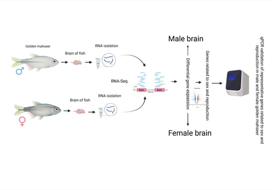
2. Materials and Methods

2.1. Ethics Statement

We carried out the sampling protocols (DCFR/IACUC/12A/06/2020/09) in line with the guidelines of, and after obtaining approval from, the Institutional Animal Care and Use Committee of the Directorate of Coldwater Fisheries Research (DCFR), Bhimtal, India. The brain sampling procedures were performed after euthanizing the fish with ethyl 3-amino-benzoate-methanesulfonic acid (MS-222, HiMedia, Maharashtra, India) to minimize pain.

2.2. Collection of Fish Sample

Between May and October 2015, a total of 13 mature golden mahseers; seven males with an average weight of 360.06 ± 101.71 g (average size of 36.9 ± 7.3 cm) and six females with an average weight of 830.06 ± 237.91 g (average size of 46.5 ± 13.8 cm), were obtained from licensed fishermen. The male displayed milt secretion, while the females were ovulating. The developmental stage of the gonads was determined based on morphological and histological analyses, as described in our previous work [19], and only the fish with mature gonads were used for brain sampling. In brief, transverse fragments of gonads from the midlobe were fixed in 4% phosphate-buffered formalin. For histological examination, the gonad tissue was dehydrated in an ascending series of ethanol and processed by standard histological methods. Gonad sections were cut at 4 µm thickness on a semi-automatic microtome (Microm HM340E, Thermo Scientific, Waltham, MA, USA) and stained with hematoxylin and eosin (H&E, HiMedia, Maharashtra, India). The stained section was examined under an upright light microscope (Leica DM500, Leica, Wetzlar, Germany). All sampled fish were in gonadal development stage IV, indicating maturity. The fish were captured using a hook and line from the Bhimtal Lake (coordinates, 29°20′ N, 79°33′ E; altitude, 4375 feet above mean sea level), Uttarakhand, India. Immediately following their capture, the fish were transported to the wet laboratory of ICAR-DCFR, Bhimtal, India, where they were euthanized using 280 mg/L MS-222 on the same sampling day. The whole brain of each fish was dissected under RNase-free conditions and stored in RNAlater RNA stabilizing reagent (QIAGEN, Hilden, Germany) at −20 °C until further use.

2.3. RNA Isolation, Library Preparation, and Sequencing

The brain tissue samples from the seven male and six female golden mahseers were pooled by sex prior to RNA isolation. The total RNA was isolated from the brain tissue using TRIzol reagent (Thermo Fisher Scientific, Waltham MA, USA) per the manufacturer’s protocols. After removing the genomic DNA from the isolated RNA using DNase I (QIAGEN, Hilden, Germany), the quantity and quality of the extracted RNA were evaluated using a Qubit 2.0 fluorometer (Invitrogen, Waltham, MA, USA) and the Agilent Bioanalyzer system (Thermo Fisher Scientific Inc., Waltham, MA, USA), respectively. RNA with an RNA integrity number between 8 and 10 and an A260/280 between 1.8 and 2.2 was used to construct the cDNA library.
To generate the cDNA library, we separated 5 µg of RNA from the male and female brain tissue and converted the samples into double-stranded cDNA using the SMARTer Ultra Low Input RNA Kit for Illumina Sequencing-HV (Clontech, Hayward, CA, USA) in line with the manufacturer’s instructions. An Illumina TrueSeq Stranded mRNA library kit was utilized to prepare a paired-end (PE) cDNA sequencing library by following the A-tailing, adapter ligation, and PCR enrichment protocols (Clontech, Hayward, CA, USA). The concentration and integrity of the cDNA library were estimated using the DNA high-sensitivity assay kit (Thermo Fisher Scientific, Waltham, MA, USA) and confirmed with the Agilent Bioanalyzer High Sensitivity DNA chip (Thermo Fisher Scientific, Waltham, MA, USA). The library was sequenced using 2 × 150 PE chemistry on the Illumina NextSeq platform at Xcelris Genomics Ltd. in Ahmedabad, India.

2.4. Transcriptome Assembly and Coding Sequence Prediction

Following the removal of low-quality reads and contamination using Trimmomatic version 0.30 [20], we assembled the male and female brain cDNA libraries with Trinity software version 2.3.2 (Broad Institute, Cambridge, MA, USA & The Hebrew University of Jerusalem, Israel) using default parameters [21,22]. Adapter trimming was carried out after the data passed quality filtration (mean quality score ≥ 25), and the trimmed reads were stored in FASTQ format. The quality of the contigs was verified by mapping the quality reads using CLC Genomics Workbench software version 11.0.1 (QIAGEN, Hilden, Germany). After concatenating the assembled transcripts into a single file, TransDecoder version 5.5.0 (www.github.com/TransDecoder/TransDecoder accessed on 18 March 2018) was used to predict the cDNA coding sequences (CDS) from the assembled transcript contigs [22]. The raw form of the nucleotide sequence data was then deposited in the National Centre for Biotechnology Information’s (NCBI) Sequence Read Archive (SRA).

2.5. Functional Annotation

The male and female golden mahseer brain contigs were analyzed separately to determine their similarity and functional characteristics. Blast2GO version 5.0.13 (Biobam, bioinformatics solutions, Valencia, Spain) [23] was used to search for matches in the nucleotide (Nt), non-redundant protein (Nr), and Swiss-Prot (curated protein sequence) databases. Matches with an E-value < 10−5 and an annotation score of 45 were considered significant hits, and the best reciprocal matches were selected based on their scores [23]. To ensure accuracy and reduce false positives, only genes that covered at least 45% of the fish references database were included for further study. Functional annotation of the contigs for male and female fish was carried out using several bioinformatics tools. Gene ontology (GO) mapping was performed using the Gene Ontology database, categorizing the contigs into Biological Process (BP), Molecular Function (MF), and Cellular Component (CC) categories. Ortholog assignment and pathway mapping were carried out using the Kyoto Encyclopedia of Genes and Genomes (KEGG) through KAAS. The coding sequences (CDS) were compared using Basic Local Alignment Search Tool X (BLASTX) against the KEGG database, assigning the best hit with a bit-score value above 60 as the GO term. The DEGs between the male and female golden mahseer brains were compared and visualized using a volcano plot. A volcano plot for male and female golden mahseer brains was generated using the edgeR program. Additionally, the mapped CDS provided insights into metabolic pathways of major biomolecules, genes involved in information processing, and cellular processes. Figure 1 presents a brief overview of the bioinformatic workflow employed in this study.
The annotation results of CDS were utilized to find genes linked to sex differentiation and reproduction by focusing on relevant keywords such as gonad, ova, ovary, fertilization, testis, male, female, milt, eggs, sperm, milt, oocyte, and steroid.

2.6. Differential Expression of Genes

The high-quality reads of the male and female golden mahseer were mapped to the CDS using the CLC Genomics Workbench software version 11.0.1 (QIAGEN, Hilden, Germany), and the fragments per million mapped reads (FPKM) were calculated from the formula:
FPKM = [RMg × 109]/[RMt × L]
where RMg represents the number of reads mapped to a particular gene, RMt is the total number of reads mapped to protein-coding sequences in the alignment, and L is the gene length in base pairs (bp).
The enrichment likelihood for each cDNA contig to identify differentially expressed genes (DEGs) in the golden mahseer male and female brains was calculated using a Poisson-based enrichment test. Using the Poisson-based enrichment test, we calculated the enrichment score by comparing the observed number of differentially expressed genes in the gene set of interest to the expected number based on the overall gene expression levels. This is performed using a Poisson distribution, which assumes that the number of differentially expressed genes follows a Poisson distribution given the average expression level. The resulting enrichment score was then assessed for statistical significance using Fisher’s exact test. This test considers the number of DEGs within the gene set and determines the probability of counts in one sex based on the counts in the other sex. A significant enrichment score suggests that the gene set or pathway is overrepresented with differentially expressed genes. Multiple testing correction was performed by false discovery rate (FDR). The Benjamini–Hochberg procedure was employed as the method for this adjustment for correcting the p-value for multiple testing. A p-value threshold of 0.001 was used to identify putative, sex-related genes from the datasets. Key terms related to reproduction and sex, such as “ovary”, “testis”, “male”, “female”, “sex”, “gonad”, “steroid”, “egg”, and “sperm”, were used to predict brain DEGs based on the annotation results. The DEGs analysis between the male and female brains was performed with edgeR of R statistical environment version 4.1.2 [24]. The heatmap was generated through FPKM value. The heatmap was generated from the FPKM value, the red shows the lower expression values, and the green shows the higher expression values.

2.7. qPCR Validation of Differential Gene Expression

2.7.1. Fish Samples

Eighteen mature golden mahseers (average weight of 960.63 ± 182.97 g; average size of 53.6 ± 16.9 cm) were caught from Bhimtal Lake with equal representation of male and female fish (n = 9 each). The fish were euthanized using MS-222 as per the details provided in Section 2.2 of this paper, and their brains were carefully collected under RNase-free conditions. Brain samples (n = 3) from each sex were pooled for further RNA isolation and analysis.

2.7.2. RNA Isolation and Synthesis of cDNA

RNA was isolated from the pooled brains using the RNeasy Plus Mini Kit (QIAGEN, Hilden, Germany). The concentration and integrity of the isolated RNA were assessed using 1.5% formaldehyde agarose gel electrophoresis with ethidium bromide and a Qubit 2.0 fluorometer (Invitrogen, Waltham, MA, USA), respectively. Approximately 500 ng of RNA was used for cDNA synthesis using the iScript cDNA Synthesis Kit (BioRad, Hercules, CA, USA).

2.7.3. qPCR Assay

Based on differences in the transcript expression between males and females, 16 genes related to gonads and reproduction were selected for qPCR validation. The selected genes were cytochrome P450, family 19, subfamily A, polypeptide 1a (cyp19a1a); double sex and mab-3 related transcription factor (dmrt2a); growth differentiation factor 9 (gdf9); SRY-box transcription factor 9b (sox9b); Wilms tumor protein 1a (wt-1a); aquaporin 1 (aqp1); C-X-C motif chemokine ligand 2 (cxcl2); anti-mullerian hormone (amh); forkhead box L3 (foxl3); nuclear receptor subfamily 0, group B, member 1 (dax1); kinesin family member 20 (kif20); transketolase (tkt); SRY-box transcription factor 9a (sox9a); steroidogenic acute regulatory protein (star); kelch-like family member 6 (klhl6), and SRY-box transcription factor 11b (sox11b).
To validate the RNA-Seq results, 24 DEGs were randomly selected, and qPCR analysis was performed. The genes selected for the qPCR analysis were cyp19a1a; 11-beta-hydroxysteroid dehydrogenase type 3 (hsd11b3); mineralocorticoid receptor (nr3c2); estrogen receptor 2a (esr2a); G protein-coupled receptors (gpcr); protein male abnormal 3 (mab3); dmrt2a; forkhead box protein F2 (foxf2); hydroxysteroid 17-beta dehydrogenase 2 (hsd17b2); sox9a; wt-1a; fibroblast growth factor 13 (fgf13); Tudor domain-containing protein 3 (tdrd3); transforming acidic coiled-coil containing protein 2 (tacc2); sperm associated antigen 16 (spag16); ruvB-like AAA ATPase 2 (ruvbl2); aqp1; nucleoporin 98 (nup98); transforming growth factor beta regulator 1 (tbrg1); DEAD-box helicase 19A (ddx19); SRY-box transcription factor 9a (sox10); IQ motif-containing protein E (iqce); amh; and star.
Primer pairs for each gene were designed using the Primer-BLAST tool available from the NCBI (https://www.ncbi.nlm.nih.gov/tools/primer-blast/ accessed on 11 February 2016). The desalted, lyophilized primers were purchased from Integrated DNA Technologies (IDT, Coralville, IA, USA). The primers and their sequences are listed in Table 1. The qPCR analysis was performed in duplicate using the ABI StepOne PCR system (Applied Biosystems, USA), and the relative expression levels of the target genes were determined using the optimized comparative Ct method (2−ΔΔCT) described by Livak and Schmittgen [25].
The qPCR reactions were conducted using 2x GoTaq qPCR Master Mix (Promega, Fitchburg, WI, USA) with a final total volume of 25 µL. Each reaction comprised 12.5 µL of 2x GoTaq Master Mix, 0.75 µL of 10 µM primers (600 nM forward and reverse primers each), 6 µL nuclease-free water, and 5 µL of 1:10 diluted cDNA. The reaction conditions consisted of the first denaturation step at 95 °C for 2 min, followed by 40 cycles of amplification at 95 °C for 15 s and 60 °C for 60 s. The primer efficiency was determined by performing serial dilutions of the male and female brain cDNA and verifying the single gene-specific fragments via a melt curve study. The transcripts of the individual gene were normalized against β-actin and presented as relative expression. Each sample was analyzed in duplicate, and the qPCR experiment was repeated twice to confirm the expression patterns. No template reactions or reverse transcriptase negative controls were included in any of the runs to detect primer-dimer formation and contamination.

2.8. Relative Expression Estimation

The relative expression levels for RNA-Seq were obtained by applying the FPKM normalization method to the raw sequence read counts. Subsequently, differential expression analysis was conducted to identify genes that exhibited significant expression differences between male and female brains. The Log2Fold changes in expression between male and female brains were determined by comparing the normalized expression values. The statistical significance of the observed fold change of differential expression between female and male samples for individual genes was assessed using a t-test of Statistical Package for Social Sciences (SPSS) version 19.0 (IBM, New York, NY, USA).
For qPCR, the relative expression levels were determined by normalizing the qPCR data, followed by calculating the relative expression by comparing the Ct values between the brains of male and female fish. The Ct values of the target gene were normalized to the Ct values of the reference gene and then compared between sexes. The fold change in expression between the brains of male and female fish was calculated based on the relative expression values.

2.9. Statistical Analysis

To determine significant differences in the relative fold change of mRNA expression of each target gene between the male and female brains by qPCR, we performed a one-way analysis of variance using the SPSS. Differences in gene expression were evaluated by comparing the mean relative quantity values for biological duplicates using the Newman–Keuls method with a 95% confidence level. The statistical significance of the fold change in relative expression, determined through RNA sequencing, was assessed using a t-test.

3. Results

3.1. Assembly and Functional Classification of Unigenes

Two cDNA libraries were generated from the brains of mature male and female golden mahseer. The read length of the cDNA libraries was 300 bp (2 × 150 bp PE), and Illumina Next-Seq generated 20.6 and 21.5 million reads from the male and female cDNA pools, respectively. After filtering low-quality (Phred score < 30) and vague sequences, 19.0 million reads (92.2%) from the male brains and 20.21 million reads (94%) from the female brains were retained for transcriptome assembly. The results of the quality check of contigs are given in Table 2, and the Q30 score was more than 92%. The RNA-Seq data of the male and female golden mahseer were deposited at NCBI GenBank SRA with accession nos. SRX2464224 and SRX2442156 for the male and female brains, respectively. Table 3 provides a summary of the RNA-Seq data assembly and unigenes statistics obtained from the male and female golden mahseer brains.
BLASTX homology was carried out with a cut-off E-value of 1 × 10⁻⁶, which resulted in most hits being against zebrafish (Danio rerio, 80%), followed by common carp (Cyprinus carpio, 12%), tilapia (Oreochromis niloticus, 2%), and other fish species (6%) (Supplementary Figure S1). The CDS of the male and female golden mahseer were analyzed using BLASTX and BLASTN, and TransDecoder, which predicted CDS annotations of 26,989 and 55,600 for the male and female samples, respectively. The assembled brain transcripts of the male and female golden mahseer were primarily 500–600 bp in length, and the distribution of the number and range of the transcripts in each range was determined (Supplementary Figures S2 and S3). Annotation and mapping of the CDS to the GO database using the Blast2GO program resulted in the successful annotation of 26,258 CDS in males and 53,446 CDS in females. The distribution of the CDS based on their length ranges is presented in Supplementary Figure S4.
The transcripts derived from the male and female golden mahseer brain samples were classified into three subcategories: BP, MF, and CC. The female fish samples had more transcripts than those of the male fish, with 17,382 transcripts under BP, 14,317 transcripts under MF, and 17,433 transcripts under CC in the female brains compared to 6306 transcripts under BP, 6309 transcripts under MF, and 4819 transcripts under CC in the male brains (Figure 2A–F). Cluster analysis identified 23,688, 20,626, and 22,252 transcripts under BP, MF, and CC, respectively. The BP domain was mainly related to cellular, single-organism, and metabolic processes (Figure 2A,D), while the CC domain had the highest number of transcripts in the subcategories related to cells, organelles, and membranes (Figure 2B,E). The dominant subcategories for the MF domain are related to binding and catalytic activities (Figure 2C,F).
Both the male and female golden mahseer CDS were compared to those of the KEGG database through BLASTX comparison with a bits score threshold of 60. The results revealed that cDNA coding sequencing is involved in various biomolecule-related pathways, including carbohydrate metabolism, lipid metabolism, nucleotide metabolism, amino acid metabolism, glycans biosynthesis and metabolism, cofactor and vitamin metabolism, terpenoids, and polyketides (Figure 3).
Among the CDS, 30.7% (2277 genes) in the male golden mahseer were classified under metabolism (M), 19.1% (1419 genes) under genetic information processing (GIP), 25.4% (1883 genes) under environmental information processing (EIP), 22.4% (1663 genes) under cellular processes (CP), and 2% (153 genes) under organismal systems (OS). In contrast, the female golden mahseer had 26.8% (2584 genes) of the CDS classified under M, 17.4% (1673 genes) under GIP, 17.4% (2884 genes) under EIP, 25.6% (2465 genes) under CP, and 2.2% (214) genes under OS.

3.2. Identification of Genes Showing Differential Sex Expression

In this study, we identified a total of 1187 upregulated DEGs in the brains of mature male and female golden mahseer, with 953 DEGs upregulated in the female brains and 234 DEGs upregulated in the male brains (Figure 4A). Additionally, 492 and 744 genes (a total of 1236 downregulated genes) were downregulated in the brain of female and male golden mahseer. The volcano plot and heat map revealed the distribution of DEGs in both female and male brains and demonstrated the patterns of upregulation and downregulation of various distinct genes (Figure 4B,C).
Among DEGs, 410 genes were related to reproductive events, such as spermiation in males and ovulation in females, followed by 181 genes for embryonic development and 110 genes for gonadal sex differentiation/determination (Figure 5). The data associated with Figure 5 is given in the Supplementary Table S1. Table 4 shows a representative selection of the reproduction and sex-biased genes that exhibited differential expression in the brains of the mature golden mahseer. The p-value and the Log2fold changes are given for each DEG. There were few female-biased and male-biased genes in the brain of the golden mahseer. In the brain of female golden mahseer, the expression of cyp19a1a, wnt5, xicof, tbrg, and aqp1 was significantly upregulated (Table 4), whereas the expression of npy, hint, sat1, spef2, and dmrt2a was significantly downregulated in the female brain.

3.3. Transcription Levels of Differential Gene Expressions Using qPCR

We evaluated the transcript expression levels of 16 gonad and reproduction-related genes. Significant differences were apparent between the male and female fish for the transcript levels of cyp19a1a, amh, foxl3, dmrt2a, gdf9, sox9b, dax1, kif20, wt-1a, aqp1, and tkt. In particular, the transcription levels of the cyp19a1a, dmrt2a, gdf9, sox9b, wt-1a, and aqp1 genes were significantly higher in the brains of the female golden mahseer, while the transcription of amh, foxl3, dax1, kif20, and tkt was significantly higher in the brains of the male golden mahseer (Figure 6).
We also evaluated the expression of the 24 selected genes (cyp19a1a, hsd11b3, nr3c2, esr2a, gpcr, mab3, dmrt2a, foxf2, hsd17b2, sox9a, wt-1a, fgf13, tdrd3, tacc2, spag16, ruvbl2, aqp1, nup98, tbrg1, ddx19, sox10, iqce, amh, and star) using qPCR (Figure 7A,B) to validate the RNA-Seq results. The qPCR results were in agreement with the RNA-Seq results, which indicated a strong statistical correlation (R2 = 0.926) between the two methods. The relative expression of mab3, dmrt2a, and wt-1a was highest in the brain of male golden mahseer, whereas in the female brain, the relative expression of spag16 was highest.

4. Discussion

Generally, the gonad transcriptome has been widely used to identify reproduction and sex-related genes in teleosts [12,26,27,28,29]. However, since the brain is the upstream regulator of the BPG axis in fish [2], we performed RNA-Seq on the brains of mature male and female golden mahseer during their annual spawning period. Notably, the brain plays a critical role in various processes by, for example, detecting day length through visual stimuli, regulating neuronal activity, facilitating neurotransmitter synthesis and release, integrating light and temperature cues to regulate circadian gene expression, influencing melanocyte function, and modulating growth and learning as well as feeding behavior [9]. In this study, we aimed to explore the genes involved in gonadal development, gametogenesis, and reproduction. Moreover, the mRNA levels of representative gonad and reproduction-related genes were validated by qPCR in male and female golden mahseer brains. Our findings provide crucial data regarding the reproductive biology of golden mahseer during the annual spawning period and offer new insights into the regulation of the BPG axis in teleost fish.
Consistent with previous research on the tissue-specific transcriptome of golden mahseer [17], the gene annotation analysis in this study revealed a high degree of homology between golden mahseer and zebrafish (80%), followed by common carp (12%) and Nile tilapia (2%). Conversely, low homology of gene annotations was observed with Atlantic salmon (Salmo salar), southern platyfish (Xiphophorus maculatus), and goldfish (Carassius auratus). Our results indicate a substantial degree of gene conservation between golden mahseer and zebrafish or common carp, which can be attributed to their shared taxonomic classification within the Cyprinidae family. We identified a total of 26,989 and 55,600 CDS in the brains of male and female golden mahseer, respectively. The higher number of CDS found in the female brain compared to the male brain may be attributed to the complexity of the oogenesis process, which requires the expression of numerous reproduction-related genes for yolk protein storage, ovarian follicle growth, chondrocyte differentiation, meiosis, fertilization, embryonic development, ovarian-related function, and the regulation of fertility [9]. This finding is consistent with previous research on female spotted scat (Scatophagus argus), which also showed a higher abundance of gene transcripts in females than in males [30]. However, more gene transcripts have been reported in mature European eel males than females [8], which suggests that the differences in gene expression during sexual maturation are specific to various types of fish.
In our study, most of the male and female golden mahseer transcripts were classified in the BP category in the GO analysis. Within the BP category, the highest number of transcripts in the males and females were related to cellular and single-organism processes as well as metabolic processes. This increase in brain activity during the spermiation and ovulation processes may be due to the involvement of several proteins in biological activities, such as localization, signaling, locomotion, flagellum formation, yolk accumulation, meiosis, fertilization, and post-fertilization developmental activities [4,8,12].
Among the mature male and female golden mahseer, we observed an increment in the expression levels of the genes involved in signal transduction, transport, and catabolism, which implies their role in spermiation and ovulation through the BPG axis. Meanwhile, we found lower levels of gene expression for membrane transport and the biosynthesis of secondary metabolites, which indicates the reallocation of resources during this stage. We further identified a total of 853 gonad and reproduction-related genes in the male and female brains of the golden mahseer, and these were associated with reproductive events, such as hormonal activity, receptor binding, embryonic development, and sex differentiation. This finding supports the concept that many reproduction and sex-related genes are expressed in the somatic tissue of the brain, as previously reported in zebrafish [31] and the rice field eel (Monopterus albus) [32].
The transcriptome analysis conducted in this study revealed that the female golden mahseer brain had a higher number of upregulated transcripts than that of the male, which is in contrast to the male-biased gene expression previously reported in the brains of other fish species, such as tropical gar (Atractosteus tropicus) [33]. This difference could be attributed to sexual selection or male gamete competition [34,35].
In the female golden mahseer, the expression of cyp19a1a, wnt5, xicof, tbrg, and aqp1 in the brain exhibited significant upregulation compared to their male counterparts. Conversely, the expression of npy, hint, sat1, spef2, and dmrt2a in the female brain displayed significant downregulation. The aromatase gene cyp19a appears to play a pivotal role in the reproduction of golden mahseer by facilitating the conversion of androgens into estrogens, thereby influencing sex determination, ovarian development, and oocyte maturation. The observed upregulation of the wnt5 gene in the female brain suggests its involvement in oogenesis and oocyte maturation. Disruptions in the wnt5 gene within golden mahseer may lead to impaired reproductive processes and sexual abnormalities in females. Furthermore, xicof genes in female fishes are recognized for their participation in oocyte development, maturation, and fertilization processes, thereby contributing to oogenesis and embryonic development [32,33,34]. The regulation of follicle growth, oocyte maturation, and ovulation in female fish is known to be governed by the tbrg gene, and the upregulation observed in our study establishes its crucial role in the spawning of female fish. The upregulated expression of the aqp1 gene in the female fish brain appears to be critical for successful oocyte growth, hydration, as well as the development and maturation of oocytes, as previously reported [31]. On the contrary, the downregulation of npy, hint, sat1, spef2, and dmrt2a gene expression in the female brain suggests their limited involvement in the reproduction of female golden mahseer.
Moreover, we analyzed the mRNA expression levels of 16 sex- and reproduction-related genes in the male and female brains of the mature golden mahseer by qPCR. Among the selected unigenes, significant differences were observed in the mRNA expression levels between the spermiating male and ovulating female golden mahseer for the genes cyp19a1a, amh, foxl3, dmrt2a, gdf9, sox9b, dax1, kif20, wt-1a, aqp1, and tkt.
Notably, the expression of the cyp19a1a transcript in the female brains was approximately three times greater than in the brains of the male golden mahseer, which is consistent with previous findings showing relatively higher cyp19a1a expression in female fish tissues [12,32]. The cyp19a1a is crucial in sex differentiation, as it catalyzes the conversion of androgens to estrogens. In several fish species, amh serves as the male sex-determining gene and regulates germ cell accumulation [36]. In our study, the relative mRNA concentration of amh in the brains of the male golden mahseer was twice that of the female brains. The expression of the dmrt2a was significantly elevated in the female golden mahseer brain, which suggests that it plays a role in ovarian development. In contrast, Xu et al. [37] reported that dmrt expression was significantly higher in the brains of male rainbow trout during late gonadal development.
Unlike the study by He et al. [12], in which aqp1 was found to be upregulated in the gonads of the male fish, aqp1 was expressed several folds higher in the brains of the female fish in our study, whereas it was only expressed marginally in the male fish brains. In fish gonads, aqp1 is essential for mediating water uptake into the gametes, but its role in the fish brain needs to be elucidated. The relative expression of foxl3, dax1, kif20, and tkt in the brains of the male fish in our study was several times higher compared to the female fish brains. This suggests their possible role in male spermatogenesis and reproduction during the breeding period of this fish species. Nonetheless, gdf9, sox9b, and wt-1a were expressed relatively higher in the female brain, which implies their involvement in ovary development and ovulation. Our study highlights the differential gene expression related to gonads and reproduction in the somatic tissue of the male and female brains of golden mahseer and suggests the possible roles of specific genes in male spermatogenesis and female ovary development. Our findings have important implications for the understanding of the reproductive biology of golden mahseer and the development of strategies for their management and conservation.

5. Conclusions

In this study, we revealed novel findings regarding the gene expression profiles of the brains of sexually mature male and female golden mahseer during the annual spawning season. The transcriptome data generated from the brain tissue of the mature golden mahseer in our study hold significant importance: they contribute to the understanding of the BPG axis, have implications for conservation, elucidate the potential for aquaculture and technical advancements, and fill gaps in the existing knowledge of this subject. The outcomes of this research have far-reaching implications for the management and conservation of golden mahseer populations and shed light on the reproductive biology of other fish species.

Supplementary Materials

The following supporting information can be downloaded at: https://www.mdpi.com/article/10.3390/fishes8070352/s1, Figure S1: BLAST search for RNA sequences obtained from the transcription study of golden mahseer to other species showing the most closely-related top hit fish species distribution; Figure S2: Distribution of transcript contig length assembled from brain of golden masheer transcriptome sequencing; Figure S3: Distribution of number of unigenes assembled from brain of golden masheer transcriptome sequencing; Figure S4: Distribution of number of cDNA (CDS) based on the length assembled from brain of golden masheer transcriptome sequencing; Table S1: Number of genes and type of gene activitiy involved in both male and female golden masheer shown in this study.

Author Contributions

The present study was conceptualized and administered by N.S., S.K.M., D.S. and W.S. The laboratory experiment was performed by N.S., B.S. and S.K.M., while the golden mahseer specimens were collected by S.K.M. and D.S. NGS data analysis was conducted by B.S. and N.S. The initial and final draft of this research article was written by N.S., B.S. and W.S. The funding for this study was obtained by N.S. and W.S. All authors have read and agreed to the published version of the manuscript.

Funding

This research was funded by the Indian Council of Agricultural Research (ICAR), grant number AQ16 and the Office of the Ministry of Higher Education, Science, Research and Innovation, and the Thailand Science Research and Innovation through the Kasetsart University Reinventing University Program 2022.

Institutional Review Board Statement

The study was conducted in accordance with the Declaration of Helsinki and approved by the Institutional Animal Care and Use Committee of the Directorate of Coldwater Fisheries Research (DCFR), Bhimtal, India (DCFR/IACUC/12A/06/2020/09).

Data Availability Statement

The transcriptome shotgun sequencing data of the male and female golden mahseer has been deposited at NCBI’s GenBank SRA under accession no. SRP095582, and experiment accession no. SRX2464224 (male) and SRX2442156 (female). Raw sequence reads can be found in the SRA database under Bioproject accession no. PRJNA358460.

Conflicts of Interest

The authors declare that they have no known competing financial interests or personal relationships that could have appeared to influence the work reported in this manuscript.

References

  1. Borella, M.I.; Chehade, C.; Costa, F.G.; de Jesus, L.W.O.; Cassel, M.; Batlouni, S.R. Chapter 14—The brain-pituitary-gonad axis and the gametogenesis. In Biology and Physiology of Freshwater Neotropical Fish; Baldisserotto, B., Urbinati, E.C., Cyrino, J.E.P., Eds.; Academic Press: Cambridge, MA, USA, 2020; pp. 315–341. [Google Scholar] [CrossRef]
  2. Devlin, R.H.; Nagahama, Y. Sex determination and sex differentiation in fish: An overview of genetic, physiological, and environmental influences. Aquaculture 2002, 208, 191–364. [Google Scholar] [CrossRef]
  3. Bar, I.; Cummins, S.; Elizur, A. Transcriptome analysis reveals differentially expressed genes associated with germ cell and gonad development in the Southern bluefin tuna (Thunnus maccoyii). BMC Genom. 2016, 17, 217. [Google Scholar] [CrossRef] [PubMed] [Green Version]
  4. Zohar, Y.; Muñoz-Cueto, J.A.; Elizur, A.; Kah, O. Neuroendocrinology of reproduction in teleost fish. Gen. Comp. Endocrinol. 2010, 165, 438–455. [Google Scholar] [CrossRef] [PubMed]
  5. Alvarado, M.V.; Carrillo, M.; Felip, A. Expression of kisspeptins and their Receptors, gnrh-1/gnrhr-II-1a and gonadotropin genes in the brain of adult male and female European sea bass during different gonadal stages. Gen. Comp. Endocrinol. 2013, 187, 104–116. [Google Scholar] [CrossRef]
  6. Ando, H.; Sasaki, Y.; Okada, H.; Urano, A. Prepubertal increases in the levels of two salmon gonadotropin-releasing hormone mRNAs in the ventral telencephalon and preoptic area of masu salmon. Neurosci. Lett. 2001, 307, 93–96. [Google Scholar] [CrossRef]
  7. Mechaly, A.S.; Viñas, J.; Piferrer, F. Sex-specific changes in the expression of kisspeptin, kisspeptin receptor, gonadotropins and gonadotropin receptors in the Senegalese sole (Solea senegalensis) during a full reproductive cycle. Comp. Biochem. Physiol. Part A Mol. Integr. Physiol. 2012, 162, 364–371. [Google Scholar] [CrossRef] [Green Version]
  8. Churcher, A.M.; Pujolar, J.M.; Milan, M.; Hubbard, P.C.; Martins, R.S.T.; Saraiva, J.L.; Huertas, M.; Bargelloni, L.; Patarnello, T.; Marino, I.A.M.; et al. Changes in the gene expression profiles of the brains of male European eels (Anguilla anguilla) during sexual maturation. BMC Genom. 2014, 15, 799. [Google Scholar] [CrossRef] [Green Version]
  9. Sahu, D.K.; Panda, S.P.; Meher, P.K.; Das, P.; Routray, P.; Sundaray, J.K.; Jayasankar, P.; Nandi, S. Construction, De-Novo Assembly and Analysis of Transcriptome for Identification of Reproduction-Related Genes and Pathways from Rohu, Labeo rohita (Hamilton). PLoS ONE 2015, 10, e0132450. [Google Scholar] [CrossRef] [Green Version]
  10. Shahi, N.; Singh, A.K.; Sahoo, M.; Mallik, S.K.; Thakuria, D. Molecular cloning, characterization and expression profile of kisspeptin1 and kisspeptin1 receptor at brain-pituitary-gonad (BPG) axis of golden mahseer, Tor putitora (Hamilton, 1822) during gonadal development. Comp. Biochem. Physiol. Part B Biochem. Mol. Biol. 2017, 205, 13–29. [Google Scholar] [CrossRef]
  11. Pan, Z.; Zhu, C.; Chang, G.; Wu, N.; Ding, H.; Wang, H. Differential expression analysis and identification of sex-related genes by gonad transcriptome sequencing in estradiol-treated and non-treated Ussuri catfish Pseudobagrus ussuriensis. Fish Physiol. Biochem. 2021, 47, 565–581. [Google Scholar] [CrossRef]
  12. He, P.; Zhu, P.; Wei, P.; Zhuo, X.; Ma, Y.; Chen, X.; Lin, Y.; Xu, Y.; Luo, H.; Peng, J. Gonadal transcriptomic analysis and differentially expressed genes between the testes and ovaries in Trachinotus ovatus. Aquac. Fish. 2022, 7, 31–39. [Google Scholar] [CrossRef]
  13. Nazari, S.; Khoshkholgh, M.; Baeza, J.A. Comparative transcriptome sequencing analysis of the narrow-clawed crayfish Pontastacus leptodactylus (Eschscholtz, 1823) and discovery of candidate sex-related genes. Aquac. Rep. 2022, 25, 101235. [Google Scholar] [CrossRef]
  14. Zhang, Y.; Cao, X.; Zou, Y.; Yan, Z.; Huang, Y.; Zhu, Y.; Gao, J. De novo gonad transcriptome analysis of elongate loach (Leptobotia elongata) provides novel insights into sex-related genes. Comp. Biochem. Physiol. Part D Genom. Proteom. 2022, 42, 100962. [Google Scholar] [CrossRef]
  15. Sun, S.; Song, F.; Shi, L.; Zhang, K.; Gu, Y.; Sun, J.; Luo, J. Transcriptome analysis of differentially expressed circular RNAs in the testis and ovary of golden pompano (Trachinotus blochii). Comp. Biochem. Physiol. Part D Genom. Proteom. 2023, 45, 101052. [Google Scholar] [CrossRef]
  16. Pinder, A.C.; Britton, J.R.; Harrison, A.J.; Nautiyal, P.; Bower, S.D.; Cooke, S.J.; Lockett, S.; Everard, M.; Katwate, U.; Ranjeet, K.; et al. Mahseer (Tor spp.) fishes of the world: Status, challenges and opportunities for conservation. Rev. Fish Biol. Fish. 2019, 29, 417–452. [Google Scholar] [CrossRef] [Green Version]
  17. Barat, A.; Kumar, R.; Goel, C.; Singh, A.K.; Sahoo, P.K. De novo assembly and characterization of tissue-specific transcriptome in the endangered golden mahseer, Tor putitora. Meta Gene 2016, 7, 28–33. [Google Scholar] [CrossRef]
  18. Kumar, R.; Sahoo, P.K.; Barat, A. Transcriptome profiling and expression analysis of immune responsive genes in the liver of Golden mahseer (Tor putitora) challenged with Aeromonas hydrophila. Fish Shellfish Immunol. 2017, 67, 655–666. [Google Scholar] [CrossRef]
  19. Shahi, N.; Mallik, S.K.; Pande, J.; Das, P.; Singh, A.K. Spermatogenesis and related plasma androgen and progestin level in wild male golden mahseer, Tor putitora (Hamilton, 1822), during the spawning season. Fish Physiol. Biochem. 2015, 41, 909–920. [Google Scholar] [CrossRef]
  20. Bolger, A.M.; Lohse, M.; Usadel, B. Trimmomatic: A flexible trimmer for Illumina sequence data. Bioinformatics 2014, 30, 2114–2120. [Google Scholar] [CrossRef] [Green Version]
  21. Grabherr, M.G.; Haas, B.J.; Yassour, M.; Levin, J.Z.; Thompson, D.A.; Amit, I.; Adiconis, X.; Fan, L.; Raychowdhury, R.; Zeng, Q.; et al. Full-length transcriptome assembly from RNA-Seq data without a reference genome. Nat. Biotechnol. 2011, 29, 644–652. [Google Scholar] [CrossRef] [Green Version]
  22. Haas, B.J.; Papanicolaou, A.; Yassour, M.; Grabherr, M.; Blood, P.D.; Bowden, J.; Couger, M.B.; Eccles, D.; Li, B.; Lieber, M.; et al. De novo transcript sequence reconstruction from RNA-seq using the Trinity platform for reference generation and analysis. Nat. Protoc. 2013, 8, 1494–1512. [Google Scholar] [CrossRef] [PubMed]
  23. Conesa, A.; Götz, S.; García-Gómez, J.M.; Terol, J.; Talón, M.; Robles, M. Blast2GO: A universal tool for annotation, visualization and analysis in functional genomics research. Bioinformatics 2005, 21, 3674–3676. [Google Scholar] [CrossRef] [PubMed] [Green Version]
  24. R Development Core Team. R: A Language and Environment for Statistical Computing. R Foundation for Statistical Computing; R Development Core Team: Vienna, Austria, 2008. [Google Scholar]
  25. Livak, K.J.; Schmittgen, T.D. Analysis of Relative Gene Expression Data Using Real-Time Quantitative PCR and the 2−ΔΔCT Method. Methods 2001, 25, 402–408. [Google Scholar] [CrossRef] [PubMed]
  26. Chen, Y.; Liu, Y.; Gong, Q.; Lai, J.; Song, M.; Du, J.; Deng, X. Gonadal transcriptome sequencing of the critically endangered Acipenser dabryanus to discover candidate sex-related genes. PeerJ 2018, 6, e5389. [Google Scholar] [CrossRef] [PubMed] [Green Version]
  27. Li, X.-Y.; Gui, J.-F. Diverse and variable sex determination mechanisms in vertebrates. Sci. China Life Sci. 2018, 61, 1503–1514. [Google Scholar] [CrossRef]
  28. Mei, J.; Gui, J.-F. Genetic basis and biotechnological manipulation of sexual dimorphism and sex determination in fish. Sci. China Life Sci. 2015, 58, 124–136. [Google Scholar] [CrossRef] [Green Version]
  29. Tian, C.; Li, Z.; Dong, Z.; Huang, Y.; Du, T.; Chen, H.; Jiang, D.; Deng, S.; Zhang, Y.; Wanida, S.; et al. Transcriptome Analysis of Male and Female Mature Gonads of Silver Sillago (Sillago sihama). Genes 2019, 10, 129. [Google Scholar] [CrossRef] [Green Version]
  30. Mustapha, U.F.; Peng, Y.-X.; Huang, Y.-Q.; Assan, D.; Zhi, F.; Shi, G.; Huang, Y.; Li, G.-L.; Jiang, D.-N. Comparative transcriptome analysis of the differentiating gonads in Scatophagus argus. Front. Mar. Sci. 2022, 9, 962534. [Google Scholar] [CrossRef]
  31. Chen, W.; Liu, L.; Ge, W. Expression analysis of growth differentiation factor 9 (Gdf9/gdf9), anti-müllerian hormone (Amh/amh) and aromatase (Cyp19a1a/cyp19a1a) during gonadal differentiation of the zebrafish, Danio rerio†. Biol. Reprod. 2017, 96, 401–413. [Google Scholar] [CrossRef] [Green Version]
  32. Chi, W.; Gao, Y.; Hu, Q.; Guo, W.; Li, D. Genome-wide analysis of brain and gonad transcripts reveals changes of key sex reversal-related genes expression and signaling pathways in three stages of Monopterus albus. PLoS ONE 2017, 12, e0173974. [Google Scholar] [CrossRef] [Green Version]
  33. Cribbin, K.M.; Quackenbush, C.R.; Taylor, K.; Arias-Rodriguez, L.; Kelley, J.L. Sex-specific differences in transcriptome profiles of brain and muscle tissue of the tropical gar. BMC Genom. 2017, 18, 283. [Google Scholar] [CrossRef] [Green Version]
  34. Danzmann, R.G.; Kocmarek, A.L.; Norman, J.D.; Rexroad, C.E.; Palti, Y. Transcriptome profiling in fast versus slow-growing rainbow trout across seasonal gradients. BMC Genom. 2016, 17, 60. [Google Scholar] [CrossRef] [Green Version]
  35. Ellegren, H.; Parsch, J. The evolution of sex-biased genes and sex-biased gene expression. Nat. Rev. Genet. 2007, 8, 689–698. [Google Scholar] [CrossRef]
  36. Yan, Y.-L.; Batzel, P.; Titus, T.; Sydes, J.; Desvignes, T.; BreMiller, R.; Draper, B.; Postlethwait, J.H. A Hormone That Lost Its Receptor: Anti-Müllerian Hormone (AMH) in Zebrafish Gonad Development and Sex Determination. Genetics 2019, 213, 529–553. [Google Scholar] [CrossRef]
  37. Xu, G.; Huang, T.; Jin, X.; Cui, C.; Li, D.; Sun, C.; Han, Y.; Mu, Z. Morphology, sex steroid level and gene expression analysis in gonadal sex reversal of triploid female (XXX) rainbow trout (Oncorhynchus mykiss). Fish Physiol. Biochem. 2016, 42, 193–202. [Google Scholar] [CrossRef]
Figure 1. Brief outline of the bioinformatics workflow conducted in this study for the RNA-Seq analysis of the brain of matured male and female golden mahseer (Tor putitora).
Fishes 08 00352 g001
Figure 2. Gene ontology (GO) classifications of the non-redundant assembled unigenes from the brains of mature male (M) and female (F) golden mahseer (Tor putitora). (A) indicates the unigenes related to the biological process (BP) of male golden mahseer; (B) unigenes related to the cellular component (CC) of male golden mahseer; (C) unigenes related to the molecular function (MF) of male golden mahseer; (D) indicates the unigenes related to the biological process (BP) of female golden mahseer; (E) unigenes related to the cellular component (CC) of female golden mahseer; (F) unigenes related to the molecular function (MF) of female golden mahseer.
Fishes 08 00352 g002aFishes 08 00352 g002b
Figure 3. Classification of the non-redundant unigenes based on the Kyoto Encyclopedia of Genes and Genomes (KEGG) pathway. The plot shows the distribution of different gene categories based on their functions in the male and female brain, and the unigenes are grouped into 23 categories based on their respective KEGG pathways. The x-axis shows the category of unigenes, whereas the y-axis shows the number of unigenes under each category.
Fishes 08 00352 g003
Figure 4. Differential expression of genes (DEGs) in the brains of matured male and female golden mahseer (Tor putitora). (A) Bar plot illustrating the differentially expressed upregulated and downregulated genes in the female and male brains of matured golden mahseer. (B) Volcano plot (edgeR test) displaying the distribution of the differentially expressed genes in female and male brains of mature golden mahseer with log2fold changes vs. −log10 (p-value). (C) Heatmap showing the differential expression of genes in the brains of mature golden mahseer categorized by sex. The heatmap was generated from the FPKM value, the red shows the lower expression values, and the green shows the higher expression values.
Fishes 08 00352 g004
Figure 5. Differential expression of the genes (DEGs) related to the gonad and reproduction events in the brains of male and female golden mahseer (Tor putitora).
Fishes 08 00352 g005
Figure 6. Quantitative real-time PCR (qPCR) validation of 16 gonad and reproduction-related genes in the brains of male and female golden mahseer (Tor putitora). cyp19a1a, cytochrome P450, family 19, subfamily A; amh, anti-mullerian hormone; foxl3, forkhead box protein 13; dmrt2a, double sex and mab-3-related transcription factor 2a; sox11b, SRY-box transcription factor 11b; gdf9, growth differentiation factor 9; sox9b, SRY-box transcription factor 9b; dax1, nuclear receptor subfamily 0, group B, member 1; kif20, kinesin family member 20; wt-1a, wilms tumor protein 1a; aqp1, aquaporin 1; tkt, transketolase; sox9a, SRY-box transcription factor 9a; klhl6, kelch-like protein 6; star, steroidogenic acute regulatory protein; cxcl2 C-X-C motif chemokine ligand 2. The data are shown as mean ± SD. * represents p < 0.05, ** p < 0.005, and *** p < 0.001.
Fishes 08 00352 g006aFishes 08 00352 g006b
Figure 7. Comparison of the expression levels of 24 differentially expressed genes measured by quantitative polymerase chain reaction (qPCR) and RNA-sequencing (RNA-Seq) in the brains of male and female golden mahseer (Tor putitora). (A) Comparison of the expression levels in the male brain. (B) Comparison of the expression levels in the female brain. cyp19a1a, cytochrome P450, family 19, subfamily A; hsd11b3, 11-beta-hydroxysteroid dehydrogenase type 3; nr3c2, mineralocorticoid receptor; esr2a, estrogen receptor 2a; gpcr, G protein-coupled receptors; mab3, protein male abnormal 3; dmrt2a, double sex and mab-3 related transcription factor 2a; foxf2, forkhead box protein F2; hsd17b2, hydroxysteroid 17-beta dehydrogenase 2; sox9a, SRY-box transcription factor 9a; wt-1a, Wilms tumor protein 1a; fgf13, fibroblast growth factor 13; tdrd3, Tudor domain-containing protein 3; tacc2, transforming acidic coiled-coil containing protein 2; spag16, sperm associated antigen 16; ruvbl2, ruvB-like AAA ATPase 2; aqp1, aquaporin 1; nup98, nucleoporin 98; tbrg 1, transforming growth factor beta regulator 1; ddx19, DEAD-box helicase 19A; sox10, SRY-box transcription factor 10; iqce, IQ motif-containing protein E; amh, anti-mullerian hormone; star, steroidogenic acute regulatory protein. The significance level, denoted by *, was set at p < 0.05.
Fishes 08 00352 g007aFishes 08 00352 g007b
Table 1. Primers used in this study for the validation of RNA-Seq results and differential expression of genes (DEGs) using quantitative real-time PCR (qPCR).
Primer NamePrimer Sequence (5′ to 3′)Amplicon Length (bp)PCR Efficiency (%)Description
cyp19a1aGCAGACGGTTCTCATACAGC
TGTCTCTTCCAGCTTCTCCA
15496RNA-Seq validation and DEGs
amhCCTGCAGACTCACAGAGTGG
CTTGAGCAGCAAAACGGACC
23299RNA-Seq validation and DEGs
foxl3CACGTGGGCTCAAAATGTCC
AGGAAAGCCTTGCGTCTGAA
11293Male and Female DEGs
dmrt2aCCCCGGCAAAACTATGAGGT
TTCCTGGAACGGATTCGGTG
169104RNA-Seq validation and DEGs
sox11bACCAAGATGACCAGTCACGGAA
CGATTGTGGTGCAGGCGAG
94105Male and Female DEGs
gdf9CAGACCTGGAGGCCAGATTC
TGCTTCTTCTCTGGTACGCC
11092Male and Female DEGs
sox9bCTCCTGGAGAACACTCCGGT
ATGAGTCTGCAGGCGTTGTG
11697RNA-Seq validation and DEGs
dax1TACGCGTACTTGAAAGGGGC
TTAAGTGCCTGGTTCGCCTC
10497Male and Female DEGs
kif20AGCCAGCTCGAAAAACCTCT
GGGTCCGTGCTGAGGATCTG
15497Male and Female DEGs
wt-1aGGGGTGTGTAAGTCCTCTCTTC
CGGATTTGGCGACCATCAAG
13899RNA-Seq validation and DEGs
aqp1CTCAGTGTTTGCCTGGGACA
ACGAATCATTGCTGGTCCGA
9097RNA-Seq validation and DEGs
tktGACCACTACCACGAAGG
AGGAACGTGGGACACAG
12898DEGs
sox9aAATCTGAAGACGGCAGCGAA
ATCGAATCGAATGGCGAGTCA
15699DEGs
klhl6GGACCAAAGTTCGTGGAGGT
AGCCACGTAAAGGAAACCGT
188103Male and Female DEGs
starGTGGAACCCCAATGTCAAACA
AAGAACCTGAGAGGGACCAAA
23299RNA-Seq validation and DEGs
cxcl2CACATCAGCGGAGGACACAT
GGGTGTAACTCCGTAGAGCG
14799Male and Female DEGs
ß-actinTGTCCCTTCCCTTATGGCCT
CATCCCAGTCCCTAAAGTGCT
72100Reference gene
hsd11b3GCATATGCGTCGCGTTCATT
AATGGTACGCCAACTGCTCA
11999RNA-Seq validation
nr3c2TGTTTTGTGGCTTAGTAAATG
GAGTTCCCTGGGTGATTGGG
11298RNA-Seq validation
esr2aCAGCTCCCGTTTGTCTCACT
GTTTAGGGTCCGTGCTGTGA
105100RNA-Seq validation
gpcrTACGGTGTTTGGGTGTTGCT
CCCCTTGCTGTGGAAGTGAA
10597RNA-Seq validation
mab3CTGATGGAGCTCAAGACGCA
GTCGACAGAGAAGGTTCCCG
10298RNA-Seq validation
foxf2ACCGCATCTGACTTCCGTTT
TTTTGGAGAGCCGAGTGCAT
152100RNA-Seq validation
hsd17b2CCTGCTTTATTTGTGAGCCA
AGCACAAAGGCCTGAGGTGA
11299RNA-Seq validation
fgf13GAGAAATCAAACGCCTGCCG
TGGAGCCGAAAAGCTTGACT
109100RNA-Seq validation
tdrd3CTACGAAGAACCTCCCCACG
GGACTTTGCCTCTTCACCGA
9396RNA-Seq validation
tacc2TTTGTGAAGATGCTGGCGTTG GTCCGAAAGGCTCGTCTCTT10299RNA-Seq validation
spag16GTTGGGCATGGGTTTGACG
TTTGGAAGGCCCAACACCTT
19297RNA-Seq validation
ruvbl2AGGTGGCAACCACAAAGGTT
GGCTCCAGAGCATCATCCAA
106103RNA-Seq validation
nup98CACCTACCGCTCAACCAACA
CCCCCATCGAAGCATTTTGC
12396RNA-Seq validation
tbrg1CTCGCAGAGGCAAACCCTTA
CAGGGCAGCTCTGGATTAGG
9992RNA-Seq validation
ddx19AGCTGCAGCGGAATCGATAA
TTGCTTCGGTCTTCGCTTCT
101102RNA-Seq validation
sox10GCTCAACTGCTACGACTGGA
AAGACCAGGTGAAAGACGTGA
187100RNA-Seq validation
iqceTACATCCGTACAGCGACGAC
CCGTTTAGAAAAGCAGCGGG
11198RNA-Seq validation
Table 2. Statistical data of the assembly quality of male and female brain transcriptome of golden mahseer (Tor putitora).
OrganRaw
Reads
Raw
Bases
Clean
Reads
Clean
Bases
Error
Rate
(%)
Q20
(%)
Q30
(%)
GC
Content
(%)
Male brain20,610,1053,762,376,80019,678,7233,367,741,6350.025798.0594.2950.08
Female brain21,541,2565,674,182,70520,456,8575,237,821,4650.024898.1293.5849.91
Table 3. Summary of the RNA-sequencing data and unigene statistics for the brains of mature male and female golden mahseer (Tor putitora).
DescriptionMale BrainFemale Brain
Number of reads20,610,10521,541,256
Total number of bases3,762,376,8005,674,182,705
Number of transcripts contigs39,04775,736
Total transcript contigs length (bp)58,794,838311,938,504
Average transcript contigs length (bp)15051742
Maximum length of transcript contigs14,97218,509
N50 value16462729
Number of unigenes26,98955,600
Maximum length of unigenes11,28918,509
Total unigenes length (bp)26,397,81387,317,771
Average unigenes length (bp)9781570
Table 4. List of the genes associated with gonad and reproductive events showing the differential expression in the brains of male and female golden mahseer. Gene descriptions are based on BLAST hits in the RefSeq database.
Gene SymbolGene Descriptionp-ValueLog2FC
Female/Male
Sex determination, gonadal development, and neuronal development
sox10SRY-box transcription factor 102.63 × 10−180.67
sox19aSRY-box transcription factor 19a3.01 × 10−111.15
sox4SRY-box transcription factor 41.15 × 10−290.72
sox11aSRY-box transcription factor 11a4.06 × 10−220.91
sox21bSRY-box transcription factor 21b3.02 × 10−61.23
sox4bSRY-box transcription factor 4b6.71 × 10−160.76
sox5x3SRY-box transcription factor 5 isoform x32.31 × 10−91.92
sox7SRY-box transcription factor 71.42 × 10−101.01
sox3SRY-box transcription factor 31.67 × 10−80.67
wnt5Wingless-type MMTV integration site family, member 53.12 × 10−611.78
dmrt2aDouble sex and mab-3-related transcription factor 2a3.89 × 10−7−17.93
dmrta2Double sex and mab-3-related transcription factor a27.55 × 10−17−19.12
dmrta1Double sex and mab-3-related transcription factor a16.35 × 10−121.56
dmrt2bDouble sex and mab-3-related transcription factor 2b3.65 × 10−19−17.04
foxl1Forkhead box protein l11.23 × 10−41.67
foxf2Forkhead box protein f28.81 × 10−153.90
foxn2Forkhead box protein n21.91 × 10−27−8.78
foxj3Forkhead box protein J35.03 × 10−227.67
foxd1Forkhead box protein D13.76 × 10−224.78
foxn2Forkhead box protein N21.63 × 10−202.99
fgf11Fibroblast growth factor 115.09 × 10⁻²²−6.76
fgf8bFibroblast growth factor 8b2.83 × 10−343.24
wt-1aWilms tumor protein 1a3.99 × 10−165.03
srySex determining region Y3.98 × 10−43−9.85
mab3Mannosyl (beta-1,4-)-glycoprotein beta-1,4-N-acetylglucosaminyltransferase1.86 × 10−346.39
alcamActivated leukocyte cell adhesion molecule2.76 × 10−91.93
hint2Histidine triad nucleotide-binding protein 22.42 × 10⁻¹⁴−11.26
hint3Histidine triad nucleotide-binding protein 32.55 × 10−57−11.39
xicof20Oocyte zinc finger protein 206.35 × 10−10019.94
xicof6Oocyte zinc finger protein 68.02 × 10−1315.45
xicof26Oocyte zinc finger protein 267.66 × 10−14813.45
xicgfGastrula zinc finger protein3.09 × 10−5−0.75
tdrdTodor and KH domain-containing protein1.51 × 10−146.55
spag16Sperm associated antigen 16 protein3.45 × 10−470.13
spag1Sperm associated antigen 15.03 × 10−200.03
spag2Sperm specific antigen 23.46 × 10−130.12
sat1Spermidine/spermine N1-acetyltransferase8.09 × 10−23−15.78
spef2Sperm flagellar protein 25.09 × 10−43−12.38
ef1a2Elongation factor 1-alpha21.11 × 10−181.88
cxcl2C-X-C motif chemokine ligand 23.74 × 10−262.96
rgs4Regulator of G protein signaling 43.10 × 10−121.40
muc13Mucin 131.87 × 10⁻¹⁷−6.89
trim39Tripartite motif containing 391.24 × 10−52.91
tacc2Transforming acidic coiled-coil-containing protein 25.75 × 10−173.15
klhl6Kelch-like protein 63.67 × 10−191.37
klhl4Kelch-like protein 46.31 × 10−20.45
bcl-2Apoptosis regulator factor 29.05 × 10−227.45
bcl-2aBcl2 related ovarian killer protein homolog a isoformx21.88 × 10−41−6.23
phb2Prohibitin-22.31 × 10−60.67
map7Ensconsin6.53 × 10−134.70
aqp1Aquaporin 11.21 × 10−1015.06
tgfb1Transforming growth factor beta 12.43 × 10−81.63
rdh10Retinol dehydrogenase 103.01 × 10−53.16
nipblNipped-b-like protein3.12 × 10−65.23
Hormone receptors and steroidogenesis
hsd17b2Hydroxysteroid 17-beta dehydrogenase 21.23 × 10−112.33
hsd17b1Hydroxysteroid 17-beta dehydrogenase 13.65 × 10⁻⁹−1.93
hsd20bHydroxysteroid 20-beta dehydrogenase5.07 × 10−116.09
hsd3b7Hydroxysteroid 3-beta dehydrogenase 73.76 × 10−262.87
hsd17b14Hydroxysteroid 17-beta dehydrogenase 142.16 × 10−92.89
hsd17b4Hydroxysteroid 17-beta dehydrogenase 45.22 × 10−134.01
hsd17b12Hydroxysteroid 17-beta-dehydrogenase 121.19 × 10−147.61
hsd17b2Hydroxysteroid 17-beta-dehydrogenase 22.76 × 10⁻⁶−1.67
esrαEstrogen receptor alpha1.99 × 10−214.54
esr1Estrogen receptor 13.41 × 10−775.90
esr2aEstrogen receptor 2a6.61 × 10−1113.98
esr2bEstrogen receptor 2b2.67 × 10−179.45
cyp19a1aCytochrome P450, family 19, subfamily A, polypeptide 1a5.51 × 10−1215.56
cyp20a1aCytochrome P450 family 20, subfamily A, polypeptide 1a6.31 × 10−1912.78
nr3c1Glucocorticoid receptor 11.99 × 10−95.13
nr3c2Mineralocorticoid receptor 22.78 × 10−193.25
tktTransketolase1.66 × 10−73.61
gpcrG-protein coupled estrogen receptor 19.23 × 10−112.37
starSteroidogenic acute regulatory protein5.87 × 10−86.91
cyp11a1Cytochrome P450, family 11, subfamily A, polypeptide 12.99 × 10−214.83
cyp11c1Cytochrome P450, family 11, subfamily C, polypeptide 13.06 × 10−15.75
cyp11b2Cytochrome P450, family 11, subfamily B, polypeptide 17.61 × 10−224.34
npbNeuropeptide B receptor1.23 × 10−12−3.56
npyNeuropeptide Y receptor7.43 × 10−26−11.09
hgfrHepatocyte growth factor receptor 15.12 × 10−12.90
fgfFibroblast growth factor receptor2.99 × 10−343.45
tbrgTransforming growth factor-beta receptor1.23 × 10−7213.76
Disclaimer/Publisher’s Note: The statements, opinions and data contained in all publications are solely those of the individual author(s) and contributor(s) and not of MDPI and/or the editor(s). MDPI and/or the editor(s) disclaim responsibility for any injury to people or property resulting from any ideas, methods, instructions or products referred to in the content.

