Article

Effect of the Potential Probiotic Vibrio proteolyticus DCF12.2 on the Immune System of Solea senegalensis and Protection against Photobacterium damselae subsp. piscicida and Vibrio harveyi

by
Alberto Medina
,
Jorge García-Márquez
,
Miguel Ángel Moriñigo
and
Salvador Arijo
*
Departamento de Microbiología, Facultad de Ciencias, Instituto Andaluz de Biotecnología y Desarrollo Azul, Universidad de Málaga, 29071 Málaga, Spain
*
Author to whom correspondence should be addressed.
Fishes 2023, 8(7), 344; https://doi.org/10.3390/fishes8070344
Submission received: 1 June 2023 / Revised: 26 June 2023 / Accepted: 27 June 2023 / Published: 30 June 2023
(This article belongs to the Special Issue Potential Application of Probiotics for Sustainable Aquaculture)

Abstract

:
Some of the characteristics of probiotics used in aquaculture are the antagonistic activity against pathogens and the activation of the immune response. Moreover, some probiotics with common antigens to pathogens can produce antibodies that react with these pathogens. One of those potential probiotics is Vibrio proteolyticus DCF12.2, a strain with the capacity to produce antibodies in soles (Solea senegalensis) that react with Photobacterium damselae subsp. piscicida and Vibrio harveyi antigens. Therefore, the objective of the work was to determine the capacity of DCF12.2 to activate the immune response in the sole, providing protection against both pathogens. For this purpose, DCF12.2 was administered to cultured soles by intraperitoneal injection, bath, and diet. The DCF12.2 strain activated the gene expression related to the fish immune response and increased the antibody production with cross-reaction to P. damselae subsp. piscicida and V. harveyi cells. Challenges with P. damselae subsp. piscicida resulted in a 30–40% relative percent survival (RPS) through DCF12.2 treatments. After challenge with V. harveyi, only intraperitoneal treatment protected the fish (67% RPS). In conclusion, the DCF12.2 strain showed great potential to be used to prevent diseases caused by both pathogens, and indicates a way to use probiotics as live vaccines.
Key Contribution: In this study, we demonstrate that the administration of Vibrio proteolyticus DCF12.2 to Senegalese sole (Solea senegalensis) activates the immune response, leading to the production of cross-reactive antibodies against Photobacterium damselae subsp. piscicida and Vibrio harveyi. Furthermore, the administration of this strain confers protection against experimental infection with both pathogens.

Graphical Abstract

1. Introduction

Probiotics, which are live microorganisms that confer health benefits on the host when administered in adequate amounts [1], have been extensively documented for their health benefits in human health and farm animal production [2,3,4]. In aquaculture, probiotics offer an alternative approach to combat fish pathogens. These beneficial microorganisms exhibit various features, including providing nutrient supply to the host, prompting colonization of mucosal surfaces, acting as a barrier against pathogens, modulation of the gut microbiota, inhibition of bacterial growth through the production of bacteriocins and other substances, and activation of the immune response [5,6,7,8,9].
While the ability of probiotics to activate the non-specific immune response is well-established, few studies have investigated their potential to activate the specific immune response. Some probiotics have been observed to stimulate the production of immunoglobulins [10]. Fish also secrete immunoglobulins on the epithelial and intestinal mucus to neutralize pathogens [11]. Additionally, oral administration of probiotics has demonstrated the production of specific antibodies [12]. On the other hand, cross-reaction between common antigens of different strains could play a significant role in host protection against pathogens [13]. Therefore, administering probiotics with common antigens to some pathogens could protect the host [14]. This opens up the possibility of using probiotics with common antigens to certain pathogens as live vaccines.
In this context, a previous work [15] selected probiotics based on their ability to generate cross-reactive antibodies against two pathogens (Vibrio harveyi and Photobacterium damselae subsp. piscicida) which affect farmed Senegalese sole (Solea senegalensis). Among these probiotics, the Vibrio proteolyticus DCF12.2 strain stood out for its high capacity to induce antibody production in fish, and for other characteristics, such as ability to inhibit pathogen growth, non-virulence for fish, and survival under storage conditions. This strain also showed enzymatic activities such as lecithinase, gelatinase, caseinase, amylase, and lipase, which allow a better digestibility of the feed, viability after feed storage, and survival in the pH conditions of the fish gut [15]. These combined attributes make the DCF12.2 strain highly promising for dual use as both probiotic and live vaccine. Consequently, it is of interest to evaluate the administration of the DCF12.2 strain in in vivo assays using the Senegalese sole as an experimental animal.
The objective of this study was to determine the effect of the probiotic strain V. proteolyticus DCF12.2, administered to the fish by different routes, on the adaptive and innate immune system of juvenile Senegalese sole (S. senegalensis). The degree of fish protection treated with probiotic potential was also evaluated through experimental infections, with virulent strains of either P. damselae subsp. piscicida Lg41.01 or V. harveyi Lg16.00.

2. Materials and Methods

2.1. In Vitro Test to Determine DCF12.2 Survival in Intestinal Tract: pH and Bile Tolerance

To determine the ability of the strain to pass through the intestinal tract, bile and pH tolerance were evaluated for V. proteolyticus DCF12.2 as a previous step in the in vivo study. V. proteolyticus was grown in tryptic soy broth medium with 2% NaCl (TSBs) at 22 °C for 24 h. Bile salt and pH tolerance were determined using the method described by Thankappan et al. [16], with some modifications. In brief, tubes with TSBs (10 mL) were prepared with 0 (control), 0.3, 0.5, 1.0, 3.0, and 5.0% of bile salt (Sigma-Aldrich, St. Louis, MO, USA) or adjusted to different pH values (1.0, 2.0, 3.0, and 4.0). The tubes were inoculated with 106 cfu/mL of V. proteolyticus DCF12.2 from an overnight growth culture, after centrifugation at 3000× g for 15 min and washing two times with sterile saline. Samples were incubated at 22 °C. Control (no bile salts) and test cultures were evaluated at 1, 2, and 3 h for the presence or absence of growth by streaking the samples (0.1 mL) onto TSAs plates.

2.2. Experimental Diets and Feeding Trial

S. senegalensis specimens (n = 360; 17.5 ± 2.5 g body weight) were provided by the Facultad de Ciencias del Mar of the University of Cadiz (Cádiz, Spain), transferred to the Centro de Experimentación de Ecología y Microbiología de Sistemas Acuáticos Controlados Grice-Hutchinson (CEMSAC) of the University of Malaga (Malaga, Spain; Spanish Operational Code REGA ES290670002043), and acclimated to experimental conditions for one week before commencement of the trial. During the adaptation period, fish were fed a commercial diet (58% protein, 17% fat, Gemma, Skretting, Burgos, Spain). Four homogeneous groups of 90 fish were randomly distributed in 500 L tanks coupled to a recirculation aquaculture system (RAS) equipped with physical and biological filters. Fish were maintained under natural photoperiod (May–July), temperature (16.5 ± 3.5 °C), and salinity (35 g L−1). Supplemental aeration was provided to maintain dissolved oxygen at 6.8 ± 0.4 mg L−1. Ammonia (<0.1 mg L−1), nitrite (<0.2 mg L−1), nitrate (<50 mg L−1) and pH (7.0–7.8) were determined weekly at 9 a.m. The four experimental groups were then established as follows:
(i)
Control diet (CTRL group): fish fed a commercial diet (Gemma, Skretting, Burgos, Spain).
(ii)
Fish intraperitoneally injected with V. proteolyticus DCF12.2 (IP group): V. proteolyticus DCF12.2 was grown on TSAs medium and resuspended in PBS at a concentration of 109 colony-forming units (cfu) mL−1. The fish were anesthetized with 100 ppm of isoeugenol (clove oil) and intraperitoneally injected using 0.1 mL of bacterial suspension [15].
(iii)
Fish immersed in a V. proteolyticus DCF12.2 suspension at 107 cfu mL−1 added in the tanks (BATH group), within the range used by other authors when probiotics were administrated though water [17]. One hour later, the water was changed to remove the probiotic cells. The treatment was repeated on the same fish 15 days later.
(iv)
Fish fed with a commercial diet (Gemma, Skretting, Burgos, Spain) supplemented with 109 cfu g−1 of V. proteolyticus DCF12.2 (DIET group). This experimental diet was prepared just before the administration by spraying the probiotic suspension on the feed surface [18].
Control, IP, and BATH groups were fed with a commercial diet (Gemma, Skretting, Burgos, Spain) at 1% of their body weight over the 28 days of the trial. The diet group was fed with the supplemented diet at 1% of their body weight for 28 days, alternating five days of feeding with the supplemented diet (109 cfu g−1 of V. proteolyticus DCF12.2) with 2 days of feeding the commercial diet. During days 28 to 56, all surviving fish were fed control feed.

2.3. Fish Sampling

At days 3, 7, 14, 21, 28, and 56 of the experiment, fish (five fish per group) were euthanized by an overdose of clove oil (200 ppm).
Serum was obtained after clotting of blood samples at room temperature (for 60 min) and subsequent centrifugation (3000× g, 20 min, 4 °C) [19]. After blood extraction, skin and whole intestinal mucus were scraped with a slide to determine the antibody titre. Skin and intestinal mucus samples were collected using the method described by Guardiola et al. [20], with some modifications. In brief, skin and intestinal mucus were obtained by gently scraping either the dorsolateral surface of specimens (skin mucus) or the whole intestine (intestinal mucus) using a cell scraper with enough care to avoid contamination with blood. The mucus was homogenized with PBS and centrifuged at 300× g for 10 min at 4 °C. The supernatant was frozen at −80 °C, lyophilized, and then redissolved in chemically pure water. The protein concentration was measured by the Bradford method [21] and adjusted at 250 μg mL−1. In addition, on days 3 and 28, head-kidney samples were taken for qPCR assays. Finally, serum, mucus, and head-kidney samples were stored at –80 °C until use.
The experimental procedures complied with the guidelines of the University of Malaga (Málaga, Spain) and the European Union Council (2010/63/EU) for the use of animals in research. The experimental procedures were previously approved by the Spanish Government’s Ethics and Animal Welfare Committee (RD53/2013).

2.4. Detection of Antibodies in Serum, Intestinal Mucus, and Skin

The detection of specific antibody titers in serum (days 14, 21, 28, and 56), and intestinal and skin mucus (days 7, 14, 21, 28, and 56) was carried out by ELISA [22]. Antigens used in ELISA were the bacteria DCF12.2 strain and V. harveyi Lg16.00 strain, incubated for 24 h at 22 °C in TSAs, and P. damselae subsp. piscicida Lg41.01 grown for 48 h at 22 °C. They were then resuspended in PBS with an optical density similar to one at an absorbance of 600 nm. Briefly, wells of microtiter plates (Nunc; Sigma-Aldrich, St. Louis, MO, USA) were coated with 200 µL of the bacteria whole-cells suspension and incubated overnight. After washing with PBS, antigen-coated wells were incubated with mucus, at a protein concentration of 250 μg mL−1, or serum at 1:500 dilution, and incubated for 2 h. After PBS washing, wells were sequentially incubated with rabbit antibody anti-sole immunoglobulin (1:1000), horseradish peroxidase-conjugated goat antibody antirabbit immunoglobulin (1:5000), and o-phenylendiamine dihydrochloride (Fast OPG; Sigma-Aldrich, St. Louis, MO, USA) as developing solution. The reaction was stopped by addition of 3M H2SO4, and absorbance was determined at 490 nm.

2.5. Determination of Genes Involved in Innate Immune Response

The effect of the different administration routes of DCF12.2 strain on the gene expression involved in the immune response was also evaluated. The expression was measured 3 (short-term effect) and 28 days (long-term effect) after starting the trial.
The determination of gene expression was carried out according to Tapia-Paniagua et al. [23]. The total RNA was isolated from the head kidney using a GeneJET RNA Purification Kit (Thermo Scientific, Madrid, Spain), according to the manufacturer’s instruction. RNA quality was checked by running an aliquot on an agarose gel and quantity was spectrophotometrically determined in Nanodrop ND-1000 (Thermo Scientific, Madrid, Spain) via A260/280nm and A260/230nm readings. DNase treatment (Thermo Scientific, Madrid, Spain) was carried out to completely eliminate the DNA. Reverse transcription was performed using a First Strand cDNA Synthesis Kit (Thermo Scientific, Madrid, Spain) with 1 µg of total RNA, and by using oligo (dT)18 primers. One microliter of each cDNA synthesis reaction was employed as the template in the qPCRs to analyze each gene transcription. Primers to detect expression of genes encoding complement components 3 and 7 (C3 and C7), C and G type-lysozyme (LYS-C and LYS-G), sequestosome 1 (SQSTM-1), caspase 6 (casp6), natural killer enhancing factor (NKEF), nicotinamide adenine dinucleotide phosphate (NADPH) oxidase (NADPHox), and three heat shock proteins (GP96, HSP90AB, and HSP70) were used as described by the authors listed in Table 1. Amplifications were performed in a final volume of 10 µL. The reaction mixture contained 5 µL of SsoAdvanced SYBR Green Supermix (Bio-Rad Laboratories, Hercules, CA, USA), 0.5 µL of each primer set (10 µM), 1 µL of template cDNA and 3 µL of DEPC-water. Real-time PCR determinations were performed in triplicate in 96-well PCR plates and carried out in a CFX96 Touch Real-Time PCR Detection System (Bio-Rad Laboratories, Hercules, CA, USA) with an initial denaturation cycle of 95 °C for 30 s, followed by 40 cycles of 95 °C for 5 s and 51 °C for 15 s. Amplification was followed by a standard melting curve from 65 °C to 95 °C, in increments of 0.5 °C for 5 s at each step, to confirm that only one product was amplified and detected. For cDNA normalization, samples were run in parallel with two reference genes, glyceraldehyde-3-phosphate dehydrogenase 1 and 2 (GADPH1 and GADPH2) [24,25].
The threshold values (Ct) were calculated with iCycler iQ v3.0 (Biorad, Hercules, CA, USA). The relative values of mRNA expression were calculated using method 2−ΔΔCt according to Pfaffl [32]. The statistical significance of the difference between the mean values was obtained by ANOVA analysis at a significance level of p ≤ 0.05.

2.6. Experimental Infection with P. damselae subsp. piscicida and V. harveyi

The degree of protection was determined by experimental infections with P. damselae subsp. piscicida (strain Lg41.01) and V. harveyi (strain Lg16.00) 30 days after starting probiotic administration. Both strains were isolated from diseased Senegalese sole and have shown pathogenicity in experimental trials [33,34]. For this, 10 fish per group were randomly chosen. Specimens were anesthetized with clove oil as previously described, and then intraperitoneally injected, either with 0.1 mL of PBS with a suspension of P. damselae subsp. piscicida Lg41.01 (dose 104 cfu g−1) or V. harveyi Lg16.00 (dose 2 × 10⁶ cfu g−1). One group was maintained as a control and was inoculated with 0.1 mL of PBS. Mortality was recorded for 21 days. The protection conferred by V. proteolyticus was assessed as described by Amend [35] by calculation of the relative percent survival (RPS) using the following Formula (1):
RPS (%) = [1 − (% mortality of fish fed with experimental diet/% mortality of fish fed with control diet)] × 100

2.7. Statistical Analysis

All statistical analyses and graphics were performed with GraphPad (version 9.3.0.463) software. Results are reported as means ± SD. Normal distribution was checked for all data with the Shapiro–Wilk test, while the homogeneity of the variances was obtained using the Levene test. When necessary, an arcsine transformation was performed. Differences between the experimental treatments (CTRL, IP, BATH, DIET) were tested using one-way ANOVA. In all statistical tests performed, p < 0.05 was considered a significant difference.

3. Results

3.1. Growth of Vibrio Proteolyticus DCF12.2 in the Presence of Bile and Different pH

The bacterium was tolerant to all bile salt concentrations tested (Figure 1a). Regarding the growth in TSB at different pH, DCF12.2 was tolerant to media at a pH greater than 4.0. The growth was inhibited in less than one hour at pH 2.0, in two hours at pH 3.0, and in three hours at pH 4.0, while at pH values of 5.0, 6.0, and 7.0, the strain remained viable all the time (Figure 1b).

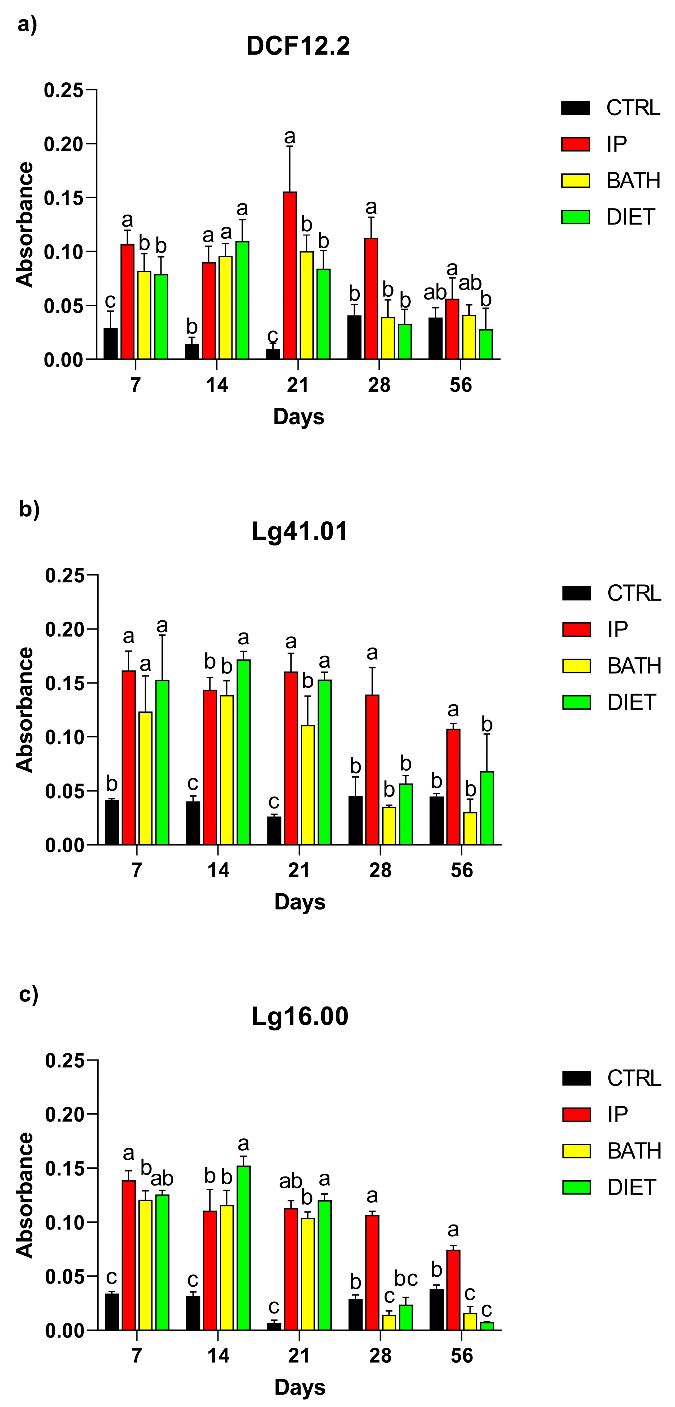
3.2. Detection of Antibodies in Serum, Intestinal Mucus, and Skin

The antibody serum levels in the IP group, when DCF12.2 cells were used as an antigen, were significantly higher than in the other groups (CTRL, BATH, and DIET groups) from day 14 to the end of the assay (Figure 2a). When P. damselae subsp. piscicida Lg41.01 cells were used as antigens, the only significant differences were found in the IP group at day 56, being higher than the other groups (Figure 2b). When V. harveyi Lg16.00 cells were used as antigens, only the IP group, on days 14 and 56, showed a significant increase compared to the control (Figure 2c).
The analysis of the intestinal mucus (Figure 3) showed a higher titer in IP, BATH, and DIET groups than the CTRL group on days 7, 14, and 21, regardless of the bacterium used as antigen (Figure 3a–c). At day 28, only the IP group showed a significant increase in antibody titer compared to the control. This significant increase was maintained at 56 days when Lg41.01 and Lg16.00 were used as antigens (Figure 3b,c).
The antibody titer in skin mucus is shown in Figure 4. When DCF12.2 cells were used as an antigen, significant increases over the control were only obtained on day 56, specifically, for the IP and BATH groups (Figure 4a). The antibody skin mucus levels in the BATH group, when P. damselae subsp. piscicida Lg41.01 cells were used as antigens, were significantly higher than control group on days 7 and 21 (Figure 4b). At the end of the experiment (day 56), the IP and DIET groups had higher antibody titers than did the CTRL and BATH groups (Figure 4b). Furthermore, no significant differences were detected in skin mucus antibody titer when V. harveyi was used as the antigen (Figure 4c).

3.3. Determination of Genes Involved in Innate Immune Response

Dietary effects were observed on several genes associated with the immune response system in the head kidney (Figure 5). On day 3, the DIET group showed downregulated expression of the sequestosome 1 gene (SQSTM) relative to the CTRL and BATH groups (Figure 5a). The expression of the caspase 6 gene (casp6) was downregulated in the IP, BATH, and DIET groups at day 3 compared to the CTRL group (Figure 5a). In contrast, the natural killer enhancing factor (NKEF) gene was upregulated in all the treatments over the control group. Four weeks later, the expression of the complement 3 (C3) and the lysozyme G-type (LYS-G) genes, as well as the SQSTM gene, were upregulated in the BATH group compared to the other groups.
In a different pathway related to stress response, on day 3 the expression of heat shock protein gp96 (GP96) was upregulated in the BATH group, while the heat shock protein 90 AB gene was overexpressed relative to the control in the IP and DIET groups (Figure 5b). The dinucleotide phosphate oxidase (NADPHox) gene was upregulated in the IP and DIET groups at 3 days compared to the CTRL group (Figure 5b). At day 28, only the heat shock protein 70 (HSP70) was upregulated in the BATH group. The rest of the groups did not generate changes with respect to the control in the other genes (Figure 5d).

3.4. Determination of the Survival of Fish Administered with Probiotic and Infected with P. damselae subsp. piscicida and V. harveyi

The fish infected with P. damselae subsp. piscicida produced an RPS value of 30% when the V. proteolyticus DCF12.2 was administered intraperitoneally or through feed, and the RPS was 40% when the strain was administered by bath (Table 2). After the challenge with V. harveyi, the RPS value obtained was 67% in fish treated with DCF12.2 intraperitoneally, while bath and diet treatments did not offer protection (Table 2).

4. Discussion

V. proteolyticus DCF12.2 was selected for this study based on its antagonistic activity against P. damselae subsp. piscicida and V. harveyi, as well as its shared common antigens with these pathogens. Furthermore, this strain is non-pathogenic to fish, demonstrates viability under storage conditions, produces beneficial enzymes for digestion, is susceptible to common antibiotics, and grows at a slightly acidic pH [15]. These characteristics open up the possibility of using the strain in cultured fish with a dual application, namely, as both a probiotic and a live vaccine against V. harveyi and P. damselae subsp. piscicida, which forms the working hypothesis of this study.
A probiotic must survive the hostile environment of the gastrointestinal system to confer beneficial effects on the host. For this reason, bile and acid tolerance are two properties commonly tested to select probiotic bacteria [36]. The gastrointestinal tract generates antimicrobial substances, acids, bile salts, and various enzymes that create a hostile environment for many microorganisms [37]. Bile, in particular, exhibits significant antimicrobial activity by destabilizing cell membranes through bile hydrolase deconjugation [38]. Balcazar et al. [39] estimated that bile concentration in fish gastrointestinal tract ranges between 0.4% and 1.3%, and in vitro tests for probiotic resistance to bile have commonly used concentrations ranging from 0.15 and 0.3% [40]. V. proteolyticus DCF12.2 demonstrated bile tolerance even at a concentration of 5%, indicating its ability to survive the attack by bile acids in the fish digestive tract. During the initial hour of the experiment, the growth of DCF12.2 decreased at pH 4.0. However, this is unlikely to be a concern for administration purposes, as the stomach of Senegalese sole has a slightly alkaline pH [41], and the evacuation time of feed is typically short [42]. Additionally, the mixing of bacteria with the food reduces the impact of stomach pH [43]. Therefore, based on these findings, the strain has demonstrated the ability to survive passage through the digestive tract of the fish.
Upon administration of V. proteolyticus DCF12.2 to fish, significant increases in antibodies were observed in the blood, skin mucus, and gut mucus, although the response differed depending on the route of administration. The antibodies generated also exhibited cross-reactivity with the two pathogens when used as antigens in ELISA. While significant concentrations of antibodies were detected in the serum of fish injected with the DCF12.2 strain throughout the sampling period, this was not observed in fish treated by bathing or diet. This discrepancy may be attributed to the relatively independent immune response systems present in mucosal tissues, including gut-associated lymphoid tissue (GALT) and skin-associated lymphoid tissue (SALT) [44]. When the sera were tested in ELISA using pathogenic strains, a significant antibody response was observed only at 56 days. This could be attributed to the improved affinity of the antibodies against the common antigens of DCF12.2, Lg41.01, and Lg16.00 [45].
The intestinal mucus exhibited the most notable differences in specific antibody concentration compared to the control group across all treatments. It is important to consider that the intestine is an area with high immune system activity, including the acquired immune response [44]. The mucus antibodies mainly consist of immunoglobulins M and T, which are present in the mucus of teleost fish [46]. In the case of fish treated by bathing, the presence of antibodies in the intestinal mucus is likely due to the entry of bacteria into the digestive tract, as oral immunization leads to a significant titer of antibodies in the intestinal mucosa [47,48]. The ability of the strain DCF12.2 to activate gut antibody production is useful, as both V. harveyi and P. damselae subsp. piscicida can enter the fish through the gut [49,50] and both may be part of the microbiota of fish [51].
Conversely, the lowest increases in specific antibodies were observed in epithelial mucus. This could be attributed to the high rate of production and elimination of epithelial mucus in soles, resulting in lower antibody concentration. Moreover, immunoglobulin levels are generally lower in the skin [52], as they are transported with difficulty through specific transport systems [53]. Additionally, the proteolytic enzymes present in the mucus itself can degrade the secreted antibodies [54,55].
The expression of immune-system-related genes varied depending on the treatment and sampling time, potentially influenced by methodological differences between treatments. Complement expression genes did not show significant changes compared to controls, except for the BATH group at 28 days, specifically, for C3. The complement system is a crucial component of both non-specific and specific immunity, playing a vital role in a fish’s innate immune response. It is involved in various immune functions such as microbial killing, phagocytosis, inflammatory reactions, immune complex clearance, and antibody production [56]. In particular, complement C3 is responsible for multiple immune effector functions, including pathogen elimination, promotion of inflammatory responses, and clearance of homeostatic cells [57]. Similarly, LYS-G was only overexpressed in the BATH group after 28 days. Lysozymes play a crucial role in the innate immune response against bacterial infection [58]. These enzymes exhibit lytic activity against both Gram-positive and Gram-negative bacteria, demonstrate opsonic activity, and stimulate the complement system and phagocytes [59]. The lack of expression of these genes in the other treatments may be attributed to their overexpression only in the initial hours of infection. Alternatively, the inoculation of a high concentration of microorganisms could have a suppressor effect on the complement expression [60].
In terms of sequestosome 1 (SQSTM1) expression, it was downregulated in the IP and DIET groups at 3 days but upregulated in the BATH group at 28 days. Sequestosome 1, also known as p62, is involved in a wide range of biological processes including inflammation, oxidative stress, apoptosis, tumorigenesis, and the degradation of ubiquitinated proteins [61]. Furthermore, p62 is a specific marker and an essential regulator of autophagy [62].
The expression of caspase (CASP6) was downregulated in all treatments compared to the control. Caspases are a family of proteins that play a significant role in cellular apoptosis [63]. They are also involved in various biological processes, including animal development, homeostasis, and immune responses to bacterial, viral, and parasitic infections [64]. The result obtained could indicate a modulatory effect of the DCF12.2 strain on apoptosis and immune response, but it is unclear whether this effect can be beneficial for fish.
On the other hand, the NKEF gene was upregulated in all treatments at 3 and 28 days. The NKEF gene encodes the natural killer cell enhancing factor protein, which is a component of peroxiredoxin, a family of antioxidant enzymes that protect cells against oxidative damage [65]. This protein also contributes to cell survival and tissue repair following injury [66]. Previous studies have indicated that the up-regulation of the NKEF gene occurs in response to various stimuli, including treatment with lipopolysaccharide (LPS) and exposure to bacterial, viral, and parasitic pathogens [67,68,69]. These findings suggest that the NKEF gene plays a crucial role in the innate immune response, particularly in response to bacterial and viral agents [70].
Regarding stress-related gene expression, the heat shock protein GP96 was only upregulated in the BATH group at 3 days after the start of treatment. GP96 is an endoplasmic reticulum heat shock protein that specifically interacts with receptors on the surface of antigen-presenting cells [71]. It is released during stress and cell necrosis, thereby inducing immunity [72].
Heat shock protein genes (HSPs) were upregulated at day 3 for IP and DIET groups (HSP90AB), whereas HSP70 was only upregulated in the BATH group at day 28. HSPs play a crucial role in the cellular stress response system, promoting protein homeostasis and cell survival in the face of different stressors [31,73]. HSP70 contributes to improved cell survival by enhancing antioxidant defenses and regulating cellular events in response to stressors such as bacterial infections [74]. HSPs also influence cell signaling and immunity through cell surface receptors expressed on various cell types, including antigen-presenting cells [75]. The up-regulation of HSP70 has been associated with the inhibition of apoptosis and is linked to its antioxidant properties [76]. Additionally, isoforms of HSP90 (hsp90aa and hsp90ab) have protective effects against various stressors and play essential roles in protein folding and translocation [77].
The NADPH-oxidase gene was only upregulated at day 3. NADPH oxidase plays a crucial role in the host defense against microbial infections and the activation of inflammatory responses triggered by microorganisms and microbial-derived components [78]. It is responsible for producing reactive oxygen species (ROS) [79], which are involved in antimicrobial activities. However, excessive ROS production can lead to oxidative stress, ultimately resulting in cell death [80].
Finally, the different routes of administration were evaluated for their effectiveness in providing protection against pathogens. Intraperitoneal administration was chosen as the experimental infection route because it is commonly used to assess protection strategies against pathogens [33,81,82]. The results showed that the intraperitoneal administration of the V. proteolyticus DCF12.2 strain provided 30% protection (relative percent survival, RPS) against P. damselae subsp. piscicida. When administered through bathing or diet, the DCF12.2 strain exhibited higher protection levels, rising to 40% RPS. These RPS values are comparable to the protection levels achieved by Arijo et al. [34] using prolonged immersion with P. damselae subsp. piscicida bacterin (38.46 and 44.44%). Furthermore, the RPS value obtained through oral administration was similar to the protection observed with the probiotic Shewanella putrefaciens PDP11 in the study by García de la Banda et al. [83], with RPS values ranging from 25.0% to 43.8%. Similar protection has been observed in rainbow trout (Oncorhynchus mykiss) through oral administration of probiotic strains Aeromonas sobria and Bacillus subtilis against Aeromonas salmonicida, Vibrio anguillarum, Vibrio ordalii [84], Streptococcus iniae and Lactococcus garvieae [85]. Similarly, rainbow trout have been protected against yersiniosis by intraperitoneal or intramuscular administration of the putative probiotics A. sobria and B. subtilis [86]. The intraperitoneal route was found to be the most effective in providing protection against V. harveyi. Arijo et al. [87] showed that the administration of A. sobria A3-51 in rainbow trout stimulated the production of specific antibodies against V. harveyi Lg16.00 through cross-reaction, resulting in protection against the pathogen. The intraperitoneal route allows for direct entry of antigens, which enhance the immune response [88,89]. On the other hand, the administration of probiotics through the diet poses challenges, as the antigens may be degraded by proteolytic enzymes in the digestive system. To overcome this issue, increasing the probiotic dosage or encapsulating the probiotic can be considered [90,91]. Overall, the results indicate that the selected administration routes, particularly intraperitoneal and oral administration, show promise in providing protection against pathogens, and further optimization of dosages and delivery methods may enhance the efficacy of probiotics as protective agents in fish.
Based on the results obtained, we propose the following mechanism of action of the DCF12.2 strain: once the bacteria comes into contact with the mucosa, or when it is directly injected intraperitoneally, it activates the innate humoral and cellular response, including NK cells. This makes the probiotic behave as an immunostimulant substance, protecting against infections with pathogens. However, the DCF12.2 strain also activates the production of specific antibodies. Since the strain has antigens in common with V. harveyi and P. damselae subsp. piscicida [15], parts of these antibodies may recognize these pathogens, favoring their opsonization and subsequent inactivation by the immune system. Thus, the strain DCF12.2 could be routinely administered to fish, as its antagonistic activity and enzymatic capacity makes it behave as a classical probiotic. In addition, its ability to activate innate and acquired immune responses, which generate antibodies that cross-react with pathogens, also allows it to act as a live vaccine. It could also be used as an adjuvant or booster mechanism in vaccination procedures, increasing the protection conferred by vaccines.

5. Conclusions

In conclusion, the DCF12.2 strain appears to offer protection against fish pathogens through the production of antibodies, as well as the activation of the cellular immune response, particularly natural killer cells. The findings suggest that V. proteolyticus DCF12.2 has significant potential for preventing diseases caused by P. damselae subsp. piscicida and V. harveyi, opening the way for the use probiotics as live vaccines. However, further research is needed to elucidate the main mechanism of protection exerted by the DCF12.2 strain.

Author Contributions

Conceptualization, A.M., M.Á.M., and S.A.; methodology, A.M., J.G.-M., and S.A.; software, J.G.-M.; validation, A.M., J.G.-M., M.Á.M., and S.A.; formal analysis, A.M., J.G.-M., and S.A.; investigation, A.M. and S.A.; resources, S.A.; data curation, J.G.-M. and S.A.; writing—original draft preparation, A.M., J.G.-M., and S.A.; writing—review and editing, J.G.-M., M.Á.M., and S.A.; visualization, A.M., J.G.-M., M.Á.M., and S.A.; supervision, S.A.; project administration, S.A.; funding acquisition, S.A. All authors have read and agreed to the published version of the manuscript.

Funding

This study was supported by the Ministry of Science of Spain (Plan Nacional de Investigación, AGL2010-17789).

Institutional Review Board Statement

The experimental procedures complied with the guidelines of the University of Malaga (Spain) and the European Union Council (2010/63/EU) for the use of animals in research. The experimental procedures were previously approved by the Spanish Government’s Ethics and Animal Welfare Committee (RD53/2013).

Informed Consent Statement

Not applicable.

Data Availability Statement

The data presented in this study are available on request from the corresponding author.

Acknowledgments

The authors would like to thank Juan Miguel Mancera and the University of Cádiz (Spain) for providing the fish used in this work.

Conflicts of Interest

The authors declare no conflict of interest.

References

  1. Hill, C.; Guarner, F.; Reid, G.; Gibson, G.R.; Merenstein, D.J.; Pot, B.; Morelli, L.; Canani, R.B.; Flint, H.J.; Salminen, S.; et al. The International Scientific Association for Probiotics and Prebiotics consensus statement on the scope and appropriate use of the term probiotic. Nat. Rev. Gastroenterol. Hepatol. 2014, 11, 506–514. [Google Scholar] [CrossRef] [PubMed] [Green Version]
  2. Kerry, R.G.; Patra, J.K.; Gouda, S.; Park, Y.; Shin, H.S.; Das, G. Benefaction of probiotics for human health: A review. J. Food Drug Anal. 2018, 26, 927–939. [Google Scholar] [CrossRef] [PubMed] [Green Version]
  3. El-Hack, M.E.A.; El-Saadony, M.T.; Shafi, M.E.; Qattan, S.Y.A.; Batiha, G.E.; Khafaga, A.F.; Abdel-Moneim, A.-M.E.; Alagawany, M. Probiotics in poultry feed: A comprehensive review. J. Anim. Physiol. Anim. Nutr. 2020, 104, 1835–1850. [Google Scholar] [CrossRef] [PubMed]
  4. Ringø, E.; Doan, H.; Van Lee, S.H.; Soltani, M.; Hoseinifar, S.H.; Harikrishnan, R.; Song, S.K. Probiotics, lactic acid bacteria and bacilli: Interesting supplementation for aquaculture. J. Appl. Microbiol. 2020, 129, 116–136. [Google Scholar] [CrossRef] [Green Version]
  5. Patel, S.; Shukla, R.; Goyal, A. Probiotics in valorization of innate immunity across various animal models. J. Funct. Foods 2015, 14, 549–561. [Google Scholar] [CrossRef]
  6. Marchesi, J.R.; Adams, D.H.; Fava, F.; Hermes, G.D.A.; Hirschfield, G.M.; Hold, G.; Quraishi, M.N.; Kinross, J.; Smidt, H.; Tuohy, K.M.; et al. The gut microbiota and host health: A new clinical frontier. Gut 2016, 65, 330–339. [Google Scholar] [CrossRef] [Green Version]
  7. Gao, C.; Ganesh, B.P.; Shi, Z.; Shah, R.R.; Fultz, R.; Major, A.; Venable, S.; Lugo, M.; Hoch, K.; Chen, X.; et al. Gut Microbe–Mediated Suppression of Inflammation-Associated Colon Carcinogenesis by Luminal Histamine Production. Am. J. Pathol. 2017, 187, 2323–2336. [Google Scholar] [CrossRef] [Green Version]
  8. Jobin, C. Precision medicine using microbiota. Science 2018, 359, 32–34. [Google Scholar] [CrossRef]
  9. El-Saadony, M.T.; Alagawany, M.; Patra, A.K.; Kar, I.; Tiwari, R.; Dawood, M.A.O.; Dhama, K.; Abdel-Latif, H.M.R. The functionality of probiotics in aquaculture: An overview. Fish Shellfish Immunol. 2021, 117, 36–52. [Google Scholar] [CrossRef]
  10. Sharifuzzaman, S.M.; Abbass, A.; Tinsley, J.W.; Austin, B. Subcellular components of probiotics Kocuria SM1 and Rhodococcus SM2 induce protective immunity in rainbow trout (Oncorhynchus mykiss, Walbaum) against Vibrio anguillarum. Fish Shellfish Immunol. 2011, 30, 347–353. [Google Scholar] [CrossRef]
  11. Palaksha, K.J.; Shin, G.W.; Kim, Y.R.; Jung, T.S. Evaluation of non-specific immune components from the skin mucus of olive flounder (Paralichthys olivaceus). Fish Shellfish Immunol. 2008, 24, 479–488. [Google Scholar] [CrossRef]
  12. Maassen, C.B.M.; Boersma, W.J.A.; Van Holten-Neelen, C.; Claassen, E.; Laman, J.D. Growth phase of orally administered Lactobacillus strains differentially affects IgG1/IgG2a ratio for soluble antigens: Implications for vaccine development. Vaccine 2003, 21, 2751–2757. [Google Scholar] [CrossRef] [Green Version]
  13. Beal, R.K.; Wigley, P.; Powers, C.; Barrow, P.A.; Smith, A.L. Cross-reactive cellular and humoral immune responses to Salmonella enterica serovars Typhimurium and Enteritidis are associated with protection to heterologous re-challenge. Vet. Immunol. Immunopathol. 2006, 114, 84–93. [Google Scholar] [CrossRef]
  14. Daniel, C.; Sebbane, F.; Poiret, S.; Goudercourt, D.; Dewulf, J.; Mullet, C.; Simonet, M.; Pot, B. Protection against Yersinia pseudotuberculosis infection conferred by a Lactococcus lactis mucosal delivery vector secreting LcrV. Vaccine 2009, 27, 1141–1144. [Google Scholar] [CrossRef]
  15. Medina, A.; Moriñigo, M.Á.; Arijo, S. Selection of putative probiotics based on antigen-antibody cross-reaction with Photobacterium damselae subsp. piscicida and Vibrio harveyi for use in Senegalese sole (Solea senegalensis). Aquac. Rep 2020, 17, 100366. [Google Scholar] [CrossRef]
  16. Thankappan, B.; Ramesh, D.; Ramkumar, S.; Natarajaseenivasan, K.; Anbarasu, K. Characterization of Bacillus spp. from the gastrointestinal tract of Labeo rohita—Towards to identify novel probiotics against fish pathogens. Appl. Biochem. Biotechnol. 2015, 175, 340–353. [Google Scholar] [CrossRef]
  17. Jahangiri, L.; Esteban, M.A. Administration of Probiotics in the Water in Finfish Aquaculture Systems: A Review. Fishes 2018, 3, 33. [Google Scholar] [CrossRef] [Green Version]
  18. Sultana, S.; Saifuddin, M.; Kaizer, M. Potential probiotic and health fostering effect of host gut-derived Enterococcus faecalis on freshwater prawn, Macrobrachium rosenbergii. Aquacul. Fish 2020, 7, 59–66. [Google Scholar] [CrossRef]
  19. García-Márquez, J.; Rico, R.M.; Sánchez-Saavedra, M.P.; Gómez-Pinchetti, J.L.; Acién, F.G.; Figueroa, F.L.; Alarcón, F.J.; Moriñigo, M.Á.; Abdala-Díaz, R.T. A short pulse of dietary algae boosts immune response and modulates fatty acid composition in juvenile Oreochromis niloticus. Aquac. Res. 2020, 51, 4397–4409. [Google Scholar] [CrossRef]
  20. Guardiola, F.A.; Cuesta, A.; Arizcun, M.; Meseguer, J.; Esteban, M.A. Comparative skin mucus and serum humoral defence mechanisms in the teleost gilthead seabream (Sparus aurata). Fish Shellfish Immunol. 2014, 36, 545–551. [Google Scholar] [CrossRef]
  21. Bradford, M.M. A rapid and sensitive method for the quantitation of microgram quantities of protein utilizing the principle of protein-dye binding. Anal. Biochem. 1976, 72, 248–254. [Google Scholar] [CrossRef] [PubMed]
  22. Robertson, P.A.W.; Xu, H.S.; Austin, B. An enzyme-linked immunosorbent assay (ELISA) for the detection of Vibrio harvey in penaeid shrimp and water. J. Microbiol. Methods 1998, 34, 31–39. [Google Scholar] [CrossRef]
  23. Tapia-Paniagua, S.T.; Vidal, S.; Lobo, C.; Prieto-Álamo, M.J.; Jurado, J.; Cordero, H.; Cerezuela, R.; García de la Banda, I.; Esteban, M.A.; Balebona, M.C.; et al. The treatment with the probiotic Shewanella putrefaciens Pdp11 of specimens of Solea senegalensis exposed to high stocking densities to enhance their resistance to disease. Fish Shellfish Immunol. 2014, 41, 209–221. [Google Scholar] [CrossRef]
  24. Manchado, M.; Infante, C.; Asensio, E.; Cañavate, J.P. Differential gene expression and dependence on thyroid hormones of two glyceraldehyde-3-phosphate dehydrogenases in the flatfish Senegalese sole (Solea senegalensis Kaup). Gene 2007, 400, 1–8. [Google Scholar] [CrossRef] [PubMed]
  25. Osuna-Jiménez, I.; Williams, T.D.; Prieto-Álamo, M.J.; Abril, N.; Chipman, J.K.; Pueyo, C. Immune- and stress-related transcriptomic responses of Solea senegalensis stimulated with lipopolysaccharide and copper sulphate using heterologous cDNA microarrays. Fish Shellfish Immunol. 2009, 26, 699–706. [Google Scholar] [CrossRef]
  26. Prieto-Álamo, M.J.; Abril, N.; Osuna-Jiménez, I.; Pueyo, C. Solea senegalensis genes responding to lipopolysaccharide and copper sulphate challenges: Large-scale identification by suppression subtractive hybridization and absolute quantification of transcriptional profiles by real-time RT-PCR. Aquat. Toxicol. 2009, 91, 312–319. [Google Scholar] [CrossRef]
  27. Fernández-Trujillo, M.; Porta, J.; Manchado, M.; Borrego, J.J.; Alvarez, M.; Béjar, J. c-Lysozyme from Senegalese sole (Solea senegalensis): cDNA cloning and expression pattern. Fish Shellfish Immunol. 2008, 25, 697–700. [Google Scholar] [CrossRef]
  28. Salas-Leiton, E.; Anguis, V.; Martín-Antonio, B.; Crespo, D.; Planas, J.V.; Infante, C.; Cañavate, J.P.; Manchado, M. Effects of stocking density and feed ration on growth and gene expression in the Senegalese sole (Solea senegalensis): Potential effects on the immune response. Fish Shellfish Immunol. 2010, 28, 296–302. [Google Scholar] [CrossRef]
  29. Sarasquete, C.; Úbeda-Manzanaro, M.; Ortiz-Delgado, J.B. Effects of the isoflavone genistein in early life stages of the Senegalese sole, Solea senegalensis: Role of the Survivin and proliferation versus apoptosis pathways. BMC Vet. Res. 2018, 14, 16. [Google Scholar] [CrossRef] [Green Version]
  30. Teles, M.; Mackenzie, S.; Boltaña, S.; Callol, A.; Tort, L. Gene expression and TNF-alpha secretion profile in rainbow trout macrophages following exposures to copper and bacterial lipopolysaccharide. Fish Shellfish Immunol. 2011, 30, 340–346. [Google Scholar] [CrossRef]
  31. Manchado, M.; Salas-Leiton, E.; Infante, C.; Ponce, M.; Asensio, E.; Crespo, A.; Zuasti, E.; Cañavate, J.P. Molecular characterization, gene expression and transcriptional regulation of cytosolic HSP90 genes in the flatfish Senegalese sole (Solea senegalensis Kaup). Gene 2008, 416, 77–84. [Google Scholar] [CrossRef]
  32. Pfaffl, M. Development and Validation of an Externally Standardised Quantitative Insulin-like Growth Factor-1 RT-PCR Using LightCycler SYBR Green I Technology. In Rapid Cycle Real-Time PCR; Meuer, S., Wittwer, C., Nakagawara, K.I., Eds.; Springer: Berlin/Heidelberg, Germany, 2001; pp. 281–291. [Google Scholar] [CrossRef]
  33. Arijo, S.; Chabrillón, M.; Díaz-Rosales, P.; Rico, R.; Martínez-Manzanares, E.; Balebona, M.C.; Toranzo, A.E.; Moriñigo, M.A. Bacteria isolated from outbreaks affecting cultured sole, Solea senegalensis (Kaup). Bull. Eur. Assoc. Fish Pathol. 2005, 25, 148–154. [Google Scholar]
  34. Arijo, S.; Rico, R.; Chabrillón, M.; Díaz-Rosales, P.; Martinez-Manzanares, E.; Balebona, M.C.; Magariños, B.; Toranzo, A.E.; Moriñigo, M.A. Effectiveness of a divalent vaccine for sole, Solea senegalensis (Kaup), against Vibrio harveyi and Photobacterium damselae subsp. piscicida. J. Fish Dis. 2005, 28, 33–38. [Google Scholar] [CrossRef]
  35. Amend, D.F. Potency testing of fish vaccines. In Fish Biologics: Serodiagnostics and Vaccines; Karger: Berlin, Germany, 1981; pp. 447–454. [Google Scholar]
  36. Pérez-Sánchez, T.; Ruiz-Zarzuela, I.; de Blas, I.; Balcázar, J.L. Probiotics in aquaculture: A current assessment. Rev. Aquac. 2014, 6, 133–146. [Google Scholar] [CrossRef]
  37. Gómez, G.D.; Balcázar, J.L. A review on the interactions between gut microbiota and innate immunity of fish. FEMS Microbiol. Immunol. 2008, 52, 145–154. [Google Scholar] [CrossRef] [Green Version]
  38. Begley, M.; Gahan, C.G.M.; Hill, C. The interaction between bacteria and bile. FEMS Microbiol. Rev. 2005, 29, 625–651. [Google Scholar] [CrossRef] [PubMed] [Green Version]
  39. Balcázar, J.L.; Vendrell, D.; de Blas, I.; Ruiz-Zarzuela, I.; Muzquiz, J.L.; Girones, O. Characterization of probiotic properties of lactic acid bacteria isolated from intestinal microbiota of fish. Aquaculture 2008, 1–4, 188–191. [Google Scholar] [CrossRef]
  40. Chen, Y.; Li, J.; Xiao, P.; Li, G.Y.; Yue, S.; Huang, J.; Zhu, W.Y.; Mo, Z.L. Isolation and characterization of Bacillus spp. M001 for potential application in turbot (Scophthalmus maximus L.) against Vibrio anguillarum. Aquac. Nut. 2016, 22, 374–381. [Google Scholar] [CrossRef]
  41. Yúfera, M.; Darías, M.J. Changes in the gastrointestinal pH from larvae to adult in Senegal sole (Solea senegalensis). Aquaculture 2007, 267, 94–99. [Google Scholar] [CrossRef]
  42. Ringø, E.; Olsen, R.E.; Mayhew, T.M.; Myklebust, R. Electron microscopy of the intestinal microflora of fish. Aquaculture 2003, 227, 395–415. [Google Scholar] [CrossRef]
  43. Nikoskelainen, S.; Ouwehand, A.; Salminen, S.; Bylund, G. Protection of rainbow trout (Oncorhynchus mykiss) from furunculosis by Lactobacillus rhamnosus. Aquaculture 2001, 198, 229–236. [Google Scholar] [CrossRef]
  44. Salinas, I. The Mucosal Immune System of Teleost Fish. Biology 2015, 4, 525–539. [Google Scholar] [CrossRef] [Green Version]
  45. Magor, B.G. Antibody Affinity Maturation in Fishes-Our Current Understanding. Biology 2015, 4, 512–524. [Google Scholar] [CrossRef] [PubMed] [Green Version]
  46. Zhang, Y.A.; Salinas, I.; Li, J.; Parra, D.; Bjork, S.; Xu, Z.; Lapatra, S.E.; Bartholomew, J.; Sunyer, J.O. IgT, a primitive immunoglobulin class specialized in mucosal immunity. Nat. Immunol. 2010, 11, 827–835. [Google Scholar] [CrossRef] [PubMed] [Green Version]
  47. Esteve-Gassent, M.D.; Fouz, B.; Barrera, R.; Amaro, C. Efficacy of oral reimmunisation after immersion vaccination against Vibrio vulnificus in farmed European eels. Aquaculture 2004, 231, 9–22. [Google Scholar] [CrossRef]
  48. Irie, T.; Watarai, S.; Iwasaki, T.; Kodama, H. Protection against experimental Aeromonas salmonicida infection in carp by oral immunisation with bacterial antigen entrapped liposomes. Fish Shellfish Immunol. 2005, 18, 235–242. [Google Scholar] [CrossRef]
  49. Núñez-Díaz, J.A.; Fumanal, M.; Mancera, J.M.; Moriñigo, M.A.; Balebona, M.C. Two routes of infection with Photobacterium damselae subsp. piscicida are effective in the modulation of the transcription of immune related genes in Solea senegalensis. Vet. Immunol. Immunopathol. 2016, 179, 8–17. [Google Scholar] [CrossRef]
  50. Zhang, X.H.; He, X.; Austin, B. Vibrio harveyi: A serious pathogen of fish and invertebrates in mariculture. Mar. Life Sci. Technol. 2020, 2, 231–245. [Google Scholar] [CrossRef] [Green Version]
  51. Tapia-Paniagua, S.T.; Fumanal, M.; Anguís, V.; Fernández-Díaz, C.; Alarcón, F.J.; Moriñigo, M.A.; Balebona, M.C. Modulation of Intestinal Microbiota in Solea senegalensis Fed Low Dietary Level of Ulva ohnoi. Front. Microbiol. 2019, 10, 171. [Google Scholar] [CrossRef] [Green Version]
  52. Lobb, C.J.; Clem, W.L. Phylogeny of immunoglobulin structure and function-XII. Secretory immunoglobulins in the bile of the marine teleost Archosargus probatocephalus. Mol. Immunol. 1981, 18, 615–619. [Google Scholar] [CrossRef]
  53. Vervarcke, S.; Ollevier, F.; Kinget, R.; Michoel, A. Oral vaccination of African catfish with Vibrio anguillarum O2: Effect on antigen uptake and immune response by absorption enhancers in lag time coated pellets. Fish Shellfish Immunol. 2004, 16, 407–414. [Google Scholar] [CrossRef]
  54. Hatten, F.; Fredriksen, Å.; Hordvik, I.; Endresen, C. Presence of IgM in cutaneous mucus, but not in gut mucus of Atlantic salmon, Salmo salar. Serum IgM is rapidly degraded when added to gut mucus. Fish Shellfish Immunol. 2001, 11, 257–268. [Google Scholar] [CrossRef]
  55. Klesius, P.H.; Shoemaker, C.A.; Evans, J.J. Efficacy of a killed Streptococcus iniae vaccine in tilapia (Oreochromis niloticus). Bull. Eur. Ass. Fish Pathol. 1999, 19, 39. [Google Scholar]
  56. Holland, M.C.H.; Lambris, J.D. The complement system in teleosts. Fish Shellfish Immunol. 2002, 12, 399–420. [Google Scholar] [CrossRef] [Green Version]
  57. Ichiki, S.; Kato-Unoki, Y.; Somamoto, T.; Nakao, M. The binding spectra of carp C3 isotypes against natural targets independent of the binding specificity of their thioester. Dev. Comp. Immunol. 2012, 38, 10–16. [Google Scholar] [CrossRef]
  58. Ye, X.; Zhang, L.; Tian, Y.; Tan, A.; Bai, J.; Li, S. Identification and expression analysis of the g-type and c-type lysozymes in grass carp Ctenopharyngodon idellus. Dev. Comp. Immunol. 2010, 34, 501–509. [Google Scholar] [CrossRef]
  59. Ragland, S.A.; Criss, A.K. From bacterial killing to immune modulation: Recent insights into the functions of lysozyme. PLoS Pathog. 2017, 13, e1006512. [Google Scholar] [CrossRef] [Green Version]
  60. Mauri, I.; Romero, A.; Acerete, L.; MacKenzie, S.; Roher, N.; Callol, A.; Cano, I.; Alvarez, M.C.; Tort, L. Changes in complement responses in Gilthead seabream (Sparus aurata) and European seabass (Dicentrarchus labrax) under crowding stress, plus viral and bacterial challenges. Fish Shellfish Immunol. 2011, 30, 182–188. [Google Scholar] [CrossRef]
  61. Moscat, J.; Diaz-Meco, M.T. p62: A versatile multitasker takes on cancer. Trends Biochem. Sci. 2012, 37, 230–236. [Google Scholar] [CrossRef] [Green Version]
  62. Miao, Z.; Miao, Z.; Wang, S.; Wu, H.; Xu, S. Exposure to imidacloprid induce oxidative stress, mitochondrial dysfunction, inflammation, apoptosis and mitophagy via NF-kappaB/JNK pathway in grass carp hepatocytes. Fish Shellfish Immunol. 2022, 120, 674–685. [Google Scholar] [CrossRef]
  63. Cui, T.; Liu, P.; Chen, X.; Liu, Z.; Wang, B.; Gao, C.; Wang, Z.; Li, C.; Yang, N. Identification and functional characterization of caspases in turbot (Scophthalmus maximus) in response to bacterial infection. Fish Shellfish Immunol. 2023, 137, 108757. [Google Scholar] [CrossRef] [PubMed]
  64. Bolívar, B.E.; Vogel, T.P.; Bouchier-Hayes, L. Inflammatory caspase regulation: Maintaining balance between inflammation and cell death in health and disease. FEBS J. 2019, 286, 2628–2644. [Google Scholar] [CrossRef] [PubMed] [Green Version]
  65. Rhee, S.G.; Chae, H.Z.; Kim, K. Peroxiredoxins: A historical overview and speculative preview of novel mechanisms and emerging concepts in cell signaling. Free Radic. Biol. Med. 2005, 38, 1543–1552. [Google Scholar] [CrossRef]
  66. Ishii, T.; Warabi, E.; Yanagawa, T. Novel roles of peroxiredoxins in inflammation, cancer and innate immunity. J. Clin. Biochem. Nutr. 2012, 50, 91–105. [Google Scholar] [CrossRef] [PubMed] [Green Version]
  67. Chen, J.; Wu, H.Q.; Niu, H.; Shi, Y.H.; Li, M.Y. Increased liver protein and mRNA expression of natural killer cell-enhancing factor B (NKEF-B) in ayu (Plecoglossus altivelis) after Aeromonas hydrophila infection. Fish Shellfish Immunol. 2009, 26, 567–571. [Google Scholar] [CrossRef]
  68. Bethke, J.; Rojas, V.; Berendsen, J.; Cárdenas, C.; Guzmán, F.; Gallardo, J.A.; Mercado, L. Development of a new antibody for detecting natural killer enhancing factor (NKEF)-like protein in infected salmonids. J. Fish Dis. 2012, 35, 379–388. [Google Scholar] [CrossRef]
  69. Loo, G.H.; Sutton, D.L.; Schuller, K.A. Cloning and functional characterisation of a peroxiredoxin 1 (NKEF A) cDNA from Atlantic salmon (Salmo salar) and its expression in fish infected with Neoparamoeba perurans. Fish Shellfish Immunol. 2012, 32, 1074–1082. [Google Scholar] [CrossRef]
  70. Esteban, M.A.; Chaves-Pozo, E.; Arizcun, M.; Meseguer, J.; Cuesta, A. Regulation of natural killer enhancing factor (NKEF) genes in teleost fish, gilthead seabream and European sea bass. Mol. Immunol. 2013, 55, 275–282. [Google Scholar] [CrossRef]
  71. Strbo, N.; Podack, E.R. REVIEW ARTICLE: Secreted Heat Shock Protein gp96-Ig: An Innovative Vaccine Approach. Am. J. Reprod. Immunol. 2008, 59, 407–416. [Google Scholar] [CrossRef]
  72. Núñez-Díaz, J.A.; García de la Banda, I.; Lobo, C.; Moriñigo, M.A.; Balebona, M.C. Transcription of immune related genes in Solea senegalensis vaccinated against Photobacterium damselae subsp. piscicida. Identification of surrogates of protection. Fish Shellfish Immunol. 2017, 66, 455–465. [Google Scholar] [CrossRef]
  73. Huo, D.; Sun, L.; Zhang, L.; Yang, H.; Liu, S.; Sun, J.; Su, F. Time course analysis of immunity-related gene expression in the sea cucumber Apostichopus japonicus during exposure to thermal and hypoxic stress. Fish Shellfish Immunol. 2019, 95, 383–390. [Google Scholar] [CrossRef]
  74. Guo, S.; Wharton, W.; Moseley, P.; Shi, H. Heat shock protein 70 regulates cellular redox status by modulating glutathione-related enzyme activities. Cell Stress Chaperones 2007, 12, 245. [Google Scholar] [CrossRef]
  75. Zininga, T.; Ramatsui, L.; Shonhai, A. Heat Shock Proteins as Immunomodulants. Molecules 2018, 23, 2846. [Google Scholar] [CrossRef] [Green Version]
  76. Azad, P.; Ryu, J.; Haddad, G.G. Distinct role of Hsp70 in Drosophila hemocytes during severe hypoxia. Free Radic. Biol. Med. 2011, 51, 530–538. [Google Scholar] [CrossRef] [Green Version]
  77. Chen, Y.M.; Kuo, C.E.; Wang, T.Y.; Shie, P.S.; Wang, W.C.; Huang, S.L.; Tsai, T.J.; Chen, P.P.; Chen, J.C.; Chen, T.Y. Cloning of an orange-spotted grouper Epinephelus coioides heat shock protein 90AB (HSP90AB) and characterization of its expression in response to nodavirus. Fish Shellfish Immunol. 2010, 28, 895–904. [Google Scholar] [CrossRef]
  78. Segal, B.H.; Grimm, M.J.; Khan, A.N.H.; Han, W.; Blackwell, T.S. Regulation of innate immunity by NADPH oxidase. Free Radic. Biol. Med. 2012, 53, 72–80. [Google Scholar] [CrossRef] [Green Version]
  79. Hoseinifar, S.H.; Yousefi, S.; Van Doan, H.; Ashouri, G.; Gioacchini, G.; Maradonna, F.; Carnevali, O. Oxidative Stress and Antioxidant Defense in Fish: The Implications of Probiotic, Prebiotic, and Synbiotics. Rev. Fish. Sci. Aquac. 2020, 29, 198–217. [Google Scholar] [CrossRef]
  80. Ben Mkaddem, S.; Pedruzzi, E.; Werts, C.; Coant, N.; Bens, M.; Cluzeaud, F.; Goujon, J.M.; Ogier-Denis, E.; Vandewalle, A. Heat shock protein gp96 and NAD(P)H oxidase 4 play key roles in Toll-like receptor 4-activated apoptosis during renal ischemia/reperfusion injury. Cell Death Differ. 2010, 17, 1474–1485. [Google Scholar] [CrossRef] [Green Version]
  81. Acosta, F.; Real, F.; Ellis, A.E.; Tabraue, C.; Padilla, D.; Ruiz De Galarreta, C.M. Influence of vaccination on the nitric oxide response of gilthead seabream following infection with Photobacterium damselae subsp. piscicida. Fish Shellfish Immunol. 2005, 18, 31–38. [Google Scholar] [CrossRef]
  82. Lin, J.H.Y.; Chen, T.Y.; Chen, M.S.; Chen, H.E.; Chou, R.L.; Chen, T.I.; Su, M.-S.; Yang, H.L. Vaccination with three inactivated pathogens of cobia (Rachycentron canadum) stimulates protective immunity. Aquaculture 2006, 255, 125–132. [Google Scholar] [CrossRef]
  83. García de la Banda, I.; Lobo, C.; Chabrillón, M.; León-Rubio, J.M.; Arijo, S.; Pazos, G.; María Lucas, L.; Moriñigo, M.Á. Influence of dietary administration of a probiotic strain Shewanella putrefaciens on senegalese sole (Solea senegalensis, Kaup 1858) growth, body composition and resistance to Photobacterium damselae subsp piscicida. Aquac. Res. 2012, 43, 662–669. [Google Scholar] [CrossRef]
  84. Brunt, J.; Newaj-Fyzul, A.; Austin, B. The development of probiotics for the control of multiple bacterial diseases of rainbow trout, Oncorhynchus mykiss (Walbaum). J. Fish Dis. 2007, 30, 573–579. [Google Scholar] [CrossRef] [PubMed]
  85. Brunt, J.; Austin, B. Use of a probiotic to control lactococcosis and streptococcosis in rainbow trout, Oncorhynchus mykiss (Walbaum). J. Fish Dis. 2005, 28, 693–701. [Google Scholar] [CrossRef]
  86. Abbass, A.; Sharifuzzaman, S.M.; Austin, B. Cellular components of probiotics control Yersinia ruckeri infection in rainbow trout, Oncorhynchus mykiss (Walbaum). J. Fish Dis. 2010, 33, 31–37. [Google Scholar] [CrossRef] [PubMed]
  87. Arijo, S.; Brunt, J.; Chabrillón, M.; Díaz-Rosales, P.; Austin, B. Subcellular components of Vibrio harveyi and probiotics induce immune responses in rainbow trout, Oncorhynchus mykiss (Walbaum), against V. harveyi. J. Fish Dis. 2008, 31, 579–590. [Google Scholar] [CrossRef]
  88. Plant, K.P.; LaPatra, S.E. Advances in fish vaccine delivery. Dev. Comp. Immunol. 2011, 35, 1256–1262. [Google Scholar] [CrossRef]
  89. Nakanishi, T.; Kiryu, I.; Ototake, M. Development of a new vaccine delivery method for fish: Percutaneous administration by immersion with application of a multiple puncture instrument. Vaccine 2002, 20, 3764–3769. [Google Scholar] [CrossRef]
  90. Harikrishnan, R.; Kim, J.S.; Balasundaram, C.; Heo, M.S. Vaccination effect of liposomes entrapped whole cell bacterial vaccine on immune response and disease protection in Epinephelus bruneus against Vibrio harveyi. Aquaculture 2012, 342–343, 69–74. [Google Scholar] [CrossRef]
  91. Mao, Z.; He, C.; Qiu, Y.; Chen, J. Expression of Vibrio harveyi ompK in the yeast Pichia pastoris: The first step in developing an oral vaccine against vibriosis? Aquaculture 2011, 318, 268–272. [Google Scholar] [CrossRef]
Figure 1. Viability of V. proteolyticus DCF12.2 after incubation in TSBs with different concentrations of bile salt (a) and different pH (b).
Fishes 08 00344 g001
Figure 2. Absorbance values obtained by ELISA in fish serum. Codes: CTRL: fish fed control diet; IP: fish intraperitoneally injected with V. proteolyticus DCF12.2; BATH: fish immersed in a V. proteolyticus DCF12.2 suspension in the tanks; DIET: fish fed a commercial diet supplemented with V. proteolyticus DCF12.2. The sera were diluted to 1/500. The ELISA were performed with Vibrio proteolyticus DCF12.2 cells (a), P. damselae subsp. piscicida Lg41.01 cells (b), or V. harveyi Lg16.00 cells (c) as antigens. Letters indicate significant differences between groups within each period (p < 0.05).
Fishes 08 00344 g002
Figure 3. Absorbance values obtained by ELISA in intestinal mucus. Codes: CTRL: fish fed control diet; IP: fish intraperitoneally injected with V. proteolyticus DCF12.2; BATH: fish immersed in a V. proteolyticus DCF12.2 suspension in the tanks; DIET: fish fed a commercial diet supplemented with V. proteolyticus DCF12.2. The ELISA were performed with Vibrio proteolyticus DCF12.2 cells (a), P. damselae subsp. piscicida Lg41.01 cells (b), or V. harveyi cells (c) as antigens. Letters indicate significant differences between groups within each period (p < 0.05).
Fishes 08 00344 g003
Figure 4. Absorbance values obtained by ELISA in skin mucus. Codes: CTRL: fish fed control diet; IP: fish intraperitoneally injected with V. proteolyticus DCF12.2; BATH: fish immersed in a V. proteolyticus DCF12.2 suspension in the tanks; DIET: fish fed a commercial diet supplemented with V. proteolyticus DCF12.2. The ELISA were performed with Vibrio proteolyticus DCF12.2 cells (a), P. damselae subsp. piscicida cells (b), or V. harveyi cells (c) as antigens. Letters indicate significant differences between groups within each period (p < 0.05).
Fishes 08 00344 g004
Figure 5. Expression of Senegalese sole head-kidney genes related with the immune system at 3 and 28 days (a,c) and stress at days 3 and 28 (b,d). Codes: CTRL: fish fed control diet; IP: fish intraperitoneally injected with V. proteolyticus DCF12.2; BATH: fish immersed in a V. proteolyticus DCF12.2 suspension in the tanks; DIET: fish fed a commercial diet supplemented with V. proteolyticus DCF12.2. Letters indicate significant differences between groups within each period (p < 0.05).
Fishes 08 00344 g005
Table 1. List of Solea senegalensis genes studied in this work.
ProteinGenReference
Immune response
Complement component 3C3Prieto-Álamo et al. [26]
Complement component 7C7Prieto-Álamo et al. [26]
Lysozyme CC-LYSFernández-Trujillo et al. [27]
Lysozyme GG-LYSSalas-Leiton et al. [28]
Sequestosome 1SQSTM-1Prieto-Álamo et al. [26]
Caspase 6Casp6Sarasquete et al. [29]
Natural killer enhancing factorNKEFPrieto-Álamo et al. [26]
Stress response
NADPH oxidaseNADPHoxTeles et al. [30]
Heat shock protein 70HSP70Salas-Leiton et al. [28]
Heat shock protein 90HSP90ABManchado et al. [31]
Heat shock protein gp96GP96Osuna-Jiménez et al. [25]
Reference genes
Gliceraldehide-3-phosfatase 1GADPH1Osuna-Jiménez et al. [25]
Gliceraldehide-3-phosfatase 2GADPH2Manchado et al. [24]
Table 2. Percentage mortality and relative percentage survival (RPS) of fish inoculated intraperitoneally with P. damselae subsp. piscicida or V. harveyi.
TreatmentP. damselae subsp. piscicidaV. harveyi
MortalityRPS (%)MortalityRPS (%)
Control100-60-
IP70302067
BATH604070-
DIET703070-
Disclaimer/Publisher’s Note: The statements, opinions and data contained in all publications are solely those of the individual author(s) and contributor(s) and not of MDPI and/or the editor(s). MDPI and/or the editor(s) disclaim responsibility for any injury to people or property resulting from any ideas, methods, instructions or products referred to in the content.

