Article

Differential Response of Olive Cultivars to Leaf Spot Disease (Fusicladium oleagineum) under Climate Warming Conditions in Morocco

by
Khaoula Habbadi
1,*,
Ilyass Maafa
2,
Abdellatif Benbouazza
1,
Faiçal Aoujil
1,
Hasnae Choukri
2,
Salma El Iraqui El Houssaini
1 and
Ahmed El Bakkali
1
1
Phytobacteriolgy and Biological Control Laboratory, Regional Center of Agricultural Research of Meknes, National Institute of Agricultural Research, Rabat 10090, Morocco
2
International Center for Agricultural Research in the Dry Areas (ICARDA), Rabat 10080, Morocco
*
Author to whom correspondence should be addressed.
Horticulturae 2023, 9(5), 589; https://doi.org/10.3390/horticulturae9050589
Submission received: 7 March 2023 / Revised: 25 April 2023 / Accepted: 28 April 2023 / Published: 16 May 2023
(This article belongs to the Section Biotic and Abiotic Stress)

Abstract

:
Olive leaf spot (OLS), also called olive scab and peacock eye, caused by Fusicladium oleagineum, is a major disease that causes significant damage to olive trees. However, we still lack information about how cultivar and environmental factors influence disease development. In this study, evaluation of the incidence and severity on twenty olive cultivars (Olea europaea L.), maintained in an ex situ collection in Morocco, was carried out monthly during the period from March to July 2021. Biochemical parameters were also evaluated for each cultivar including leaf chlorophyll, polyphenols and flavonoid contents. Results revealed that the OLS incidence was highly correlated with severity (r = 0.94) and found to be related to climatic conditions and cultivars. The studied cultivars were classified into four major groups, i.e., susceptible, moderately susceptible, moderately resistant and resistant. Finally, our investigations revealed a partial relationship between resistance to the OLS disease and phenolic and flavonoid leaf contents, supporting the assumption of the potential involvement of such components in cultivar resistance to the disease. Overall, our work highlights the importance of characterizing olive cultivar resistance to OLS in driving the choice of the best varieties for an effective control of the disease in specific warming regions such as Morocco.

1. Introduction

Climate change is a global concern for sustainable agriculture production [1]. More frequent extreme weather events such as heatwaves and high temperatures significantly impact ecosystems and threaten plant health and global food security [1,2]. In the Mediterranean region, climate warming is 20% faster than the global average, which results in more severe impacts on agricultural plant species of economic importance such as the olive tree [3,4].
Olive (Olea europaea L., Oleaceae) is a fundamental crop in the Mediterranean countries. Its cultivation offers ecosystemic services and, most importantly, constitutes a major source of highly nutritious and economically valued edible oil [5,6]. Much like in other countries experiencing global warming challenges, the biological and physiological development of the olive tree is heavily dysregulated by changing climatic conditions in Morocco [7]. For instance, rising temperatures can affect developmental stages in the olive tree and its susceptibility to fungal pathogens [8].
Olive leaf spot (OLS, also called repilo, olive leaf scab or peacock’s eye) is a devastating disease of the olive tree in the Mediterranean basin and worldwide [9]. The disease is caused by the fungus Fusicladium oleaginum (Castagne), also called Venturia oleaginea, Spilocaea oleaginea, and Cycloconium oleaginum [10]. This pathogen belonging to the phylum Ascomycota (family of Venturiaceae) has long been reported in all olive-growing countries. It has the ability to develop at the cutaneous part of the leaf, resulting in circular lesions surrounded by yellow halos of 3 to 10 mm in diameter on the upper side of the leaves [11]. The color of the spots varies from gray-brownish to yellow-orange. Under climatic favorable conditions, the spot becomes covered with a brownish down consisting of conidiophores and conidia [12]. Affected leaves become partially chlorotic, then necrotic, and drop prematurely, leading to a complete defoliation of the tree and resulting in a general weakening of the tree with subsequent remarkable reduction in yield [9,13].
In Morocco, the disease causes significant damage in most olive-growing regions, reaching up to 20% damage in terms of olive production and oil quality [14,15]. A large body of work has been carried out on the biology, ecology and control methods, including genetic resistance [12,16,17,18,19,20]. However, results from these studies cannot be generalized across a wide range of environmental conditions, nor host genetic diversity. In the prospect of selecting locally adapted and highly resistant varieties, in-depth context-specific research with regard to local climate change is needed to unravel the implications for pathogen behavior and host susceptibility. In fact, more than 1200 olive cultivars have been described worldwide [21], but their susceptibility to OLS disease is not fully understood [19] as in the dominant variety in Morocco ‘Picholine marocaine’ that is known to be highly susceptible [14,15,22,23].
This work aims to study the susceptibility of some Mediterranean olive cultivars to OLS and to examine the relationship between the environmental and the biological factors underpinning cultivars’ differential reactions to the pathogen. Specifically, this study sets out to: (i) assess the monthly progression of the disease symptoms, (ii) rank varieties based on their resistance and susceptibility levels, and (iii) study the effect of some leaf biochemical contents on the fungus occurrence.

2. Materials and Methods
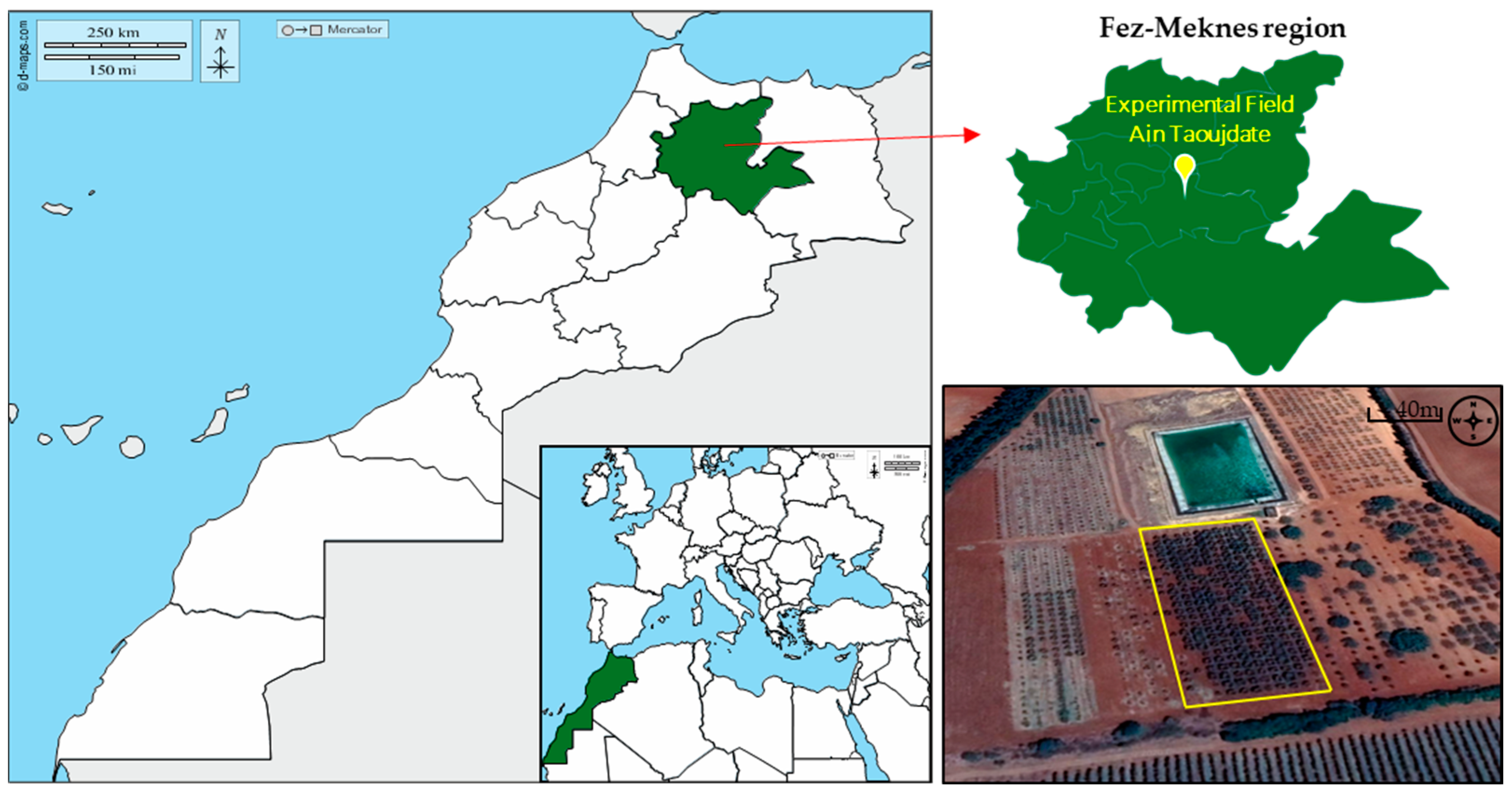
2.1. Study Area

The epidemiological study of the OLS disease was carried out in an ex situ collection maintained in the experimental station of National Institute of Agronomic Research (INRA), Ain Taoujdate, Morocco, (Region of Fez-Meknes; altitude: 550 m, latitude: 33.931031, longitude: 5.274508; Figure 1). This collection is made of more than eighty olive accessions from nine countries and was established in 2006 [24]. The density of the plantation is 5 × 5 m and maintenance cultural practices are limited to pruning, annual ploughing and drip irrigation. A panel of twenty true-to-type olive cultivars, genetically identified using microsatellite molecular markers in a previous study [24], were selected for this study (Table S1). Climate data (temperature, precipitation and relative humidity) were collected from the weather station of the experimental field.

2.2. Plant Materiel Sampling

Disease symptom progression rate was evaluated by monthly leaf sampling over a five-month period from March to July 2021. For every studied variety, 200 leaves were randomly sampled from the four cardinal points of two trees, 100 leaves each, disinfected with 90% alcohol, and placed in labelled sterile bags. Samples were kept in the laboratory under 4 °C for further investigation.

2.3. Assessment of Disease Incidence

Disease incidence, known as the percentage of infected leaves per variety, was determined according to the Teviotdale et al. protocol [27]. For each tree/variety, disease incidence was assessed as the percentage of symptomatic leaves including both visible and latent lesions. To determinate latent lesions, the olive leaves spots revealed by soaking symptomless samples in 5% NaOH solution for 15–20 min at room temperature as described by Shabi et al. [28]. The leaves were then examined and the numbers of infected leaves (recognizable by the appearance of dark circular spots) was recorded.

2.4. Assessment of Disease Severity

Disease severity was rated using a 0–8 scale. The scale considers the number of lesions per leaf with: 0 = no symptoms, 1 = 12.5%, 2 = 12.6% to 25%, 3 = 26% to 37%, 4 = 38% to 50%, 5 = 51% to 62%, 6 = 63% to 75%, 7 = 76% to 87% and 8 = 88% to 100% of the upper surface covered with black lesions [29].
Finally, the area under the disease progress curve (AUDPC) [30], a quantitative summary of disease intensity over time, was calculated based on the incidence and the severity evaluated monthly using the following formula:
A U D P C = i = 1 n 1 y i + y i + 1 2 t i + 1 t i
where:
  • yi+1 is the cumulative disease incidence in the i observation,
  • ti is the time at the observation and,
  • n is the total number of observations.

2.5. Assessment of Chlorophyll Content

Leaves were washed with distilled water and dried using a lyophilizer freeze dryer for 72 h. Leaves were then ground and 20 mg of the resulting fine powder material was stirred in 2 mL of 80% acetone for 1.5 h until all the pigments were extracted. The suspension was centrifuged at 14,000 rpm for 15 min at 4 °C. The optical density (OD) of supernatant was measured at both wavelengths 645 nm and 663 nm (UV-1700 Spectrophotometer, Shimadzu, Tokyo, Japan). The concentrations of chlorophyll pigments ‘a’ and ‘b’ were given by the following formulas [31]:
Cha = 12.7 (OD663) − 2.69 (OD645), and
Chb = 22.9 (OD645) − 4.86 (OD663)

2.6. Assessment of Total Phenolic and Flavonoid Contents

Leaf samples were collected in April where the conditions are optimal for the pathogen development. Total phenolic and flavonoid contents were assessed following the protocol previously described by Sanders et al. [32] and Xie and Bolling [33]. First, an amount of 1 g of leaf powder was transferred to polypropylene tubes and homogenized in 20 mL of ethanol and ultrapure water (80:20, v/v) at 4 °C for 15 min using an IKA T-18 Basic Ultra-Turrax homogenizer (IKAWerke GmbH & Co., Staufen, Germany). The homogenate was then centrifuged at 3000× g for 10 min at 4 °C. For each tree, three extractions were performed separately, and the supernatants were then pooled and filtered through Whatman No. 1 filter paper.
Total phenolic (TP) content was determined using the Folin–Ciocalteu micro method [34]. Three Folin’s reaction replicates were made for each sample. The reaction medium contained 40 μL of extract, 3160 μL of ultrapure water, 200 μL of the Folin–Ciocalteu reagent and 600 μL of 20% sodium carbonate solution. After 30 min of incubation at 40 °C, absorbance was measured at OD765. The TP content is expressed as gallic acid equivalent per dry weight (mg GAE/gdw).
Total flavonoid (TF) content was similarly measured on three replicates per sample using the colorimetric method with aluminum chloride [35]. Absorbance was measured at OD510 and the results were expressed as catechin equivalent per dry weight (mg CE/gdw).

2.7. Data analysis

All statistical analyses were performed using R statistical software. Analysis of variance and Turkey’s HSD tests with p < 0.05 as the significance level were used to determine the significance level of cultivars and sampling time on incidence, severity and AUDPC. In addition, Pearson’s correlation coefficients between different parameters were computed. Finally, Hierarchical Cluster Analysis (HCA) using Ward’s method based on Euclidian distance was performed on the twenty cultivars, and the dendrogram was constructed using the “dendextend” R package.

3. Results

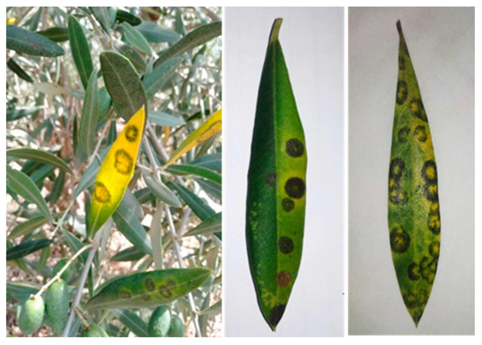
3.1. Field Observation and Disease Symptoms Identification

Field observations showed that the extent of OLS damage is cultivar-dependent. Typical OLS symptoms were observed in the field, mostly on the lower part of the trees, and consist of circular spots on the upper side of old leaves surrounded by a yellow halo, hence the name “peacock eye” (Figure 2). The color of these spots ranges from gray to brown or dark-brown to orange-yellow. These spots are often accompanied by chlorosis and yellowing of the leaves. Field observations revealed that the proportion of symptomatic leaves changed over time during the period of the study. At the beginning of the experiment, i.e., from March to May, the number of symptomatic leaves spiked, then dropped in late summer following trees’ defoliation.

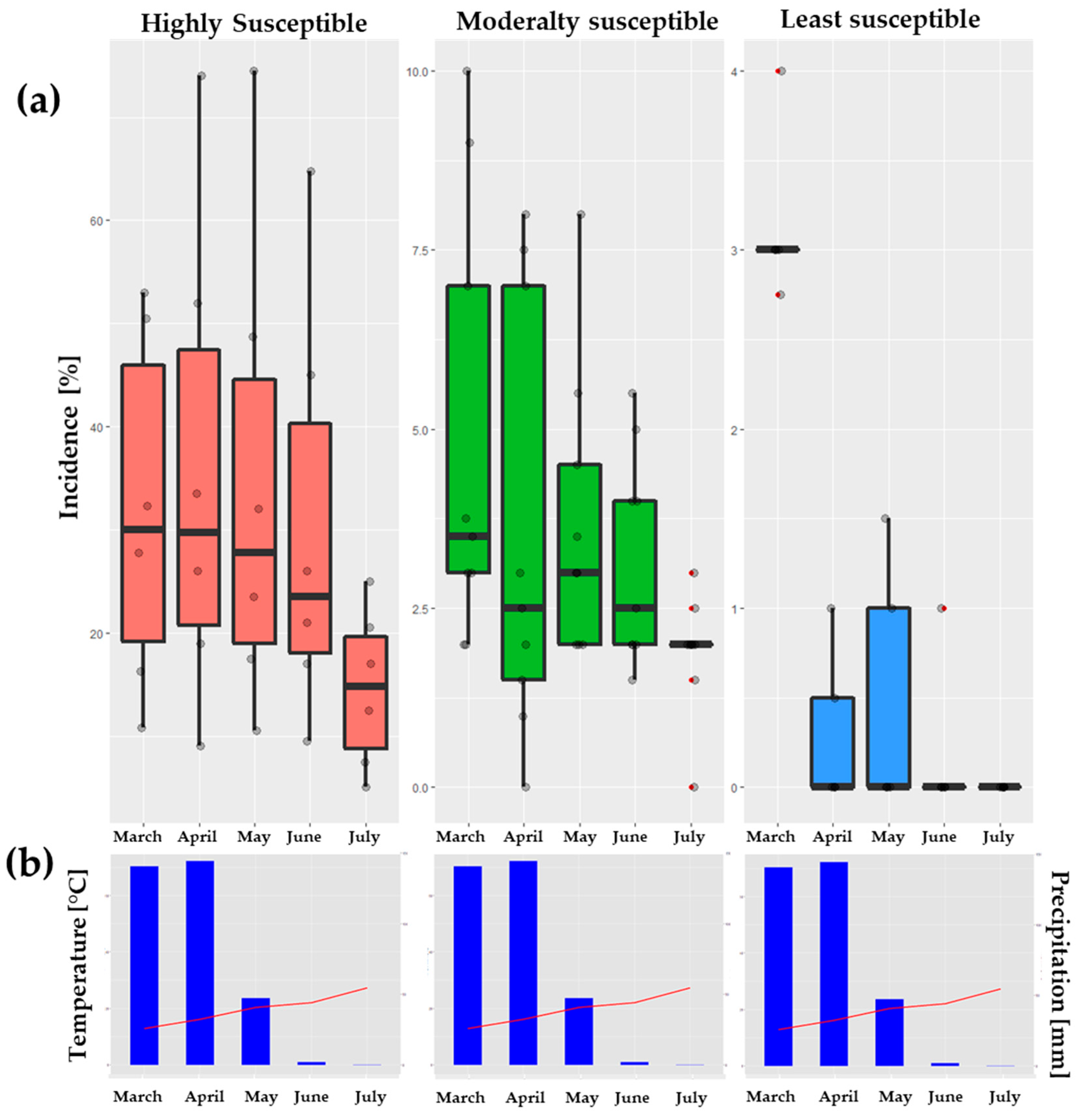
3.2. Assessment of Disease Incidence and Severity

The assessment of disease level is usually expressed by incidence and/or severity [36]. These indices are usually shown as indicators of the aggressiveness of the pathogen and/or of the effectiveness of the control treatments applied [37]. Here, disease incidence and severity varied significantly among varieties and sampling months (Table 1). Disease severity ranged from 0 to 74% and was highly and significantly correlated with incidence (r = 0.94, p-value < 0.001; Figure S1), indicating differential impact of OLS on olive cultivars. Indeed, four major groups were identified as susceptible, moderately susceptible, resistant and highly resistant based on hierarchical clustering analysis (Figure 3; Table S1). ‘Blanqueta’ cultivar is classified as the most susceptible with 74% of infected leaves. The cultivars ‘Picholine marocaine’, ‘Arbequine’, ‘Verdial Tansmontana’, ‘Piangente’ and ‘Tabelout’ had 9 to 52% infection rate and were classified as moderately susceptible. In contrast, the cultivars ‘Maurino’, ‘Carmelitana’, ‘Amellau’, ‘Bouchouk Soummam’, ‘Sevillenca’, ‘Galega Vulgar’, ‘Madonna Dell’ Impruneta’, ‘Meslala’ and ‘Grappolo’ were found to be moderately resistant, whereas ‘Ascolana Tenera’, ‘Frantoio’, ‘Leucocarpa’, ‘Changlot Real’ and ‘Chetoui’ proved to be the most resistant cultivars.

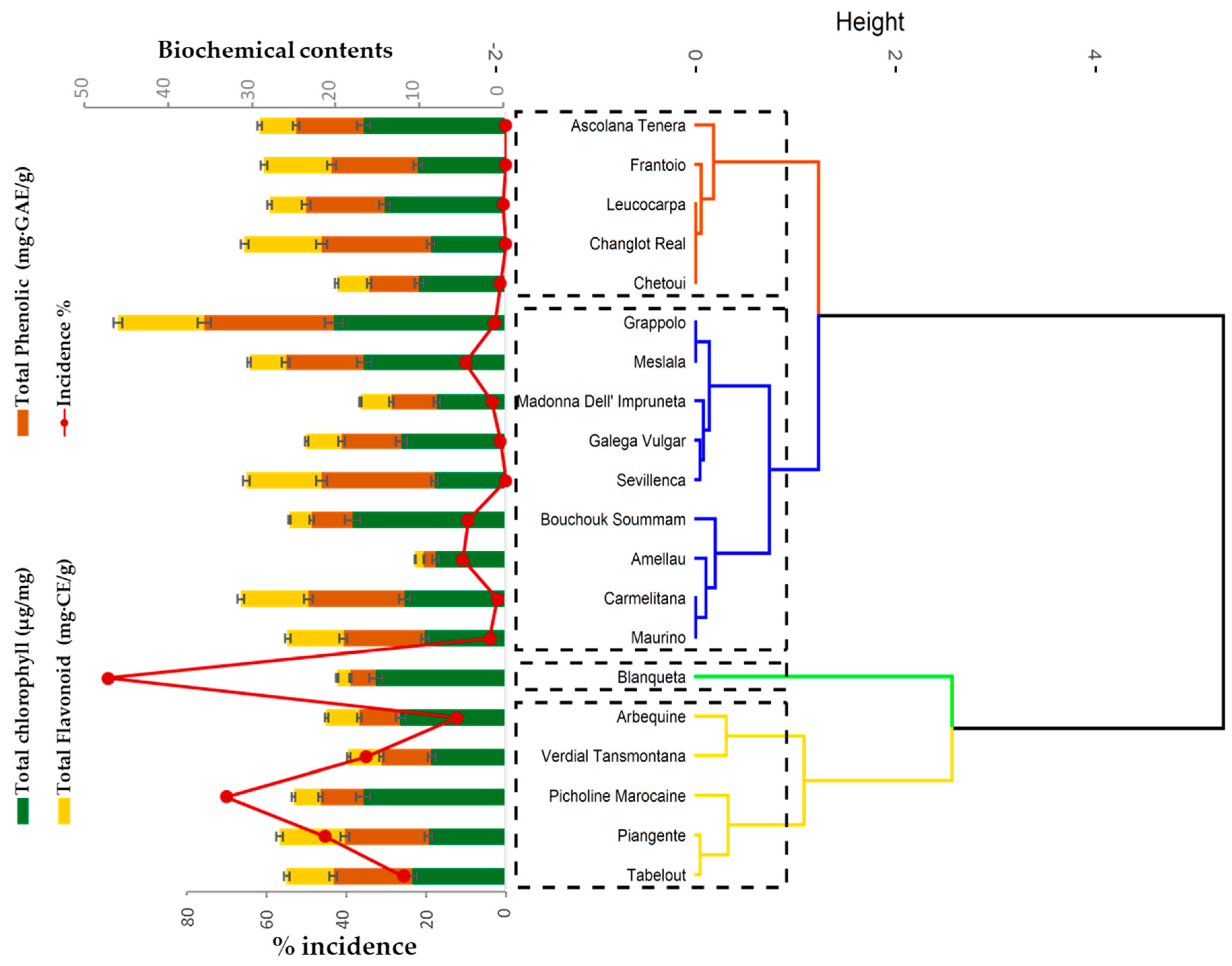
3.3. Determination of Total Chlorophyll, Phenolic and Flavonoid Contents

Total chlorophyll, phenolic and flavonoid leaf contents are reported in Figure 3 and Figure S1. Cultivars differed significantly in their chlorophyll content. ‘Grappolo’ cultivar had the greatest content in chlorophyll ‘a’ (0.65 µg/mg) and chlorophyll ‘b’ (0.32 µg/mg). ‘Verdial Tansmontana’ cultivar contained the least amount of chlorophyll ‘a’ (0.27 µg/mg) and ‘Madonna Dell Impruneta’ cultivar contained the least amount of chlorophyll ‘b’ (0.09 µg/mg Table S1).
Cultivar had a significant effect on phenolic and flavonoid contents. Specifically, ‘Grappolo’, ‘Sevillenca’, ‘Changlot Real’, ‘Carmelitana’, ‘Frantoio’, ‘Maurino’, and ‘Piangente’ showed the highest phenolic (1.4–15.03 mg·GAE/g) and flavonoid contents (1.01 and 10.01 mg·CE/g). Conversely, the lowest contents were found in the cultivars ‘Amellau’, ‘Picholine marocaine’, ‘Arbequine’ and ‘Blanqueta’. Interestingly, incidence was negatively correlated with polyphenol and flavonoid contents when computed across all cultivars, though this correlation was not significant (Figure S2). However, when determined among moderately susceptible cultivars only, phenols and flavonoids were significantly correlated to disease incidence (Figure S2). Overall, cultivars with high and/or moderate resistance to the OLS disease, such as ‘Leucocarpa’, had greater phenolic and flavonoid contents than susceptible cultivars such as ‘Picholine marocaine’ (Figure 3).

3.4. OLS Disease Progression over Time

AUDPC varied significantly among cultivars (Table 2) and was influenced by environmental conditions. Disease symptom development was more pronounced in spring (March and April) than in summer, suggesting the effect of environmental conditions such as temperatures, precipitation and relative humidity RH on disease progression within a campaign (Figure 4). The prevalence of infections increased significantly over time. Disease incidence dropped with decreasing rainfall between March (70.4 mm) and June (1 mm). High temperatures in June and July of more than 27 °C resulted in weak infection rate and thus low levels of disease incidence and severity (Figure 4).

4. Discussion

Climate changes, caused by the variation of temperatures and precipitations, increase the risk of fungal diseases on olive trees [38]. The present study investigated the resulting effect of various factors on the development of OLS disease in twenty olive cultivars maintained under a temperate Mediterranean climate with warm, dry summers (average of 17 °C) and rainy, cool winters (average of 470 mm).
Climatic conditions in the station are optimal for the development of F. oleagineum, the sporulation and germination of conidia, which explains the significant level of leaf damages encountered in our study. Our results revealed a strong relationship between climatic conditions and disease development indices over time. The variability of disease indices was highly affected by the sampling period and cultivar. A considerable disease occurrence (incidence and severity) was detected in spring when the conditions, principally precipitations, were favorable for OLS development.
Our findings show a substantial influence of weather condition on cultivar response to OLS. Previous investigations reported similar results on the effect of climatic conditions on the susceptibility of olive cultivars [20,29,38,39]. In fact, Obanor et al. [40] and Al-Khatib et al. [41] have reported that the variation in susceptibility of olive cultivars under field conditions is due to the effect of environmental conditions such as temperature, relative humidity and light. They also reported that conidia production is optimal at 15 °C under high humidity (100%), while conidia germination and infection require continuous free humidity for 12 to 24 h and temperatures ranging from 5 °C to 25 °C. Guechi and Girre [42] reported that the most favorable period for OLS infection is late autumn and spring. Other studies also reported that F. oleagineum infection occurs at the beginning of autumn [43,44]. In the absence of favorable conditions, particularly hot and dry summer, infection by F. oleagineum is latent without visible symptoms. The pathogen survives as mycelium in lesions on living leaf weeds resulting in the appearance of black spots as a response to NaOH reaction [45,46].
Regardless of climatic conditions, our work showed different levels of susceptibility of the studied cultivars to F. oleagineum. The susceptibility/resistance of cultivars is usually expressed by the percentage of infected leaves [27,28], the number of lesions and the diseased leaf area [29] occurring in low levels in resistant varieties. The studied varieties were classified into four main groups: susceptible, moderately susceptible, resistant and highly resistant to OLS. Our findings are in accordance with previous studies such as those reported in the OLEA database ([47], Table S1). In fact, Rahioui et al. [48] reported that ‘Picholine marocaine’ is a very susceptible cultivar and Ouerghi et al. [49] that ‘Chetoui’ is a resistant cultivar while Barranco et al. [50] reported that this latter is very susceptible to the OLS. Moreover, Carla et al. [51] highlighted that the variety ‘Galega vulgar’ is known to be moderately susceptible to OLS, which is consistent with our results, whereas Rallo et al. [52] reported that this variety is tolerant. Finally, ‘Frantoio’ is classified as tolerant cultivar [53] and ‘Leucocarpa’ as highly resistant to F. oleagineum [12].
The discordance found in different studies, including our research, may be explained by the mis-identification of true-to-type varieties, variation in virulence among pathogen populations, and/or environmental conditions where the studies were conducted [54]. In the present work, the studied cultivars were identified using molecular markers [24] and therefore the susceptibility/resistance observed is mainly related to the ontogenic resistance (OR: the ability of the cultivar to resist or tolerate the OLS under the same conditions). The OR is generally controlled by two factors: physical and chemical characteristics of the olive leaf cuticle [18,55]. Some authors have attested that old leaves are more susceptible than young leaves [13], while others have reported that young leaves are more susceptible than old leaves [56], which is in accordance with our filed observations. These results are consistent with those of Lόpez-Doncel et al. [57] who noted that young leaves are highly susceptible to OLS disease compared to older leaves. The age-related resistance may be associated with water-repellent waxes accumulated on leaves surface which prevent the formation of an optimal water film for the pathogen germination. In fact, older leaves are more cutinized than the younger ones and are more resistant to OLS [49,58]. The thicker cuticle of older leaves may block the penetration of the pathogen compared to younger leaves [59]. Moreover, according to previous studies, the cultivar susceptibility may be determined by the presence/absence of several genes where their expression occurs after infection and the resistance to OLS may be due to an active defense response genotype dependent (162 cDNA fragments; [46,60]).
Leaf chlorophyll content was smaller in the symptomatic leaves of some cultivars compared to healthy ones. The major alteration induced by fungal invasion appears to be the degradation of chloroplast in palisade parenchyma cells and progressively devoid of their cytoplasmic content. Consequently, the chlorophyll pigments disappeared gradually at advanced stages of infection, which gives a yellowish appearance to the diseased leaves [61]. This explains the significant drop in the chlorophyll content in the leaves severely infected by the causal agent.
Cultural practices represent other important factors in the development of the disease. Our field investigations revealed that the proportion of infected leaves depends on the position of the leaves in the canopy [15]. The severity of the disease decreases going from the lower part to the upper part of the tree. This finding may be explained by the fact that the lower parts of the tree retain more humidity level, while in the upper parts leaves are more exposed to light and aeration [29]. The exposition of leaves is known to be another factor of the pathogen development. In fact, the proportion of damaged leaves exposed to the north is usually higher than the leaves exposed to the south. The northern side, the less sunny in the tree is the most contested, followed by the western, southern and eastern sides [13,15,62,63]. The unpruned trees have a high incidence compared to pruned trees where tree thinning creates conditions for aeration of the inner parts of the canopy with less relative humidity [13,18]. Soil moisture management and irrigation control are also very important factors to control, and many studies concluded the effect of increased fertilization on the development of OLS. In fact, high severity is recorded in trees fertilized with high levels of nitrogen [64]. This may be explained by the fact that a high nitrogen supply decreases the polyphenol content and therefore the increase in the severity of the disease [65]. Hence, the need for a more rational and sustainable use of nitrogen fertilizers.

5. Conclusions

Olive tree cultivars respond differently to OLS. This variability is a function of time, environmental conditions, and phenols and flavonoids leaf contents. These findings shed light on mechanisms that govern OLS development and can guide the best choice of varieties to be grown in specific olive-growing regions. Additionally, this work paves the way for a better understanding of the factors involved in the susceptibility/resistance of olive cultivars to boost breeding programs for olives, aiming to select highly resistant cultivars. Finally, elucidating the relationship between disease progression and climatic conditions will allow developing prediction models based on climatic conditions to foresee the risk of OLS in different regions. Overall, our study contributes significant insights into the dynamic of the OLS and the functioning of the olive–OLS pathosystem in the prospect of developing effective and sustainable management methods against this disease.

Supplementary Materials

The following supporting information can be downloaded at: https://www.mdpi.com/article/10.3390/horticulturae9050589/s1, Figure S1: Pearson correlation between the incidence and severity disease indices in each sampling month; Figure S2: Pearson correlation between the OLS disease incidence and biochemical contents in each sampling month of the study; Table S1: List of cultivars used in the study, results of different parameters evaluated and their susceptibility to OLS disease.

Author Contributions

Conceptualization, K.H. and A.E.B.; methodology, K.H., A.B. and F.A.; software, I.M. and H.C.; validation, K.H. and A.E.B.; formal analysis, I.M and H.C.; investigation, K.H. and S.E.I.E.H.; resources, A.E.B.; data curation, K.H. and I.M.; writing—original draft preparation, K.H. All authors have read and agreed to the published version of the manuscript.

Funding

This research was funded by PRIMA FREECLIMB project 2020/2022 (Fruit Crops Resilience to Climate Change in the Mediterranean Basin). Coordinated by Dr. Ahmed El Bakkali from National Institute of Agronomic Research, Meknes-Morocco.

Institutional Review Board Statement

Not applicable.

Data Availability Statement

The original contributions presented in the study are included in the article/Supplementary Material, further inquiries can be directed to the corresponding author.

Acknowledgments

This research was funded by the project FREECLIMB “Fruit Crops Adaptation To Climate Change in The Mediterranean Basin” in the frame of the Partnership for Research and Innovation in the Mediterranean Area (PRIMA; call 2018). Authors warmly thank Adil Essarioui for the helpful comments on the manuscript and for his English revision.

Conflicts of Interest

The authors declare no conflict of interest.

References

  1. Ouraich, I.; Tyner, W.E. Moroccan agriculture, climate change, and the moroccan green plan: A CGE analysis. Afr. J. Agric. Resour. Econ. 2014, 13, 307–330. [Google Scholar]
  2. Cramer, W.; Guiot, J.; Fader, M.; Garrabou, J.; Gattuso, J.P.; Iglesias, A.; Lange, M.A.; Lionello, P.; Llasat, M.C.; Paz, S.; et al. Climate change and interconnected risks to sustainable development in the Mediterranean. Nat. Clim. Chang. 2018, 8, 972–980. [Google Scholar] [CrossRef]
  3. Portner, H.O.; Roberts, D.C.; Tignor, M.M.B.; Poloczanska, E.; Mintenbeck, K.; Alegria, A.; Craig, M.; Langsdorf, S.; Loschke, S.; Moller, V.; et al. Climate Change 2022: Impacts, Adaptation and Vulnerability. Contribution of Working Group II to the Sixth Assessment Report of the Intergovernmental Panel on Climate Change. 2022. Available online: https://www.ipcc.ch/report/ar6/wg2/ (accessed on 28 February 2022).
  4. Barbuzano, J. Climate change will reduce spanish olive oil production. Eos 2020, 101. [Google Scholar] [CrossRef]
  5. Kaniewski, D.; Van Campo, E.; Boiy, T.; Terral, J.F.; Khadari, B.; Besnard, G. Primary domestication and early uses of the emblematic olive tree: Palaeo botanical, historical and molecular evidences from the Middle East. Biol. Rev. 2012, 87, 885–899. [Google Scholar] [CrossRef] [PubMed]
  6. Kalua, C.M.; Allen, M.S.; Bedgood, D.R.; Bishop, A.G.; Prenzler, P.D.; Robards, K. Olive oil volatile compounds, flavour development and quality: A critical review. Food Chem. 2007, 100, 273–286. [Google Scholar] [CrossRef]
  7. Cannon, J. Cradle of transformation: The Mediterranean and Climate Change. Mongabay. 2022. Available online: https://news.mongabay.com/2022/04/cradle-of-transformation-the-mediterranean-and-climate-change/ (accessed on 28 April 2022).
  8. Abdou, Z.S. Impact of climate change on plant diseases and IPM strategies. In Plant Diseases-Current Threats and Management Trends; IntechOpen: London, UK, 2019. [Google Scholar] [CrossRef]
  9. Rodrigues, Â.M.; Valentim, C.; Margarida, A.; Eugénia, G.; Raimundo, S.; Carlos, M.; Correia, A.; Albino, B. The effect of nitrogen fertilization on the incidence of olive fruit fly, olive leaf spot and olive anthracnose in two olive cultivars grown in rainfed conditions. J. Sci. Hortic. 2019, 256, 108658. [Google Scholar] [CrossRef]
  10. Castagne, J.L.M. Catalogue des Plantes Qui Croissent Naturellement Aux Environs de Marseille; Nicot et Pardigon: Pont-Moreau, France, 1845; p. 220. [Google Scholar]
  11. González-Lamothe, R.; Segura, R.; Trapero, A.; Baldoni, L.; Botella, M.A.; Valpuesta, V. Phylogeny of the fungus Spilocaea oleagina, the causal agent of peacock leaf spot in olive. FEMS Microbiol. Lett. 2002, 210, 149–155. [Google Scholar] [CrossRef]
  12. Renaud, P. Ecologie de la maladie de l’œil de paon et résistance variétale dans leurs incidences sur la culture de l’olivier dans le pays. Afr. Med. Al-Awamia 1968, 26, 55–74. [Google Scholar]
  13. Issa, T.; Almadi, L.; Jarrar, S.; Tucci, M.; Buonaurio, R.; Famiani, F. Factors affecting Venturia oleaginea infections on olive and effects of the disease on floral biology. Phytopathol. Med. 2019, 58, 221–229. [Google Scholar]
  14. ONSSA: Office National de Sécurité Sanitaire et Alimentaire. Bulletin de Veille Phytosanitaire; 2016. Available online: https://www.onssa.gov.ma/evaluation-des-risques/surveillance-des-risques-phytosanitaires/ (accessed on 28 February 2022).
  15. Rhimini, Y.; Chliyeh, M.; Ouazzani, A.; Chahdi, A.; Touati, J.; Ouazzani, A.; Touhami, R.; Benkirane, R.; Douira, A. Influence of certain cultural practices and variable climatic factors on the manifestation of Spilocaea oleagina, olive peacock spot agent in the north western region of Morocco. Inter J. Pure Appl. Biosci. 2014, 2, 1–9. [Google Scholar]
  16. Ater, M.; Essalou, L.; Ilbert, H.; Moukhli, A.; Khadari, B. L’Oléiculture au maroc de la préhistoire à nos jours: Pratiques, diversité, adaptation, usages, commerce et politiques. Options Méditerranéennes Séminaires Méditerranéennes 2016, 2, 224. [Google Scholar]
  17. Couanon, W.; Le Verge, S.; Pinatel, C. L’OEIL DE PAON, les Fiches Techniques de l’AFIDOL; Centre Technique de l’Olivier: Aix-en-Provence, France, 2018; pp. 1–2. [Google Scholar]
  18. Roubal, C. Tavelures du Pommier et de L’olivier: Réalisation de Modèles Epidémiologiques Par des Méthodes Exploitant des Observations Biologiques Acquises au Verger. Sciences Agricoles; Université d’Avignon: Avignon, France, 2017; p. 186. [Google Scholar]
  19. Trapero, A.; Roca, R.; Luis, F.; Moral, J.; Trapero, C.; López-Escudero, F.J. El Olivar Ecologico, Chapter: Capítulo 7: Las Enfermedades y su Manejo en el Olivar Ecológico; Junta de Andalucía: Andalusia, Spain, 2011; pp. 206–256.
  20. Salman, M.; Hawamda, A.; Amarni, A.; Rahil, M.; Hajjeh, H.; Natsheh, B.; Abuamsha, R. Evaluation of the incidence and severity of olive leaf spot caused by Spilocaea oleagina on olive trees in Palestine. Am. J. Plant Sci. 2011, 2, 457–460. [Google Scholar] [CrossRef]
  21. Bartolini, G.; Prevost, G.; Messeri, C.; Carignani, C. Olive germplasm: Cultivars and world-wide collections. In FAO SaPGRSo; FAO: Rome, Italy, 2005. [Google Scholar]
  22. Khadari, B.; Charafi, J.; Moukhli, A.; Ater, M. Substantial genetic diversity in cultivated Moroccan olive despite a single major cultivar: A paradoxical situation evidenced by the use of SSR loci. Tree Genet. Genomes 2008, 4, 213–221. [Google Scholar] [CrossRef]
  23. Barguigua, A.; Zahir, I.; Youss, S.; Fikri, N.; Youss, B. Prospection des maladies microbiennes de l’olivier dans la région Tadla-Azilal. Rev. Mar. Sci. Agron. Vét. 2020, 8, 331–338. [Google Scholar]
  24. El Bakkali, A.; Mekkaoui, A.; El Iraqui, E.S.; Essarioui, A.; Khadari, B. Addressing the challenge of cultivars identification and authentication in mediterranean olive collections: A case study in morocco. Eur. Sci. J. 2020, 16, 339–355. [Google Scholar] [CrossRef]
  25. D-map. Available online: https://d-maps.com/index.php?lang=en (accessed on 31 August 2009).
  26. Google-Earth. Available online: https://www.google.com/intl/fr/earth/ (accessed on 20 March 2021).
  27. Teviotdale, B.L.; Sibbett, G.S.; Harper, D. Several copper fungicides control olive leaf spot. Cali. Agric. 1989, 43, 30–31. [Google Scholar]
  28. Shabi, E.; Birger, R.; Lavee, S. Leaf spot (Spilocaea oleagina) of olive in israel and it control. Acta Hortic. 1994, 356, 390–394. [Google Scholar] [CrossRef]
  29. Macdonald, A.J.; Walter, M.; Trought, M.; Frampton, C.M.; Burnip, G. Survey of olive leaf spot in New Zealand. Plant Prot. 2000, 53, 126–132. [Google Scholar] [CrossRef]
  30. Jeger, M.J.; Viljanen-Rollinson, S.L.H. The use of the area under disease-progress curve (AUDPC) to assess quantitative disease resistance in crop cultivars. Theor. Appl. Genet. 2001, 102, 32–40. [Google Scholar] [CrossRef]
  31. Singh, P.V.; Billore, R.P. Relationship between chlorophyll and energy contents of the Andropogon grassland community. Photosynthetica 1975, 9, 93–95. [Google Scholar]
  32. Sanders, T.H.; McMichael, R.W.; Hendrix, K.W. Occurrence of resveratrol in edible peanuts. J. Agric. Food Chem. 2020, 48, 1243–1246. [Google Scholar] [CrossRef] [PubMed]
  33. Xie, L.; Bolling, B.W. Characterisation of stilbenes in California almonds (Prunus dulcis) by UHPLC–MS. Food Chem. 2014, 148, 300–306. [Google Scholar] [CrossRef] [PubMed]
  34. Waterhouse, A.L. Determination of total phenolics. Food Annal. Chem. 2002, 6, 111–118. [Google Scholar]
  35. Barreira, J.C.; Ferreira, I.C.; Oliveira, M.; Pereira, J.A. Antioxidant activities of the extracts from chestnut fower leaf skins and fruit. Food Chem. 2018, 107, 1106–1113. [Google Scholar] [CrossRef]
  36. Bock, C.H.; Chiang, K.S.; Del Ponte, E.M. Plant disease severity estimated visually: A century of research, best practices, and opportunities for improving methods and practices to maximize accuracy. Tropi. Plant Pathol. 2021, 47, 25–42. [Google Scholar] [CrossRef]
  37. Seem, R. Disease incidence and severity relationships. Ann. Rev. Phytopathol. 1984, 22, 133–150. [Google Scholar] [CrossRef]
  38. Sanei, S.J.; Razavi, S.E. Differentiation of olive Colletotrichum gloeosporioides populations on the basis of vegetative compatibility and pathogenicity. Afri. J. Agricul. Res. 2011, 6, 2099–2107. [Google Scholar]
  39. Kleef, F.; Salman, M. Antifungal effect of Ambrosia artemisiifolia L. Extract and chemical fungicide against Spilocaea oleagina causing olive leaf spot. Arab. J. Sci. Eng. 2022, 43, 113–117. [Google Scholar] [CrossRef]
  40. Obanor, F.O.; Jaspers, M.; Jones, E.; Walter, M. Green house and field evaluation of fungicides for control of olive leaf spot in New Zealand. Crop. Prot. 2008, 120, 211–222. [Google Scholar]
  41. Al-Khatib, M.; Alhussaen, K.; El-Banna, N.; Zyadeh, M. Biological control of olive leaf spot (peacock spot disease) caused by Cycloconium oleaginum (Spilocea oleagina). J. Microbiol. 2010, 2, 64–67. [Google Scholar]
  42. Guechi, A.; Girre, L. Sources of Cycloconium oleaginum (Cast.) conidia for infection of olive leaves and conditions determining leaf spot disease development in the region of Setif (Algeria). J. Mycopathol. 1994, 125, 163–171. [Google Scholar] [CrossRef]
  43. Obanor, F.O.; Walter, M.; Jones, E.; Jaspers, M.V. Sources of variation in a field evaluation of the incidence and severity of olive leaf spot. N. Z. Plant. Prot. 2005, 58, 273–277. [Google Scholar] [CrossRef]
  44. Graniti, A. Olive scab: A review. Bull. OEPP 1993, 23, 377–384. [Google Scholar] [CrossRef]
  45. Roca, L.F.; Juan, M.M.; Jose, R.V.; Arantxa, A.; Rodrigues, O.; Trapero, A. Copper Fungicides in the Control of Olive Diseases; FAO Olive Network: Rome, Italy, 2007. [Google Scholar]
  46. Benítez, Y.; Botella, M.A.; Trapero, A.; Alsalimiya, M.; Caballero, J.L.; Dorado, G.; Muñoz-Blanco, J. Molecular analysis of the interaction between Olea europaea and the biotrophic fungus Spilocaea oleagina. Mol. Plant Pathol. 2005, 6, 425–438. [Google Scholar] [CrossRef]
  47. Bartolini, G. OLEA Databases. Available online: http://www.oleadb.it/ (accessed on 31 August 2009).
  48. Rahioui, B.; Aissam, S.; Messaouri, H.; Moukhli, A.; Khadari, B.; El Modafar, C. Role of phenolic metabolism in the defense of the olive-tree against leaf-spot disease caused by Spilocaea oleagina. Int. J. Agric. Biol. 2013, 15, 273–278. [Google Scholar]
  49. Ouerghi, F.; Rhouma, A.; Rassaa, N.; Hennachi, I.; Bouzid, N. Factors affecting resistance of two olive cultivars to leaf spot disease in the North-West of Tunisia. Eur. J. Adv. Res. Biol. Life Sci. 2017, 4, 39–51. [Google Scholar]
  50. Barranco, N.D.; Cimato, A.; Fiorino, P.; Rallo, R.L.; Touzani, A.; Castañeda, C.; Serafini, E.; Trujillo, N.I. World Catalogue of Olive Varieties, COI; Mundi-Prensa: Madrid, Spain, 2000; p. 360. [Google Scholar]
  51. Carla, M.R.; Varanda, A.; Patrick, M.; Miguel, L.; Maria, D.; Campos, A.; Maria, R. Fungal communities associated with peacock and cercospora leaf spots in olive. Plant 2019, 8, 169. [Google Scholar]
  52. Rallo, L.; Barranco, D.; Caballero, J.M.; Del Rio, C.; Martin, A.; Tous, J.; Trujillo, I. Variedades de Olivoen Espana; Mundi-Prensa: Madrid, Spain, 2005; p. 480. (In Spanish) [Google Scholar]
  53. Hadiddou, A.; Oukabli, A.; Moudaffar, C.; Mamouni, A.; Gaboun, F.; Mekaoui, A.; El Fechtali, M. Evaluation des performances de production de 14 variétés de l’olivier (Olea europaea L.) Nationales et méditerranéennes dans deux systèmes contrastés de culture (pluvial et irrigué) au Maroc. Al Awamia 2013, 127, 22–43. [Google Scholar]
  54. Moral, J.; Ávila, L.M.; López-Doncel, M.; Alsalimiya, R.; Oliveira, F.; Gutiérrez, N.; Benali, A.; Roca, L.F.; Navarro, N.; Bouhmidi, K.; et al. Resistencia a los repilos de distintas variedades de olivo. Vida Rural. 2005, 208, 34–41. [Google Scholar]
  55. Adawi, A.; Jarrar, S.; Almadi, L.; Alkowni, R.; Gallo, M.; D’Onghia, A.M.; Buonaurio, R.; Famiani, F. Effectiveness of low copper-containing chemicals against olive leaf spot disease caused by Venturia oleaginea. Agriculture 2022, 12, 326. [Google Scholar] [CrossRef]
  56. Trigui, M.; Ben Hsouna, A.; Hammami, I.; Culioli, G.; Ksantini, M.; Tounsi, S.; Jaoua, S. Efficacy of Lawsonia inermis leaves extract and its phenolic compounds against olive knot and crown gall diseases. Crop. Prot. 2013, 45, 83–88. [Google Scholar] [CrossRef]
  57. López-Doncel, L.M.; Viruega, J.R.; Trapero, A. Respuesta del olivo a la inoculación con Spilocaea oleagina, agente del repilo. Bol. San. Veg. Plagas. 2000, 26, 349–363. [Google Scholar]
  58. Sergeeva, V.; Braun, U.; Spooner-Hart, R.; Nair, N. Observations on spot caused by Fusicladium oleagineum on olives (Olea europaea) in new south Wales, Australia. Australas. Plant Dis. 2009, 4, 26–28. [Google Scholar]
  59. Jayaramraja, P.R.; Pius, P.K.; Manian, S.; Meenakshi, T.S. Certain factors associated with blister blight resistance in Camellia sinensis (L.). Physiol. Mol. Plant Pathol. 2005, 67, 291–295. [Google Scholar] [CrossRef]
  60. Rhouma, A.; Chttaoui, M.; Krid, S.; Elbsir, H.; Msallem, M.; Triki, M.A. Evaluation of susceptibility of an olive progeny (Picholine x Meski) to olive leaf spot disease caused by Fusicladium oleagineum. Eur. J. Plant Pathol. 2013, 135, 23–33. [Google Scholar] [CrossRef]
  61. Lanza, B.; Di Serio, M.G.; Di Giovacchino, L. Long-term spreading of olive mill wastewater on olive orchard: Effects on olive production, oil quality, and soil properties. Commun. Soil. Sci. Plant Anal. 2017, 48, 2420–2433. [Google Scholar] [CrossRef]
  62. Viruega, J.R.; Roca, L.F.; Moral, J.; Trapero, A. Factors affecting infection and disease development on olive leaves inoculated with Fusicladium oleagineum. Plant Dis. 2011, 95, 1139–1146. [Google Scholar] [CrossRef]
  63. Tajnari, H. Étude bio-écologique d’Euphyllura olivina Costa (Hom. Psyllidae) Dans les Régions du Haouz et d’Essaouira: Mise en Evidence d ‘un état de Diapause Ovarienne. Master’s Thesis, École Nationale D’agriculture, Meknes, Maroc, 1992; 153p. [Google Scholar]
  64. Veresoglou, S.D.; Barto, E.K.; Menexes, G.; Rillig, M.G. Fertilization affects severity of disease caused by fungal plant pathoges. Plant Pathol. 2012, 62, 961–969. [Google Scholar] [CrossRef]
  65. Fernandez, E.R.; Beltran, G.; Sanchez-Zamora, M.A.; García-Novelo, J.; Aguilera, M.; Uceda, M. Olive oil quality decreases with nitrogen over fertilization. Hort. Sci. 2006, 41, 215–219. [Google Scholar]
Figure 1. Sampling site of the study [25,26].
Horticulturae 09 00589 g001
Figure 2. Symptoms and signs of olive leaf spot disease (Fusicladium oleagineum).
Horticulturae 09 00589 g002
Figure 3. Hierarchical clustering contructed for the twenty olive cultivars as classified into four groups based on their susceptibility to OLS disease. Biochemical parameters contents and disease incidence recorded in April are indicated for each cultivar.
Horticulturae 09 00589 g003
Figure 4. Disease incidence in olive cultivars according to sampling months and climatic conditions. Extent of disease incidence over cultivars according to sampling months is shown as boxplots (a). Cultivars (round spots) were classified according to their level of susceptibility: red column: cultivars with a high susceptibility to OLS, green column: cultivars with a moderate susceptibility, and blue column: resistant cultivars. Temperature and precipitation data during the period of the study are indicated (b).
Horticulturae 09 00589 g004
Table 1. Analysis of variance (ANOVA) for the two disease indices.
Incidence (%)Severity (%)
Cultivars352.34 ***94.36 ***
Month1180.59 ***242.77 ***
Cultivars—Month113.19 ***31.30 **
Error59.83416.843
R20.7720.755
**, *** Significance at p-value < 0.01 and p-value < 0.001, respectively.
Table 2. Analysis of variance of AUDPC for the two disease indices.
Incidence (%)Severity (%)
Cultivars693,963.05 *223,763.39 *
Error304,815.1996,429.702
R20.6840.688
*, Significance at p-value < 0.05.
Disclaimer/Publisher’s Note: The statements, opinions and data contained in all publications are solely those of the individual author(s) and contributor(s) and not of MDPI and/or the editor(s). MDPI and/or the editor(s) disclaim responsibility for any injury to people or property resulting from any ideas, methods, instructions or products referred to in the content.

