Article

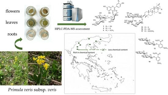
Exploring the Chemical Content of Primula veris L. subsp. veris Wild-Growing Populations along a Climate Gradient: An HPLC-PDA-MS Quality Assessment of Flowers, Leaves and Roots for Sustainable Exploitation

by
Ilias Stefanis
1,
Paschalina Chatzopoulou
2,
Nikos Krigas
2,* and
Anastasia Karioti
1,*
1
Laboratory of Pharmacognosy, School of Pharmacy, Aristotle University of Thessaloniki, University Campus, 54124 Thessaloniki, Greece
2
Hellenic Agricultural Organization—DEMETER, Institute of Breeding and Plant Genetic Resources, 57001 Thessaloniki, Greece
*
Authors to whom correspondence should be addressed.
Horticulturae 2023, 9(10), 1120; https://doi.org/10.3390/horticulturae9101120
Submission received: 10 September 2023 / Revised: 2 October 2023 / Accepted: 7 October 2023 / Published: 10 October 2023

Abstract

:
Primula veris (cowslip) is an important medicinal–aromatic plant used traditionally for its expectorant and anti-inflammatory properties, as well as a valuable horticultural plant with ornamental value and agroalimentary interest (edible flowers and leaves). With extensive illegal harvesting across northern Greece to date, the long-term survival of Greek cowslip wild-growing populations seems compromised. With the aim to characterize the phytochemical content of locally adapted native genotypes of P. veris subsp. veris, we examined samples from 13 wild-growing populations sourced from 8 mountain areas along a longitudinal gradient of northern Greece. Flowers, leaves, and roots were separately evaluated with HPLC-PDA-MS for their chemical content to select the genotypes associated with the best phytochemical traits. The flowers were found to contain mainly flavonoids, isorhamnetin, quercetin, and kaempferol triglycosides, with varied population contents, generally higher in northwestern population samples with a colder and more humid climate; however, all in line with the European Medicines Agency’s (EMA) reports. The leaves were characterized by the prevalence of kaempferol and quercetin triglycosides, being generally higher than that of the flowers. In the roots, saponins were detected in relatively low percentages, and the phenolic glycosides were found up to nearly 2%. The results of this study suggest a potential specificity of the marginal Greek genotypes being at the edge of the species’ native distribution range in Europe and Asia. The phytochemical characterization herein supports the domestication process of Greek native cowslip genotypes. In turn, this may alleviate the overharvesting pressure in wild populations, thus contributing to species conservation and offering an incentive for farmers to sustainably cultivate at local scales well-adapted indigenous genotypes of high value.

Graphical Abstract

1. Introduction

Primula veris L. (Primulaceae), commonly known as cowslip, is an herbaceous perennial plant which is native almost across Europe (from the northern parts of the Mediterranean to Scandinavia) and in a large part of Asia, extending from Siberia and Manchuria to the Caucasus and Iran [1,2]. With four subspecies across its range, this species has been referred to as model plant by Charles Darwin, who was fascinated by its complex reproductive system, owing to the natural coexistence of different flower forms with the diversified positioning of female and male sex organs (S- and L-morph); this is known to date as heteromorphy (or heterostyly), being a form of genetically controlled flower polymorphism [3,4]. Since then, P. veris has continued to attract the attention of the scientific community, especially of environmentalists, as it is considered an important index of human-mediated environmental changes (including climate change). Anthropogenic impact, overexploitation of land, change in land uses, along with the drastic warming of Arctic or alpine regions can lead to the alteration of P. veris habitats, with detrimental effects on its survival [5]. In this line, an initiative was recently launched by Estonian scientists at the pan-European level with the aim to detect possible morph-frequency aberrations from the natural 1:1 ratio of flower morphs (isoplethy of S- and L-morphs) of P. veris across Europe and at inferior regional scales [6,7].
Besides the botanic, genetic, ecological, and evolutionary interest, P. veris is also an important medicinal plant, and is additionally associated with a high ornamental and agroalimentary value. Infusions and decoctions have been traditionally used as expectorants in cases of cough, asthma, bronchitis, and catarrh [8,9,10]. Triterpenoid saponins, such as primulic acids I/II and priverosaponin B, are considered the main bioactive molecules reported in the literature, involved in a mechanism of action which has not yet been adequately explained [8]. The latter employs the irritation of the gastric mucosa and mucous membranes in the throat and respiratory tract, followed by an increase in bronchial secretion, as well as a decrease in the viscosity of the sputum due to the surface-tension-lowering action of the saponins. To date, the official registration of this traditionally used herbal drug in the European Medicines Agency (EMA) has paved the way to the commercialization of P. veris herbal medicinal products. Consequently, extracts from cowslip roots and flowers, usually in combination with thyme, appear as components of many herbal medicinal products available on the market, such as Bronchicum®, Bronchosol®, and Bronchipret®. More recent pharmacological data bring to light other interesting applications of P. veris to consider in the future; for example, the lipophilic extracts of cowslip flowers in combination with Cistus creticus L. subsp. creticus, the latter of which has recently been demonstrated to exert an anti-influenza virus activity [11]. In addition, a solid extract from the aerial parts of the plant has been found to increase the myocardial contractility in an animal model of chronic heart failure [12], exerting cardioprotective activity in a chronic alcohol intoxication rat model [13]. The latter has been attributed by the authors to the strong antioxidant effects of cowslip flavonoids. Cowslip flowers have also been traditionally used against nervousness, headache, and rheumatism [10], besides their use as edible (floral) elements in gastronomy. The extracts from cowslip flowers also find modern applications in cosmetics due to their anti-inflammatory and antioxidant properties [14,15,16], whereas their delicate scent has long ago been exploited in perfumery [17]. Besides their pleasant scent and flavor, cowslip flowers are a good source of vitamin C, and together with the leaves, they have been used in culinary applications [18].
From another point of view, P. veris is praised for its early-spring bright-yellow flower color and delicate perfume, which renders this ornamental species as popular for home or pot gardening and landscaping with native plants [19]. Indeed, Primula spp. are among the most popular ornamental plants in Europe, especially for the late winter and early spring market. In 2012, Primula sales ranked fifth in the market of a major consumer of ornamental products, namely Germany [20]. To date, P. veris includes four infraspecific taxa, namely: subsp. canescens (Opiz) Hayek ex Ludi, found in central and southcentral European countries; subsp. columnae (Ten.) Maire & Petitm., occurring in the mountains of southern Europe (excepting southern Balkan countries, such as Greece) and Turkey; subsp. macrocalyx (Bunge) Lüdi, found in the Asiatic part of its range; the typical subsp. veris, recorded across its European range [1], all with distinct morphological variations that are appreciated in the ornamental industry.
For all the above-mentioned reasons, P. veris subsp. veris has gained popularity, which in turn has led to its uncontrollable harvesting from the wild. In Greece, for example, where P. veris subsp. veris is limited in the cooler higher-altitude zones, extensive illegal harvesting events have been reported to occur, which may further compromise the long-term survival of its wild-growing populations [21,22,23]. Therefore, both in situ (natural reserves, Natura 2000 sites, Important Plant Areas) and ex situ (botanic gardens, ex situ field collections, and seed banks) conservation strategies need to be employed for its protection. To this end, the cultivation and adaptation of P. veris in man-made settings is an imperative action to ensure its survival in the wild habitats of Greece and beyond, producing adequate quantities of plant material and addressing the market demand for its sustainable exploitation in the pharma–cosmetic or the agroalimentary and the ornamental–horticultural sectors. To this end, an applied research project was undertaken by the local Greek authorities of Epirus (northwestern Greece) in collaboration with the Hellenic Agricultural Organization Demeter (ELGO Dimitra) for the development of a sustainable exploitation strategy involving the domestication process of Greek native P. veris subsp. veris and its phytochemical characterization. In the framework of this project, flowers of P. veris subsp. veris belonging to three populations of Epirus were initially studied for the first time regarding their chemical content, and an analytical protocol has been established for the qualitative and quantitative analysis of its flowers [23]. The encouraging results prompted us to further investigate different populations of P. veris subsp. veris from northern Greece. In this framework, the present study focused on the first-time phytochemical content exploration of Greek wild-growing populations of P. veris subsp. veris along a climatic and longitudinal gradient in northern Greece with the aim to characterize and select at later stages promising genotypes associated with the best phytochemical traits. To this end, a targeted HPLC-PDA-MS metabolic analysis was employed to assess separately, for the first time, the phytochemical profiles of Greek populations of P. veris subsp. veris flowers, leaves, and roots in plants of similar developmental stages.

2. Materials and Methods

2.1. Chemicals

HPLC-grade solvents were used for the HPLC analyses. All solvents were purchased from Sigma-Aldrich (Darmstadt, Germany). Water was purified by a Milli-Qplus system from Millipore (MilliporeSigma, Darmstadt, Germany). Rutin trihydrate (purity 95%), used for the quantitative HPLC analyses of flowers and leaves, was purchased from Sigma Aldrich. Primeverin (purity 100%), primulaverin (purity 98.63%), primulasaponin I (purity 90.11%), and primulasaponin II (purity 86.60%) were purchased from PhytoLab (Vestenbergsgreuth, Germany).

2.2. Plant Samples

Plant samples of P. veris subsp. veris were collected along a longitudinal gradient of northern Greece, extending from the northeast (Mt. Karpouzi in Thrace, Mt. Belles, and Mt. Pangaio in eastern Macedonia) to the northcentral (Mt. Chortiatis and Mt. Vermio) and the northwest parts of the mainland (Metsovo, Mt. Vasilitsa, Mt. Varnous). The collection sites and related details are reported in Table 1.
All plant samples were authenticated with taxonomic identification using standard references [24]. Living specimens are currently maintained at the premises of the Institute of Plant Breeding and Genetic Resources, Hellenic Agricultural Organization Demeter, for propagation, ex situ conservation, and sustainable exploitation using standardized IPEN (International Plant Exchange Network) unique accession numbers (e.g., GR-1-BBGK-19,414; GR-1-BBGK-19,112; GR-1-BBGK-22,12; GR-1-BBGK-22,35).

2.3. Sample Preparation for HPLC Analysis

To enable phytochemical comparison among the collected samples across the Greek bioclimatic gradient explored, we only analyzed plant individuals at the same developmental stage, i.e., we analyzed flowers only from samples at full flowering (samples from out-flowered or fruiting plant individuals were not analyzed); similarly, only samples including totally green leaves were analyzed for their chemical content (samples from plant individuals with naturally desiccated leaves were not analyzed). The plant samples (Table 1) after harvesting were air-dried in the dark, at ambient temperature, were divided into different plant parts, namely the flowers, leaves, and roots, and they were subsequently ground to obtain homogenous powdered samples. For the qualitative/quantitative analyzes of the flowers and leaves, the analytical and validated protocol developed by Chintiroglou et al. [23] was followed with some modifications, i.e., approximately 200 mg of the dried and powdered P. veris flowers were ultrasonicated at room temperature (Transsonic 660/H, Elma Hans Schmidbauer, operating at 35 kH, Singen, Germany) with 70% (v/v) aqueous methanol for 10 min four times (4 × 10 mL in total). The samples were filtered through a paper filter and the filtrates were combined and adjusted to 100.0 mL using 70% (v/v) aqueous methanol. The solutions were then filtered through nylon filters (0.45 µm pore size) and immediately injected. Each experiment was repeated three times (three extractions/sample).
Regarding the qualitative/quantitative analyses of the roots, the samples were prepared according to the method by Müller et al. [25], with the following modification: approximately 125 mg of the dried and powdered P. veris roots were ultrasonicated at room temperature with 50% (v/v) aqueous methanol for 10 min five times (5 × 10 mL in total). The supernatants obtained after centrifugation (10 min at 3000 rpm) were combined in a 50 mL volumetric flask and filled up with 50% (v/v) aqueous methanol. The solutions were then filtered through nylon filters (0.45 µm pore size) and immediately injected. Each extraction was performed in triplicate (three extractions/sample).

2.4. HPLC Analysis

2.4.1. HPLC-PDA-MS Analysis Instrumentation

All analyses were carried out on an LC-PDA-MS Thermo Finnigan system (LC Pump Plus, Autosampler, Surveyor PDA Plus Detector, Thermo Scientific, Waltham, MA, USA) interfaced with an ESI MSQ Plus (Thermo Scientific) and equipped with Xcalibur 2.1 software (Thermo Scientific, Waltham, MA, USA). For each plant material, different HPLC conditions were applied to achieve maximum separation and HPLC repeatability.
Analyses of flowers: The mass spectrometer operated in negative ionization mode in the range from m/z 100 to 1000. The gas temperature was at 350 °C, the nitrogen flow rate at 10 L/min, and the capillary voltage was set at 3000 V. The cone voltage was in the range 60–100 V. An SB-Aq RP-C18 column (3.5 µm, 150 mm × 3 mm, Agilent, Santa Clara, CA, USA) maintained at 30 °C was used for separation. The mobile phase consisted of H2O containing 0.05% formic acid (pH 2.8–3.0) (A) and acetonitrile (B) at a flow rate of 0.4 mL/min. The samples were analyzed with the following gradient: 0–5 min, 85% A; 5–10 min, to 80% A; 10–15 min, 80% A; 15–20 min, to 75% A; 20–25 min, to 50% A; 25–30 min, to 85% A; 30–35 min, 85% A. A volume of 5 μL was injected. The UV–vis spectra were recorded between 220 and 600 nm and the chromatographic traces were registered at 280 and 354 nm.
Analyses of leaves: The mass spectrometer operated in negative ionization mode in the range from m/z 100 to 800. The gas temperature was at 350 °C, the nitrogen flow rate at 10 L/min, and the capillary voltage was set at 3000 V. The cone voltage was 110 V. A Luna RP-C18 column (3 µm, 150 × 3 mm, Phenomenex, Torrance, CA, USA) maintained at 30 °C was used for separation. The mobile phase consisted of H2O containing 0.05% formic acid (pH 2.8–3.0) (A) and acetonitrile (B) at a flow rate of 0.35 mL/min. The samples were analyzed with the following gradient: 0–15 min, 82% A; 15–28 min, to 55% A; 28–35 min, to 82% A; 35–40 min, 82% A. A volume of 5 μL was injected. The UV–vis spectra were recorded between 200 and 600 nm and the chromatographic traces were registered at 280 and 354 nm.
Analyses of roots: The mass spectrometer operated in negative ionization mode in the range from m/z 850 to 1300. The gas temperature was at 350 °C, the nitrogen flow rate at 10 L/min, and the capillary voltage was 3000 V. The cone voltage was 180 V. A Luna RP-C18 column (3 µm, 150 × 3 mm, Phenomenex) maintained at 30 °C was used for separation. The mobile phase consisted of H2O containing 0.05% formic acid (pH 2.8–3.0) (A) and acetonitrile (B) at a flow rate of 0.35 mL/min. The samples were analyzed with the following gradient: 0–5 min, to 80% A; 5–20 min, to 55% A; 20–25 min, to 50% A; 25–35 min, to 40% A; 35–40 min, to 83% A; 40–45 min, 83% A. A volume of 5 μL was injected. The UV–vis spectra were recorded between 200 and 600 nm and the chromatographic traces were registered at 270 and 321 nm.

2.4.2. Identification and Quantitative Determination of Flavonoids in Flowers and Leaves

The identification of flavonoids was performed by comparing the UV, MS spectra, and retention times with those of authentic reference samples or previously isolated compounds from the same plant species [23] and a chemical library [26], or by comparing fragmentation patterns with those available in the literature. Peak purity was determined by examination of the UV and MS spectra. For the quantitative determination of flavonols, the external standard method was applied using rutin as secondary standard and employing a correction response factor or a molecular weight correction factor, where necessary. All measurements were performed at 354 nm, which is the mean maximum absorbance of 3-O-substituted flavonols. Quantitative results are expressed as the percentage of dry weight of herbal material, according to the European Pharmacopoeia and European Medicines Agency specifications [27,28].

2.4.3. Identification and Quantitative Determination of Roots’ Phenolic Glycosides and Saponins

The identification of phenolic glycosides (compounds 13, 14) was performed by comparing their retention times and the UV spectra with those of primary reference standards with certified absolute purity, along with the MS data (Figure 1, Figures S1 and S2). For their quantitative determination, measurements were performed at 321 nm and at 270 nm, respectively. Saponins were detected by MS (Supplementary Materials Figure S3) by use of a combination of full scan (for identification) and SIM mode (for quantification) at an optimal cone voltage of 180 V. Peak purity was checked by the examination of the MS and/or UV spectra. For the quantification of saponins 15, 16, the external standard method was applied using primulasaponin I (syn. primulic acid I) as a secondary standard and applying a weight correction factor, where necessary. Quantitative results are expressed as the percentage of dry weight of herbal drug (% w/w), according to the European Pharmacopoeia’s and the European Medicines Agency’s standardization.

3. Results

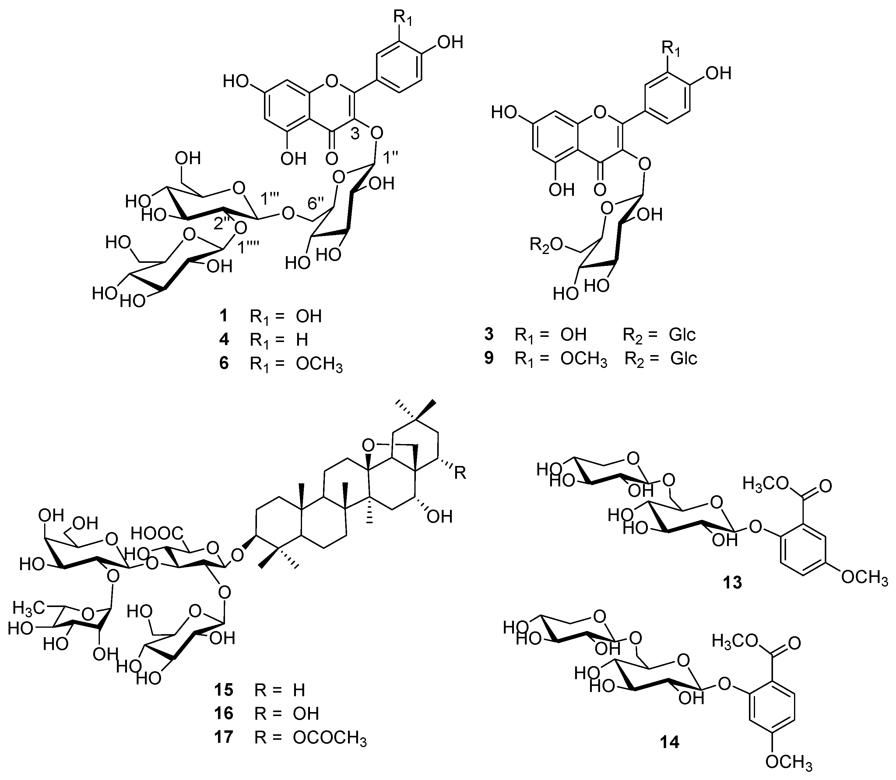
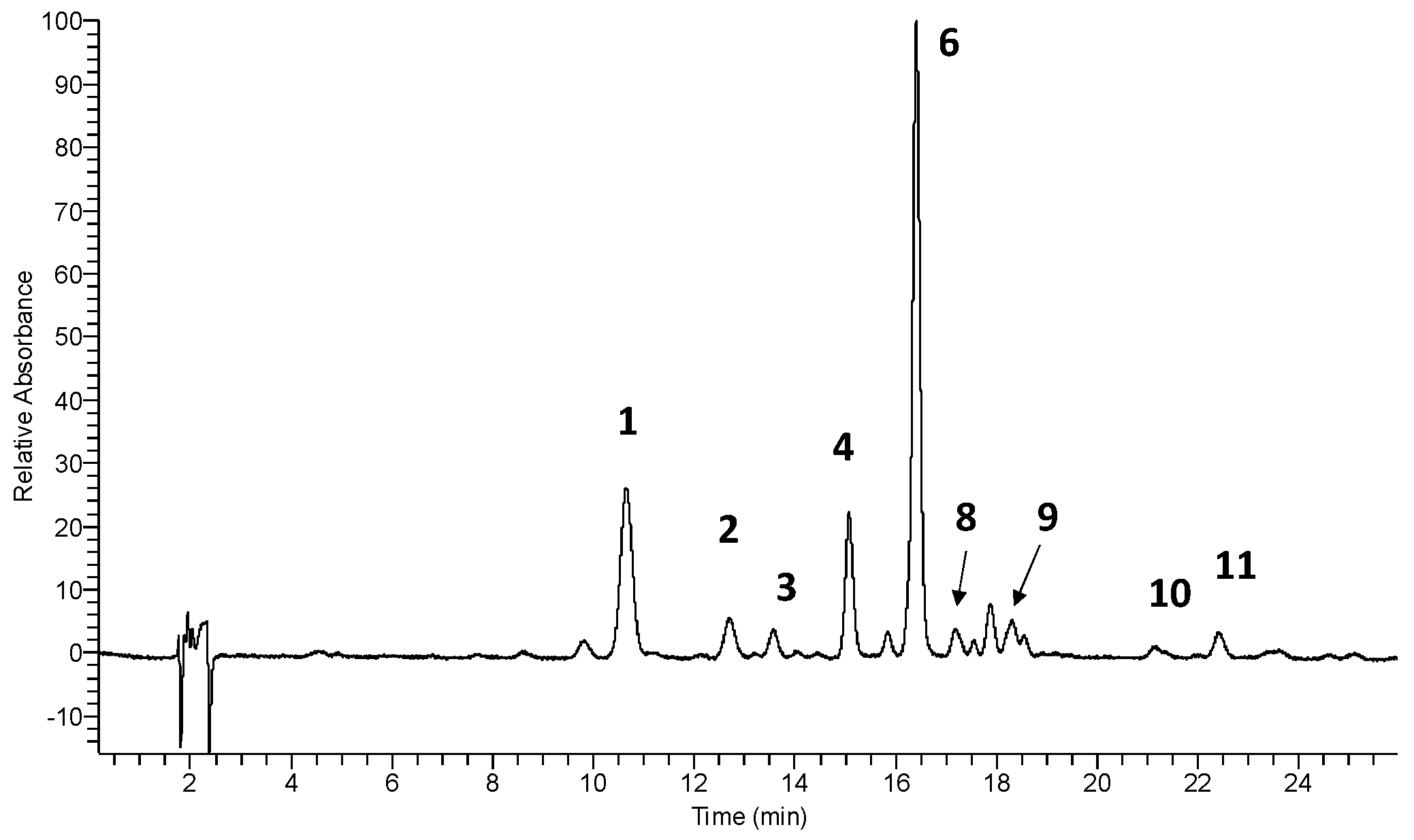
3.1. Identification and Quantitative Determination of Flavonols in Cowslip Flowers

The HPLC-PDA-MS analysis of the examined flower samples sourced from different wild-growing Greek populations of P. veris subsp. veris revealed the presence of flavonol triglycosides and diglycosides (Table 2, Figure 1 and Figure 2). In general, the flower samples studied herein had similar chromatographic patterns among them (only small differences), resembling also the previously reported ones of P. veris subsp. veris from Epirus [23]. The secondary metabolites 1, 3, 4, 6, and 9 were unambiguously identified by comparing retention times, UV, and fragmentation patterns with those of a previously developed chemical library [23]. Among them, the constituents 1, 4, and 6 were 3-O-triglucosides (3-O-β-glucopyranosyl-(1→2)-β-glucopyranosyl-(1→6)-β-glucopyranoside) of quercetin, kaempferol, and isorhamnetin, respectively. These were the main metabolites occurring in all flower samples from the herein examined P. veris subsp. veris populations, similarly to the populations studied previously from Epirus [23]. Compounds 3 and 9 were two diglucosides identified as 3-O-β-glucopyranosyl-(1→6)-β-glucopyranosides of quercetin and isorhamnetin, respectively, and should be referred to as minor constituents. A series of rutinosides was also detected, peaks 8, 10, and 11, which were identified as rutin, nicotiflorin, and narcissin, respectively. Their identification was based on comparison of their retention times, UV and MS spectral data with those of authentic reference samples, or previously developed chemical library [23,26]. These compounds were also previously detected in the populations from Epirus, northwestern Greece. Finally, a series of triglycosides were detected (peaks 2, 5, and 7) and their identification was tentative, mainly based on their spectral features and literature fragmentation reports. All three metabolites were characterized by a neutral loss of 454 amu, indicating the presence of a dirhamnosylhexoside, whereas the aglycon fragments at m/z = 300.3, 285.0, and 314.9 suggested the presence of quercetin, kaempferol, and isorhamnetin, respectively. The UV spectral data were in accordance with this assumption: a shoulder at ~266 nm in Band II indicated the presence of ortho-substituted flavonols, while the UV maximum not more than 360 nm indicated the presence of sugars in position 3. Such dirhamnosylhexosides are common in Primula spp., such as P. farinosa L., P. halleri J.F. Gmel. [29,30], but also P. veris subsp. columnae (Ten.) [31] and P. veris (subspecies not specified) [32]. Recently, the kaempferol triglycoside clitorin was also reported in a P. veris subsp. veris infusion originating from Prespa Lake Park, northern Greece [33].
Concerning the quantitative data in our study, the investigation of 13 samples of widely scattered wild-growing populations of P. veris subsp. veris represents only the second report from Greece, with the first one being restricted to wild populations from N. W Greece–Epirus [23]. The obtained results are shown in Table 3. The main metabolite in the Greek cowslip flowers investigated was isorhamnetin-3-O-β-glucopyranosyl-(1→2)-β-glucopyranosyl-(1→6)-β-glucopyranoside (6), followed by quercetin-3-O-β-glucopyranosyl-(1→2)-β-glucopyranosyl-(1→6)-β-glucopyranoside (1) and kaempferol-3-O-β-glucopyranosyl-(1→2)-β-glucopyranosyl-(1→6)-β-glucopyranoside) (4), whilst all other flavonoids were minor compounds and were not considered for quantification. Chemical analyses showed that the two populations of Mt. Vermio (Verm-1, Verm-2) had a high content in flavonol glycosides and similar chemical features both qualitatively and quantitatively, having the same ratios of isorhamnetin, quercetin, and kaempferol. The two population samples from Mt. Vasilitsa (Vasi-1, Vasi-2) were also rich in flavonol glycosides, although Vasi-2 was richer in terms of the quercetin-3-O-β-glucopyranosyl-(1→2)-β-glucopyranosyl-(1→6)-β-glucopyranoside (1). In contrast, populations from Mt. Chortiatis (Chor-1, Chor-2), Mt. Belles (Bell), Mt. Karpouzi (Karp), and Mt. Pangaio (Pang) exhibited low amounts of the above-mentioned metabolites.

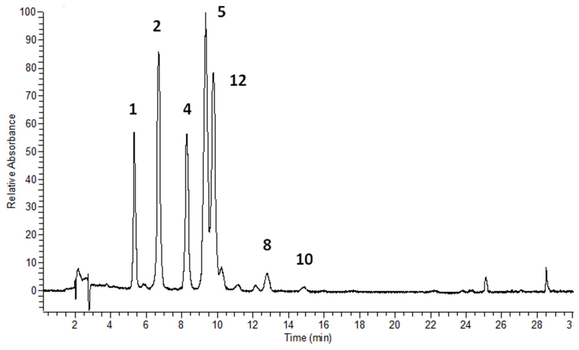
3.2. Flavonols’ Identification and Flavonoid Quantitative Determination in Leaves

The HPLC-PDA-MS analysis of the leaves’ samples revealed the presence mainly of flavonol triglycosides, while diglycosides were only minor compounds present in nonquantifiable amounts (Table 2 and Table 3, Figure 3). This is the first report on Greek cowslip leaves, and qualitatively, they exhibited a simpler chromatographic profile compared to the flowers, comprising solely triglycosides of quercetin and kaempferol, while isorhamnetin derivatives were barely traced. Indeed, quantitative analysis showed the predominance of kaempferol derivatives, in contrast to the flowers. Specifically, the main metabolites in Primula veris subsp. veris leaves were kaempferol-3-O-β-glucopyranosyl-(1→2)-β-glucopyranosyl-(1→6)-β-glucopyranoside) (4) together with a mixture of kaempferol-dirhamnosylhexosides. In some cases, their amount ranged between 67 and 80% of the total flavonoids in leaves. Quantitative results disclosed the same trend with flowers. Populations from Mt. Vermio and Mt. Vasilitsa exhibited a high productivity in flavonol triglycosides in contrast to populations from Mts. Chortiatis, Belles, Karpouzi, and Pangaion. The contents of quercetin and kaempferol were diversified among the samples of the different populations studied. The population samples from Mt. Belles, Mt. Pangaio, and Pisoderi of Mt. Varnous contained mainly kaempferol triglycosides, whereas that of Mt. Karpouzi was distinguished from all populations due to its poorest flavonol content and similar concentrations of quercetin and kaempferol. The population samples of Mt. Chortiatis, on the other hand, were diversified; Chor-1 had extremely low amounts of the target metabolites, which consisted almost exclusively of kaempferol-3-O-β-glucopyranosyl-(1→2)-β-glucopyranosyl-(1→6)-β-glucopyranoside (4, K3G), while Chor-2 and Chor-3 had slightly higher contents of quercetin than kaempferol triglycosides.

3.3. Identification and Quantitative Determination of Roots’ Phenolic Glycosides and Saponins

The identification of the phenolic glycosides primulaverin and primeverin (compounds 13, 14) was performed by comparing their retention times and the respective UV spectra with those of primary reference standards along with the MS data. In the Mt. Pangaio population sample, a minor peak was also observed at early retention times (Figures S1 and S2), and, since its UV and MS data (m/z = 491 [M + HCOO]-, 445.2 [M-H]-, 293, 151.0 [A-H]-) were in accordance with the literature data, the peak was tentatively identified as gaultherin [34]. Peaks at longer elution times were also observed in all samples, but they were not identified; however, given their UV absorbances at 330 and 296 nm, it is suggested that these compounds are of a phenolic nature. Chromatographic isolations are currently underway, and their complete spectral identification is expected shortly. Saponins were specifically detected by MS in the SIM mode at an optimal cone voltage of 180 V. The identification of primulasaponin I (15) was based on the use of a reference standard, while priverosaponin B (compound 16) and priverosaponin B 22-acetate (compound 17), were assigned based on the pseudomolecular ions observed at m/z = 1119.7 and 1161.8 (Figure S3), respectively, in combination with the literature reports. Both saponins were previously isolated from the roots of another subspecies of P. veris, namely subsp. macrocalyx [35]. Regarding the quantitative analysis, preliminary extractions with 100% methanol, as well as 70% and 50% (v/v) aqueous methanol, showed that the latter was the best solvent system for the extraction of the biologically active saponins (over the nonactive/marker constituents phenolics). Therefore, the extraction protocol proposed by Müller et al. [25] was adopted. Quantitative results (Table 4) demonstrated that populations from Metsovo and Mt. Vermio exhibited a high productivity in primulic acid I, followed by populations from Mts. Chortiatis (Chor-2, Chor-3). Interestingly, the latter also had a high content in priverosaponin B.

4. Discussion

This investigation explored, for the first time, the phytochemical profile of wild-growing Greek populations of Primula veris subsp. veris, examining separately the samples of flowers, leaves, and roots. In the studied flower samples, from the examined populations, the results were in line with our previous findings [23], indicating the presence of mainly isorhamnetin, quercetin, and kaempferol triglycosides (in a decreasing scale of amounts), while diglycosides of the same flavonols were found only in small amounts (traces). The presence of isorhamnetin-3-O-β-glucopyranosyl-(1→2)-β-glucopyranosyl-(1→6)-β-glucopyranoside (6) as the main metabolite in Greek cowslip flowers is herein confirmed, as in another previous own report [23], thus probably indicating a typical flower feature of the wild-growing Greek populations of P. veris subsp. veris. Additionally, isorhamnetin and kaempferol dirhamnosylhexosides were also detected in small amounts. Interestingly, the flavonoid contents in the flower samples of the different populations in the present study were not as rich as those originated from Epirus—the northwestern part of Greece (4.26–6.36%) [23]. However, their contents were within the 3% limit reported by Wichtl [9] and suggested by the EMA [10].
Context-wise, the present study is the first to explore phytochemically Greek cowslips along a large geographical scale (ca. 300 km in line) of a longitudinal gradient in northern Greece. The samples of the studied Greek populations showed great variability in terms of their examined secondary metabolites. The previously reported samples from Epirus [23], along with the herein examined samples from Mt. Vermio located in the northcentral part of mainland Greece, and those of Mt. Vasilitsa lying in the northwestern part (all originating from the colder and more humid regions of Greece) [36], appeared to be richer in terms of flavonol glycosides as compared to all other examined samples from populations originating from the comparatively warmer northeastern part of Greece [36]. Given that all samples were sourced from mountain areas of a similar latitude (see Table 1), it seems likely that the local longitudinal climatic gradient (from the west to the east) and concomitant stress conditions may influence the productivity of the main secondary metabolites of P. veris subsp. veris. The latter suggests the following tendency: the more western the P. veris subsp. veris population in mainland Greece, the richer in flavonol glycosides, or vice versa. Although further studies are needed to confirm this predisposition herein reported for the first time, such a trend might be of use for the artificial selection of highly productive mother plants of wild origin as basic starting materials for future breeding purposes and the cultivation of elite materials of desirable constitution.
The examination of leaf tissues in the present study is the first ever conducted in Greek populations of P. veris subsp. veris. Leaf samples, analyzed with the same protocol as flower samples, were characterized almost exclusively by the presence of kaempferol and quercetin triglycosides. Kaempferol derivatives represented the main constituents in nearly all cases, whereas the isorhamnetin derivatives, which predominate in flowers, were detected only in traces. The flavonoid content once again showed variability among the examined population samples but was generally higher than that of the flowers. Mt. Vermio and Mt. Vasilitsa samples appeared again as two populations with better chemical characteristics, while the most northwestern population sample from Mt. Varnous (Pisoderi) stood out with the highest percentages of kaempferol and quercetin triglycosides, reaching up to 6.35%. The literature review composes a rather multifaceted picture. Tarapatskyy et al. [37] investigated the polyphenolic content in leaf extracts (aq. ethanol 70%) of P. veris from Podkarpacie, Poland (not specified to subspecies), and revealed thatkaempferol-3-O-rutinoside-7-O-rhamnoside was the prominent constituent in all leaf extracts. This compound was considered as characteristic of the leaves, since much lower concentrations of this triglycoside have been detected in the extracts from flowers and stalks. In the same study, quercetin-3-O-rutinoside (rutin) was reported to be the second most important constituent in dried leaf extracts. As the latter study aimed only to characterize the different extracts and not the plant material itself, any quantitative comparison with our results is not feasible, apart from the deduction that kaempferol derivatives may predominate in the leaves of P. veris, as also reported herein for the studied Greek populations. In another study, Apel et al. [33] focused on the secondary metabolite profiles of flowers, leaves, and roots in three flower color variants (yellow, orange, and red) of P. veris (subspecies not mentioned) in southern Germany using HPLC-DAD-MS. The methanolic leaf extracts of the three P. veris variants were reported to exhibit flavonoid profiles comparable to those of flowers, with eight out of the ten flavonoid glycosides identified in the petals being also detected in the leaves (thus obscuring quantitative differences), with kaempferol-3-O-galactoside-rhamnoside-7-O-rhamnoside being reported as the major leaf metabolite in each case. Generally, quercetin and kaempferol derivatives are characteristic in the leaves of P. veris [32], whilst leaves of three different P. veris subsp. columnae populations exposed to different UV conditions [31] were rich in kaempferol (such as kaempferol-3-O-dideoxyhexose-hexoside and kaempferol-3,7-O-trihexoside) derivatives. On the contrary, the same study has reported two prominent flower flavonoids, i.e., quercetin-3-O-dihexoside and isorhamnetin-3-O-dideoxyhexose-hexoside. Another study has demonstrated that kaempferol, quercetin, and isorhamnetin triglycosides are rather a common feature in the leaves of alpine Primula species, such as P. auricula L., P. farinosa, and P. helleri [29]. Despite the phytochemical investigations of the above-mentioned Primula species, no quantitative data are available for comparison with those obtained herein. Some chemical investigations on P. auricula subsp. auricula [38] leaves have focused mainly on the exudate flavones, with leaf tissue flavonols, including isorhamnetin and kaempferol derivatives with combinations of xylose, glucose, and rhamnose. Again, no quantitative information is available to allow comparisons with the quantitative data generated herein. Nevertheless, the present study evidenced specific populations of Primula veris subsp. veris with a high flavonoid content in flowers and leaves. In view of the culinary applications of these plants [18], this is an important finding in terms of the antioxidant/nutritional value.
The roots of P. veris subsp. veris contain characteristic metabolites, such as the triterpenic saponins, namely primulic acids (or primulasaponins), which are considered as active compounds, and the phenolic glycosides, namely primeverin and primulaverin. The latter degrade during storage in the presence of the primverase enzyme, and release fragrant salicylates, serving in this way as indicators of the age of the plant material [25]. Due to the different chemical and physical properties of the metabolites present in P. veris roots, a different analytical protocol was applied, combining a metabolite-specific extraction according to Müller et al. [25] with the HPLC-UV-MS methodology, which permits the simultaneous analysis of saponins and phenolic glycosides. It should be mentioned that the present study is the first chemical investigation of root tissues from different Greek populations of P. veris subsp. veris. In the studied root samples of P. veris subsp. veris, primulasaponin I was the prominent saponin in all populations, followed by priverosaponin B, while priverosaponin B 22-acetate was found in traces. Primulasaponin II was not detected in any of the studied samples. Furthermore, the phenolic glycoside primulaverin was detected in all populations, while primeverin was detected in all root samples except those originating from Mt. Chortiatis and Mt. Belles. This might also be due to the extraction protocol used favoring saponin extraction.
An examination of the literature indicates primulasaponin I as the main saponin in P. veris roots, with primulasaponin II found in much lower amounts [25,32,39]. The root extracts from three color variants of P. veris (subspecies not mentioned) originating from a natural habitat in Germany and cultivated in a botanical garden [32] have shown identical qualitative profiles consisting of primulasaponin I, II, and priverosaponin B 22-acetate, but no quantitative data were reported. In addition, P. veris root samples collected from the wild (several locations) in Austria and Germany [25] exhibited high amounts of primulasaponin I (major saponin in all samples, 5.71–8.26%), followed by priverosaponin B 22-acetate (1.39–1.87%), and primulasaponin II (up to 0.32%), with another constituent of an unknown chemical structure appearing in all root samples (3.81–5.30%). The authors of the latter study attributed this constituent as a saponin, taking into consideration its properties (e.g., mass range, no UV absorption). More recently, Włodarczyk et al. [39] reported that primulasaponin II can be detected in traces below 0.2% in roots of commercially available P. veris, while primulasaponin I can be the main saponin comprising 2.84% of root dry mass. Likewise, the EMA reports that saponins in the roots of P. veris may vary between 3 and 10%, even up to 12% [8].
The phenolic glycosides primulaverin and primeverin are also characteristic components of the roots, typically found in variable amounts up to 2.3% according to the EMA [8]. Indeed, Muller et al. [25] reported that the cumulative percentages of primulaverin and primeverin may vary between 0.70 and 1.48%, with the latter being the major representative in all examined samples. Similar observations have been reported by Bączek et al. [40], who studied wild-growing populations of P. veris (subspecies not mentioned) and P. elatior (L.) Hill. from eastern Poland, outlining that primulaverin and primeverin are detected only in the roots, and their content is approximately 10 times higher in P. veris than in P. elatior. Primeverin was reported again as the main phenolic glycoside (1183.32 mg/100 g DW of primeverin over 536.16 mg/100 g DW of primulaverin).
One way or another, it should be noted that P. veris across its native range includes four infraspecific taxa, namely: subsp. canescens, found in central and southcentral European countries; subsp. columnae, occurring in mountains of Turkey and southern Europe (except Greece); subsp. macrocalyx, found in the Asiatic part of the species’ range; the typical subsp. veris, recorded across the European range of P. veris [1]. The latter means that all the above-mentioned phytochemical studies related to P. veris and the comparisons made with the results furnished in this investigation should be first envisaged according to the exact subspecies they actually refer to. Thus, the taxonomic identity of all the above-mentioned literature data and the concomitant comparisons should be interpreted with caution, since the results obtained herein clearly refer exclusively to P. veris subsp. veris and not to other subspecies. From another viewpoint, it is also well established that the levels of secondary metabolites reported in phytochemical studies of different plant species are known to be affected by both inherent factors, such as the developmental stage, age, genome, and genetic lineage, as well as from external factors, either biotic, such as plant–animal or microbe/mycorrhiza interactions, or nonbiotic, such as altitude, solar radiation, environmental temperature, humidity, soil composition, and salinity [41,42]. Such issues should also be taken into consideration when comparing the secondary metabolites in plant materials sourced directly from the wild.

5. Conclusions

The current investigation assessed for the first time, separately and in a comparative fashion, the chemical composition of flowers, leaves, and roots sourced from 13 wild-growing populations of P. veris subsp. veris located along a large longitudinal gradient, covering eight different areas in northern Greece spanning ca. 300 km in line. In this way, this study indicated a high productivity in certain phytoconstituents and locally adapted native genotypes from relatively cooler and warmer Greek regions of a similar latitude, thus discerning some peculiar characteristics of the local genotypes, i.e., being richer westwards or poorer eastwards. Given that the range of P. veris is centered in rather colder areas of Europe and Asia, and is represented by four subspecies, it should be noticed that the results of this study refer only to the examined marginal Greek genotypes of subsp. veris, being at the edge of its native distribution range in the Mediterranean context of the Greek peninsula. In general, almost all Greek cowslip populations examined in this study were characterized by a low content of saponins. Being typical across almost all studied population samples, a peculiar feature associated with low-saponin content can possibly be inferred for the studied marginal Greek genotypes. It also appears from our studies on flowers of Greek genotypes that they have a rather uniform chemical profile, with isorhamnetin-3-O-β-glucopyranosyl-(1→2)-β-glucopyranosyl-(1→6)-β-glucopyranoside as the main metabolite, thus distinguishing Greek populations of P. veris subsp. veris from those of other European subspecies. Unfortunately, comparison with other Greek indigenous genotypes is not feasible, since the literature reports are missing to date. Nonetheless, further investigation is required prior to drawing solid conclusions.
The phytochemical insight furnished in this study may also aid the further selection of desirable Greek native genotypes during the ongoing domestication process of P. veris subsp. veris, and can further facilitate conservation strategies aiming to alleviate the overharvesting pressure on wild-growing populations in Greece and other countries. Overall, this study contributes to the phytochemical characterization of cowslip plants in the Eurasiatic context of P. veris, while offering an incentive for domestic farmers to cultivate, at local scales, indigenous P. veris subsp. veris genotypes of high added value.

Supplementary Materials

The following supporting information can be downloaded at: https://www.mdpi.com/article/10.3390/horticulturae9101120/s1, Figure S1: Representative HPLC-PDA chromatogram of the Primula veris subsp. veris root extracts from the wild-growing population of Mt. Pangaio; Figure S2: UV and MS spectra of the compound designated as gaultherin; Figure S3: Representative HPLC-MS chromatogram (SIM mode) of the Primula veris subsp. veris root extracts from the wild-growing population of Mt. Pangaio.

Author Contributions

Conceptualization, A.K. and N.K.; methodology, A.K. and N.K.; investigation, A.K. and I.S.; taxonomic identification, N.K.; resources, A.K. and N.K.; data curation, P.C., I.S. and A.K.; writing—original draft preparation, I.S., A.K. and N.K; writing—review and editing, A.K., N.K. and P.C.; supervision, A.K.; funding acquisition, A.K. and P.C. All authors have read and agreed to the published version of the manuscript.

Funding

This study has been partly funded by the Region of Epirus (PRIMULA-PINDOS—project).

Data Availability Statement

The data presented in this study are available on request from the corresponding author.

Conflicts of Interest

The authors declare no conflict of interest.

References

  1. Plants of the World Online. Kew Botanical Gardens. Available online: https://powo.science.kew.org/taxon/urn:lsid:ipni.org:names:702751-1 (accessed on 1 September 2023).
  2. Brys, R.; Jacquemyn, H. Biological Flora of the British Isles: Primula veris L. J. Ecol. 2009, 97, 581–600. [Google Scholar] [CrossRef]
  3. Zhao, Z.; Luo, Z.; Yuan, S.; Mei, L.; Zhang, D. Global transcriptome and gene co-expression network analyses on the development of distyly in Primula oreodoxa. Heredity 2019, 123, 784–794. [Google Scholar] [CrossRef] [PubMed]
  4. Gilmartin, P.M. On the origins of observations of heterostyly in Primula. New Phytol. 2015, 208, 39–51. [Google Scholar] [CrossRef] [PubMed]
  5. Bai, Y.-H.; Zhang, S.-Y.; Guo, Y.; Tang, Z. Conservation status of Primulaceae, a plant family with high endemism, in China. Biol. Conser. 2020, 248, 108675. [Google Scholar] [CrossRef]
  6. Aavik, T.; Reitalu, T.; Kaldra, M.; Reinula, I.; Hool, K.; Zobel, M.; Pärtel, M.; Uuemaa, E.; Kmoch, A.; Moges, D.M.; et al.; (Institute of Ecology and Earth Sciences, University of Tartu, Tartu, Estonia) Personal communication, 2023.
  7. Looking for Cowslips. Available online: https://cowslip.science/en (accessed on 30 August 2023).
  8. EMA/HMPC/113577/2012. Assessment Report on Primula veris L. and/or Primula elatior (L.) Hill, Radix. Available online: https://www.ema.europa.eu/en/documents/herbal-report/final-assessment-report-primula-veris-l/primula-elatior-l-hill-radix_en.pdf (accessed on 25 August 2023).
  9. Wichtl, M. Primulae radix. In Herbal Drugs and Phytopharmaceuticals; Wichtl, M., Ed.; Medpharm Scientific Publishers: Stuttgart, Germany, 2004; pp. 472–474. [Google Scholar]
  10. EMA/HMPC/136583/2012. EMA Assessment Report on Primula veris L. and/or Primula elatior (L.) Hill, Flos. Available online: https://www.ema.europa.eu/en/documents/herbal-report/final-assessment-report-primula-veris-l/primula-elatior-l-hill-flos_en.pdf (accessed on 25 August 2023).
  11. Eliopoulos, A.G.; Angelis, A.; Liakakou, A.; Skaltsounis, L.A. In vitro anti-influenza virus activity of non-polar Primula veris subsp. veris Extract. Pharmaceuticals 2022, 15, 1513. [Google Scholar] [CrossRef] [PubMed]
  12. Latypova, G.M.; Bychenkova, M.A.; Katayev, V.A.; Perfilova, V.N.; Tyurenkov, I.N.; Mokrousov, I.S.; Prokofiev, I.I.; Salikhov, S.h.M.; Iksanova, G.R. Composition and cardioprotective effects of Primula veris L. solid herbal extract in experimental chronic heart failure. Phytomedicine 2018, 54, 17–26. [Google Scholar] [CrossRef]
  13. Prokofiev, I.I.; Kustova, M.V.; Nesterova, A.A.; Perfilova, V.N.; Khusainova, G.H.; Borodkina, L.E.; Tivon, Y.V.; Tyurenkov, I.N.; Kataev, V.A.; Latypova, G.M. Solid herbal extract of Primula veris L. improves morphofunctional condition of rats’ myocardium in chronic alcohol intoxication. J. Tradit. Complement. Med. 2023, 13, 306–314. [Google Scholar] [CrossRef]
  14. Ghédira, K.; Goetz, P. Primevère officinale Primula veris L. (Primulaceae). Phytothérapie 2017, 15, 240–244. [Google Scholar] [CrossRef]
  15. Kahraman, C.; Sari, S.; Küpeli Akkol, E.; Tatli Cankaya, I. Bioactive Saponins of Primula vulgaris Huds. promote wound healing through inhibition of collagenase and elastase enzymes: In vivo, in vitro and in silico evaluations. Chem Biodivers. 2022, 19, e202200582. [Google Scholar] [CrossRef]
  16. Cosmetic Ingredients. SpecialChem, The Material Selection Platform. Available online: https://cosmetics.specialchem.com/inci/primula-veris-extract (accessed on 31 August 2023).
  17. Jiang, S.; Deng, L.; Luo, H.; Li, X.; Guo, B.; Jiang, M.; Jia, Y.; Ma, J.; Sun, L.; Huang, Z. Effect of fragrant primula flowers on physiology and psychology in female college students: An empirical study. Front. Psychol. 2021, 12, 607876. [Google Scholar] [CrossRef]
  18. Plantes Sauvages Comestibles. Available online: https://plantes-sauvages-comestibles.com/la-primevere-mangeons-du-coucou/ (accessed on 31 August 2023).
  19. Primulas for Scent. The Royal Horticultural Society. Available online: https://www.rhs.org.uk/plants/primula/for-scent (accessed on 31 August 2023).
  20. Hayta, S.; Smedley, M.A.; Li, J.; Harwood, W.A.; Gilmartin, P.M. Plant regeneration from leaf-derived callus cultures of primrose (Primula vulgaris). Hort. Sci. 2016, 51, 558–562. [Google Scholar] [CrossRef]
  21. Arrest for Illegal Collection of Primroses in Grammos. Available online: https://dasarxeio.com/2023/05/08/125161/ (accessed on 18 August 2023).
  22. Arrests of Foreigners for Illegal Collection of Primroses. Available online: https://dasarxeio.com/2023/06/01/125781/ (accessed on 18 August 2023).
  23. Chintiroglou, P.I.; Krigas, N.; Chatzopoulou, P.; Karioti, A. Development and validation of an HPLC method for the analysis of flowers of wild-growing Primula veris from Epirus, Greece. Planta Med. 2021, 87, 1219–1230. [Google Scholar] [CrossRef] [PubMed]
  24. Strid, A. Atlas of the Aegean Flora Part 1 (Text & Plates) & Part 2 (Maps), 1st ed.; Englera 33 (1 & 2); Botanic Garden and Botanical Museum: Berlin, Germany; Freie Universität: Berlin, Germany, 2016. [Google Scholar]
  25. Müller, A.; Ganzera, M.; Stuppner, H. Analysis of phenolic glycosides and saponins in Primula elatior and Primula veris (primula root) by liquid chromatography, evaporative light scattering detection and mass spectrometry. J. Chromatogr. A 2006, 1112, 218–223. [Google Scholar] [CrossRef] [PubMed]
  26. Khalfallah, A.; Karioti, A.; Berrehal, D.; Kabouche, A.; Lucci, M.; Bilia, A.R.; Kabouche, Z. A new flavonol triglycoside and other flavonol glycosides from Astragalus armatus Willd. (Fabaceae). Rec. Nat. Prod. 2013, 8, 12–18. [Google Scholar]
  27. EMA/HMPC/CHMP/CVMP/201116/20051 Rev. 3 Guideline on Quality of Herbal Medicinal Products2/Traditional Herbal Medicinal Products. Available online: https://www.ema.europa.eu/en/documents/scientific-guideline/final-guideline-quality-herbal-medicinal-products/traditional-herbal-medicinal-products-revision-3_en.pdf (accessed on 5 September 2023).
  28. Guide for the Elaboration of Monographs on Herbal Drugs and Herbal Drug Preparations European Pharmacopoeia. 2nd Edition. 2023. Available online: https://www.edqm.eu/documents/52006/66555/Guide+for+the+elaboration+of+monographs+on+HERBAL+DRUGS+AND+HERBAL+DRUG+PREPARATIONS.pdf/bef3251e-fafc-581b-93ef-3c1a95e2793c?t=1684932003926 (accessed on 5 September 2023).
  29. Colombo, P.S.; Flamini, G.; Christodoulou, M.S.; Rodondi, G.; Vitalini, S.; Passarella, D.; Fico, G. Farinose alpine Primula species: Phytochemical and morphological investigations. Phytochemistry 2014, 98, 151–159. [Google Scholar] [CrossRef]
  30. Bhutia, T.D.; Valant-Vetschera, K.M.; Adlassnig, W.; Brecker, L. Flavonoids in selected Primula spp.: Bridging micromorphology with chemodiversity. Nat. Prod. Commun. 2012, 7, 1469–1473. [Google Scholar] [CrossRef]
  31. El Morchid, E.M.; Torres Londoño, P.; Papagiannopoulos, M.; Gobbo-Neto, L.; Müller, C. Variation in flavonoid pattern in leaves and flowers of Primula veris of different origin and impact of UV-B. Biochem. Syst. Ecol. 2014, 53, 81–88. [Google Scholar] [CrossRef]
  32. Apel, L.; Kammerer, D.R.; Stintzing, F.C.; Spring, O. Comparative metabolite profiling of triterpenoid saponins and flavonoids in flower color mutations of Primula veris L. Int. J. Mol. Sci. 2017, 18, 153. [Google Scholar] [CrossRef]
  33. Graikou, K.; Mpishinioti, A.; Tsafantakis, N.; Maloupa, E.; Grigoriadou, K.; Chinou, I. Comparative phytochemical analyses of flowers from Primula veris subsp. veris growing wild and from ex situ cultivation in Greece. Foods 2023, 12, 2623. [Google Scholar] [CrossRef]
  34. Michel, P.; Granica, S.; Rosińska, K.; Glige, M.; Rojek, J.; Poraj, Ł.; Olszewska, M.A. The effect of standardised leaf extracts of Gaultheria procumbens on multiple oxidants, inflammation-related enzymes, and pro-oxidant and pro-inflammatory functions of human neutrophils. Molecules 2022, 27, 3357. [Google Scholar] [CrossRef]
  35. Çaliş, I.; Yürüker, A.; Rüegger, H.; Wright, A.D.; Sticher, O. Triterpene saponins from Primula veris subsp. macrocalyx and Primula elatior subsp. meyeri. J. Nat. Prod. 1992, 55, 1299–1306. [Google Scholar] [CrossRef]
  36. Climatic Atlas of Greece. Available online: http://climatlas.hnms.gr/sdi/ (accessed on 7 September 2023).
  37. Tarapatskyy, M.; Gumienna, A.; Sowa, P.; Kapusta, I.; Puchalski, C. Bioactive phenolic compounds from Primula veris L.: Influence of the extraction conditions and purification. Molecules 2021, 26, 997. [Google Scholar] [CrossRef]
  38. Holzbach, B.; Reuter, V.; Bacher, M.; Schinnerl, J.; Brecker, L.; Rosenau, T.; Valant-Vetschera, K. Flavonoid diversification in different leaf compartments of Primula auricula (Primulaceae). Biochem. Syst. Ecol. 2021, 98, 104310. [Google Scholar] [CrossRef]
  39. Włodarczyk, M.; Pasikowski, P.; Osiewała, K.; Frankiewicz, A.; Dryś, A.; Gleńsk, M. In Search of High- Yielding and Single-Compound-Yielding Plants: New Sources of Pharmaceutically Important Saponins from the Primulaceae Family. Biomolecules 2020, 10, 376. [Google Scholar] [CrossRef] [PubMed]
  40. Bączek, K.; Przybyl, J.L.; Mirgos, M.; Kosakowska, O.; Szymborska-Sandhu, I.; Węglarz, Z. Phenolics in Primula veris L. and P. elatior (L.) Hill raw materials. Int. J. Anal. Chem. 2017, 2017, e2871579. [Google Scholar] [CrossRef] [PubMed]
  41. Li, Y.; Kong, D.; Fu, Y.; Sussman, M.R.; Wu, H. The effect of developmental and environmental factors on secondary metabolites in medicinal plants. Plant Physiol. Biochem. 2020, 148, 80–89. [Google Scholar] [CrossRef] [PubMed]
  42. Elkady, W.M.; Gonaid, M.H.; Yousif, M.F.; El-Sayed, M.; Omar, H.A.N. Impact of altitudinal variation on the phytochemical profile, anthelmintic and antimicrobial activity of two Pinus species. Molecules 2021, 26, 3170. [Google Scholar] [CrossRef]
Figure 1. Representative metabolites from flowers, leaves and roots of P. veris subsp. veris from wild-growing populations in northern Greece: (1) quercetin-3-O-β-glucopyranosyl-(1→2)-β-glucopyranosyl-(1→6)-β-glucopyranoside (Q3G); (3) quercetin-3-O-β-glucopyranosyl-(1→6)-β-glucopyranoside; (4) kaempferol-3-O-β-glucopyranosyl-(1→2)-β-glucopyranosyl-(1→6)-β-glucopyranoside (K3G); (6) isorhamnetin-3-O-β-glucopyranosyl-(1→2)-β-glucopyranosyl-(1→6)-β-glucopyranoside (I3G); (9) isorhamnetin-3-O-β-glucopyranosyl-(1→6)-β-glucopyranoside; (13) primulaverin; (14) primeverin; (15) primulasaponin I; (16) priverosaponin B; (17) priverosaponin B 22-acetate.
Horticulturae 09 01120 g001
Figure 2. Representative HPLC-UV chromatogram of the Primula veris subsp. veris flower extracts at 354 nm from the Vas-2 wild-growing population of Mt. Vasilitsa. Column: Zorbax SBAq. Gradient system: (A) H2O (with 0.05% formic acid, v/v), and (B) acetonitrile, as follows: 0–5 min, 85% A; 5–10 min, to 80% A; 10–15 min, 80% A; 15–20 min, to 75% A; 20–25 min, to 50% A. 1: quercetin-3-O-β-glucopyranosyl-(1→2)-β-glucopyranosyl-(1→6)-β-glucopyranoside; 2: quercetin-3-O-dirhamnosylhexoside; 3: quercetin-3-O-β-glucopyranosyl-(1→6)-β-glucopyranoside; 4: kaempferol-3-O-β-glucopyranosyl-(1→2)-β-glucopyranosyl-(1→6)-β-glucopyranoside; 6: isorhamnetin-3-O-β-glucopyranosyl-(1→2)-β-glucopyranosyl-(1→6)-β-glucopyranoside; 8: rutin (quercetin-3-O-rutinoside); 9: isorhamnetin-3-O-β-glucopyranosyl-(1→6)-β-glucopyranoside; 10: nicotiflorin (kaempferol-3-O-rutinoside); 11: narcissin (isorhamnetin-3-O-rutinoside). Minor peaks between constituents 8 and 9 are acetylated derivatives of flavonol triglycosides.
Horticulturae 09 01120 g002
Figure 3. Representative HPLC-UV chromatogram of the Primula veris subsp. veris leaf extracts at 354 nm from the Ver-2 wild-growing population of Mt. Vermio. Column: Luna RP-C18 (3 µm, 150 × 3 mm, Phenomenex). Gradient system: (A) H2O (with 0.05% formic acid, v/v) and (B) acetonitrile, as follows: 0–15 min, 82% A; 15–28 min, to 55% A; 28–35 min, to 82% A; 35–40 min, 82% A. 1: quercetin-3-O-β-glucopyranosyl-(1→2)-β-glucopyranosyl-(1→6)-β-glucopyranoside; 2: quercetin-3-O-dirhamnosylhexoside; 4: kaempferol-3-O-β-glucopyranosyl-(1→2)-β-glucopyranosyl-(1→6)-β-glucopyranoside; 5: kaempferol-3-O-dirhamnosylhexoside; 8: rutin (quercetin-3-O-rutinoside); 10: nicotiflorin (kaempferol-3-O-rutinoside); 12: kaempferol-dirhamnosylhexoside. Peaks between 24 and 30 min belong to flavones.
Horticulturae 09 01120 g003
Table 1. Collection data regarding the studied wild-growing genotypes of Primula veris subsp. veris along the longitudinal gradient of northern Greece (geographical coordinates in the WGS84 system) and plant parts examined in this investigation (R: roots; L: leaves; F: flowers).
Abbreviation/CodeArea and LocalityCollection DateAltitude (m)Latitude (North)Longitude (East)Plant Part Examined
Mets-1/PV-71 Metsovo, Aoos springs 22.5.2021 *136039.8157321.09162R
Mets-2/PV-8Metsovo, Aoos springs22.5.2021 *136539.8436521.09584R
Verm-1/PV-9Mt Vermio 23.5.2021184040.64011221.956557F, L, R
Verm-2/PV-10Mt Vermio, Tsanaktsi 23.5.2021182640.57187021.965445F, L, R
Vasi-1/PV-11Mt Vasilitsa, Smixi 21.5.2021134340.05921221.115840F, L, R
Vasi-2/PV-122 Mt Vasilitsa, summit area21.5.2021185040.06418721.078943F, L, R
Chor-1/PV-13Mt Chortiatis, north8.4.202190040.59750023.103083F, L, R
Chor-2/PV-14Mt Chortiatis, east8.4.202196540.59208323.121972F, L, R
Chor-3/PV-213 Mt Chortiatis, south18.4.2022 *91540.59302823.102278L, R
Bell/ PV-15Mt Belles (Kerkini) 23.05.2021127241.31298523.021752F, L, R
Karp/PV-16Mt Karpouzi 13.6.202187041.118306 24.752167F, L, R
Pang/PV-17Mt Pangaio, Akrovouni13.6.2021154040.91666724.166667F, L, R
Piso/PV-224 Mt Varnous, Pisoderi4.6.2022 *149540.78391721.261972L, R
Ex situ living specimens—1: GR-1-BBGK-19,414; 2: GR-1-BBGK-19,112; 3: GR-1-BBGK-22,12; 4: GR-1-BBGK-22,35 * Specimens naturally out-flowered and/or with desiccated leaves not analyzed for flower and/or leaf content in secondary metabolites.
Table 2. MS fragmentation and UV–vis absorption data of the compounds detected in the studied flower and leaf samples of Primula veris subsp. veris wild-growing populations (see Table 1).
FlowersLeaves
Νο.Rt (min)
Zorbax®
Rt (min)
Luna®
UV (nm)m/z (-) Negative ModeIdentificationReferences
110.635.35256, 268 sh, 357300.8 [A-H]-, 787.2 [Μ-H]-quercetin-3-O-β-glucopyranosyl-(1→2)-β-glucopyranosyl-(1→6)-β-glucopyranoside (Q3G)[23] *
212.686.72255, 266 sh, 356300.3 [A-H]-, 755.1 [Μ-H]-quercetin-3-O-dirhamnosylhexoside[29]
313.55-255, 266 sh, 356301.0 [A-H]-, 625.0 [Μ-H]-quercetin-3-O-β-glucopyranosyl-(1→6)-β-glucopyranoside[23] *
415.058.33268, 352285.0 [A-H]-, 771.1 [Μ-H]-kaempferol-3-O-β-glucopyranosyl-(1→2)-β-glucopyranosyl-(1→6)-β-glucopyranoside (K3G)[23] *
515.099.39268, 352285.0 [A-H]-, 739.1 [Μ-H]-kaempferol-3-O-dirhamnosylhexoside (admixture with 4)[29,33]
616.38-254, 268 sh, 357314.9 [A-H]-, 801.1 [Μ-H]-isorhamnetin-3-O-β-glucopyranosyl-(1→2)-β-glucopyranosyl-(1→6)-β-glucopyranoside (I3G)[23] *
716.38-254, 268 sh, 357314.9 [A-H]-, 769.1 [Μ-H]-isorhamnetin-3-O-dirhamnosylhexoside (admixture with 6)[29,33]
817.1612.87255, 265 sh, 354301.0 [A-H]-, 609.0 [Μ-H]-rutin (quercetin-3-O-rutinoside)std
918.28-254, 270 sh, 356314.9 [A-H]-, 639.0 [Μ-H]-isorhamnetin-3-O-β-glucopyranosyl-(1→6)-β-glucopyranoside[23] *
1021.1214.92265, 350285.1 [A-H]-, 592.9 [Μ-H]-nicotiflorin (kaempferol-3-O-rutinoside)[26]
1122.38-254, 265 sh, 356314.9 [A-H]-, 622.9 [Μ-H]-narcissin (isorhamnetin-3-O-rutinoside)[26]
12-9.83265, 348284.0 [A-H]-, 739.3 [Μ-H]-kaempferol-dirhamnosylhexoside[29,33]
A: aglycon; * P. veris subsp. veris isolate.
Table 3. Content (% w/w ± SD (RSD)) of flavonoids in flower (shaded) and leaf (not shaded) samples of Primula veris subsp. veris wild-growing populations (see Table 1).
I3G *Q3G *K3G *Q-Dirha-Hex *Mixture K-Dirha-Hex *TotalTotal
Sample FlowersFlowersLeavesFlowersLeavesLeavesLeavesFlowersLeaves
Verm-11.63 ± 0.05
(2.74)
0.59 ± 0.02
(2.55)
0.63 ± 0.01
(0.67)
0.30 ± 0.01
(1.16)
1.37 ± 0.01
(0.69)
1.23 ± 0.01
(0.82)
2.99 ± 0.01
(0.13)
2.53 ± 0.06
(2.17)
6.23 ± 0.02
(0.26)
Verm-21.79 ± 0.07
(4.00)
0.58 ± 0.01
(1.86)
0.59 ± 0.01
(1.55)
0.40 ± 0.01
(3.49)
1.21 ± 0.03
(2.82)
1.17 ± 0.02
(2.01)
3.26 ± 0.08
(2.56)
2.78 ± 0.07
(2.47)
6.23 ± 0.15
(2.33)
Vasi-11.25 ± 0.04
(2.99)
0.53 ± 0.02
(3.10)
0.73 ± 0.02
(2.44)
0.28 ± 0.01
(2.62)
2.86 ± 0.02
(0.66)
0.20 ± 0.01
(3.60)
0.93 ± 0.01
(0.93)
2.05 ± 0.06
(2.85)
4.72 ± 0.02
(0.52)
Vasi-21.78 ± 0.05
(2.71)
0.93 ± 0.02
(1.76)
1.22 ± 0.01
(1.06)
0.48 ± 0.01
(1.60)
2.77 ± 0.03
(0.91)
0.33 ± 0.01
(1.69)
1.04 ± 0.02
(1.64)
3.19 ± 0.07
(2.09)
5.36 ± 0.02
(0.44)
Chor-10.81 ± 0.03
(3.94)
0.27 ± 0.01
(1.30)
0.03 ± 0.01
(2.74)
0.22 ± 0.01
(2.11)
1.37 ± 0.03
(1.83)
1.31 ± 0.03
(2.36)
1.40 ± 0.03
(1.85)
Chor-20.67 ± 0.01
(2.14)
0.19 ± 0.01
(3.12)
0.18 ± 0.01
(3.27)
0.07 ± 0.01
(1.93)
0.06 ± 0.01
(3.58)
1.07 ± 0.02
(1.89)
0.96 ± 0.01
(1.09)
0.93 ± 0.02
(2.04)
2.26 ± 0.03
(1.51)
Chor-3--0.42 ± 0.01
(2.23)
-0.21 ± 0.01
(1.70)
1.12 ± 0.02
(2.00)
0.91 ± 0.02
(1.66)
-2.66 ± 0.04
(1.33)
Bell0.70 ± 0.02
(2.78)
0.22 ± 0.01
(2.85)
0.14 ± 0.01
(2.80)
0.18 ± 0.01
(3.18)
2.57 ± 0.05
(2.04)
nq0.81 ± 0.01
(1.25)
1.10 ± 0.03
(2.68)
3.51 ± 0.06
(1.83)
Karp0.35 ± 0.01
(2.29)
0.06 ± 0.01
(4.20)
0.18 ± 0.01
(0.46)
0.05 ± 0.01
(2.36)
0.16 ± 0.01
(2.32)
0.81 ± 0.02
(1.93)
0.73 ± 0.01
(0.78)
0.45 ± 0.01
(2.11)
1.87 ± 0.03
(1.36)
Pang0.46 ± 0.02
(3.59)
0.26 ± 0.01
(3.37)
0.80 ± 0.02
(2.75)
0.10 ± 0.01
(1.54)
2.05 ± 0.03
(1.29)
0.28 ± 0.01
(3.09)
0.60 ± 0.01
(1.10)
0.82 ± 0.03
(3.17)
3.74 ± 0.05
(1.37)
Piso --0.66 ± 0.01
(1.92)
-4.30 ± 0.04
(0.85)
0.21 ± 0.01
(3.39)
1.18 ± 0.01
(0.53)
-6.35 ± 0.06
(0.96)
nq: nonquantifiable. * I3G: isorhamnetin-3-O-β-glucopyranosyl-(1→2)-β-glucopyranosyl-(1→6)-β-glucopyranoside; Q3G: quercetin-3-O-β-glucopyranosyl-(1→2)-β-glucopyranosyl-(1→6)-β-glucopyranoside; K3G: kaempferol-3-O-β-glucopyranosyl-(1→2)-β-glucopyranosyl-(1→6)-β-glucopyranoside); Q-dirha-hex: quercetin-3-O-dirhamnosyl-hexoside; mixture K-dirha-hex: mixture of kaempferol–dirhamnosyl–hexosides.
Table 4. Content (% w/w ± SD (RSD)) of saponins and phenolic glycosides in examined root samples of Primula veris subsp. veris wild-growing populations (see Table 1).
Sample Primulasaponin ΙPriverosaponin BPriverosaponin B 22-AcetatePrimeverinPrimulaverin
Mets-11.60 ± 0.07 (4.43)nqnq1.74 ± 0.01 (0.56)0.22 ± 0.01 (3.35)
Mets-21.24 ± 0.06 (4.82)0.09 ± 0.01 (1.81)nq1.57 ± 0.02 (1.16)0.29 ± 0.01 (3.72)
Verm-11.38 ± 0.01 (0.20)0.23 ± 0.01 (2.19)nq0.96 ± 0.03 (3.60)0.28 ± 0.01 (3.70)
Verm-21.52 ± 0.03 (2.20)nqnq1.27 ± 0.01 (0.19)0.51 ± 0.01 (1.33)
Vasi-10.99 ± 0.04 (4.24)nqnq0.78 ± 0.03 (3.52)0.16 ± 0.01 (3.53)
Vasi-20.80 ± 0.03 (3.89)nqnq1.17 ± 0.03 (2.97)0.16 ± 0.01 (2.64)
Chor-11.08 ± 0.05 (4.81)nqnq0.16 ± 0.01 (2.86)
Chor-21.29 ± 0.02 (1.38)0.83 ± 0.04 (4.53)nq0.95 ± 0.02 (2.13)
Chor-31.17 ± 0.04 (3.78)0.37 ± 0.01 (1.27)nq1.31 ± 0.03 (1.91)
Bell0.79 ± 0.03 (3.58)0.13 ± 0.01 (3.39)nq0.32 ± 0.01 (3.10)
Karp1.07 ± 0.01 (1.24)nqnq0.15 ± 0.07 (4.53)1.01 ± 0.04 (4.17)
Pang0.27 ± 0.01 (3.77)nqnq0.97 ± 0.02 (2.48)0.56 ± 0.02 (4.43)
Piso1.15 ± 0.05 (4.17)0.10 ± 0.01 (1.45)nq0.35 ± 0.02 (4.63)0.62 ± 0.03 (4.37)
nq: nonquantifiable.
Disclaimer/Publisher’s Note: The statements, opinions and data contained in all publications are solely those of the individual author(s) and contributor(s) and not of MDPI and/or the editor(s). MDPI and/or the editor(s) disclaim responsibility for any injury to people or property resulting from any ideas, methods, instructions or products referred to in the content.

