Article

Autochthonous Microbes to Produce Ligurian Taggiasca Olives (Imperia, Liguria, NW Italy) in Brine

1
Department of Environmental Life and Earth Science, University of Genoa, Corso Europa 26, 16132 Genoa, Italy
2
MICAMO LAB srl, Spinoff of University of Genoa, Via XX Settembre 33/10, 16121 Genova, Italy
*
Author to whom correspondence should be addressed.
Fermentation 2023, 9(7), 680; https://doi.org/10.3390/fermentation9070680
Submission received: 30 June 2023 / Revised: 15 July 2023 / Accepted: 18 July 2023 / Published: 19 July 2023
(This article belongs to the Special Issue Development and Application of Starter Cultures)

Abstract

:
Table olives are considered high-quality food, and Italy has a wealth of varieties and typical features that are truly unique in the world (about eighty cultivars of table olives or dual-purpose olives, four of which are protected by the protected designation of origin—PDO), and it is the second largest European consumer, behind Spain. The Taggiasca olive does not have a PDO, but it is very appreciated not only in the region of production (Liguria), but also in all the Italian regions and abroad. Autochthonous microbes (bacteria, yeasts, and filamentous fungi) are essential in the fermentative processes for brine olive production. However, these microbial communities that colonised the olive drupes are affected by the environmental conditions and the fermentation treatments. Hence the importance of studying and comparing olive microbes from different farms and investigating the relationships between bacteria, yeasts, and filamentous fungi to speed up the deamarisation process. Our results showed that yeasts are dominant relative to lactobacteria in all three brines studied, and Wickerhamomyces anomalus was the most performant fungus for the oleuropein degradation. The latter represents the best candidate for the realisation of a microbial starter.

1. Introduction

The Taggiasca black olive variety is typical of the Liguria region (Northern Italy), where it is cultivated to produce both oil and table olives [1,2,3]. This variety is recognised as high-quality and takes its name from Taggia, a small town in the province of Imperia; its cultivation is limited to the provinces of Imperia and Savona [2,4]. To date, the production of table olives includes three main methods: i. Spanish process for green olives, with a lye chemical treatment of the drupes before fermentation in brine; ii. Californian process that incorporates lye treatment and air oxidation; iii. Greek process that consists of the natural fermentation of black olives in brine [1,5]. In terms of Taggiasca olives, they are harvested and sorted when they become black. Every producer follows the same general protocol to produce Taggiasca olives in brine, but some steps may be affected by the farm’s climatic exposure and structure and by the autochthonous microbial colonisation of the drupes. In general, olives are rinsed with water on site and then placed in barrels. These are later filled with freshly prepared brine with a salt concentration of 8–12% (w/v) [6,7]. For safety reasons, the practice requires a reduction in pH by the addition of lactic or citric acid [6,7]. Olives are marinated in the brine until they lose their bitter taste, and after 6 or more months, they are placed in jars filled with fresh brine and pasteurised [6,7]. During the processing period, the barrels are stored at room temperature and can also be stocked outdoors. In this method, the removal of bitter compounds, mainly represented by oleuropein and its aglycons, occurs due to the enzymatic activity (β-glucosidase and esterase) of the fruits and the microorganisms (bacteria and yeasts), as well as the diffusion of the phenolic compounds into the brine [8,9].
This procedure, however, is often affected by problems. For example, the processing period is strongly affected by climatic conditions, and during cold winters, the fermentation activity can slow down, increasing the time of storage of the olives in the barrels (sometimes up to 8 months) [6,7]. Another problem is represented by the possible growth of undesirable microbes (mainly yeasts and moulds), which can produce “gas pockets” or biofilm on the top layer of the brine [9]. Hence the importance of studying the microbial (bacteria, yeasts, and moulds) community of the drupes as well as the microbial composition of the brine, which can change from farm to farm. In fact, the knowledge of autochthonous microorganisms is essential to avoid and prevent “crazed brines” or taste defects, and to allow the creation of microbial starters employable in fermentation processes for the decreasing of the olives’ storage time in barrels [7]. Moreover, the investigation of the Taggiasca drupes funga led to an understanding of the fungal role in the protection of the olive tree against biotic adversities through the production of bioactive compounds and the stimulation of the defence reaction, as well as the application of these microorganisms as potential biopesticides and biofertilisers [10].
This work aims to: i. characterise the fungal community of Taggiasca olives harvested in three different farms in Liguria in winter 2020 and 2021, ii. characterise the microbial community of the brine of these farms, and iii. investigate the potentiality of autochthonous strains as starters.

2. Materials and Methods

2.1. Olives Sampling

Sampling sites were selected by altitude, exposure, and location. The reference sites for the investigation coincide with the olive-processing locations and their respective agricultural farms. Samples were, therefore, collected in the olive groves in the towns of Pompeiana (Imperia, Liguria, Italy—43.85331° N, 7.88898° E), Lucinasco (Imperia, Liguria, Italy—43.96766° N, 7.96472° E), and Diano Arentino (Imperia, Liguria, Italy—43.94692° N, 8.04026° E) in three farms identified by the codes DB, CS, and RF, respectively. The collected olives were characterised based on the indications provided by the producers themselves.
The sampling activities were carried out during November 2020 and 2021 and did not only concern the drupes, but also the processing conditions of the olives in brine. In fact, brine samples were taken to contextualise the presence and development of the microorganisms responsible for the product’s fermentation process.
During the sampling operations, environmental parameters were recorded, and information was collected on the operational methods for the production of the brine used by each company, in order to identify technical and technological differences between different producers.
For each farm, two samples were prepared: the first of drupes washed in situ before stocking, and the second of drupes harvested without washing.
After sampling, stocks were prepared for medium-term storage. The olives, as received, were distributed into containers placed at a temperature of 4 °C in the laboratory of Active Cells S.r.l. at the Center for Advanced Biotechnologies. Another part was stored, under similar conditions, in the laboratories of MICAMO and Mycology at DISTAV—UNIGE. A further fraction was placed at −20 °C for long-term storage.

2.2. Fungal Characterisation of Drupes and Brines

All the harvested olives were briefly washed in sterile water and then inoculated in plates of 150 mm in diameter. The culture media employed were Agar Water (AW, SigmaAldrich®, St. Louis, MO, USA) and Potato Dextrose Agar (PDA, SigmaAldrich®). Olives were plated in duplicate for each farm and incubated in the dark at 24 ± 1 °C. The plates were monitored weekly for 21 days.
As for olives in brine, they were inoculated in AW and PDA plates (150 mm diameter) enriched with 5% salt concentration. Moreover, 0.5 mL of brine from each farm was inoculated on PDA plates (90 mm diameter) enriched with 10% salt concentration. All the samples were plated in duplicate, incubated in the dark at 24 ± 1 °C, and monitored weekly for 21 days.
After the fungal growth, colonies were isolated in axenic culture on PDA and Malt Extract Agar (MEA, SigmaAldrich®) plates (60 mm diameter) and finally cryopreserved at −20 °C in the culture collection of the Mycological Laboratory of the Department of Earth, Environment, and Life Sciences of the University of Genoa (ColD-DISTAV-UNIGE JRU MIRRI-IT).
All fungal morphotypes were identified by a polyphasic approach (morphological and molecular). Macro-micromorphological characteristics were studied by stereomicroscopy (×10–50) and optical microscopy (×40–100).
Genomic DNA was extracted from 100 mg of fresh fungal culture using the cetyltrimethylammonium bromide (CTAB) method modified by [11]. The PCR amplification of the ITS region was performed using universal primers ITS1F and ITS4 [12,13]. The PCR protocol was as follows: 1 cycle of 5 min at 95 °C; 40 s at 94 °C; 45 s at 55 °C; 35 1 min cycles at 72 °C; 1 10 min cycle at 72 °C. Later, PCR products were purified and sequenced using Macrogen Inc. (Seoul, Republic of Korea). The sequence assembly and editing were performed using Sequencher® (Gene Codes Corporation, Centerville, MA, USA, version 5.2). The taxonomic assignment of the sequenced samples was carried out using the BLASTN algorithm to compare the sequences obtained against the GenBank database. We took a conservative approach to a species-level assignment (identity ≥ 97%) and verified the accuracy of the results by also studying the macro- and micro-morphological features of the colonies. The nomenclature of the species was checked by Index Fungorum (http://www.indexfungorum.org, accessed on 27 February 2023) and Mycobank (https://www.mycobank.org, accessed on 27 February 2023). The sequences obtained were deposited in GenBank with accession numbers ranging from SUB12938672 006_D9 OQ589871 to SUB12938672 008_F1 OQ589911.

2.3. Bacteria Characterisation of the Brines

The microbiological analysis of the brines was carried out in non-selective conditions both aero- and anaerobically.
A pre-enrichment test was used to isolate the bacteria present in the early stages. Since the olive production procedure involves treatment in 10–12% brine, pre-enrichment is used by means of brines at different concentrations to isolate the bacterial flora capable of resisting and proliferating at high brine concentrations. For this purpose, all samples were pre-enriched with 10% brine.
Furthermore, to simulate the natural evolution of the bacterial flora, two series of containers were prepared with a greater quantity of olives from each farm, 120 g of olives and 110 g of brine.
The first experimental series was placed in aerobic conditions. Subsequently, a second experimental series was prepared in airtight jars with limited headspace. Samples were taken from these jars during the first days of the debittering phase and cultivated on special nutrient media. In particular, the deMan Rogosa Sharpe (MRS) culture medium (specific for Lactobacillus spp.) and the Thioglycollate Fluid Medium were used.
Once grown on the different media for the isolation of the lactobacilli, we operated according to the following scheme:
A total of 1 mL of the liquid sample, or 1 mL of the stock suspension if solid, and 1 mL of the successive decimal dilutions were placed in Petri dishes, and 10–15 mL of medium was added.
Depending on the type of lactobacilli, the following were incubated:
  • Thermophilic lactobacilli: 42 °C for 48 h;
  • Mesophilic lactobacilli: 35 °C for 48 h;
  • Psychrophilic lactobacilli: 25 °C for 5 days;
  • Mesophiles and psychrophiles: 30 °C for 48 h + 22 °C for 24 h.
Microbial strains developed earlier in a sodium thioglycolate broth are potentially anaerobic, as the medium limits oxygen concentration. Then, the positive cultures were transferred to MRS medium and kept in a confined incubation in a CO2-enriched GasPack. In parallel, the positive cultures were grown on a MRS medium in liquid form.
The GasPack anaerobic system was used to create a low-oxygen, low-CO2 environment for the growth of anaerobic microorganisms.
The colonies grown on MRS agar or grown in MRS broth were subjected to biochemical tests for the identification of lactobacilli according to the scheme suggested by Sharpe, Fryer, and Smith [14].

2.4. Evaluation of the Autochthonous Microbial Strains’ Properties of Oleuropein Degradation

To select the microorganisms capable of debittering the olives (both bacteria and yeasts), a method was developed to evaluate the effectiveness of the selected strains in eliminating oleuropein. This method allowed us to highlight the enzymatic activity of β-D-glucosidase, responsible for the hydrolysis of oleuropein. The detection principle is based on the specific visualisation of β-D-glucosidase through a chromogenic reaction of 5-bromo-4-chloro-3-indolyl-β-D-glucopyranoside, which is diffused to the surface in the isolation medium itself, MRS agar. The medium used in the method is under patent, and it was developed thanks to the project. The nutritional aspect is guaranteed by the enzymatic digestion of casein, glucose, meat, and yeast extract, while the growth stimulus consists of polyoxyethylene sorbitan monooleate, magnesium, and manganese phosphate.
The method developed is under patent and is inspired by the test by Kneifel and Pacher [15], who developed an agar medium, designated X-Glu agar, for the selective counting of Lb. acidophilus in yogurt-related dairy products containing a mixed microflora of lactobacilli, streptococci, and bifidobacteria.
The enzyme splits 5-bromo-4-chloro-3-indolyl-β-D-glucopyranoside, a chromogenic substrate included in the formulation of the medium; therefore, the colonies showing the active enzyme β-D-glucosidase are identified because they take on the colour blue.
To accurately evaluate the degradation activity of oleuropein implemented by the isolated strains, high-performance liquid chromatography (HPLC) analyses were also carried out in addition to the colourimetric test, as reported by Servili et al. [16].
The phenolic extract was obtained from the olive brines using liquid–liquid extraction by ethyl acetate: 100 mL of fresh brines filtered through a 0.45-µm CA syringe filter (Whatman, Clifton, NJ, USA) were mixed with 100 mL of ethyl acetate, and after the two separation phases, the organic solvent was recovered, dehydrated through the passage of a column filled with anhydrous sodium sulphate, then evaporated by rotavapor, and finally, the phenolic extract was recovered with 5 mL of methanol and separated by HPLC.
The brines were first filtered through a 0.45-µm CA syringe filter (Whatman). The SPE procedure was followed for both olive brine loading with 2 mL of sample and a 5 g/25 mL Extraclean highload C18 cartridge (Alltech Italia S.r.l., Sedriano, Italy) using 200 mL of methanol as eluting solvent. An Inertsil ODS-3 column (150 mm, 4.6 mm i.d.) (Alltech) was employed for HPLC analysis.
The HPLC system was composed of a Varian 9010 solvent delivery system (Varian Associates, Inc., Walnut Creek, CA, USA) with a 150 × 4.6 mm i.d. Inertsil ODS-3 column (Alltech Italia Srl) coupled with a Varian Polychrom 9065 ultraviolet (UV) diode array detector operating in the UV region. The samples were dissolved in methanol, and a sample loop of 20 µL capacity was used. The mobile phase was a mixture of solution A (0.2% acetic acid, pH 3.1) and methanol (B), and the flow rate was 1.5 mL/m. The total run time was 55 min, and the gradient changed as follows: 95% A/5% B for 2 min; 75% A/25% B for 8 min; 60% A/40% B for 10 min; 50% A/50% B for 10 min; 0% A/100% B for 10 min; the mixture was maintained for 5 min, and then returned to 95% A/5% B for 10 min.

3. Results

3.1. Fungal Characterisation

The list of fungal species isolated both from drupes and brines is reported in Table 1.
A total of 19 species were isolated from the 2020 samples, while 21 species were isolated from the 2021 samples.
Nine species were isolated in both years: Alternaria alternata, Aspergillus niger, Cladosporium sp., Epicoccum nigrum, Fusarium oxysporum, Fusarium sp., Mucor racemosus, Penicillium sp., and Trichoderma sp.
Concerning the farms, DB showed a total of 12 species on the drupes not washed, 6 species were isolated from the washed drupes, and only 1 yeast species (Wickerhamomyces anomalus) was isolated from the brine samples. Regarding the CS farm, 13 species were isolated from the not washed samples, while 6 were from the washed drupes, and from the brine samples, 2 species were isolated: a yeast (Wickerhamomyces anomalus) and a filamentous fungus (Penicillium carneum).
As far as the RF farm, 13 species were isolated from the not-washed drupes, 5 from the washed ones, and from the brine samples, the same 2 species were found isolated from the brine samples of the CS farm.
In general, seven species were isolated from both washed and not washed samples, such as Alternaria alternata, Aureobasidium pullulans, and A. microstictum, and species belonging to the genera Cladosporium and Trichoderma. A total of 17 species were only isolated from not-washed olives (Acrodontium crateriforme, Alternaria infectoria, Apiospora sacchari, Ascochyta rabiei, Aspergillus species, Chaetomium sp., Didymella pinodella, Fusarium acuminatum, Mucor racemosus, Neocucurbitaria juglandicola, Penicillium brevicompactum, and Trichoderma gamsii), while 5 species were isolated only from washed drupes (Fusarium brachygibbosum, F. oxysporum, Nigrospora sp., Pyrenophora avenicola, and Rhodotorula sp.).

3.2. Bacteria

Aero- and anaerobic microbiological analyses highlighted a heterogeneous load with a high concentration of fungi, moulds, and yeasts, which mask the bacterial component, while the pre-enrichment treatment showed a remarkable superficial growth of fungi, which, in our case, limited the possibility of isolating bacterial strains. Table 2 and Table 3 summarise the results of the cultures.
As for the anaerobic positive cultures analysed by the GasPack system, after the incubation period, many colonies have grown on the specific MRS medium for Lactobacillus sp. At the same time, growth with the probable presence of lactic flora was observed in liquid culture.
As far as the batch of olives kept in aerobic conditions is concerned, a non-bacterial component is mainly evident: transparent soil with a superficial growth of fungi.
The enzymatic tests showed negative results, underlining the presence of only three lactobacilli strains with different colonies’ morphologies: i. smooth, ii. little smooth, iii. wrinkled.

3.3. Evaluation of the Autochthonous Microbial Strains’ Properties of Oleuropein Degradation

As far as yeasts, the strains isolated from the drupes and brines samples were identified by the following codes: A5R, A5L, B2, C6, and D5, were tested.
The percentage of oleuropein degradation was measured by evaluating the decrease in the concentration of the molecule of interest in solutions containing the isolated strains, starting from a known concentration identified by HPLC. As it is possible to see first in the summary table (Table 4) and then in the graph, even if A5R is able to split oleuropein, the yeast strains B2, C6, and D5 are certainly more effective in this activity, while the A5L strain does not show a good ability to hydrolyse oleuropein in time.
The graph (Figure 1) shows the degradation of oleuropein by yeasts starting from a known concentration and decreasing over time (as the graph visually suggests).
With regards to lactic bacteria, the first following graph (Figure 2) shows the degradation of oleuropein by lactic acid bacteria, which starts at a known concentration and decreases over time (as the graph visually suggests).
The other two graphs (Figure 3 and Figure 4) refer to two peaks due to the presence of molecules that may probably be the degradation products of oleuropein (hydroxytyrosol and elenolic acid). Their increase over time coincides with the decrease in the oleuropein peak.
Figure 5 shows the outputs of the HPLC analyses.
A commercial strain, Probify—L. plantarum, was used as a control.
The results are also reported in the summary tables (Table 5, Table 6 and Table 7).

4. Discussion

Results showed that some fungal species found were common to both sampling years, evidencing that there is a typical funga of the Taggiasca olive. Many of these species, in fact, were also common to the three farms studied (Alternaria species, Cladosporium species, Fusarium species, and Trichoderma species). Nicoletti et al. [10] in their work listed the main species of endophytic fungi of Olea europea, and many of those isolated in our work were present. Moreover, results showed that the in situ washing of olives significantly reduced the number of isolates, highlighting that only a few species were typical epiphytic fungi. Among these, some species were well-known potential pathogens or parasitic fungi (e.g., Aspergillus niger, Didymella pinodella, Fusarium sp., Pyrenophora avenicola), while others were noted biocontrol fungi (e.g., Aureobasidium pullulans, Epicoccum nigrum, Trichoderma sp.) [10,17].
Many studies show that the microbiota of processed olives and/or brine are composed of a complex association of bacteria, such as lactic acid bacteria, Enterobacteriaceae, Clostridium, Staphylococcus, yeasts, and, occasionally, moulds [18,19].
During the ripening phases of the olives in brine, the presence of autochthonous microorganisms conferred an assimilative or degrading capacity which was naturally used, without control, with the possible development of anomalous ripening processes.
The maturation process derives from the interaction between the microbial population and the concentration of brine used [20]. It had to be emphasised that both the microbial population and the substances present in the drupes were heterogeneous groups made up of many families of microorganisms and different substances [21]. Among the microorganisms, we found species of bacteria, fungi, and yeasts, all endowed with complex metabolic activities that often acted in syntrophic conditions. The products of such interactions led to debittering.
Oleuropein was the main polyphenol present in the leaves and fruits of the olive tree; its activity in humans was anti-inflammatory, antioxidant, and immunomodulatory, but it also had antimicrobial properties, which have proven effective in the course of infections with Gram-positive and Gram-negative bacteria [22,23].
Oleuropein is the main constituent responsible for the bitter taste of olives and olive leaves [22,24]. It, like all phytoalexins, possessed antimicrobial, fungicidal, and insecticidal activities, acting as a defence against infections and infestations [22].
In our study, several nutritional substrates were tested to allow the isolation of most of the species present. In fact, it is known that by isolating microorganisms directly from the environment on a synthetic substrate, the fastest microorganisms were often chosen, leaving out those that, due to their slowness, had the best degradation activities of oleuropein. The samples we tested fall into this category; in fact, there were numerous culture media, but the composition of the flora found indicates that the olives have a very low presence of lactic flora.
The main characteristics sought in the bacteria isolated that could potentially be used as starter cultures to produce table olives, included the ability to degrade oleuropein (Figure 2), the ability to grow in the presence of high concentrations of chloride of sodium (NaCl) and phenols, and the ability to withstand low temperatures [25,26]. These bacteria had to exhibit homolactic fermentation for carbohydrates and the ability for rapid acidification of brines. Other important characteristics for strain selection were represented by the expression of specific enzymes such as β-glucosidase, by the expression of antimicrobial substances and flavouring metabolites [26]. Recently, mixed starter cultures of lactobacilli and enterococci have been used for this type of fermentation, but the use of enterococci has been severely limited due to the possible presence of transferable factors of resistance to antibiotics [27].
However, the vitality of the lactic acid bacteria was found to be low, confirming the information found in the scientific literature, which sees yeasts as protagonists of the natural fermentation of Taggiasca olives [26,27].
Regarding yeast isolation in brine, during olive fermentation, they can be associated with the production of volatile compounds (e.g., alcohols, ethyl acetate, and acetaldehyde), metabolites that improve the taste and aroma, and olives preservation characteristics [7]. It was very interesting how only one species was found: Wickerhamomyces anomalus (anamorph Candida pelliculosa). However, this was later isolated from all the farms’ brines. Many studies have highlighted how this yeast is essential in the fermentation process of many products, in particular, the olives in brine [5,9]. Moreover, this species is not only characterised by β-glucosidase enzyme, but also by the production of antioxidant compounds and lethal toxins against human pathogens and biodeteriogen microorganisms [9]. It was noted for its ability to grow under stressful environmental conditions, such as extremes of pH, low water activity, and anaerobic conditions [28]. This yeast had many roles in the agricultural and food industries; it was often among the “film-forming” yeasts associated with beer spoilage and it had been extensively tested for biocontrol of mould growth that developed during post-harvest storage of apples and airtight-storage of grain [28]. Despite these important characteristics, the results evidenced how these properties, and in particular, the debittering capability, were strain specific. In fact, among the isolated strains, only two yeast strains (D5 and B2) showed a considerable degradation of oleuropein, among them the fastest was D5 which was the most efficient. Hence the importance of testing and conserving each isolated strain. The choice and selection, in fact, of the most performant strain were essential for the preparation of microbial starters employable in the olive brine processes. The use of starter cultures for table olive fermentation was highly recommended [9,29]. The inoculum reduced the effects of spoilage microorganisms, inhibited the growth of pathogenic microorganisms, and helped to achieve a controlled process, reducing debittering time and improving the sensory and hygienic quality of the final product [8,30,31]. The employment of local and autochthonous microbial strains was important to produce not reproducible starters. They enriched the final product with unique and specific sensory characteristics [32]. However, only a few studies reported the application of autochthonous starter cultures [6,32,33]. Three main stages of table olive fermentation can be identified: i. high pH level (6–11) with Enterobacteriaceae as the predominant microbial group together with few Gram-positive bacteria; ii. the reduction in pH level up to 5 and the beginning of the fermentation phase due to Lactobacillus species, which are dominant; iii. pH levels are reduced below 5 and some strains of yeast species, especially Candida, Pichia, and Saccharomyces are dominant [9,19]. So, the development of mixed starters (bacteria and yeasts) for the acceleration of the process and the reduction in storage time for olives, could have positive effects compared to the use of a single strain. This method, in fact, mimicked the real succession of microorganisms involved in the fermentation process [30,34]. However, recent studies focused mainly on the development mainly of yeast starter cultures, probably due to their better adaptability to the pH level’s strong variation during the fermentation processes [9] and the lower vitality of lactic bacteria.

5. Conclusions

This work investigated the microbial flora of Taggiasca olives and its brine to discover and select high-performing bacteria and yeasts employable in the deamarisation and fermentation processes of brine. Moreover, the study of the funga that inhabit and colonise the olives’ drupes allows us to understand that there are some similarities in the fungal communities of Taggiasca olive trees located in different towns, and how many fungi and which ones survive during the fermentation processes. The results showed that yeasts are dominant in all phases of Taggiasca brine production, while Lactobacteria are weaker and cannot tolerate the low pH values.

Author Contributions

Conceptualisation, M.Z. and M.T.; methodology, S.D.P., G.C., E.R., F.D.V. and M.S.S.; validation, M.Z. and M.T.; formal analysis, S.D.P., G.C. and E.R.; investigation, S.D.P., G.C. and E.R.; resources, S.D.P., G.C. and E.R.; data curation, G.C.; writing—original draft preparation, G.C.; writing—review and editing, M.Z. and M.T.; visualisation, S.D.P., E.R., F.D.V., J.V.R. and M.S.S.; supervision, M.Z.; project administration, M.T.; funding acquisition, M.T. All authors have read and agreed to the published version of the manuscript.

Funding

This research was funded by European Agricultural Fund for Rural Development: Regione Liguria, Programma di Sviluppo Rurale 2014–2020, Fondo Europeo Agricolo per lo Sviluppo Rurale: l’Europa investe nelle zone rurali Misura 16.2—cooperazione. Supporto per progetti pilota e per lo sviluppo di nuovi prodotti, pratiche, processi e tecnologie Progetto STAMOIL: STARTER DAL MICROBIOTA DELLE OLIVE domanda n. 12786.

Institutional Review Board Statement

Not applicable.

Informed Consent Statement

Not applicable.

Data Availability Statement

Data are private but can be available asking to the corresponding author.

Acknowledgments

Coldiretti Imperia (Capofila), Micamo Lab srl, Active Cells srl, Az. Agr. Siffredi, Az. Agr. Fazio, Az. Agr. Bassan.

Conflicts of Interest

The authors declare no conflict of interest.

References

  1. Pistarino, E.; Aliakbarian, B.; Casazza, A.A.; Paini, M.; Cosulich, M.E.; Perego, P. Combined effect of starter culture and temperature on phenolic compounds during fermentation of Taggiasca black olives. Food Chem. 2013, 138, 2043–2049. [Google Scholar] [CrossRef]
  2. Aceto, M.; Calà, E.; Musso, D.; Regalli, N.; Oddone, M. A preliminary study on the authentication and traceability of extra virgin olive oil made from Taggiasca olives by means of trace and ultra-trace elements distribution. Food Chem. 2019, 298, 125047. [Google Scholar] [CrossRef]
  3. Senizza, B.; Ganugi, P.; Trevisan, M.; Lucini, L. Combining untargeted profiling of phenolics and sterols, supervised multivariate class modelling and artificial neural networks for the origin and authenticity of extra-virgin olive oil: A case study on Taggiasca Ligure. Food Chem. 2023, 404, 134543. [Google Scholar] [CrossRef]
  4. Rellini, I.; Demasi, M.; Scopesi, C.; Ghislandi, S.; Salvidio, S.; Pini, S.; Stagno, A. Evaluation of the environmental components of the taggiasca “terroir” olive (imperia, Italy). BELS-Bull. Environ. Life Sci. 2022, 4, 1. [Google Scholar]
  5. Penland, M.; Pawtowski, A.; Pioli, A.; Maillard, M.B.; Debaets, S.; Deutsch, S.M.; Coton, M. Brine salt concentration reduction and inoculation with autochthonous consortia: Impact on Protected Designation of Origin Nyons black table olive fermentations. Food Res. Int. 2022, 155, 111069. [Google Scholar] [CrossRef]
  6. Ciafardini, G.; Zullo, B.A. Use of air-protected headspace to prevent yeast film formation on the brine of Leccino and Taggiasca black table olives processed in industrial-scale plastic barrels. Foods 2020, 9, 941. [Google Scholar] [CrossRef]
  7. Ciafardini, G.; Venditti, G.; Zullo, B.A. Yeast dynamics in the black table olives processing using fermented brine as starter. Food Res. 2021, 5, 92–106. [Google Scholar] [CrossRef]
  8. Corsetti, A.; Perpetuini, G.; Schirone, M.; Tofalo, R.; Suzzi, G. Application of starter cultures to table olive fermentation: An overview on the experimental studies. Front. Microbiol. 2012, 3, 248. [Google Scholar] [CrossRef] [Green Version]
  9. Perpetuini, G.; Prete, R.; Garcia-Gonzalez, N.; Khairul Alam, M.; Corsetti, A. Table olives more than a fermented food. Foods 2020, 9, 178. [Google Scholar] [CrossRef] [Green Version]
  10. Nicoletti, R.; Di Vaio, C.; Cirillo, C. Endophytic fungi of olive tree. Microorganisms 2020, 8, 1321. [Google Scholar] [CrossRef] [PubMed]
  11. Doyle, J.J.; Doyle, J.L. A rapid DNA isolation procedure for small quantities of fresh leaf tissue (No. RESEARCH). Phytochem. Bull. 1987, 19, 11–15. [Google Scholar]
  12. White, T.J.; Bruns, T.; Lee, S.; Taylor, J. Amplification and direct sequencing of fungal ribosomal RNA genes for phylogenies. In PCR Protocols: A Guide to Methods and Applications; Innis, M.A., Gelfand, D.H., Sninsky, J.J., White, T.J., Eds.; Academic Press: San Diego, CA, USA, 1990; pp. 315–322. [Google Scholar]
  13. Gardes, M.; Bruns, T.D. ITS primers with enhanced specificity for basidiomycetes-application to the identification of mycorrhizae and rusts. Mol. Ecol. 1993, 2, 113–118. [Google Scholar] [CrossRef]
  14. Sharpe, M.E.; Fryer, T.F.; Smith, D.G. Identification of the lactic acid bacteria. In Identification Methods for Microbiologists Part.A. The Society for Applied Bacteriology; Gibbs, B.M., Skinner, F.A., Eds.; Technical Series, 1; Academic Press: London, UK, 1966; pp. 65–79. [Google Scholar]
  15. Kneifel, W.; Pacher, B. An X-glu based agar medium for the selective enumeration of Lactobacillus acidophilus in yogurt-related milk products. Int. Dairy J. 1993, 3, 277–291. [Google Scholar] [CrossRef]
  16. Servili, M.; Baldioli, M.; Selvaggini, R.; Miniati, E.; Macchioni, A.; Montedoro, G.F. HPLC evaluation of phenols in olive fruit, virgin olive oil, vegetation waters and pomace and 1D and 2DNMR characterization. J. Am. Oil Chem. Soc. 1999, 76, 873882. [Google Scholar] [CrossRef]
  17. Lorenzini, M.; Zapparoli, G. Occurrence and infection of Cladosporium, Fusarium, Epicoccum and Aureobasidium in withered rotten grapes during post-harvest dehydration. Antonie van Leeuwenhoek 2015, 108, 1171–1180. [Google Scholar] [CrossRef]
  18. Almeida, M.; Hébert, A.; Abraham, A.L.; Rasmussen, S.; Monnet, C.; Pons, N.; Delbès, C.; Loux, V.; Batto, J.M.; Leonard, P.; et al. Construction of a dairy microbial genome catalog opens new perspectives for the metagenomic analysis of dairy fermented products. BMC Genom. 2014, 15, 1101. [Google Scholar] [CrossRef] [Green Version]
  19. Demirci, H.; Kurt-Gur, G.; Ordu, E. Microbiota profiling and screening of the lipase active halotolerant yeasts of the olive brine. World J. Microbiol. Biotechnol. 2021, 37, 23. [Google Scholar] [CrossRef]
  20. Hurtado, A.; Reguant, C.; Esteve-Zarzoso, B.; Bordons, A.; Rozès, N. Microbial population dynamics during the processing of Arbequina table olives. Food Res. Int. 2008, 41, 738–744. [Google Scholar] [CrossRef]
  21. Botta, C.; Cocolin, L. Microbial dynamics and biodiversity in table olive fermentation: Culture-dependent and-independent approaches. Front. Microbiol. 2012, 3, 245. [Google Scholar] [CrossRef] [Green Version]
  22. Omar, S.H. Oleuropein in olive and its pharmacological effects. Sci. Pharm. 2010, 78, 133–154. [Google Scholar] [CrossRef] [Green Version]
  23. Sun, W.; Frost, B.; Liu, J. Oleuropein, unexpected benefits! Oncotarget 2017, 8, 17409. [Google Scholar] [CrossRef]
  24. Ozdemir, Y.; Guven, E.; Ozturk, A. Understanding the characteristics of oleuropein for table olive processing. J. Food Process. Technol. 2014, 5, 1. [Google Scholar]
  25. Bonatsou, S.; Tassou, C.C.; Panagou, E.Z.; Nychas, G.E. Table Olive Fermentation Using Starter Cultures with Multifunctional Potential. Microorganisms 2017, 5, 30. [Google Scholar] [CrossRef] [PubMed] [Green Version]
  26. Portilha-Cunha, M.F.; Macedo, A.C.; Malcata, F.X. A Review on Adventitious Lactic Acid Bacteria from Table Olives. Foods 2020, 9, 948. [Google Scholar] [CrossRef] [PubMed]
  27. Anagnostopoulos, D.A.; Bozoudi, D.; Tsaltas, D. Enterococci Isolated from Cypriot Green Table Olives as a New Source of Technological and Probiotic Properties. Fermentation 2018, 4, 48. [Google Scholar] [CrossRef] [Green Version]
  28. Kurtzman, C.P. Phylogeny of the ascomycetous yeasts and the renaming of Pichia anomala to Wickerhamomyces anomalus. Antonie van Leeuwenhoek 2011, 99, 13–23. [Google Scholar] [CrossRef]
  29. Cosmai, L.; Campanella, D.; De Angelis, M.; Summo, C.; Paradiso, V.M.; Pasqualone, A.; Caponio, F. Use of starter cultures for table olives fermentation as possibility to improve the quality of thermally stabilized olive-based paste. LWT 2018, 90, 381–388. [Google Scholar] [CrossRef]
  30. De Angelis, M.; Campanella, D.; Cosmai, L.; Summo, C.; Rizzello, C.G.; Caponio, F. Microbiota and metabolome of un-started and started Greek-type fermentation of Bella di Cerignola table olives. Food Microbiol. 2015, 52, 18–30. [Google Scholar] [CrossRef]
  31. Randazzo, C.L.; Todaro, A.; Pino, A.; Corona, O.; Caggia, C. Microbiota and metabolome during controlled and spontaneous fermentation of Nocellara Etnea table olives. Food Microbiol. 2017, 65, 136–148. [Google Scholar] [CrossRef]
  32. Medina, E.; Ruiz-Bellido, M.A.; Romero-Gil, V.; Rodríguez-Gómez, F.; Montes-Borrego, M.; Landa, B.B.; Arroyo-López, F.N. Assessment of the bacterial community in directly brined Aloreña de Málaga table olive fermentations by metagenetic analysis. Int. J. Food Microbiol. 2016, 236, 47–55. [Google Scholar] [CrossRef] [Green Version]
  33. Campus, M.; Degirmencioglu, N.; Comunian, R. Technologies and Trends to Improve Table Olive Quality and Safety. Front. Microbiol. 2018, 9, 617. [Google Scholar] [CrossRef] [PubMed] [Green Version]
  34. Tıraş, Z.E.; Yıldırım, H.K. Application of mixed starter culture for table olive production. Grasas Y Aceites 2021, 72, e405. [Google Scholar] [CrossRef]
Figure 1. Yeast oleuropein degradation trend.
Fermentation 09 00680 g001
Figure 2. Bacteria degradation trend of oleuropein. Colour lines refer to: light green—Probify strain; light blue—smooth colony; orange—little smooth colony; green—wrinkled colony.
Fermentation 09 00680 g002
Figure 3. Oleuropein degradation and presence of degradation product: Hydroxytyrosol rates. Colour lines refer to: light green—Probify strain; light blue—smooth colony; orange—little smooth colony; green—wrinkled colony.
Fermentation 09 00680 g003
Figure 4. Oleuropein degradation and presence of degradation product: Elenolic acid rates. Colour lines refer to: light green—Probify strain; light blue—smooth colony; orange—little smooth colony; green—wrinkled colony.
Fermentation 09 00680 g004
Figure 5. HPLC chromatograms of ethyl acetate extract 1, hydroxytyrosol; 2, tyrosol; 3, caffeic acid; 4, elenolic acid; 5, oleuropein; 6, luteolin. (A) is the enlargement portion of (B) spectrum.
Fermentation 09 00680 g005
Table 1. List of fungal species isolated from the olives’ surface (washed in situ and not washed) and from the brine of the three farms studied during November 2020 and 2021.
SpeciesYear 2020Year 2021Farm 1 (DB)Farm 2 (CS)Farm 3 (RF)
WashNot WashBrineWashNot WashBrineWashNot
Wash
Brine
Acrodontium crateriforme (J.F.H. Beyma) de HoogX X
Alternaria alternata (Fr.) Keissl.XXXX X X
Alternaria infectoria E.G. Simmons X X
Alternaria longipes (Ellis and Everh.) E.W. MasonX XX
Apiospora sacchari (Speg.) Pintos and P. AlvaradoX X X X
Ascochyta rabiei (Pass.) Labr.X X
Aspergillus heyangensis Z.T. Qi, Z.M. Sun and Yu X. Wang X X
Aspergillus niger Tiegh.XX X X
Aspergillus pseudoustus Frisvad, Varga and Samson X X
Aureobasidium microstictum (Bubák) W.B. CookeX X X X
Aureobasidium pullulans (de Bary) G. Arnaud XX XX X
Chaetomium sp. X X
Cladosporium cladosporioides (Fresen.) G.A. de Vries XX X
Cladosporium perangustum Bensch, Crous and U. Braun X X X
Cladosporium sp.XX X X X
Didymella pinodella (L.K. Jones) Qian Chen and L. Cai X X
Epicoccum nigrum LinkXX X X
Fusarium acuminatum Ellis and Everh.X X
Fusarium brachygibbosum Padwick XX
Fusarium oxysporum Schltdl.XXX
Fusarium sp.XXXX X X
Mucor racemosus Bull.XX X X
Neocucurbitaria juglandicola Jaklitsch and VoglmayrX X
Nigrospora sp.X X
Penicillium brevicompactum DierckxX X
Penicillium carneum (Frisvad) Frisvad X X X
Penicillium sp.XX X X
Pyrenophora avenicola Y. Marín and Crous X X
Rhodotorula sp.X X
Trichoderma gamsii Samuels and Druzhin. X X
Trichoderma sp.XX X X X
Wickerhamomyces anomalus (E.C. Hansen) Kurtzman, Robnett and Basehoar-Powers X X XX X
Table 2. Samples cultured on MRS medium jars.
Jar-DB22-WashJar-DB2-Not WashJar-CS05-WashJar-CS02-Not WashJar-RF05-WashJar-RF02-Not Wash
Presence of fermentation and microbial filmPresence of few bubbles and little microbial filmFew bubbles and microbial filmbubbles and microbial film on the surfacePresence of filaments on the surface: mouldsPresence of filaments on the surface: moulds
Growth in abundant culture after 7 days in MRS brothAbundant growth in culture after 7 days in MRS broth, presence of strong fermentationGrowth in abundant culture after 7 days in MRS brothNOT Growth in culture after 7 days in MRS brothGrowth in abundant culture after 24 h in MRS brothGrowth in abundant culture after 4 days in MRS broth
Table 3. Samples cultured on tubes of Thioglycollate Fluid Medium and then plated on MRS.
DB2-Not WashCS05-WashRF05-WashRF05-Wash
High growth in tubes after 4 days in MRS broth.High growth in tubes after 4 days in MRS broth. Presence of filaments on the surface.High growth in tubes after 48 h in MRS broth. Presence of filaments on the surface.Weak growth in tubes after 7 days in MRS broth. Presence of filaments on the surface.
MRS plate:
diffuse growth after 7 days.
MRS plate: diffuse growth after 7 days.MRS plate: diffuse growth after 7 days.MRS plate: no growth after 7 days.
Table 4. Percentage of oleuropein degradation by the selected yeast strains during time.
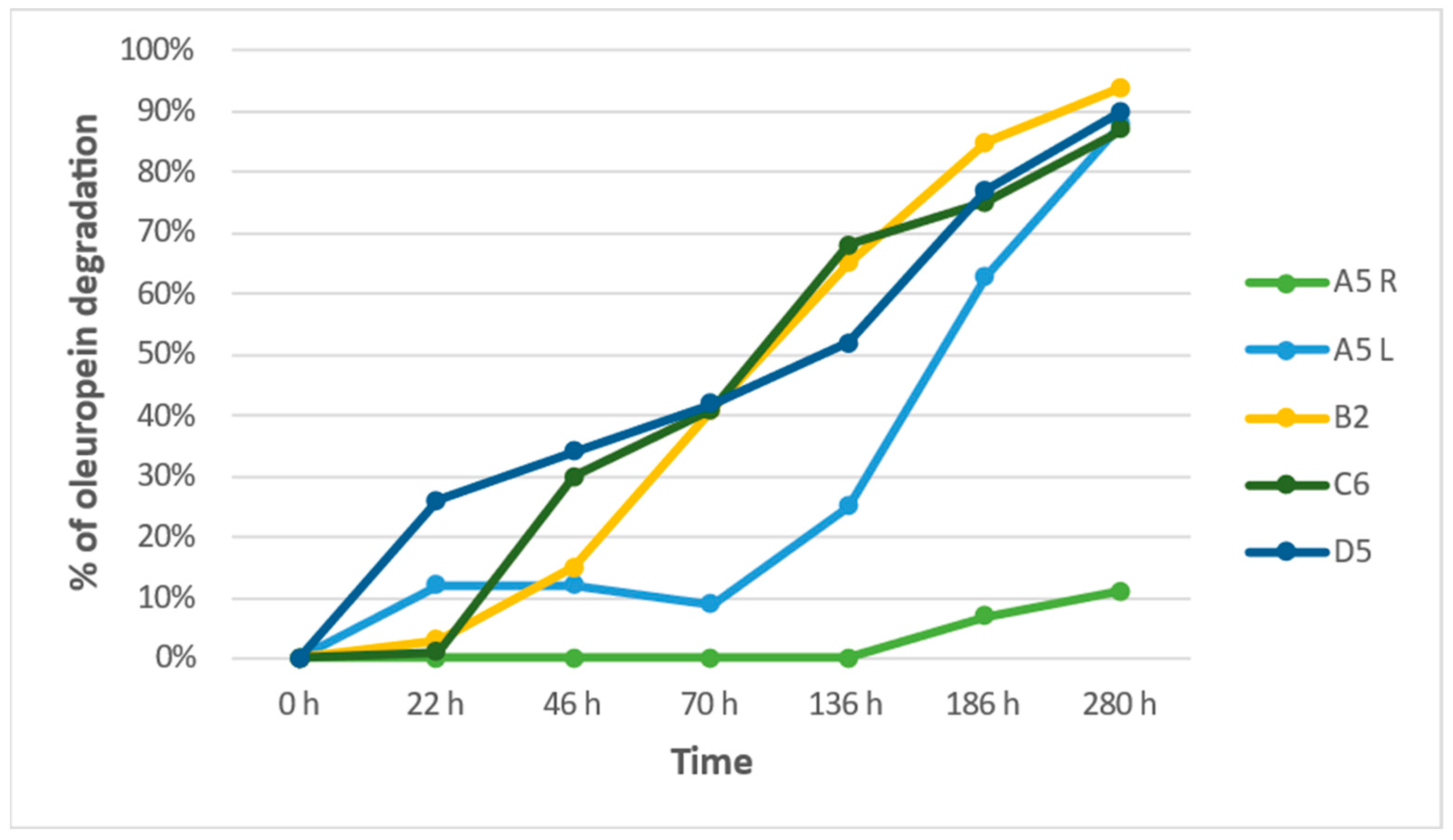
% of Oleuropein Degradation
HoursA5 RA5 LB2C6D5
0 h0%0%0%0%0%
22 h0%12%3%1%26%
46 h0%12%15%30%34%
70 h0%9%41%41%42%
136 h0%25%65%68%52%
186 h7%63%85%75%77%
280 h11%88%94%87%90%
Table 5. Oleuropein degradation by bacteria strains.
Total
HoursProbify g/LSmooth g/LLittle Smooth g/LWrinkled g/L
0 h1.141.141.141.14
18 h1.120.640.630.69
42 h0.580.690.670.56
114 h0.530.470.470.49
Table 6. Oleuropein degradation expressed by the presence of Hydroxytyrosol.
First Peak
HoursProbify g/LSmooth g/LLittle Smooth g/LWrinkled g/L
0 h0000
18 h0.350.230.250.24
42 h0.280.320.310.26
114 h0.240.230.230.22
Table 7. Oleuropein degradation expressed by the presence of Elenolic acid.
Second Peak
HoursProbify g/LSmooth g/LLittle Smooth g/LWrinkled g/L
0 h0000
18 h0.770.410.380.45
42 h0.30.370.360.3
114 h0.310.30.270.29
Disclaimer/Publisher’s Note: The statements, opinions and data contained in all publications are solely those of the individual author(s) and contributor(s) and not of MDPI and/or the editor(s). MDPI and/or the editor(s) disclaim responsibility for any injury to people or property resulting from any ideas, methods, instructions or products referred to in the content.

