Article

Effect of Complex Prebiotics on the Intestinal Colonization Ability of Limosilactobacillus fermentum DALI02

1
College of Food Science and Technology, Yangzhou University, Yangzhou 225000, China
2
Key Laboratory of Dairy Biotechnology and Safety Control, Yangzhou 225000, China
3
New Hope Dairy Co., Ltd., Chengdu 610000, China
4
Yangzhou Food and Drug Inspection and Testing Center, Yangzhou 225000, China
*
Authors to whom correspondence should be addressed.
Fermentation 2023, 9(1), 25; https://doi.org/10.3390/fermentation9010025
Submission received: 16 November 2022 / Revised: 9 December 2022 / Accepted: 20 December 2022 / Published: 28 December 2022
(This article belongs to the Section Fermentation for Food and Beverages)

Abstract

:
Intestinal colonization is beneficial to the role of probiotics, and prebiotics can promote the adhesion and colonization of probiotics in the intestine. This study optimized the combination of complex prebiotics that could improve the growth ability and adhesion ability of Limosilactobacillus fermentum (L. fermentum) DALI02 to Caco-2 cells in vitro and determined the effect of its colonization quantity and colonization time in the immunocompromised rat model. The results showed that all five prebiotics (fructo-oligosaccharides (FOS), galacto-oligosaccharides (GOS), inulin, stachyose, and xylo-oligosaccharides (XOS)) significantly promoted the growth and adhesion of L. fermentum DALI02. It was found that 0.5% (w/w) inulin had the best growth promotion effect, and 0.5% FOS had the strongest adhesion promotion (the adhesion rate was increase by 1.75 times). In addition, 0.05% FOS, 0.20% GOS, 0.30% inulin, 0.20% stachyose, and 0.30% XOS could significantly improve the adhesion rate of L. fermentum DALI02 from 1.72% to 3.98%. After 1 w of intervention, the quantity of colonization in the fermented broth with prebiotics group was significantly higher than that in the fermented broth group. The intervention time was extended from 1 d to 4 w, and the amount of colonization of L. fermentum DALI02 in the fermented broth with prebiotics group increased significantly from 4.32 lgcopies/g to 5.12 lgcopies/g. After the intervention, the serum levels of lipopolysaccharide (LPS) and D-lactic acid in rats were significantly reduced, and the most significant was in the fermented broth with prebiotics group, with LPS and D-lactic acid levels of 74.11 pg/mL and 40.33 μmol/L, respectively. Complex prebiotics can promote the growth and adhesion of L. fermentum DALI02 and significantly increase the quantity of colonization and residence time of the strain in the intestine, which helps the restoration of intestinal barrier function and other probiotic effects.

1. Introduction

Probiotics are live microorganisms that provide health benefits to the host when given in appropriate amounts [1]. They have probiotic functions, such as improving the structure of the host intestinal flora as well as antioxidants, anti-obesity, and immune enhancement [2]. Adhesion and colonization in the intestinal tract will help the strain resist intestinal peristalsis and food erosion, thus allowing probiotics to play a healthy role [3]. Colonization implies a prolonged residence time of probiotics in the intestine and an increased opportunity to interact with the host. On the one hand, it is beneficial for probiotics to reduce the amount of harmful microorganisms in the gut by seizing colonization sites. On the other hand, it can regulate probiotic metabolites, such as short-chain fatty acids, which serve as key signaling molecules to maintain intestinal health, thus benefiting host health [4].
Prebiotics are indigestible substances that provide health benefits to the host, which are selectively utilized by microorganisms in the gut [5]. They can enhance the viability and probiotic properties of probiotics, such as promoting their reproduction, metabolism, and tolerance to the gastrointestinal tract [6,7,8]. In addition, prebiotics can enhance the adhesion ability of probiotics [9] by changing their surface film properties. In addition, the enhancement effect is specific [10]. Prebiotics can also modulate the repulsion of probiotic cell membranes to intestinal epithelial cells by increasing the proportion of unsaturated fatty acids, thus improving the adherence of the strain to HT-29 [11]. Prebiotics, as a carbon source, can enhance the adhesion to Caco-2 by highly expressing the gene, encoding its adhesion protein during the growth of Bifidobacterium DNG6 [12].
Many studies have been conducted to investigate the effect of prebiotics on the adhesion ability of probiotics in vitro through the Caco-2 cell model [13], HT-29 cell model [14], or Caco-2 and HT-29 symbiosis model [15]. However, colonization in the intestine is very complex. The peristalsis of the intestine, moving of food, and intervention of other flora can affect the initial adhesion and long-term colonization of strains. Although the ability of the strains to adhere to intestinal epithelial cells is an important criterion to screen probiotics in vitro [16], it is crucial to explore the colonization ability of strains in vivo. At the same time, considering the practical application of probiotics, it is of more practical significance to explore the colonization ability of strains in the model of intestinal injury. Therefore, this study investigated the complex prebiotics that can improve the growth quantity and the adhesion ability of L. fermentum DALI02 to Caco-2 cells. The complex prebiotics (fructo-oligosaccharides (FOS), galacto-oligosaccharides (GOS), inulin, stachyose, and xylo-oligosaccharides (XOS)) on the colonization ability of the strain was investigated by evaluating the effect of the fermentation broth with prebiotics on the quantity and time of intestinal colonization of L. fermentum DALI02 in immunocompromised model rats.

2. Materials and Methods

2.1. Microorganism and Cell Strains

L. fermentum DALI02 was isolated from traditional fermented dairy products by Key Laboratory of Dairy Biotechnology and Safety Control and stored in China General Microbial Strain Conservation Management Center (No. CGMCC 16064). The lyophilized strain was cultured in de Man Rogosa and Sharpe (MRS) media at 37 °C for 24 h.
Caco-2 cells (Procell CL-0050, Procell Life Science & Technology Co., Ltd., Wuhan, China) were maintained in modified Eagle’s medium (MEM, Life Technologies Corporation, New York, NY, USA) with high glucose supplemented with 20% fetal bovine serum (FBS, PAN-Biotech, Bavaria, Germany), 1% nonessential amino acids (NEAA, Life Technologies Corporation, New York, NY, USA), 1% pyruvate (Life Technologies Corporation, New York, NY, USA), 1% GlutaMAX (Life Technologies Corporation, New York, NY, USA), and 1% penicillin/streptomycin/ neomycin (PSN, Thermo Fisher Scientific Inc., New York, NY, USA) and incubated at 37 °C in a 5% CO2 incubator (Thermo Fisher Scientific Inc., New York, NY, USA).

2.2. Preparation of Whey Hydrolysate and Prebiotics

Whey protein powder (Fonterra Brands (NZ) Ltd., Auckland, New Zealand) was adjusted with NaOH (0.1 mol/L, China Pharmaceutical Group Co., Ltd., Beijing, China) at 10% (w/w), pH 7.00, and papain (Beijing Solarbio Science & Technology Co., Ltd., Beijing, China) at 6000 U/g was added, hydrolysis temperature was 65 °C, and enzymatic digestion time was 2 h. Control of the degree of hydrolysis was 20%, high temperature inactivation was performed, and the supernatant was extracted after centrifugation at 6500 r/min for 15 min, heat-treated at 105 °C for 10 min, and placed at 4 °C.
Different weights of prebiotics (FOS, GOS, Inulin, Stachyose, and XOS, Jinfuyuan Biotechnology Co., Ltd., Shenzhen, China) were weighed and dissolved in sugar-free MRS and whey hydrolysate, respectively, sterilized at 105 °C for 10 min, and then prepared for use.

2.3. The Effects of Probiotics on L. fermentum DALI02

Activated L. fermentum DALI02 was inoculated at 3% (v/v) in the prebiotic content of 2% (w/w) MRS-Lactose medium, MRS-FOS medium, MRS-GOS medium, MRS-Inulin medium, MRS-Stachyose medium, MRS-XOS medium, as well as sugar-free MRS and incubated at 37 °C for 24 h. The OD600 was measured by enzyme marker (Thermo Fisher Scientific Inc., New York, NY, USA).
The activated L. fermentum DALI02 was inoculated into enzymatic skim milk with a prebiotic content of 0.5% (w/w) at 3% (v/v) inoculum for 24 h at 37 °C. The number of viable bacteria and their ability to adhere to Caco-2 cells were determined by dilution-plate method.

2.4. Experimental Design, Modelling, and Optimization

According to the results of the pre-experiment (Figure S1), the five prebiotics at different concentrations in this experiment had a significant effect (p < 0.05) on the adhesion capacity of L. fermentum DALI02. Therefore, a combination of five levels of quadratic regression orthogonal rotation combination design (QRORCD) and response surface methodology (RSM) were used to optimize the addition of complex prebiotics for the high adhesion capacity of L. fermentum DALI02. The coded and natural values of the independent variables X1 (FOS addition, %), X2 (GOS addition, %), X3 (Inulin addition, %), X4 (Stachyose addition, %), and X5 (XOS addition, %) are shown in Table 1. The QRORCD included 10 replicate centroids and a set of axial points selected to allow for rotatability, which ensured that the variance of the model predictions was constant at all points equidistant from the design center. The experimental runs were randomized to minimize the effect of unexpected variation in the observed responses.

2.5. Adhesion Ability to Caco-2 Cell Lines

The cultured Caco-2 cells (Procell CL-0050, Procell Life Science & Technology Co., Ltd., Wuhan, China) were carried out and the concentration was adjusted to 2 × 10⁵ cell/mL, and then the cells with the adjusted concentration were placed in a 24-well plate (Costar, Corning Inc., New York, NY, USA) and continued to be cultured until they grew into monolayer cells. The samples were diluted to a viable count of 106 CFU/mL added to each well and continued to be incubated at 37 °C in a 5% CO2 incubator for 2 h. The fermented broth was discarded, and the cells were washed twice with PBS solution and then scraped off and resuspended in sterile phosphate buffered saline (PBS) (pH 7.2; Sangon Biotech Co., Ltd., Shanghai, China) to count the viable count. The equation of adhesion rate is shown as follows:
Adhesion rate = number of live bacteria after 2 h of adhesion/number of live bacteria before adhesion × 100%.

2.6. Adhesion Parameters in Rat Model

2.6.1. Experimental Design Based on the Animal Model

To investigate the effect of gavage time on the number and residence time of L. fermentum DALI02 intestinal colonization, 36 Wistar male mice (Yangzhou University Medical Animal Experiment Center, Yangzhou, China), aged 4 weeks and weighing 140–160 g were studied. They were housed under a 12 h light/12 h dark cycle in a controlled room with a temperature of 24 ± 2 °C and a humidity of 50 ± 10%. All rats were allowed free access to food and water. All 5 rats were placed in a cage and were fed a diet (flour 20%, rice flour 10%, corn 20%, drum skin 26%, soy material 20%, fish meal 2%, and bone meal 2%). All rats were randomly divided into 4 groups (n = 15): control group, model group, fermented broth group, and fermented broth with prebiotics group. The immunocompromised rat model was established by intraperitoneal injection of 80 mg/kg of CTX for 3 consecutive days. During the next 4 weeks, all rats received the following treatments by gavage: control group and model group—whey hydrolysate (1 mL/100 g), fermented broth group—L. fermentum DALI02 fermented broth without prebiotics (1 mL/100 g, 1 × 10⁹ CFU/mL), and fermented broth with prebiotics group—L. fermentum DALI02 fermented broth with prebiotics (1 mL/100 g, 1 × 10⁹ CFU/mL). At the end of the 4th week, rats underwent 12 h of fasting prior to being anaesthetized and dissected. All rats were euthanized at the anestrus period following anesthesia under 1% sodium pentobarbital.

2.6.2. Colonization of L. fermentum DALI02

The ability of L. fermentum DALI02 to colonize the rat intestine was determined by measuring the number of L. fermentum in rat feces. Mouse fecal DNA was extracted using the Fecal Genomic DNA Extraction Kit (Beijing Solarbio Science & Technology Co., Ltd., Beijing, China) according to the manufacturer’s instructions (F: GACCAGCGCACCAAGTGATA; R: AGCGTAGCGTTCGTGGTAAT) and PCR-amplified using primers specific to L. fermentum from the existing literature [17]. The total volume was 10 μL and was run on StepOnePlus™ Real-Time PCR System with Tower (Applied Biosystems, Inc., New York, NY, USA) using SYBR Premix Ex Taq II (Takara Bio Inc., Tokyo, Japan). All measurements were performed in triplicate.
Standard curves were constructed using PCR products of L. fermentum DALI02 and the 16S rRNA gene of E. coli, and then the obtained PCR products were cloned into the T vector (Takara Bio Inc., Tokyo, Japan) and transfected into DH-5α and incubated at 37°C for 6 h (100–140 rpm) as standard samples. The recombinant plasmids were used for real-time fluorescent quantitative PCR (qPCR), and 103, 104, 105, 106, 107, 108, 109, and 1010 copies of each plasmid were used for calibration for each reaction. The copy number K = (p*6.02*1023)/Mw of DALI02 was calculated using the following metric, where p (g/L) and Mw (base pair) are the plasmid DNA concentration and relative molecular mass, respectively. Three standard curves were generated from at least five 10-fold plasmid dilutions, and Ct values were obtained by StepOne Plus software, with the Ct value as the y-axis and the logarithm of the copy number as the x-axis (Figure S2) [18]. The quantity of colonized L. fermentum DALI02 in the intestine of different groups of rats was expressed by CT values.

2.6.3. Residence Time of L. fermentum DALI02 in the Intestine

After gavage, rat feces were picked up every 12 h in sterile EP tubes and stored at -80 °C for storage. Using L. fermentum specific primers, the amount of strains in the fresh feces of rats was monitored at different times after cessation of the intervention until the quantity of colonization in the intervention group was not significantly different from that in the blank group, which was the time of colonization.

2.7. Determination of the Integrity of the Intestinal Barrier in Rats

Rat intestinal barrier integrity was determined by measuring lipopolysaccharide (LPS) and D-lactic acid levels in rat serum using an endotoxin assay kit (Shanghai Hualan Electronic Co., Ltd., Shanghai, China) and a D-lactic acid assay kit (Shanghai Hualan Electronic Co., Ltd., Shanghai, China).

2.8. Statistical Methods

Statistical analysis was performed using GraphPad Prism 9 (San Diego, CA, USA). The results are presented as the means and standard deviations, and comparisons among different groups were assessed by analysis of variance (ANOVA) with the Tukey post hoc test (one-way ANOVA-Tukey). Values of p < 0.05 were considered statistically significant. The graphics were created using GraphPad Prism 9 (San Diego, CA, USA).

3. Results

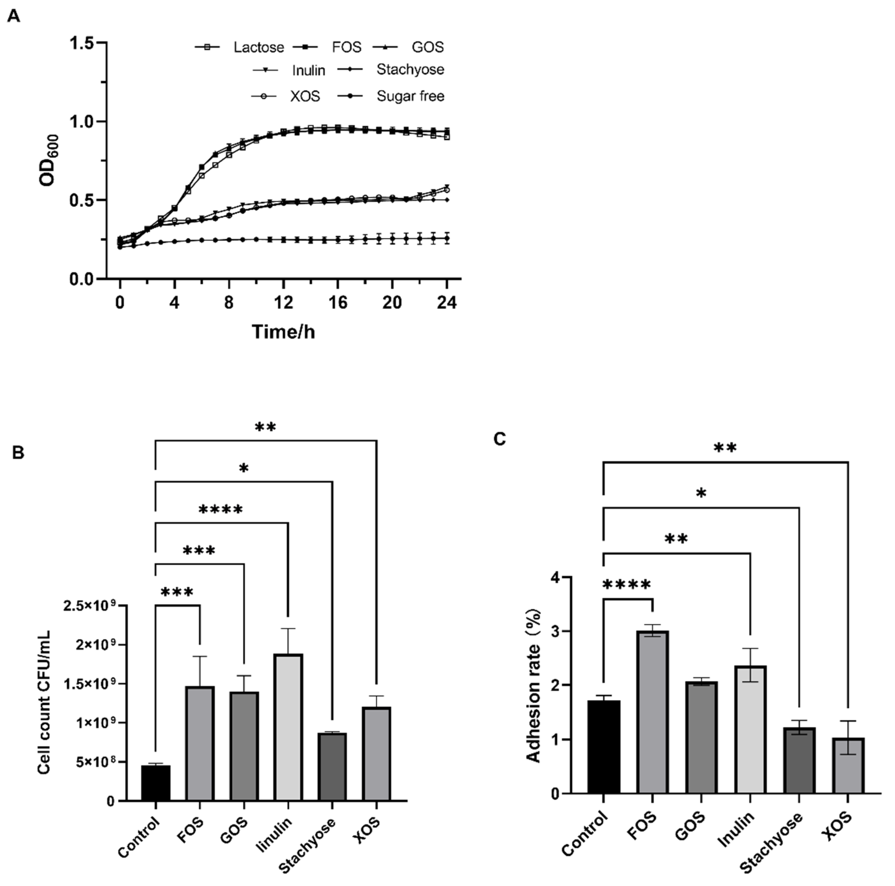
3.1. Effects of Probiotics on the Growth and Adhesion of L. fermentum DALI02

The growth curves of L. fermentum DALI02 in MRS medium with different prebiotics as the sole carbon source are shown in Figure 1A. The OD600 value in the sugar-free medium was always 0.21, indicating that a carbon source is necessary for the growth of L. fermentum DALI02. In addition, all five target prebiotics were utilized by L. fermentum DALI02, and the growth curves under 2% lactose, FOS, and GOS were more closely matched, with growth better than that of stachyose, inulin, and XOS.
The viable bacterial counts of L. fermentum DALI02 in enzymatic whey supplemented with an additional 0.5% (w/w) prebiotics after 24 h are shown in Figure 1B. The addition of all five prebiotics significantly increased the viable bacterial counts of L. fermentum DALI02 (p < 0.05), with inulin being the most significant, with the viable count increasing from 4.92 × 10⁸ ± 0.91 CFU/mL to 1.88 × 10⁹ ± 0.32 CFU/mL, up to 3.8-fold, followed by FOS, GOS, and XOS. The potentiation effect of different prebiotics on the in vitro adhesion of L. fermentum DALI02 is shown in Figure 1C. FOS and inulin were able to significantly increase the adhesion of L. fermentum DALI02 to Caco-2 cells (p < 0.01), and the adhesion rate increased from 1.72% ± 0.09 (control group) to 3.01% ± 0.11 and 2.37% ± 0.31, respectively, but stachyose and XOS significantly decreased the adhesion rate (p < 0.05).

3.2. Response Criteria for the RSM Analysis

The results of the single-factor experiment (Figure S1) show that the optimal concentration of prebiotics added varied among the different prebiotics, with the highest adhesion rates reached at 0.5% FOS, 1.5% GOS, 1.0% inulin, 1.0% stachyose, and 3.0% XOS, respectively, with 3.01%, 2.83%, 2.89%, and 1.91%, and 3.12%.
The experimental values obtained for the 36 experimental runs of the five-factor quadratic regression orthogonal rotational combination design are shown in Table S1. The parameter coefficients for each term in the mathematical model are presented in Table 2, which shows the specific parameters for the effects of linear, quadratic, and interactive effects of the five independent variables (FOS, GOS, inulin, stachyose, and XOS) on the adhesion rate of L. fermentum DALI02. The models exhibited non-significant misfit values (p > 0.05) and sufficient precision, which indicated that the model equations adequately described the effects of the independent variables on the final results. As shown in Table 2, the coefficient R2 = 0.7793, indicating that the response values can be explained by the independent variables.
Y = 2.75632 − 15.02972 X1 + 5.90181 X3 + 1.06449 X5 − 88.62500 X1X3 + 26.29167 X1X5 + 28.37500 X2X4
According to Equation (1), the effects of these five prebiotics on the adhesion rate of L. fermentum DALI02 were in the order of FOS > inulin > XOS > stachyose > GOS. The optimal formulations were obtained as 0.05% FOS, 0.20% GOS, 0.3% inulin, 0.20% stachyose, and 0.30% XOS. The theoretical adhesion rate of the strain was 4.04%, and the actual adhesion rate was 3.98 ± 0.12%, as verified by the experiment, which was not significantly different from the predicted value.

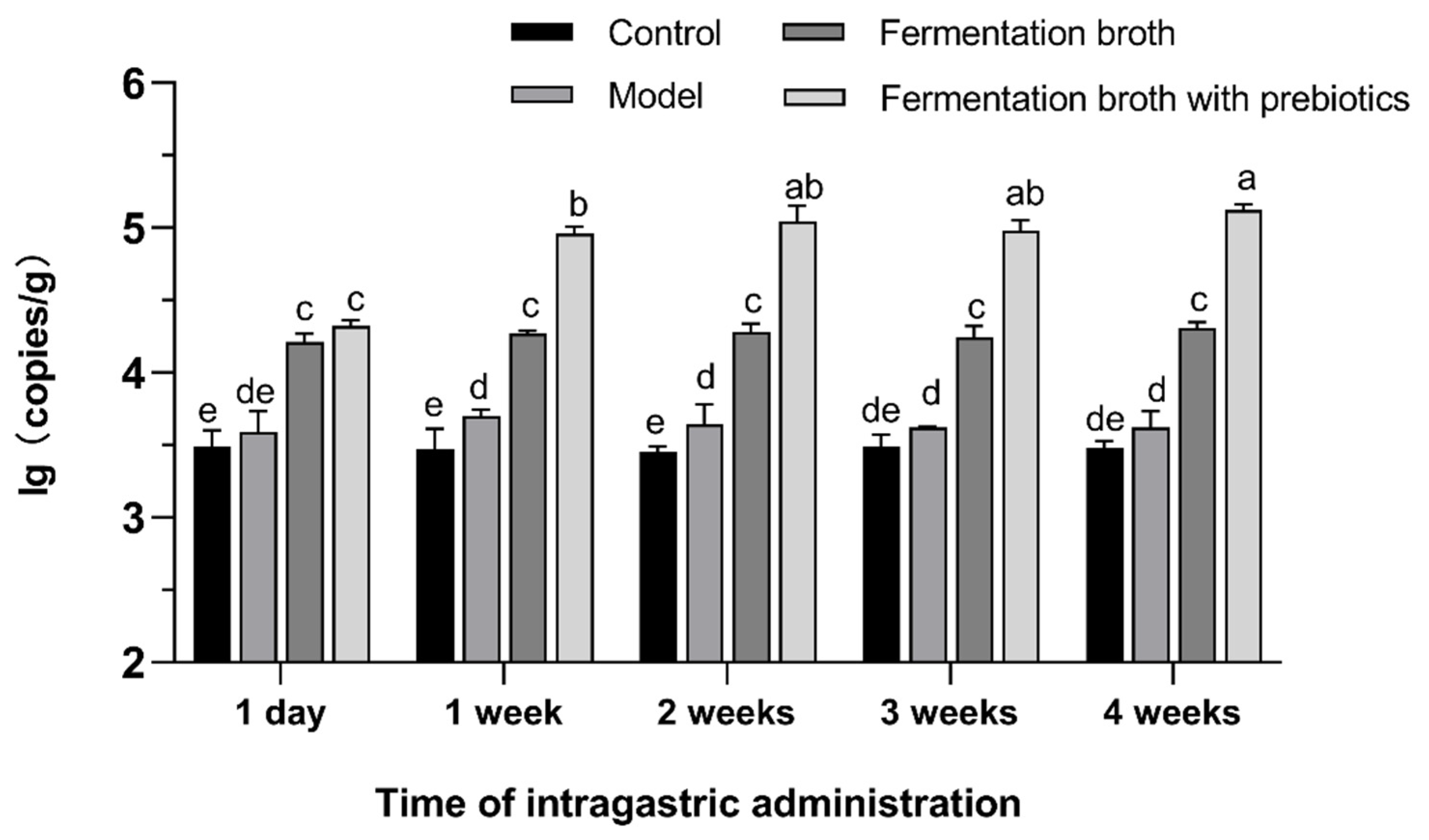
3.3. Effect of Intragastric Administration on Colonization Quantity

The content of L. fermentum in the feces of rats at 1 d, 1 w, 2 w, 3 w, and 4 w of intervention was measured, and the results are shown in Figure 2. The results showed that the fermented broth group and the fermented broth with prebiotics group were always significantly higher than the model group as well as the blank group (p < 0.05), and the fermented broth with prebiotics group was significantly higher than the fermented broth group for the remainder of the intervention time, except for 1 d of the intervention. This indicates that the presence of prebiotics can significantly increase the number of colonized strains (p < 0.05) and that a certain time is needed for this effect to take place. In addition, there was no significant difference in the time of intervention on the colonization of the fermented broth group (p > 0.05). The quantity of colonization was significantly higher after 1 w of the fermentation broth with prebiotics intervention (p < 0.05), but the quantity of colonization reached the maximum after 2 w and was not significantly different from 3 w and 4 w (p > 0.05).

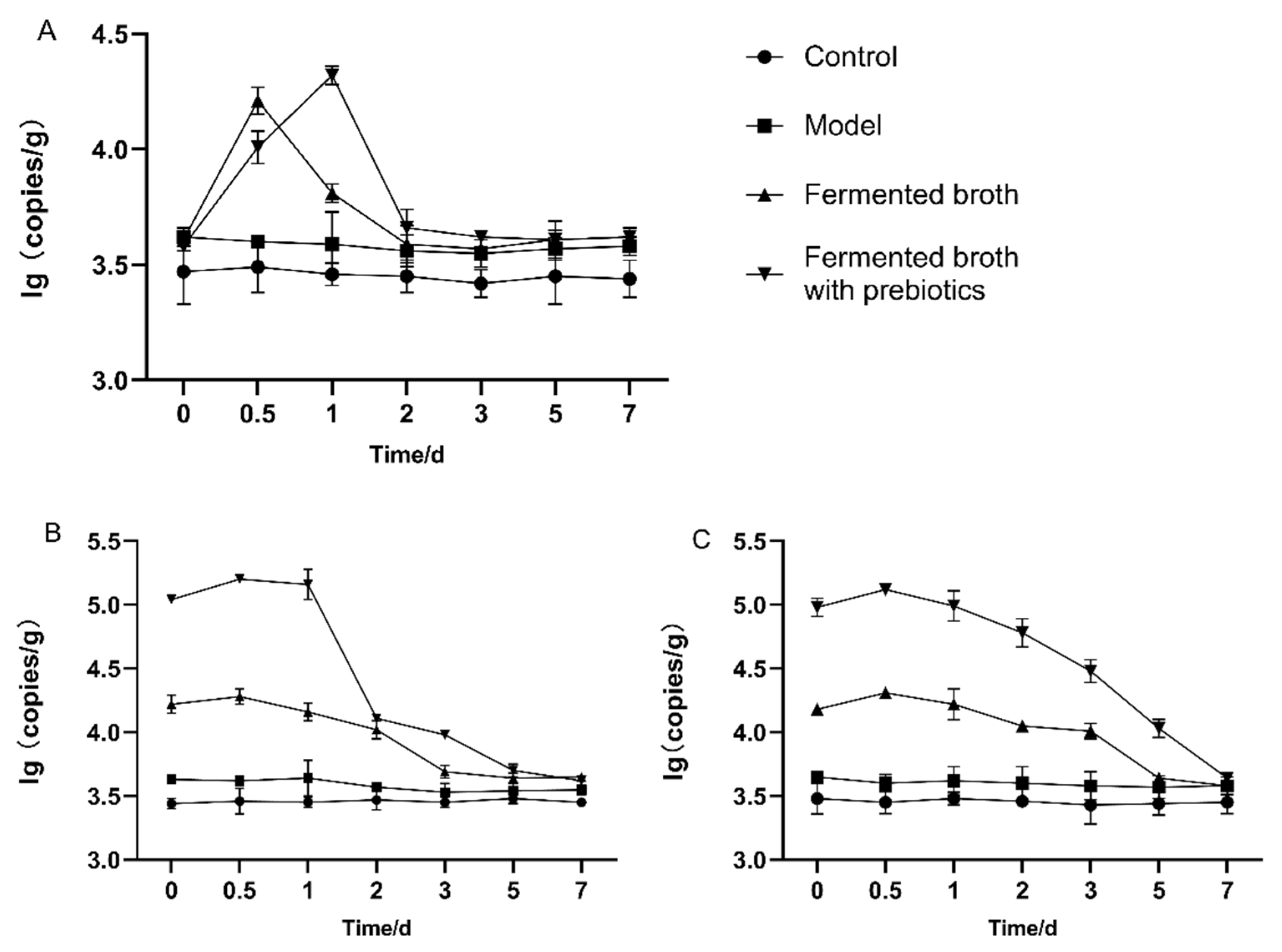
3.4. Effect of Intragastric Administration Time on Intestinal Residence Time

Gavage was stopped after 1 d, 2 w, and 4 w of intervention, and feces were collected at 0 d, 0.5 d, 1 d, 2 d, 3 d, 5 d, and 7 d, and the content of L. fermentum was measured (Figure 3). The intestinal residence of the strains at 1 d of intervention was approximately equal to that at 2 d. The residence time was longer in the fermented broth with prebiotics group than in the fermented broth group. After 2 w of intervention, the initial quantity of colonization was significantly higher in the fermented broth with prebiotics group (5 d) and fermentation broth group (3 d) than in the rest of the groups, and the residence time was extended. The residence time at 4 w of intervention was 7 d for the fermented broth with prebiotics group and 5 d for the fermented broth group. The results showed that with the extension of the intervention time, the intestinal residence time was also gradually extended, and the fermented broth with prebiotics group was always better than the fermented broth group, that is, the addition of complex prebiotics could increase the adhesion capacity and intestinal residence time of L. fermentum DALI02.

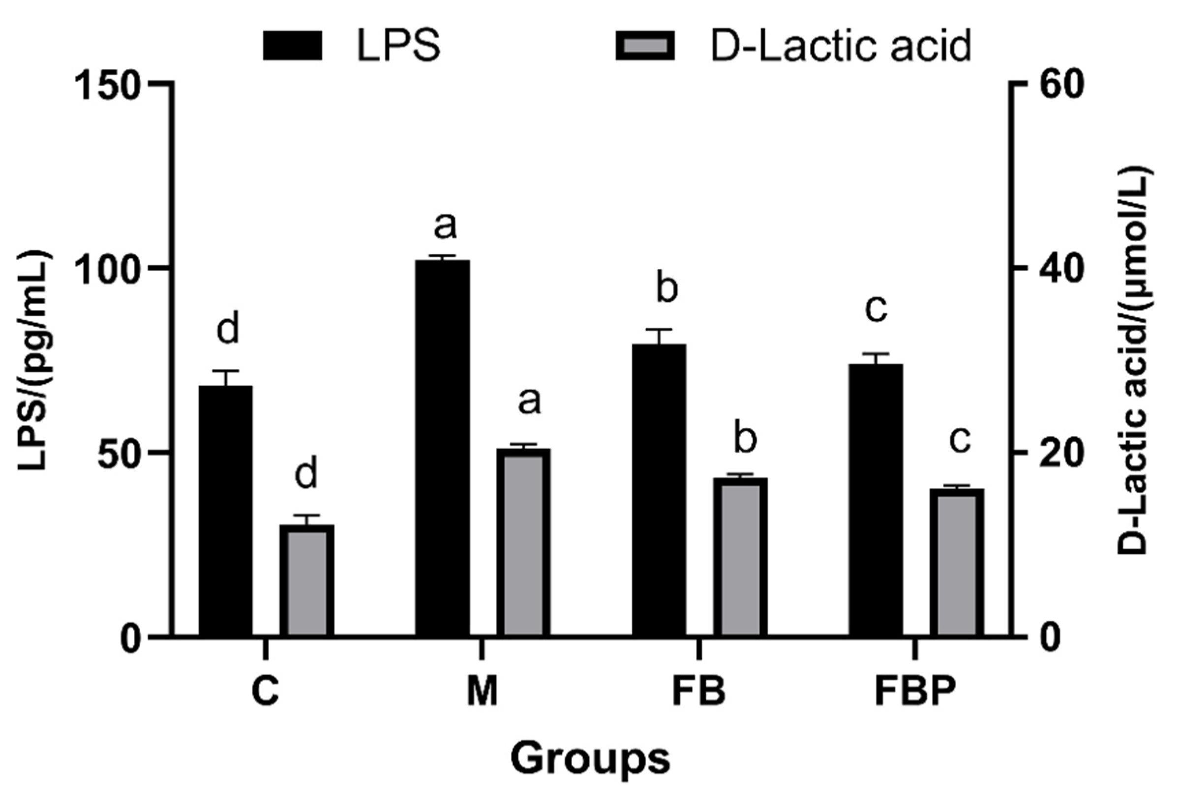
3.5. Intestinal Barrier Integrity in Rats

At the end of 4 weeks of gavage, fresh blood was taken from rats of different groups and their serum levels of LPS and D-lactic acid were measured (Figure 4). The levels of LPS and D-lactic acid in the serum of rats in the model group were 102.14 pg/mL and 51.03 μmol/L, respectively, which were significantly increased compared with the blank group (p < 0.05), indicating that the intake of CTX causes some damage to the intestinal barrier integrity of rats. After the intervention of the fermented broth group and the fermented broth with prebiotics group, the serum levels of LPS and D-lactic acid in rats were significantly downregulated compared with the model group (p < 0.05), being most significant in the fermented broth with prebiotics group, with LPS and D-lactic acid levels of 74.11 pg/mL and 40.33 μmol/L, respectively.

4. Discussion

It is a key indicator to identify the adhesion in vitro when screening candidate probiotics. Adherence will promote the transient stay of the strain in the intestine and provide the possibility of effective colonization, thus providing sufficient response time for the strains to exert their functional properties in the intestine [19]. The addition of prebiotics is known to enhance the health benefits of probiotics, including the regulation of the structure of the intestinal flora via competing for colonization sites [20]. The number of colonized strains and their residence time are crucial for the realization of probiotic function. In this study, we found that the optimized complex prebiotics could significantly increase the viable number, the adherence in vitro, and colonization in vivo. The continuous intervention for 4 weeks would effectively prolong the colonization quantity and residence time of the strain.
There is variability in the proliferative effect of different prebiotics on strains [21]. Thus, it is a critical first step to explore the utilization of prebiotics by strains, which was usually determined by measuring the ability of strains to grow in a medium with prebiotics as a single carbon source [22]. L. fermentum DALI02 in this study was able to utilize all five prebiotics (FOS, GOS, inulin, stachyose, and XOS), but there were significant differences in utilization efficiency. The strain showed the best condition of growth in FOS and GOS, and the growth curve of the strain was close to the situation in lactose (Figure 1A). This is in agreement with the previous results that the strains utilized oligofructose better [23], while the strains utilized inulin less efficiently [24]. GOS and FOS, which were utilized less efficiently in this study, showed better results in previous studies [25]. These results once again suggested that the utilization of prebiotics by strains was specific. This has been revealed by previous studies by comparing genomic differences in transporter proteins, degradation enzymes, and transcriptional regulation of strains, which may be related to operons controlling the strains involved in metabolic enzymes [26]. Such results call for the need for researchers to screen for the selection of suitable prebiotics for specific probiotics.
It was found that additional prebiotics also significantly increased the number of viable bacteria in the presence of a carbon source (Figure 1B). Stachyose and XOS, which performed poorly in a single carbon source, showed promising results here. This suggested that there was a prioritization of carbon source use by the strains, and there was a synergistic effect among the different carbon sources. However, it was more noteworthy that the addition of different prebiotics positively modulated the adherence of the strains in vitro. The strains showed an increase and decrease in adherence compared with the blank group (Figure 1C). The adhesion of probiotics to intestinal epithelial cells is triggered mainly by the interaction between the mucosal layer secreted by the intestinal epithelium, the surface structures, and secretions of the bacteria. The mucosal layer of intestinal epithelial cells consists mainly of mucoprotein, glycolipid, electrolyte, etc. The substances associated with adhesion on the surface of the strain are mainly lipophosphatidic acid, surface-layer-associated protein, and mucin-binding protein [27]. In this study, prebiotics were involved in the fermentation process of the strain, which would produce a large number of metabolites, such as short-chain fatty acids, which would increase the viability of the intestinal epithelial cells. It would enhance the adhesion ability of the strain [28]. In addition, the structure of prebiotics may have a similar polysaccharide composition to the cell wall of Limosilactobacillus. This would also increase the adhesion efficiency of cell wall polysaccharides [13]. Prebiotics may enhance the receptor interaction with intestinal epithelial cells by modulating the ratio of unsaturated fatty acids in the cell membrane of the strain and by regulating the secretion of cell membrane surface proteins [11]. In addition, the decrease in adhesion rate could be due to the dose-dependent effect of prebiotic potentiation [29], which was verified in the subsequent concentration one-way experiments (Figure S1).
The effectiveness of probiotic function is dose-dependent. The concentration of probiotics required to obtain clinical effects is generally considered to require a concentration of 106 CFU/mL [30], a value that has become the lowest dose group in many studies exploring effective doses of probiotic interventions. Many studies have shown that higher dose experimental groups tend to have better results [31]; thus, this study used 109 CFU/mL of fermentation broth for the intervention. In this study, we investigated the effect of prebiotics on the colonization of probiotic bacteria in the damaged intestine in an immunocompromised model of rats induced by CTX. The intestinal flora is disturbed and the intestinal mucosa is damaged [32]. In addition, it would affect the adhesion and colonization of exogenous probiotic bacteria. Gavage was performed according to the body weight of the rats at 100 g/mL. While the dose of a single intervention is important, there is, however, a significant effect on the frequency regarding the number of colonized strains and the residence time. It has been shown that the target strains could not be detected after 48 h of single gavage [33]. This is consistent with the results of the present study (Figure 3A), where at 48 h, the intervention groups were all close to the blank group. The residence time of strains also obtained an extension with the increase of the intervention time (intervention 1 d (48 h), intervention 2 w (72 h), and intervention 4 w (96 h)), and the residence time of the fermented broth group with prebiotics also obtained an extension from 72 h to 7 d. The prolongation of the intervention time may lead to some recovery of the already damaged intestinal flora of the rats, thus reaching relative homeostasis. According to the serum levels of LPS and D-lactic acid in rats, after 4 weeks of intervention, the levels in the fermented broth group and the fermented broth with prebiotics group were significantly reduced (p < 0.05), indicating that their intestinal barrier function was significantly restored and the intestinal mucosa was repaired, which may provide a basis for intestinal adhesion and colonization of the strain.
In terms of the quantity of colonization, there was no significant difference between the presence or absence of prebiotics after 1 intervention (p > 0.05). With the extension of the intervention time, there was no significant effect of the intervention time on the fermented broth group, and the number of L. fermentum was significantly higher in the fermented broth with prebiotics group than in the rest of the groups (p < 0.05), which indicated that prebiotics had a significant effect on the number of colonized strains (p < 0.05). In addition to the multiple reasons mentioned above, the addition of prebiotics would also lead to a sustained proliferation of L. fermentum already colonized in the intestinal tract.

5. Conclusions

In the present study, we report that the complex prebiotics can increase the adhesion capacity of L. fermentum DALI02 and their colonization parameters in immunocompromised model rats. We found that all five prebiotics (FOS, GOS, inulin, stachyose, and XOS) studied were utilized by L. fermentum DALI02, and the addition of all five prebiotics to enzymatically defatted milk increased the viable count of L. fermentum DALI02. The optimized fermentation broth with prebiotics increased the adherence of the strains to Caco-2 cells, the quantity of intestinal colonization, and the duration of colonization. Furthermore, the prolongation of the intervention time did not affect the quantity of colonization in the fermented broth group, but it did affect the quantity of colonization in the fermented broth with prebiotics group. The prolongation of the intervention time also prolonged the intestinal colonization time of L. fermentum DALI02. These results suggest that the complex prebiotics enhance the intestinal colonization of L. fermentum DALI02. This may be a strategy for developing functional foods.

Supplementary Materials

The following supporting information can be downloaded at: https://www.mdpi.com/article/10.3390/fermentation9010025/s1, Figure S1: Effect of different prebiotic additions on the adhesion rate of L. fermentum DALI02 A FOS B GOS C Inulin D Stachyose E XOS; Figure S2: Standard curve of plasmid of L. fermentans; Table S1: Results of quadratic regression rotation orthogonal experiment.

Author Contributions

Conceptualization, X.L., D.C. and Q.L.; methodology, X.L., C.Z. and D.C.; software, X.L. and Q.L.; validation, X.L. and Y.Z.; formal analysis, X.L.; investigation, H.Q. and W.W.; resources, Y.H.; data curation, L.Z.; writing—original draft preparation, X.L.; writing—review and editing, X.L. and R.G.; visualization, X.L.; supervision, R.G.; project administration, D.C. and L.X.; funding acquisition, R.G. All authors have read and agreed to the published version of the manuscript.

Funding

This research was funded by National Natural Science Foundation of China number 32272362, National Natural Science Foundation of China number 31972094, Science and Technology Plan Project of Jiangsu Provincial Market Supervision Administration number KJ21125045 and City-school cooperation to build science and Technology Innovation Platform number YZ2020265.

Institutional Review Board Statement

The study was conducted in accordance with the U.S. National Institutes of Health guidelines for the care and use of laboratory animals (NIH Publication No. 85-23 Rev. 1985) and approved by the Animal Care Committee of the Center for Disease Control and Prevention (Jiangsu, China) (No. 202103262, 2021-03-05).

Informed Consent Statement

Not applicable.

Data Availability Statement

The data supporting the findings reported here are available on reasonable request from the corresponding author.

Conflicts of Interest

The authors declare that they have no conflict of interest.

References

  1. Pandey, K.R.; Naik, S.R.; Vakil, B.V. Probiotics, prebiotics and synbiotics—A review. J. Food Sci. Technol.-Mysore 2015, 52, 7577–7587. [Google Scholar] [CrossRef] [PubMed] [Green Version]
  2. Nagpal, R.; Kumar, A.; Kumar, M.; Behare, P.V.; Jain, S.; Yadav, H. Probiotics, their health benefits and applications for developing healthier foods: A review. FEMS Microbiol. Lett. 2012, 334, 1–15. [Google Scholar] [CrossRef] [PubMed] [Green Version]
  3. Celebioglu, H.U.; Svensson, B. Dietary Nutrients, Proteomes, and Adhesion of Probiotic Lactobacilli to Mucin and Host Epithelial Cells. Microorganisms 2018, 6, 90. [Google Scholar] [CrossRef] [Green Version]
  4. Plaza-Diaz, J.; Ruiz-Ojeda, F.J.; Gil-Campos, M.; Gil, A. Mechanisms of Action of Probiotics. Adv. Nutr. 2019, 10, S49–S66. [Google Scholar] [CrossRef] [PubMed] [Green Version]
  5. Gibson, G.R.; Hutkins, R.; Sanders, M.E.; Prescott, S.L.; Reimer, R.A.; Salminen, S.J.; Scott, K.; Stanton, C.; Swanson, K.S.; Cani, P.D.; et al. The International Scientific Association for Probiotics and Prebiotics (ISAPP) consensus statement on the definition and scope of prebiotics. Nat. Rev. Gastroenterol. Hepatol. 2017, 14, 491–502. [Google Scholar] [CrossRef] [Green Version]
  6. Ashaolu, T.J.; Ashaolu, J.O.; Adeyeye, S.A.O. Fermentation of prebiotics by human colonic microbiotain vitroand short-chain fatty acids production: A critical review. J. Appl. Microbiol. 2021, 130, 677–687. [Google Scholar] [CrossRef]
  7. Brink, M.; Todorov, S.D.; Martin, J.H.; Senekal, M.; DicksL, M.T. The effect of prebiotics on production of antimicrobial compounds, resistance to growth at low pH and in the presence of bile, and adhesion of probiotic cells to intestinal mucus. J. Appl. Microbiol. 2006, 100, 813–820. [Google Scholar] [CrossRef]
  8. Nagpal, R.; Kaur, A. Synbiotic Effect of Various Prebiotics on In Vitro Activities of Probiotic Lactobacilli. Ecol. Food Nutr. 2011, 50, 63–68. [Google Scholar] [CrossRef]
  9. Russo, P.; López, P.; Capozzi, V.; De Palencia, P.F.; Dueñas, M.T.; Spano, G.; Fiocco, D. Beta-Glucans Improve Growth, Viability and Colonization of Probiotic Microorganisms. Int. J. Mol. Sci. 2012, 13, 6026–6039. [Google Scholar] [CrossRef] [Green Version]
  10. Pan, M.; Kumaree, K.K.; Shah, N.P. Physiological Changes of Surface Membrane in Lactobacillus with Prebiotics. J. Food Sci. 2017, 82, 744–750. [Google Scholar] [CrossRef]
  11. Cai, G.; Wu, D.; Li, X.; Lu, J. Levan from Bacillus amyloliquefaciens JN4 acts as a prebiotic for enhancing the intestinal adhesion capacity of Lactobacillus reuteri JN101. Int. J. Biol. Macromol. 2020, 146, 482–487. [Google Scholar] [CrossRef] [PubMed]
  12. Zhang, G.; Zhao, J.; Wen, R.; Zhu, X.; Liu, L.; Li, C. 2′-Fucosyllactose promotes Bifidobacterium bifidum DNG6 adhesion to Caco-2 cells. J. Dairy Sci. 2020, 103, 9825–9834. [Google Scholar] [CrossRef] [PubMed]
  13. Liu, M.; Sun, Z.; Shi, C.; Wang, J.; Wang, T.; Dziugan, P.; Zhang, B.; Zhao, H.; Jia, G. How do Lycium barbarum polysaccharides promote the adhesion of Lactobacillus to Caco-2 cells? J. Funct. Foods 2022, 89, 104929. [Google Scholar] [CrossRef]
  14. Anand, S.; Mandal, S.; Singh, K.S.; Patil, P.; Tomar, S.K. Synbiotic combination of Lactobacillus rhamnosus NCDC 298 and short chain fructooligosaccharides prevents enterotoxigenic Escherichia coli infection. LWT-Food Sci. Technol. 2018, 98, 329–334. [Google Scholar] [CrossRef]
  15. Krausova, G.; Hynstova, I.; Svejstil, R.; Mrvikova, I.; Kadlec, R. Identification of Synbiotics Conducive to Probiotics Adherence to Intestinal Mucosa Using an In Vitro Caco-2 and HT29-MTX Cell Model. Processes 2021, 9, 569. [Google Scholar] [CrossRef]
  16. Zhang, L.; Qu, H.; Liu, X.; Li, Q.; Liu, Y.; Wang, W.; Chen, D.; Xiao, L.; Gu, R. Comparison and selection of probiotic Lactobacillus from human intestinal tract and traditional fermented food in vitro via PCA, unsupervised clustering algorithm, and heat-map analysis. Food Sci. Nutr. 2022, 10, 4247–4257. [Google Scholar] [CrossRef]
  17. Kim, E.; Yang, S.M.; Lim, B.; Park, S.H.; Rackerby, B.; Kim, H.Y. Design of PCR assays to specifically detect and identify 37 Lactobacillus species in a single 96 well plate. BMC Microbiol. 2020, 20, 1–14. [Google Scholar] [CrossRef] [Green Version]
  18. Zhu, S.; Li, X.; Song, L.; Huang, Y.; Xiao, Y.; Chu, Q.; Kang, Y.; Duan, S.; Wu, D.; Ren, Z. Stachyose inhibits vancomycin-resistant Enterococcus colonization and affects gut microbiota in mice. Microb. Pathog. 2021, 159, 105094. [Google Scholar] [CrossRef]
  19. de Melo Pereira, G.V.; de Oliveira Coelho, B.; Júnior AI, M.; Thomaz-Soccol, V.; Soccol, C.R. How to select a probiotic? A review and update of methods and criteria. Biotechnol. Adv. 2018, 36, 2060–2076. [Google Scholar] [CrossRef]
  20. Liu, P.; Liu, M.; Liu, X.; Xue, M.; Jiang, Q.; Lei, H. Effect of alpha-linolenic acid (ALA) on proliferation of probiotics and its adhesion to colonic epithelial cells. Food Sci. Technol. 2022, 42. [Google Scholar] [CrossRef]
  21. Murtini, D.; Aryantini NP, D.; Sujaya, I.N.; Urashima, T.; Fukuda, K. Effects of prebiotic oligosaccharides consumption on the growth and expression profile of cell surface-associated proteins of a potential probiotic Lactobacillus rhamnosus FSMM15. Biosci. Microbiota Food Health 2016, 35, 41–49. [Google Scholar] [CrossRef] [PubMed] [Green Version]
  22. Kunova, G.; Rada, V.; Lisova, I.; Ročková, Š.; Vlkova, E. In vitro Fermentability of Prebiotic Oligosaccharides by Lactobacilli. Czech J. Food Sci. 2011, 29, S49–S54. [Google Scholar] [CrossRef] [Green Version]
  23. Hernández-Hernández, O.; Muthaiyan, A.; Moreno, F.J.; Montilla, A.; Sanz, M.L.; Ricke, S.C. Effect of prebiotic carbohydrates on the growth and tolerance of Lactobacillus. Food Microbiol. 2012, 30, 355–361. [Google Scholar] [CrossRef] [PubMed] [Green Version]
  24. Adebola, O.O.; Corcoran, O.; Morgan, W.A. Synbiotics: The impact of potential prebiotics inulin, lactulose and lactobionic acid on the survival and growth of lactobacilli probiotics. J. Funct. Foods 2014, 10, 75–84. [Google Scholar] [CrossRef]
  25. Abouloifa, H.; Khodaei, N.; Rokni, Y.; Karboune, S.; Brasca, M.; D’Hallewin, G.; Salah, R.B.; Saalaoui, E.; Asehraou, A. The prebiotics (Fructo-oligosaccharides and Xylo-oligosaccharides) modulate the probiotic properties of Lactiplantibacillus and Levilactobacillus strains isolated from traditional fermented olive. World J. Microbiol. Biotechnol. 2020, 36, 185. [Google Scholar] [CrossRef]
  26. Arnold, J.W.; Simpson, J.B.; Roach, J.; Bruno-Barcena, J.M.; Azcarate-Peril, M.A. Prebiotics for Lactose Intolerance: Variability in Galacto-Oligosaccharide Utilization by Intestinal Lactobacillus rhamnosus. Nutrients 2018, 10, 1517. [Google Scholar] [CrossRef] [Green Version]
  27. Monteagudo-Mera, A.; Rastall, R.A.; Gibson, G.R.; Charalampopoulos, D.; Chatzifragkou, A. Adhesion mechanisms mediated by probiotics and prebiotics and their potential impact on human health. Appl. Microbiol. Biotechnol. 2019, 103, 6463–6472. [Google Scholar] [CrossRef] [Green Version]
  28. Chen, D.W.; Chen, C.M.; Qu, H.X.; Ren, C.Y.; Yan, X.T.; Huang, Y.J.; Guan, C.R.; Zhang, C.C.; Li, Q.M.; Gu, R.X. Screening of Lactobacillus strains that enhance SCFA uptake in intestinal epithelial cells. Eur. Food Res. Technol. 2021, 247, 1049–1060. [Google Scholar] [CrossRef]
  29. Jiang, Q.; Xu, N.; Kong, L.; Wang, M.; Lei, H. Promoting effects of 6-Gingerol on probiotic adhesion to colonic epithelial cells. Food Sci. Technol. 2021, 41, 678–686. [Google Scholar] [CrossRef]
  30. Minelli, E.B.; Benini, A. Relationship between number of bacteria and their probiotic effects. Microb. Ecol. Health Dis. 2008, 20, 180–183. [Google Scholar] [CrossRef]
  31. Larsen, C.N.; Nielsen, S.; Kaestel, P.; Brockmann, E.; Bennedsen, M.; Christensen, H.R.; Eskesen, D.C.; Jacobsen, B.L.; Michaelsen, K.F. Dose-response study of probiotic bacteria Bifidobacterium animalis subsp lactis BB-12 and Lactobacillus paracasei subsp paracasei CRL-341 in healthy young adults. Eur. J. Clin. Nutr. 2006, 60, 1284–1293. [Google Scholar] [CrossRef] [PubMed] [Green Version]
  32. Ding, Y.; Yan, Y.; Chen, D.; Ran, L.; Mi, J.; Lu, L.; Jing, B.; Li, X.; Zeng, X.; Cao, Y. Modulating effects of polysaccharides from the fruits of Lycium barbarum on the immune response and gut microbiota in cyclophosphamide-treated mice. Food Funct. 2019, 10, 3671–3683. [Google Scholar] [CrossRef] [PubMed]
  33. Saxami, G.; Ypsilantis, P.; Sidira, M.; Simopoulos, C.; Kourkoutas, Y.; Galanis, A. Distinct adhesion of probiotic strain Lactobacillus casei ATCC 393 to rat intestinal mucosa. Anaerobe 2012, 18, 417–420. [Google Scholar] [CrossRef] [PubMed]
Figure 1. Effects of probiotics on growth, number of living bacteria, and adhesion rate of L. fermentum DALI02. (A) Effects on growth condition; (B) Effects on living bacteria; and (C) Effects on adhesion rate. * indicates a significant difference between groups (p < 0.05), ** indicates a significant difference between groups (p < 0.01), *** indicates a significant difference between groups (p < 0.001), **** indicates a significant difference between groups (p < 0.0001).
Fermentation 09 00025 g001
Figure 2. Effect of intervention time on colonization quantity of L. fermentum DALI02. a, b, c, d, e indicates a significant difference between groups (p < 0.05).
Fermentation 09 00025 g002
Figure 3. Effect of intragastric administration time on intestinal residence time. (A) 1 day; (B) 2 weeks; and (C) 4 weeks.
Fermentation 09 00025 g003
Figure 4. Effect of different intervention groups on the integrity of the intestinal barrier in rats. C: Control; M: Model; FB: Fermentation broth; and FBP: Fermentation broth with prebiotics. a, b, c, d indicates a significant difference between groups (p < 0.05).
Fermentation 09 00025 g004
Table 1. Codes and levels of factors.
Code XExtrusion Condition
X1 (%)X2 (%)X3 (%)X4 (%)X5 (%)
−20.000.000.000.000.00
−10.050.150.100.100.30
00.100.300.200.200.60
10.150.450.300.300.90
20.200.600.400.401.20
Table 2. Analysis of variance of regression equation for adhesion rate of L. fermentum DALI02.
Sum of SquaresdfMean SquareF Valuep ValueSignificance
Model28.98201.452.650.0296*
X15.3915.399.850.0068**
X20.007010.00700.01280.9114
X32.9912.995.460.0337*
X40.057010.05700.10430.7512
X52.6612.664.860.0435*
X1X20.445610.44560.81460.3810
X1X33.1413.145.740.0300*
X1X40.273010.27300.49910.4907
X1X52.4912.494.550.0499*
X2X30.252510.25250.46160.5072
X2X42.9012.905.300.0361*
X2X50.187110.18710.34200.5674
X3X40.832710.83271.520.2363
X3X50.628110.62811.150.3009
X4X50.029710.02980.05440.8187
X120.096610.09660.17660.6803
X221.3811.382.520.1334
X321.4111.412.580.1290
X422.1412.143.920.0665
X521.6711.673.060.1007
Residual8.20150.5470
Lack of fit1.1360.18840.23980.9519
Pure error7.0790.7860
Cor total37.1835
* indicates a significant difference between groups (p < 0.05), ** indicates a significant difference between groups (p < 0.01).
Disclaimer/Publisher’s Note: The statements, opinions and data contained in all publications are solely those of the individual author(s) and contributor(s) and not of MDPI and/or the editor(s). MDPI and/or the editor(s) disclaim responsibility for any injury to people or property resulting from any ideas, methods, instructions or products referred to in the content.

