Article

The Biological Role of the S-Layer Produced by Lactobacillus helveticus 34.9 in Cell Protection and Its Probiotic Properties

by
Iulia-Roxana Angelescu
,
Medana Zamfir
*,
Emanuela-Cătălina Ionetic
and
Silvia-Simona Grosu-Tudor
Institute of Biology Bucharest of the Romanian Academy, Splaiul Independentei No. 296, P.O. Box 56-53, 060031 Bucharest, Romania
*
Author to whom correspondence should be addressed.
Fermentation 2024, 10(3), 150; https://doi.org/10.3390/fermentation10030150
Submission received: 8 February 2024 / Revised: 1 March 2024 / Accepted: 4 March 2024 / Published: 6 March 2024
(This article belongs to the Section Microbial Metabolism, Physiology & Genetics)

Abstract

:
Lactobacillus helveticus 34.9 was isolated from a sample of Romanian home-made fermented milk, producing both surface layer proteins and a class III bacteriocin. The present study aimed to investigate the biological and functional role of the S-layer in correlation with its probiotic properties. The presence of S-layer proteins resulted in various degrees of co-aggregation of L. helveticus 34.9 with pathogens and with other lactic acid bacteria, but the removal of these proteins reduced the co-aggregation with all the tested strains. Moreover, the S-layer proved to be involved in cell wall hydrophobicity and cellular protection during freeze-drying. In the simulated passage through the gastrointestinal tract, S-layer depleted cells exhibited increased vulnerability, with greater viability loss in low pH and pepsin treatment compared to control cells. Subsequently, in the small intestine simulation, these cells lost all viability, underscoring the vital role of extracellular proteins for cell protection. The morphological effects of these treatments were observed by scanning electron microscopy. Severe structural damage was noticed when the S-layer was absent, including loss of cell shape and integrity as well as many ghost cells emptied of their content. Finally, the elimination of surface proteins reduced the interaction between L. helveticus 34.9 and mammalian cells.

1. Introduction

Probiotics are live microorganisms which confer benefits to the host when administered in adequate amounts [1,2]. They confer many health advantages such as improving the balance of the intestinal microbiota, modulation of the immune system, inhibition of adhesion and growth of pathogens, and lowering cholesterol levels [3,4,5]. The selection of new probiotic strains includes assessing some properties such as: (a) strain identity; (b) non-pathogenicity; (c) high auto-aggregation; (d) co-aggregation with pathogens; (e) adherence to different surfaces; (f) resistance to conditions in the gastrointestinal tract (GIT) such as low pH, bile, and digestive enzymes. In fact, survival in the GIT is an essential condition for the probiotic strains to reach the intestines and exert their functional properties [2]. One of the systems developed by bacteria that prevents the loss of viability in stressful environments and promotes probiotic potential is the production of surface-layer (S-layer) proteins [6]. The S-layer is a structure of paracrystalline, bi-dimensional arrays of protein monomers fully covering the surface of a bacterium [7]. These proteins were previously described to have a molecular mass of 25–71 kDa, a pI of 9.3–10.4, an oblique or hexagonal symmetry, and to self-assemble and re-form [6,8,9]. Biologically, the S-layer is involved in cell protection, determination of cell shape, surface recognition, and adhesion to surfaces [10,11,12,13]. It is found in many bacteria, including many lactobacilli, like L. helveticus, L. brevis, L. acidophilus, and L. crispatus, among others [7,14]. Due to their structure and functions, S-layer proteins have great potential in (nano)biotechnology, biomedicine, and synthetic biology.
During our previous studies, we observed that L. helveticus 34.9, isolated from a sample of Romanian home-made fermented milk, produces S-layer proteins [9]. These proteins are present during the entire bacterial growth cycle and are able to re-form in the first hours of cultivation after being removed with 5 M LiCl. Moreover, they are involved in cell survival in stressful conditions and in auto-aggregation [9]. In addition, L. helveticus 34.9 has been shown to produce a class III bacteriocin, able to inhibit the growth of closely related species but also some (potentially) pathogenic strains [15]. All the data previously obtained regarding this strain indicate its applicative potential for nutrition and health and encourage new and more in-depth studies.
The aim of this study was to investigate the biological role of S-layer proteins synthesized by L. helveticus 34.9 in correlation with the probiotic properties of this strain. Therefore, all the tests performed during this research were made by comparison between S-layer carrying and S-layer depleted cells. The tests focused on: co-aggregation with putative probiotic lactic acid bacteria (LAB) and pathogenic bacteria, respectively; microbial adhesion to hydrocarbons, to determine the biochemical properties of S-layer for cell adhesion; the influence of the S-layer for survival during freezing and freeze-drying; the protective role of the S-layer for bacterial survival in the GIT; and the impact of these proteins for the adhesion to mammalian cells.

2. Materials and Methods

2.1. Bacterial Strains and Growth Conditions

All the strains used in this study were preserved in optimal growth medium supplemented with 25% glycerol (v/v) for cryo-protection, at −80 °C. The LAB strains, namely L. helveticus 34.9, Lactiplantibacillus plantarum BR9, Lactococcus lactis 19.3, and Lactobacillus acidophilus IBB801 were cultivated in MRS medium (Merck, Germany) and incubated at 37 °C to obtain fresh cultures. The pathogenic strains, namely Listeria monocytogenes ATCC 1911-1, Staphylococcus aureus ATCC 25923, Salmonella enterica ATCC 14028, Bacillus cereus CBAB, Bacillus subtilis B17, and Escherichia coli ATCC 25922 were grown in Brain Heart Infusion medium (BHI, Merck, Darmstadt, Germany), at 37 °C, except for the two bacilli, grown at 30 °C.

2.2. S-Layer Extraction

For the extraction of the S-layer, L. helveticus 34.9 cells were treated with 5 M LiCl [16]. Briefly, cells were recovered by centrifugation (10,000× g, 4 °C) from 50 mL of fresh, overnight culture, were washed with phosphate buffered saline (PBS) (VWR Chemicals, Radnor, PA, USA), and resuspended in 10 mL of 5 M LiCl (Carl Roth GmbH + Co. KG, Karlsruhe, Germany). The suspension was incubated for 2 h at 37 °C, with gentle shaking. After centrifugation (10,000× g, 4 °C), the cells were washed twice with PBS and resuspended in 50 mL of PBS for further study. In parallel, the control culture was treated with the same extraction protocol, except that 5 M LiCl was replaced by PBS. Cell viability was determined for both control and treated suspensions by counting the CFU/mL on MRS agar medium. The survival rate after the extraction was calculated according to the formula:
Survival (%) = (N/N0) × 100,
where N represents the number of viable cells/mL after the corresponding treatment and N0 refers to the number of viable cells at the beginning of the experiment.

2.3. Co-Aggregation

The co-aggregation assay was performed according to Han et al., [17], with some modifications. For this, three functional LAB strains, namely Lc. lactis 19.3—a nisin producing strain [18], the potential probiotic L. plantarum BR9 [19], and the S-layer and acidophilin 801 producing strain, L. acidophilus IBB 801 [20,21], were used. The assay was also performed with pathogenic/potential pathogenic strains: Staph. aureus ATCC 25923, S. enterica ATCC 14028, B. cereus CBAB, B. subtilis B17, E. coli ATCC 25922, and List. monocytogenes ATCC 1911-1. The bacterial cultures were washed and resuspended in PBS buffer to an OD600 nm of 0.4. Suspensions of L. helveticus 34.9 control cells and S-layer depleted cells, respectively, were brought to the same OD600 nm of 0.4. Afterwards, the latter two suspensions were mixed (1/1, v/v) with the LAB/pathogen suspensions, vortexed thoroughly for 30 s, and incubated for 5 h at room temperature. The final OD600 nm was measured, and the co-aggregation (%) was determined according to the formula:
Co-aggregation (%) = [(Ax + Ay)/2 − Ax+y]/[(Ax + Ay)/2] × 100,
where, Ax, Ay represent the absorbances of the individual cultures, and Ax+y represents the absorbance of mixture.

2.4. Microbial Adhesion to Solvents (MATS)

The role of the S-layer for MATS was determined by analyzing the bacterial cell wall binding ability to xylene (Scharlab, Sentmenat, Spain) and chloroform (Adra Chim, Bucharest, Romania), respectively, according to Hernández-Alcántara et al., [22] with some modifications. A total of 2 mL of bacterial suspensions containing control/S-layer depleted cells, respectively, both with an OD600 nm of 0.4, were mixed with 2 mL of chloroform or xylene, vortexed thoroughly for 2 min, and incubated at room temperature. After 1 h, the aqueous phase was removed, and its optical density was measured at 600 nm. The MATS was calculated according to the formula:
Adhesion (%) = [(A0 − A)/A0] × 100,
where A0 and A represent the absorbances before and after the treatment with xylene/chloroform.

2.5. Influence of S-Layer on Bacterial Resistance during Freezing and Freeze-Drying

The role of S-layer proteins in bacterial resistance to freezing and freeze-drying was also studied by comparison between the S-layer carrying and S-layer depleted cells. On the one hand, 500 µL aliquots of each suspension were kept at −20 °C for 3, 7, or 14 days, and the bacterial survival was determined by CFU counting on MRS agar plates. On the other hand, to assess the impact of the S-layer on bacterial survival during freeze-drying, 2 mL of each bacterial suspension of untreated or treated cells were frozen at −80 °C for 4 h and subsequently freeze-dried for 24 h. Afterwards, the freeze-dried cells were resuspended in 2 mL sterile, pure water, and the number of viable cells was determined by CFU counting. The survival rate was calculated as described above.

2.6. Influence of the S-Layer on the Survival of the Producing Cells under Conditions Simulating the Passage through the Gastrointestinal Tract (GIT)

Suspensions of both LiCl-treated and control cells were centrifuged, washed, and resuspended in PBS buffer (pH 3.0) supplemented with pepsin (3 mg/mL, final concentration) (Sigma-Aldrich Chemie GmbH, Steinheim, Germany), and incubated for 3 h at 37 °C. The bacterial survival after this treatment was determined by CFU counting on MRS agar plates. Next, the bacterial suspensions were centrifuged, and the cells were washed and resuspended in PBS buffer (pH 8.0) supplemented with pancreatin (1 mg/mL, final concentration) (Serva electrophoresis GmbH, Heidelberg, Germany) and Ox-bile (1%, final concentration) (Sigma-Aldrich). The suspensions were incubated for 3 h at 37 °C and the final CFU/mL was determined. The survival rate was calculated as described above.

2.7. Scanning Electron Microscopy

Samples of the control and S-layer depleted cells were taken after each step of the simulation of passage through the GIT, namely after pepsin treatment and after pancreatin plus Ox-bile, respectively, and were prepared for microscopic observation. The cells were fixed with 4% glutaraldehyde (VWR Chemicals, Fontenay-sous-Bois, France) in 0.1 M potassium phosphate buffer (pH 7.4) for 2 h at 4 °C. Afterwards, the samples were washed with PBS buffer and dehydrated consecutively with ethanol: 50–75–100%, then ethanol/acetone (1/1, v/v), and, finally, resuspended in acetone. Each dehydration step was performed once for 20 min. A drop of each sample was spotted on a microscopic blade, air-dried, and coated with gold. The morphological changes were observed with a JSM-6610LV scanning electron microscope (Jeol Europe, Nieuw Vennep, The Netherlands).

2.8. HT-29 Cell Culture

HT-29 human colon carcinoma cells were grown in 75 cm2 flasks (IsoLab, Eschau, Germany) in Dulbecco’s Modified Eagle Medium (DMEM) (Gibco, Life Technologies Corp., Grand Island, NY, USA) supplemented with 10% heat-inactivated fetal bovine serum (Biowest, Nuaillé, France), 1% non-essential amino acids (Gibco), and 1% penicillin/streptomycin (Gibco) at 37 °C in 5% CO2 atmosphere, in a Sanyo CO2 incubator (Sanyo Electric Co., Ltd., Osaka, Japan). The medium in the flasks was refreshed every 48 h.

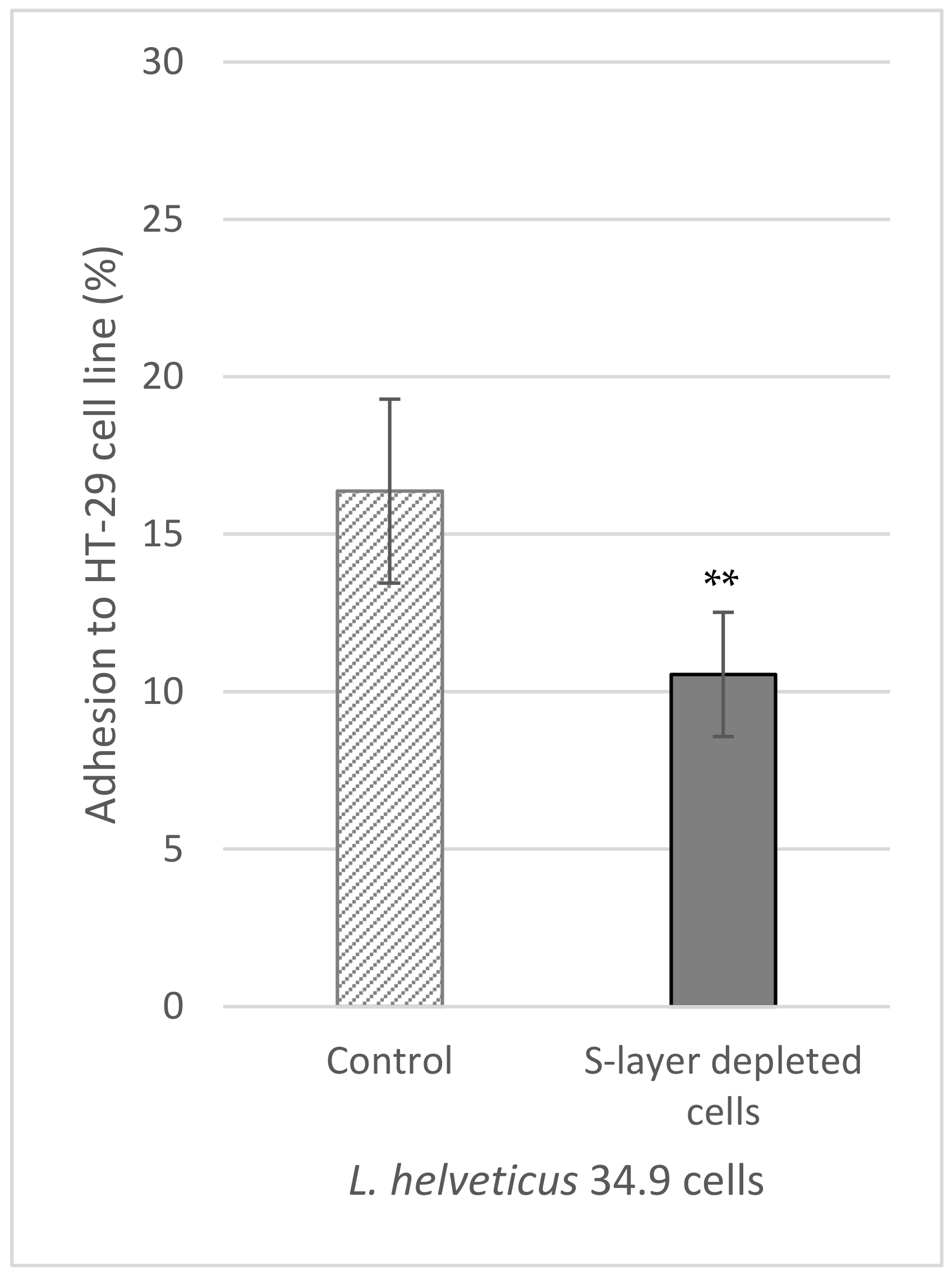
2.9. In Vitro Adhesion of L. helveticus 34.9 to HT-29 Cells

HT-29 cells were seeded on 6-well cell-culture plates (IsoLab) at a concentration of about 1.5 × 10⁵ cells/mL and grown to a final confluence of 50–60%, at 37 °C, in air with 5% CO2.
Control and S-layer depleted L. helveticus 34.9 cells were washed and resuspended in warm DMEM without antibiotics to a final concentration of about 108 CFU/mL. Simultaneously, the HT-29 monolayers were gently washed twice with PBS buffer for the removal of antibiotics. Two ml of bacterial suspensions were added in each well and the plates were incubated at 37 °C, for 2 h, in air with 5% CO2. After the incubation period, the supernatants were discarded and the wells were gently washed twice with warm PBS to remove the non-adhered bacteria. In the end, the monolayers were trypsinized (Trypsin-EDTA, Grisp, Porto, Portugal), and the adhered bacteria were counted on MRS plates. The adhesion percentage was determined as:
Adhesion (%) = (CFUadh/CFUadd) × 100,
where CFUadh represents the bacterial counts of the adhered bacteria, and CFUadd represents the bacterial counts of the added bacteria. The adhesion tests were performed in triplicate, in three wells for each bacterial suspension for every replication.

2.10. Statistical Analysis

All the experiments were performed in triplicate unless otherwise stated and the quantitative data are presented as mean and standard deviations. The results were analyzed with GraphPad Prism (GraphPad Software LLC, San Diego, CA, USA) using the t-test. The statistical differences between control and S-layer-depleted cells were considered significant at p < 0.05.

3. Results

3.1. S-Layer Extraction

The treatment with 5 M LiCl did not affect the cell viability, the survival rate after this step being similar (approx. 100%) for both the control and treated cells (results not shown). This result shows that the extraction method used in this study is safe for L. helveticus 34.9 and does not affect the cell integrity and viability.

3.2. Co-Aggregation

The percentage of co-aggregation between the control cells of L. helveticus 34.9 and the other selected strains was strain-dependent and ranged from 30.7% to 76.1% (Figure 1).
Among the functional/potential probiotic bacteria selected in this study, the highest co-aggregation capacity was observed in the case of L. acidophilus IBB 801 (56.2%) and the lowest for L. plantarum BR9 (30.7%). On the other hand, control cells of L. helveticus 34.9 showed similar co-aggregation rates with most pathogenic strains (between 48.7% and 58.7%), except for B. cereus CBAB for which the co-aggregation potential was higher (76.1%). After removal of the surface proteins, the co-aggregation capacity of L. helveticus 34.9 with all strains used in the experiment decreased in various amounts. Overall, the most significant decreases were recorded in combination with L. plantarum BR9 (from 30.7% to 5.65%) and B. cereus CBAB (from 76.1% to 39.5%). These results demonstrate that the S-layer contributes to the co-aggregation ability of L. helveticus 34.9 with most tested bacteria.

3.3. Bacterial Adhesion to Solvents

As shown in Figure 2a, the microbial adhesion to xylene, also known as the hydrophobicity, of L. helveticus 34.9 control cells is about 38.6%; however it decreases significantly (p < 0.01) to about 23.5% after LiCl treatment. When comparing the affinity to chloroform, we observed that control cells had a significantly (p < 0.0001) higher capacity to adhere to this solvent, of about 95% as compared with that of LiCl treated cells, of about 83.4% (Figure 2b). These results demonstrate that the S-layer proteins influence the hydrophobicity and affinity to chloroform of the producing cells and, consequently, the capacity of binding to different substrates/surfaces.

3.4. Influence of the S-Layer on Bacterial Resistance during Freezing and Freeze-Drying

Table 1 shows that both control and LiCl-treated cells had good viability (over 98%) even after 14 days of storage at −20 °C. The differences of the survival rates between the two cell types at the time intervals under study are insignificant (p = 0.4927). In contrast, the freeze-drying significantly (p < 0.0001) affected the viability of the cells. In the control culture, there was a decrease of cell survival of more than 30%, while the treated cells suffered a greater loss of viability; over 50% compared with the cells before freezing.

3.5. The Importance of the S-Layer for the GIT Survival

Table 2 shows the survival rate of L. helveticus 34.9 control and LiCl treated cells under the conditions simulating the stomach (acidic pH and pepsin) and small intestine (pH 8.0, pancreatin, and bile salts).
As can be observed, control cells maintain a good viability of about 70% in the first step and about 66% in the second step of the treatment simulating passage through the GIT. On the contrary, S-layer depleted cells showed a significantly (p < 0.0001) lower survival rate (about 55%) in the stomach simulating conditions and completely lost their viability in the treatment that followed, simulating passage through the small intestine.

3.6. Scanning Electron Microscopy

The SEM observations revealed several morphological changes of the bacterial cells during the treatments simulating passage through the GIT (Figure 3).
While the LiCl extraction showed no morphological changes (Figure 3B) as compared with control cells (Figure 3A), incubation under conditions simulating passage through the stomach resulted in some modifications on the cells surface (Figure 3C,D) such as loss of a smooth appearance and well-defined shape. Moreover, the treatment simulating the small intestine conditions caused more severe damage on cell integrity in the absence of the surface layer proteins compared to the control cells. As shown in Figure 3F, we could detect the presence of holes resembling transmembrane pores on the cell surface, but also many ghost cells emptied of their content, with accumulation of cell debris outside the cells.
The in-vitro adherence of control and S-layer depleted L. helveticus 34.9 cells to the HT-29 cell line was studied. As seen in Figure 4, the adherence, of about 16%, was more efficient in the presence of the surface proteins, while the removal of this protein layer by 5 M LiCl treatment significantly (p = 0.0016) reduced the adhesion ability, to about 10.5%.

4. Discussion

L. helveticus 34.9 caught our attention due to its probiotic potential, as demonstrated by its inhibitory activity towards several (potentially) pathogenic bacteria and by its good resistance to stress conditions [9,15,23]. In addition, the production of S-layer proteins by this strain may be important for maintaining its functional properties under less favorable conditions. Therefore, our study focused on the role of these proteins on bacterial protection and survival in different growth conditions, with more attention on particular aspects related to the probiotic effect. In order to investigate the importance of the S-layer, all the tests were performed by comparison between control cells and cells treated with 5 M LiCl for S-layer extraction.
The co-aggregation between probiotics and other bacterial strains plays an important role, especially for human gut colonization. Co-aggregation enables lactobacilli to manipulate the nearby environment and to inhibit the growth or prevent the colonization of pathogens in the gut by releasing antimicrobial molecules nearby or by competitive exclusion [24,25]. L. helveticus 34.9 showed a very good co-aggregation capacity associated to several bacteria, but removal of the S-layer led to a significant decrease in this capacity, demonstrating the biological consequences of these proteins in the competitiveness for substrate colonization. These properties are essential for probiotics as they act as a barrier against intestinal surface infections with pathogens [26]. Our results are in agreement with previous studies performed with potential probiotic lactobacilli [14,27,28,29]. Moreover, a strong aggregation of L. helveticus 34.9 with different probiotic/functional lactobacilli can bring benefits to the host by increasing the colonization potential if they are to be used in mixed probiotics products [25].
Hydrophobicity is another limiting factor in probiotic potential as it may influence the ability of a strain to adhere and grow on different surfaces and it can offer a competitive advantage in the adhesion of LAB [17,30,31]. Therefore, a higher cell surface hydrophobicity can be regarded as a useful feature for adherence to human intestinal epithelial cells. The xylene adhesion assay was performed to evaluate the hydrophobicity of L. helveticus 34.9 [32]. According to some researchers, the hydrophobicity or microbial adhesion to hydrocarbons (MATH) can be classified as low (MATH < 33%), medium (33% < MATH < 66%), or high (MATH > 66%) [33]. In this case, L. helveticus 34.9 can be considered to have a medium hydrophobic character. On the other hand, other authors present hydrophobicity as microbial adhesion to solvents (MATS), classifying the bacterial surface as hydrophobic (MATS ≥ 55.00%), amphiphilic (45.00% ≤ MATS ≤ 54.99%), or hydrophilic (MATS ≤ 44.99%) [34]. According to this classification, L. helveticus 34.9 can be considered a hydrophilic strain, which is in accordance with the results obtained for L. helveticus T103 [35], L. helveticus R0052 [36], and L. helveticus ATCC 12046 [37]. On the other hand, other L. helveticus strains studied previously showed more hydrophobic cellular characteristics [35,38,39]. Once again, these results show the variability of cellular characteristics between bacteria, even at the strain level. Moreover, depletion of S-layer proteins led to a decrease of bacterial adhesion to xylene compared with the control cells, which proves that S-layer proteins are involved in bacterial surface hydrophobicity. These results are in agreement with other studies on L. helveticus [28], as well as for other lactobacilli [28,38,40,41].
Finally, the strong microbial adhesion to chloroform of L. helveticus 34.9 suggests that this strain is a strong electron donor [42]. Overall, these results demonstrate that the S-layer proteins influence the hydrophobicity and affinity to chloroform of the producing cells and, consequently, the capacity of binding to different substrates/surfaces.
Freeze-drying is one of the most used techniques for long-term bacterial preservation. However, this is a harsh process that can lead to cellular damage, due to the removal of water, which can cause changes in the structure of proteins, the cell wall, and the cytoplasmic membrane [27,43]. For L. helveticus 34.9, the freeze-drying led to a more pronounced decrease of viability of the S-layer depleted cells than for the control cells. Therefore, we may state that the outer protein envelope acts as a protective barrier against cell damage, which was also stipulated in other studies carried out on lactobacilli [27]. On the other hand, for short-term storage (14 days) at −20 °C, there were no significant differences of viability between the two types of cells, which means that L. helveticus 34.9 is not sensitive to this type of storage.
Survival in the gastrointestinal tract is a pre-requisite for colonization and functional activity of probiotic LAB in the host [27]. Therefore, it is essential for bacteria to possess defense systems to withstand the typical harsh conditions encountered in this environment, like low pH, different digestive enzymes, and bile [44,45]. Our results clearly showed that S-layer proteins protect cells from these unfavorable conditions, maintaining a good viability of L. helveticus 34.9 cells during the treatments simulating the passage through the stomach and small intestine. Depletion of these surface proteins caused a significant decrease in bacterial viability, even leading to cell death in the presence of pancreatic enzymes and bile salts. The viability results were sustained by the microscopic observations. Scanning electron microscopy revealed the morphological changes during the simulated GIT conditions and offered an overall view of the role of the S-layer for cell protection and survival in stressful environmental conditions. Broadly, the absence of the external protein coat led to more morphological damage during the simulated digestion. Altogether, this research showed that surface proteins play an important role in the protective mechanisms of L. helveticus 34.9 against enzymes of the digestive tract and acidic pH. The protective role of S-layer in the GIT was also highlighted for other lactobacilli like L. brevis D6, L. helveticus M92, L. plantarum D13, L. casei DA4, L. coryniformis DA263, and L. acidophilus NCFM [14,27,46].
Finally, the adhesion of probiotic bacteria to the intestinal cells of the host is an important feature in the conferral of health benefits [47]. In our study, the HT-29 cell line was used as an in vitro model for predicting the ability of L. helveticus 34.9 to adhere to the human small intestine. This cell line was previously used in adherence studies to screen putative probiotic cultures [29,48,49]. The decreased adhesion of the S-layer depleted cells, as compared to the control cells, confirms the hypothesis that the S-layer is involved in bacterial binding to HT-29 cells. However, the adherence was not completely lost, which suggests that other factors are also involved in the interaction of lactobacilli with intestinal cells. The literature acknowledges that besides surface layer proteins, other surface structures such as lipoteichoic acids and pili, passive forces, electrostatic interactions, and hydrophobicity affects the probiotics’ adherence to the host [28,50]. Moreover, many previous in vitro studies showed that the bacterial adhesion to human cells is species and strain related; therefore, it seems that the interaction between probiotics and the host is a multifactorial process implicating different potential mechanisms [29].

5. Conclusions

The comparative results obtained in this study between untreated and S-layer depleted cells indicate that the surface layer proteins produced by L. helveticus 34.9 contribute to the probiotic potential of this strain. The S-layer synthesized by this strain is involved in cell hydrophobicity, co-aggregation with pathogens or other LAB, but also in bacterial adhesion to mammalian cells. Moreover, the decreased survival of S-layer depleted L. helveticus 34.9 cells during freeze-drying and in the simulated GIT conditions suggests that the outer protein envelope of this strain has an important role in cell protection. Corroborating the results obtained so far, it can be suggested that S-layer producing L. helveticus 34.9 is a great candidate for a probiotic strain.

Author Contributions

Conceptualization: I.-R.A.; methodology: E.-C.I. and S.-S.G.-T.; formal analysis: S.-S.G.-T. and M.Z.; writing—original draft preparation: E.-C.I. and I.-R.A.; writing—review and editing, M.Z.; supervision: I.-R.A. All authors have read and agreed to the published version of the manuscript.

Funding

Financially: this work was supported by a grant of the Romanian Ministry of Education and Research, CNCS—UEFISCDI, project number PN-III-P1-1.1-PD-2019-0808, within PNCDI III and by the Project No. RO1567-IBB05/2024 from the Institute of Biology Bucharest of Romanian Academy.

Institutional Review Board Statement

Not applicable.

Informed Consent Statement

Not applicable.

Data Availability Statement

The data presented in this study are available in the manuscript.

Acknowledgments

The authors of this paper want to thank Ionela Sarbu from the Faculty of Biology Bucharest for her support and help with the HT-29 cell line and Gabriel Maria for his help regarding the SEM pictures acquisition.

Conflicts of Interest

The authors declare no conflicts of interest.

References

  1. Martin, R.; Langella, P. Emerging Health Concepts in the Probiotics Field: Streamlining the Definitions. Front. Microbiol. 2019, 10, 1047. [Google Scholar] [CrossRef] [PubMed]
  2. FAO/WHO. Probiotic in Foods: Health and Nutritional Properties and Guidelines for Evaluation; FAO Food and Nutrition; FAO: Rome, Italy, 2006; p. 85. [Google Scholar]
  3. Ohashi, Y.; Ushida, K. Health-beneficial effects of probiotics: Its mode of action. Anim. Sci. J. Nihon Chikusan Gakkaiho 2009, 80, 361–371. [Google Scholar] [CrossRef] [PubMed]
  4. Wang, J.; Wang, J.; Yang, K.; Liu, M.; Zhang, J.; Xinyuan, W.; Fan, M. Screening for potential probiotic from spontaneously fermented non-dairy foods based on in vitro probiotic and safety properties. Ann. Microbiol. 2018, 68, 803–813. [Google Scholar] [CrossRef]
  5. Bodke, H.; Jogdand, S. Role of Probiotics in Human Health. Cureus 2022, 14, e31313. [Google Scholar] [CrossRef] [PubMed]
  6. Hynönen, U.; Palva, A. Lactobacillus surface layer proteins: Structure, function and applications. Appl. Microbiol. Biotechnol. 2013, 97, 5225–5243. [Google Scholar] [CrossRef] [PubMed]
  7. Palomino, M.M.; Allievi, M.C.; Gordillo, T.B.; Bockor, S.S.; Fina Martin, J.; Ruzal, S.M. Surface layer proteins in species of the family Lactobacillaceae. Microb. Biotechnol. 2023, 16, 1232–1249. [Google Scholar] [CrossRef] [PubMed]
  8. Mobarak Qamsari, E.; Kasra Kermanshahi, R.; Erfan, M.; Ghadam, P.; Sardari, S.; Eslami, N. Characteristics of surface layer proteins from two new and native strains of Lactobacillus brevis. Int. J. Biol. Macromol. 2017, 95, 1004–1010. [Google Scholar] [CrossRef] [PubMed]
  9. Grosu-Tudor, S.S.; Angelescu, I.R.; Brinzan, A.; Zamfir, M. Characterization of S-layer proteins produced by lactobacilli isolated from Romanian artisan fermented products. J. Appl. Microbiol. 2023, 134, lxac063. [Google Scholar] [CrossRef]
  10. Deepika, G.; Charalampopoulos, D. Chapter 4—Surface and Adhesion Properties of Lactobacilli. In Advances in Applied Microbiology; Academic Press: Cambridge, MA, USA, 2010; pp. 127–152. [Google Scholar]
  11. Martínez, M.G.; Prado Acosta, M.; Candurra, N.A.; Ruzal, S.M. S-layer proteins of Lactobacillus acidophilus inhibits JUNV infection. Biochem. Biophys. Res. Commun. 2012, 422, 590–595. [Google Scholar] [CrossRef]
  12. Sleytr, U.B.; Schuster, B.; Egelseer, E.-M.; Pum, D. S-layers: Principles and applications. FEMS Microbiol. Rev. 2014, 38, 823–864. [Google Scholar] [CrossRef]
  13. Gerbino, E.; Carasi, P.; Mobili, P.; Serradell, M.A.; Gomez-Zavaglia, A. Role of S-layer proteins in bacteria. World J. Microbiol. Biotechnol. 2015, 31, 1877–1887. [Google Scholar] [CrossRef] [PubMed]
  14. Alp, D.; Kuleaşan, H.; Korkut Altıntaş, A. The importance of the S-layer on the adhesion and aggregation ability of Lactic acid bacteria. Mol. Biol. Rep. 2020, 47, 3449–3457. [Google Scholar] [CrossRef] [PubMed]
  15. Angelescu, I.R.; Grosu-Tudor, S.S.; Cojoc, L.R.; Maria, G.M.; Chiritoiu, G.N.; Munteanu, C.V.A.; Zamfir, M. Isolation, characterization, and mode of action of a class III bacteriocin produced by Lactobacillus helveticus 34.9. World J. Microbiol. Biotechnol. 2022, 38, 220. [Google Scholar] [CrossRef] [PubMed]
  16. Lortal, S.; Van Heijenoort, J.; Gruber, K.; Sleytr, U.B. S-layer of Lactobacillus helveticus ATCC 12046: Isolation, chemical characterization and re-formation after extraction with lithium chloride. J. Gen. Microbiol. 1992, 138, 611–618. [Google Scholar] [CrossRef]
  17. Han, Q.; Kong, B.; Chen, Q.; Sun, F.; Zhang, H. In vitro comparison of probiotic properties of lactic acid bacteria isolated from Harbin dry sausages and selected probiotics. J. Funct. Foods 2017, 32, 391–400. [Google Scholar] [CrossRef]
  18. Zamfir, M.; Stefan, I.R.; Stancu, M.M.; Grosu-Tudor, S.S. Production, mode of action and sequencing of the corresponding gene of a bacteriocin produced by Lactococcus lactis 19.3. Int. J. Food Sci. Technol. 2016, 51, 2164–2170. [Google Scholar] [CrossRef]
  19. Angelescu, I.-R.; Zamfir, M.; Stancu, M.-M.; Grosu-Tudor, S.-S. Identification and probiotic properties of lactobacilli isolated from two different fermented beverages. Ann. Microbiol. 2019, 69, 1557–1565. [Google Scholar] [CrossRef]
  20. Zamfir, M.; Callewaert, R.; Cornea, P.C.; Savu, L.; Vatafu, I.; De Vuyst, L. Purification and characterization of a bacteriocin produced by Lactobacillus acidophilus IBB 801. J. Appl. Microbiol. 1999, 87, 923–931. [Google Scholar] [CrossRef]
  21. Grosu-Tudor, S.S.; Brown, L.; Hebert, E.M.; Brezeanu, A.; Brinzan, A.; Fadda, S.; Mozzi, F.; Zamfir, M. S-layer production by Lactobacillus acidophilus IBB 801 under environmental stress conditions. Appl. Microbiol. Biotechnol. 2016, 100, 4573–4583. [Google Scholar] [CrossRef]
  22. Hernández-Alcántara, A.; Wacher, C.; Llamas-Arriba, M.; López, P.; Pérez-Chabela, M. Probiotic properties and stress response of thermotolerant lactic acid bacteria isolated from cooked meat products. LWT 2018, 91, 249–257. [Google Scholar] [CrossRef]
  23. Grosu-Tudor, S.S.; Ștefan, I.R.; Zamfir, M. Growth/survival of some functional lactic acid bacteria under different stress conditions. AgroLife Sci. J. 2016, 5, 71–78. [Google Scholar]
  24. Beldarrain-Iznaga, T.; Villalobos-Carvajal, R.; Sevillano-Armesto, E.; Leiva-Vega, J. Functional properties of Lactobacillus casei C24 improved by microencapsulation using multilayer double emulsion. Food Res. Int. 2021, 141, 110136. [Google Scholar] [CrossRef] [PubMed]
  25. Kos, B.; Šušković, J.; Vuković, S.; Šimpraga, M.; Frece, J.; Matošić, S. Adhesion and aggregation ability of probiotic strain Lactobacillus acidophilus M92. J. App. Microbiol. 2003, 94, 981–987. [Google Scholar] [CrossRef]
  26. Abdulla, A.A.; Abed, T.A.; Saeed, A.M. Adhesion, autoaggregation and hydrophobicity of six Lactobacillus strains. Br. Microbiol. Res. J. 2014, 4, 381–391. [Google Scholar] [CrossRef]
  27. Uroić, K.; Novak, J.; Hynönen, U.; Pietilä, T.E.; Leboš Pavunc, A.; Kant, R.; Kos, B.; Palva, A.M.; Suskovic, J. The role of S-layer in adhesive and immunomodulating properties of probiotic starter culture Lactobacillus brevis D6 isolated from artisanal smoked fresh cheese. LWT Food Sci. Technol. 2016, 69, 623–632. [Google Scholar] [CrossRef]
  28. Banić, M.; Uroić, K.; Pavunc, A.; Novak, J.; Zorić, K.; Durgo, K.; Petković, H.; Jamnik, P.; Kazazić, S.; Kazazić, S.; et al. Characterization of S-layer proteins of potential probiotic starter culture Lactobacillus brevis SF9B isolated from sauerkraut. LWT 2018, 93, 257–267. [Google Scholar] [CrossRef]
  29. Gharbi, Y.; Fhoula, I.; Ruas-Madiedo, P.; Afef, N.; Boudabous, A.; Gueimonde, M.; Ouzari, H.-I. In-vitro characterization of potentially probiotic Lactobacillus strains isolated from human microbiota: Interaction with pathogenic bacteria and the enteric cell line HT29. Ann. Microbiol. 2019, 69, 61–72. [Google Scholar] [CrossRef]
  30. Hojjati, M.; Behabahani, B.A.; Falah, F. Aggregation, adherence, anti-adhesion and antagonistic activity properties relating to surface charge of probiotic Lactobacillus brevis gp104 against Staphylococcus aureus. Microb. Pathog. 2020, 147, 104420. [Google Scholar] [CrossRef]
  31. Vanitha, P.R.; Somashekaraiah, R.; Divyashree, S.; Pan, I.; Sreenivasa, M.Y. Antifungal activity of probiotic strain Lactiplantibacillus plantarum MYSN7 against Trichophyton tonsurans. Front. Microbiol. 2023, 14, 1192449. [Google Scholar] [CrossRef]
  32. Yadav, R.; Puniya, A.K.; Shukla, P. Probiotic Properties of Lactobacillus plantarum RYPR1 from an Indigenous Fermented Beverage Raabadi. Front. Microbiol. 2016, 7, 1683. [Google Scholar] [CrossRef]
  33. Bouchard, D.S.; Seridan, B.; Saraoui, T.; Rault, L.; Germon, P.; Gonzalez-Moreno, C.; Nader-Macias, F.M.; Baud, D.; François, P.; Chuat, V.; et al. Lactic Acid Bacteria Isolated from Bovine Mammary Microbiota: Potential Allies against Bovine Mastitis. PLoS ONE 2015, 10, e0144831. [Google Scholar] [CrossRef] [PubMed]
  34. Sandes, S.; Alvim, L.; Silva, B.; Acurcio, L.; Santos, C.; Campos, M.; Santos, C.; Nicoli, J.; Neumann, E.; Nunes, Á. Selection of new lactic acid bacteria strains bearing probiotic features from mucosal microbiota of healthy calves: Looking for immunobiotics through in vitro and in vivo approaches for immunoprophylaxis applications. Microbiol. Res. 2017, 200, 1–13. [Google Scholar] [CrossRef] [PubMed]
  35. Wasko, A.; Polak-Berecka, M.; Paduch, R.; Jozwiak, K. The effect of moonlighting proteins on the adhesion and aggregation ability of Lactobacillus helveticus. Anaerobe 2014, 30, 161–168. [Google Scholar] [CrossRef] [PubMed]
  36. Johnson-Henry, K.C.; Hagen, K.E.; Gordonpour, M.; Tompkins, T.A.; Sherman, P.M. Surface-layer protein extracts from Lactobacillus helveticus inhibit enterohaemorrhagic Escherichia coli O157:H7 adhesion to epithelial cells. Cell. Microbiol. 2007, 9, 356–367. [Google Scholar] [CrossRef] [PubMed]
  37. Boonaert, C.J.P.; Rouxhet, P.G. Surface of Lactic Acid Bacteria: Relationships between Chemical Composition and Physicochemical Properties. Appl. Environ. Microbiol. 2000, 66, 2548–2554. [Google Scholar] [CrossRef] [PubMed]
  38. Rong, J.; Zheng, H.; Liu, M.; Hu, X.; Wang, T.; Zhang, X.; Jin, F.; Wang, L. Probiotic and anti-inflammatory attributes of an isolate Lactobacillus helveticus NS8 from Mongolian fermented koumiss. BMC Microbiol. 2015, 15, 196. [Google Scholar] [CrossRef] [PubMed]
  39. Samet, K.; Icen, T.I. Screening of Potential Probiotic Properties of Lactobacillus and Lactococcus strains. Res. J. Biotechnol. 2022, 17, 119–126. [Google Scholar]
  40. Schar-Zammaretti, P.; Dillmann, M.L.; D’Amico, N.; Affolter, M.; Ubbink, J. Influence of fermentation medium composition on physicochemical surface properties of Lactobacillus acidophilus. Appl. Environ. Microbiol. 2005, 71, 8165–8173. [Google Scholar] [CrossRef]
  41. Van der Mei, H.C.; van de Belt-Gritter, B.; Pouwels, P.H.; Martinez, B.; Busscher, H.J. Cell surface hydrophobicity is conveyed by S-layer proteins—A study in recombinant lactobacilli. Colloids Surf. B Biointerfaces 2003, 28, 127–134. [Google Scholar] [CrossRef]
  42. Xu, H.; Jeong, H.S.; Lee, H.-Y.; Ahn, J. Assessment of cell surface properties and adhesion potential of selected probiotic strains. Lett. Appl. Microbiol. 2009, 49, 434–442. [Google Scholar] [CrossRef]
  43. Corcoran, B.M.; Ross, R.P.; Fitzgerald, G.F.; Dockery, P.; Stanton, C. Enhanced survival of GroESL-overproducing Lactobacillus paracasei NFBC 338 under stressful conditions induced by drying. Appl. Environ. Microbiol. 2006, 72, 5104–5107. [Google Scholar] [CrossRef] [PubMed]
  44. Davoodabadi, A.; Soltan Dallal, M.M.; Rahimi Foroushani, A.; Douraghi, M.; Sharifi Yazdi, M.K.; Amin Harati, F. Antibacterial activity of Lactobacillus spp. isolated from the feces of healthy infants against enteropathogenic bacteria. Anaerobe 2015, 34, 53–58. [Google Scholar] [CrossRef] [PubMed]
  45. Jabbari, V.; Rezaei Mokarram, R.; Khiabani, M.; Askari, F.; Ahmadi, E.; Hassanzadeh, A.; Aghazadeh, S.B.; Asgharzadeh, M.; Kafilet, H.S. Molecular Identification of Lactobacillus acidophilus as a probiotic potential from traditional doogh samples and evaluation of their antimicrobial activity against some pathogenic bacteria. Biomed. Res. 2017, 28, 1458–1463. [Google Scholar]
  46. Johnson, B.; Selle, K.; O’Flaherty, S.; Goh, Y.J.; Klaenhammer, T. Identification of extracellular surface-layer associated proteins in Lactobacillus acidophilus NCFM. Microbiology 2013, 159, 2269–2282. [Google Scholar] [CrossRef] [PubMed]
  47. Ruiz, L.; Margolles, A.; Sanchez, B. Bile resistance mechanisms in Lactobacillus and Bifidobacterium. Front. Microbiol. 2013, 4, 396. [Google Scholar] [CrossRef] [PubMed]
  48. Duary, R.K.; Batish, V.K.; Grover, S. Relative gene expression of bile salt hydrolase and surface proteins in two putative indigenous Lactobacillus plantarum strains under in vitro gut conditions. Mol. Biol. Rep. 2012, 39, 2541–2552. [Google Scholar] [CrossRef] [PubMed]
  49. Wang, B.; Wei, H.; Yuan, J.; Li, Q.; Li, Y.; Li, N.; Li, J. Identification of a Surface Protein from Lactobacillus reuteri JCM1081 That Adheres to Porcine Gastric Mucin and Human Enterocyte-Like HT-29 Cells. Curr. Microbiol. 2008, 57, 33–38. [Google Scholar] [CrossRef]
  50. Jakava-Viljanen, M.; Palva, A. Isolation of surface (S) layer protein carrying Lactobacillus species from porcine intestine and faeces and characterization of their adhesion properties to different host tissues. Vet. Microbiol. 2007, 124, 264–273. [Google Scholar] [CrossRef]
Figure 1. Co-aggregation (%) of control and S-layer depleted L. helveticus 34.9 cells with other LAB and with (potential) pathogens. Differences between control cells and S-layer depleted cells were statistically analyzed (t-test) and a p-value summary was marked with asterisks on the graph; no asterisk means no significant difference, while ** and *** mean p < 0.01, and p < 0.001, respectively.
Fermentation 10 00150 g001
Figure 2. Adhesion (%) of control and S-layer depleted L. helveticus 34.9 cells to xylene (A) and chloroform (B). Differences between control cells and S-layer depleted cells were statistically analyzed (t-test) and a p-value summary was marked with asterisks on the graph; ** and *** mean p < 0.01 and p < 0.001, respectively.
Fermentation 10 00150 g002
Figure 3. Scanning electron microscopy of L. helveticus 34.9 control cells (A,C,E) and S-layer depleted cells (B,D,F) subjected to different conditions simulating passage through the GIT: (A,B)—no treatment; (C,D)—pepsin and low pH treatment; (E,F)—pancreatin and bile salts treatment. The arrows show the morphological changes that occurred during the experiments.
Fermentation 10 00150 g003
Figure 4. Bacterial adhesion (%) of control and S-layer depleted L. helveticus 34.9 cells compared to the HT-29 cell line. Differences between control cells and S-layer depleted cells were statistically analyzed (t-test) and the p-value summary was marked with asterisks on the graph; means p < 0.01.
Fermentation 10 00150 g004
Table 1. Bacterial survival (%) after freezing and freeze-drying.
Bacterial Survival (%)
Storage at −20 °C
3 Days7 Days14 DaysFreeze-Drying
Control cells99.8 ± 1.1299.2 ± 0.27 99.0 ± 2.3768.6 ± 0.04
S-layer depleted cells99.4 ± 1.2898.2 ± 1.3597.9 ± 3.7641.8 ± 1.75
Table 2. Bacterial survival (%) after exposure to conditions similar to the gastrointestinal tract.
Bacterial Survival (%)
pH 3 + PepsinpH 8 + Pancreatin + Bile Salts
Control cells70.3 ± 0.08 66.2 ± 0.29
S-layer depleted cells55.6 ± 1.070
Disclaimer/Publisher’s Note: The statements, opinions and data contained in all publications are solely those of the individual author(s) and contributor(s) and not of MDPI and/or the editor(s). MDPI and/or the editor(s) disclaim responsibility for any injury to people or property resulting from any ideas, methods, instructions or products referred to in the content.

