
Three-Dimensional Printing Strategies for Enhanced Hydrogel Applications


Barry and Judy Silverman College of Pharmacy, Nova Southeastern University, Fort Lauderdale, FL 33314, USA
*
Author to whom correspondence should be addressed.
Gels 2024, 10(4), 220; https://doi.org/10.3390/gels10040220
Submission received: 23 February 2024
/
Revised: 13 March 2024
/
Accepted: 21 March 2024
/
Published: 25 March 2024
(This article belongs to the Special Issue 3D Printing of Gels: Applications and Properties)
Abstract
:This study explores the dynamic field of 3D-printed hydrogels, emphasizing advancements and challenges in customization, fabrication, and functionalization for applications in biomedical engineering, soft robotics, and tissue engineering. It delves into the significance of tailored biomedical scaffolds for tissue regeneration, the enhancement in bioinks for realistic tissue replication, and the development of bioinspired actuators. Additionally, this paper addresses fabrication issues in soft robotics, aiming to mimic biological structures through high-resolution, multimaterial printing. In tissue engineering, it highlights efforts to create environments conducive to cell migration and functional tissue development. This research also extends to drug delivery systems, focusing on controlled release and biocompatibility, and examines the integration of hydrogels with electronic components for bioelectronic applications. The interdisciplinary nature of these efforts highlights a commitment to overcoming material limitations and optimizing fabrication techniques to realize the full potential of 3D-printed hydrogels in improving health and well-being.
1. Introduction
The field of 3D-printed hydrogels has emerged as a dynamic and rapidly evolving area of research, driven by a diverse array of needs and challenges across various applications (Scheme 1). At the core of this innovative domain is the pursuit of advancing the customization, fabrication, and functionalization of hydrogels to meet specific requirements in biomedical engineering, soft robotics, tissue engineering, and beyond.
Customization of biomedical scaffolds for optimal tissue regeneration [1] remains a pivotal challenge, underscoring the importance of tailored solutions for individual patient needs. This necessity merges with the advancements in multimaterial 3D printing, enabling the creation of structures with unprecedented complexity and functionality. Researchers are also focused on enhancing the mechanical properties of bioinks [2], crucial for the realistic replication of biological tissues and the development of bioinspired actuators [3].
Fabrication challenges are pronounced in the domain of soft robotics, where the replication of soft material 3D printing [4] and embedding long fibers in hydrogels [5] are explored to mimic complex biological structures and functions. The quest for high-resolution, multimaterial, 3D hydrogel structures [6] further exemplifies the technical hurdles in achieving precise and scalable manufacturing techniques.
In tissue engineering, the creation of supportive environments for cell migration [7], nutrient/oxygen transport [8], and vasculature networks [9] is essential for the development of functional tissue constructs. This includes overcoming the natural limitations of hydrogel materials, such as the spread of wet droplets [10] and rapid solidification for photocrosslinking [11], to achieve desired geometries and properties.
The application of 3D-printed hydrogels extends into the domain of drug delivery [12,13], where controlled release mechanisms and biocompatibility are paramount. Similarly, the development of high-strength and ultratough hydrogels [14,15] addresses the need for materials that can withstand mechanical stresses while maintaining flexibility and biocompatibility for implantable devices.
Emerging applications also highlight the integration of hydrogels with electronic components [16,17], paving the way for innovative bioelectronics that can monitor and interact with biological systems in real-time. The development of conductive hydrogels [18,19] and self-healing materials [20,21] reflects the interdisciplinary nature of current research efforts, blending materials science with electrical engineering to create multifunctional devices.
The challenges and needs identified across these diverse applications demonstrate the interdisciplinary and innovative nature of research in 3D-printed hydrogels. From enhancing biocompatibility and mechanical strength [22,23] to achieving precision in nanoparticle arrangement [24] and optimizing scaffold fabrication [25], the field is characterized by a relentless pursuit of solutions that bridge the gap between theoretical potential and practical application. As researchers continue to address these challenges, the field of 3D-printed hydrogels stands on the border of significant breakthroughs that promise to transform a wide range of industries and improve human health and well-being.
2. Recent Studies on 3D-Printed Hydrogels
The study of 3D-printed hydrogels in the fields of material science and engineering is a rapidly evolving area focused on enhancing processing techniques, improving material properties, and integrating functional features. This work aims to expand the use of these materials across various disciplines. Key areas of research and development include the following:
- Advancing photopolymerization methods and creating both thermoresponsive and biodegradable hydrogels to enhance their mechanical and physical characteristics.
- Adding functional additives and creating new composites to address the limitations of hydrogels, thus improving their utility in sophisticated manufacturing and design applications.
- Designing materials that replicate biological systems for use in biofabrication and tissue engineering, which involves improving mechanical properties, responsiveness to stimuli, and functional complexity. This opens new possibilities in regenerative medicine, soft robotics, and other areas.
- Developing materials for tissue engineering and scaffolding that include bioactive molecules and porous structures, which support cell activity and closely resemble the natural tissue environment for advanced tissue regeneration.
- Innovating in the field of biomedical devices and drug delivery by integrating bioactive molecules, enhancing mechanical and functional properties, and devising controlled release mechanisms tailored to specific medical treatments.
- Advancing sensor technology and electronics with the use of conductive, responsive, and bioactive materials to create devices that are flexible, sensitive, and multifunctional. These devices have applications in wearable technology, soft robotics, and bioelectronics.
- Focusing on the creation of advanced actuators and robotic systems for soft robotics, which are made from materials that exhibit superior mechanical properties and responsiveness for complex movements and mimicking biological functions.
- Investigating the use of hydrogels in environmental and energy sectors to support sustainable energy production, agricultural development, and environmental clean-up, making use of their unique characteristics for innovative and sustainable solutions.
These initiatives demonstrate a strategic approach to leveraging 3D-printed hydrogels, emphasizing the continuous effort to refine material properties, broaden the scope of applications, and tackle significant challenges in science and technology.
3. Materials, Technologies, and Process Optimization of Three-Dimensional Printing
3.1. Hydrogel Crosslinking
Hydrogel crosslinking plays a pivotal role in hydrogel bioprinting, serving as the cornerstone for designing scaffolds that mimic the extracellular matrix, enabling the controlled release of bioactive molecules, and encapsulating living cells. Crosslinking methods, both chemical and physical, are integral for tailoring hydrogel properties such as degradation rates, mechanical strength, and biocompatibility, which are crucial for various biomedical applications.
Chemical crosslinking is highlighted for its versatility in creating hydrogels with robust mechanical stability. This method allows for the precise design of hydrogels that can degrade under physiological conditions into non-toxic products, ensuring biocompatibility. However, the use of potentially toxic crosslinking agents necessitates their complete removal to avoid adverse reactions with bioactive substances present within the hydrogel matrix [26]. In contrast, physical crosslinking methods offer an alternative that circumvents the use of toxic chemicals. These methods involve interactions that do not require covalent bond formation, thereby avoiding potential negative effects on the bioactive components embedded within the hydrogels. This advantage makes physically crosslinked hydrogels attractive for applications requiring high biocompatibility and functionality [26].
The development of interpenetrating networks through double crosslinking (covalent followed by ionic) not only enhances the hydrogel features but also allows for the modulation of hydrogel properties through the adjustment of preparation parameters. The use of neural network models to simulate and optimize these parameters further highlights the complexity and potential of hydrogel crosslinking in creating tailored matrices for drug delivery and cell encapsulation [27].
Exploring multifunctional crosslinking molecules has opened new avenues for regulating the degradation rates and mechanical properties of hydrogels. By comparing hydrogels formed with multifunctional versus bi-functional crosslinking molecules, it is evident that the former can provide superior mechanical stiffness and slower degradation rates, attributed to the increased crosslinking density and interaction points within the hydrogel network. This method demonstrates the importance of crosslinking chemistry in enhancing the performance of hydrogels for various biomedical applications [28].
Enzyme-catalyzed crosslinking represents an innovative and cell-friendly approach to hydrogel formation, offering a mild alternative to traditional chemical and physical crosslinking methods. This strategy enables the formation of covalently crosslinked hydrogels with complex architectures, mimicking the natural extracellular matrix more closely and providing dynamic scaffolds for tissue engineering and regenerative medicine. The shift towards enzymatic crosslinking features the evolving landscape of hydrogel preparation, where biocompatibility and functionality are paramount [29].
In situ crosslinking strategies for the 3D bioprinting of photo-crosslinkable hydrogels further illustrate the advancements in hydrogel technology. This approach enables the creation of complex, cell-laden constructs with high embedded cell viability and tunable cell behavior, featuring the critical role of crosslinking in the development of next-generation biofabricated tissues and organs [30].
Overall, the evolution of hydrogel crosslinking techniques—from chemical and physical to innovative enzymatic and in situ methods—demonstrates the field’s continuous advancement towards creating more sophisticated, biocompatible, and functional hydrogel systems for a myriad of biomedical applications.
3.2. Three-Dimensional and 4D Bioprinting
The transition from three-dimensional (3D) printing to four-dimensional (4D) printing represents a significant advancement in additive manufacturing, shifting from static constructs to dynamic, responsive structures. This evolution is characterized by the development of platforms that can change their properties, shape, or functionality over time in response to external stimuli, leveraging the capabilities of stimuli-responsive materials [31].
Three-dimensional printing has laid the groundwork for this advancement by enabling the precise layer-by-layer construction of complex structures, where control over the dimensions and properties of the printed objects is paramount. Among the materials employed in 3D printing, hydrogels have emerged as particularly promising due to their biocompatibility and the ability to undergo reversible transformations. These properties have made hydrogels a key material in the progression towards 4D printing, where the goal is to create structures capable of adapting to their environment [32].
The core of 4D printing technology lies in the use of stimuli-responsive hydrogels. These materials can expand, contract, or otherwise change shape in response to environmental triggers, such as temperature changes or the presence of specific chemicals. This responsiveness to stimuli has opened new avenues for creating devices and structures with applications ranging from actuators and cellular scaffolds to controlled drug release systems. The versatility and low cost of manufacturing these hydrogel-based structures further enhance their appeal for a wide range of applications [31].
An innovative approach in the field involves the development of composite inks, such as those combining cellulose with hydrogels. These composites not only maintain the responsive characteristics of hydrogels but also incorporate the mechanical strength of cellulose fibers. This combination enables the fabrication of structures that can morph according to predetermined designs in response to environmental stimuli, such as moisture levels. Such advancements illustrate the potential of 4D printing in creating more complex, responsive, and durable structures [33].
Among the specific applications of 4D bioprinting, the development of smart valves stands out. These valves, fabricated from a blend of alginate and poly(N-isopropylacrylamide), exemplify the practical application of 4D printing in producing structures that are not only mechanically robust but also capable of actuating in response to temperature changes. This highlights the potential of 4D-printed hydrogels in creating dynamic, responsive systems with possible uses in fluid management and soft robotics [34].
A notable technique in this evolution involves printing stimuli-responsive hydrogel structures with internal gaps using a combination of responsive and non-responsive pre-gel solutions. By printing these materials in a supportive medium and then solidifying them with ultraviolet light, it is possible to create complex structures capable of dramatic transformations in response to thermal stimuli. This method highlights the sophisticated control over material properties and structural design that 4D printing offers, paving the way for the fabrication of intricate, responsive patterns and shapes [35].
3.3. Hydrogel Bioprinting Techniques
Bioprinting is an emerging field that merges engineering, biology, and medicine to innovate in areas such as tissue engineering and regenerative medicine. This field is notable for its application of several key techniques, extrusion-based bioprinting, inkjet bioprinting, and vat photopolymerization, each with unique mechanisms and uses [36].
Extrusion-based bioprinting (EBB) is akin to 3D printing, where biomaterials mixed with cells are pushed through a nozzle to build three-dimensional structures layer by layer. This method supports a broad viscosity range, accommodating various hydrogels and cell-laden bioinks, making it adaptable for creating large, intricate structures while preserving high cell viability. Despite its versatility, it struggles with lower resolution and potential shear stress that may harm cell viability and functionality. The performance of EBB is affected by factors such as the viscosity of the hydrogel, the diameter of the nozzle, and the speed of printing. Recent progress has expanded its applications to include printing tissues, organ modules, and microfluidic devices. Nevertheless, challenges persist, including limitations in organ fabrication, feature resolution, and regulatory hurdles, indicating a need for advanced bioprinting solutions and new bioink development to bridge the gap from laboratory to clinical use, aiming to produce viable products for tissue engineering and regenerative medicine [37,38]. Extrusion printing can be carried out on a surface or into a suspension bath or media. Figure 1 below shows how a bioink is deposited onto a surface or within a suspension bath with a pattern defined by the user.
Inkjet bioprinting utilizes thermal or piezoelectric actuators to place droplets of bioink on a substrate, noted for its high precision and resolution. This method is cost-effective and straightforward but is limited to bioinks with low viscosity and faces potential cell damage from heat or mechanical stress. Factors like bioink viscosity and minimizing cell damage are vital for its performance. The drop-on-demand jetting approach allows for the non-contact deposition of materials and cells, optimizing cell-to-matrix and cell-to-cell interactions. Despite its advantages, the effect of bioink properties on printing performance and cell health is not fully understood. Research indicates that bioink viscoelasticity and viscosity are crucial for printing accuracy and cell viability, emphasizing the importance of bioink formulation for precise cell deposition and the creation of adjustable cell spheroids [39,40].
Vat photopolymerization involves the selective curing of photosensitive polymers or hydrogels with light to create solid 3D structures. It is distinguished by its high resolution and ability to produce complex shapes but is limited to photosensitive materials and the potential toxicity from photoinitiators. The technique’s performance depends on the choice and concentration of photoinitiators, as well as optimizing light intensity and exposure time. Recent developments in vat polymerization-based 3D printing and bioprinting have introduced new biomaterial ink formulations and system designs, with the expectation of their swift application in biomedical fields. This indicates a concerted effort to combine innovative vat polymerization techniques with biomaterial inks for better medical outcomes [41,42]. The patterns projected are typically derived from computerized 3D drawings or from CT scans or MRIs of patients’ resources. These patterns are designed using vat polymerization before remodeling to create patient-specific medical devices, functional human tissues for regeneration, and in vitro human-based tissue models for therapeutic screening, as illustrated in Figure 2 below.
In hydrogel bioprinting, selecting a bioprinting technique usually depends on the requirements of the desired application, such as the complexity of the structure, mechanical properties, and biological functionality. The composition of the hydrogel, the type of cells used, and the purpose of the application are crucial in determining the most appropriate bioprinting method. Ongoing research and development aim to address current limitations and expand the possibilities of bioprinting technologies, leading to the creation of more complex and functional bioprinted tissues and organs [43].
Recent advancements in 3D printing technologies, particularly in material and process optimization, have led to significant developments across various fields, including tissue engineering, regenerative medicine, soft robotics, and biomedical applications. These innovations demonstrate a concerted effort towards creating more sustainable, efficient, and customized solutions. Figure 3 below shows sample morphologies printed with GT–AT–MMT bioinks of variable concentrations under predetermined pressures.
In tissue engineering and regenerative medicine, the customization of bio-based scaffolds has been a notable achievement. These scaffolds, with controlled pore sizes and gradient structures, are designed to enhance tissue regeneration capabilities [1]. The optimization of 3D inkjet printing techniques for alginate bioinks has facilitated the creation of structures that closely mimic tissue, supporting physiological flows [10]. Additionally, the introduction of hydrogel-based technologies has marked a significant stride in cell therapy and tissue engineering applications, offering precise size control for cell therapy and tissue engineering applications [44] and optimizing cell-friendly fabrication for physiological models [9]. This is complemented by developments in bone regeneration, where MXene composite hydrogels have demonstrated synergistic antibacterial and osteogenic effects [45], and in drug delivery systems, where thermosensitive hydrogels show potential for controlled drug release [13].
The field of soft and marine robotics has also seen innovative applications of 3D printing technologies. The fabrication of jellyfish-mimic soft robot actuators [4] and biodegradable hydrogel actuators [46] illustrates the potential of these technologies in developing devices that operate effectively in challenging environments. Furthermore, the advancement in material properties, such as full-color luminescence and opacity tuning [3], enhances the adaptability and utility of robotic systems.
Hydrogel technologies have been central to overcoming fabrication limitations and introducing novel functionalities. The development of multiscale, multimaterial 3D hydrogel structures [6] and the improvement in mechanical properties through hydrogel composites [47] are exemplary. Moreover, photocrosslinking techniques for silk-fibroin-based hydrogels [11] and the use of a hydrogel pen for nanometer precision in 3D printing [48] highlight the ongoing efforts in process optimization.
Material science has played a crucial role in these advancements, with the exploration of hydrogel printability [49], rapid hydrogel bead fabrication [44], and the engineering of perfusable vasculature networks on-chip [9] being notable examples. These efforts not only enhance the practical applications of 3D printing technologies but also contribute to the broader understanding of material interactions and fabrication processes.
In conclusion, the advancements in 3D printing technologies stress the dynamic and interdisciplinary nature of this field. By pushing the boundaries of material and process optimization, these innovations offer promising solutions to complex challenges across science, medicine, and engineering. Table 1 below shows the different printing technologies, materials used and their applications.
4. Scaffold Mechanical Enhancement and Biocompatibility
The recent advancements in scaffold design, particularly in enhancing mechanical properties and biocompatibility, signify a critical trend in tissue engineering, regenerative medicine, and material science. These advancements, as evidenced by numerous studies, are foundational to the development of scaffolds that support cellular growth and tissue integration effectively. For instance, the improvement in the mechanical properties of sodium alginate through the incorporation of agarose has been noted to facilitate cell growth, thanks to the scaffold’s designed micro-pores [56]. Additionally, the enhancement in hydrogel scaffolds with microtubes has shown promising results in terms of mechanical strength and biocompatibility, demonstrating the potential for application in soft tissue engineering [23]. Figure 4 below shows the core–sheath spinneret set up for the production of the microtubules and the SEM images of the microtubules produced.
The role of 3D printing technology in scaffold fabrication has evolved significantly, enabling the creation of complex structures tailored for specific applications. Innovations such as multiple-layered hydrogel scaffold printing [57], 3D-printed metal foams integrated with hydrogel for bone replacement [58], and the use of photopolymerizable copolypeptides for producing mechanically robust 3D objects [59] exemplify the versatility and precision offered by 3D printing techniques. These advancements highlight the industry’s move towards employing manufacturing technologies that allow for customized scaffold designs with enhanced functionality.
There is a growing emphasis on developing scaffolds that offer multifunctional capabilities, such as improved osseointegration, antibacterial properties [60], and conductivity conducive to neuron growth [61]. This trend towards multifunctionality reflects a shift in focus towards creating scaffolds that not only support tissue growth but also integrate additional features to enhance the overall therapeutic outcome.
Environmental sustainability and safety in scaffold materials have emerged as important considerations. The development of biodegradable cellulose hydrogel inks for 3D printing metal structures [62] represents a step towards more environmentally friendly and safer additive manufacturing processes. This focus on sustainability indicates a broader commitment within the field to reduce environmental impact and enhance patient safety through innovative material choices.
Moreover, the exploration of scaffold materials for applications beyond traditional tissue engineering showcases the adaptability and potential of these materials in cutting-edge fields. For example, the utilization of scaffolds in soft robotics [63], flexible electronics [16], and space agriculture [64] demonstrates the broad applicability and versatility of scaffold materials, paving the way for novel technological advancements.
In summary, the trends highlighted by recent research in scaffold mechanical enhancement and biocompatibility reflect a dynamic field characterized by innovation in material properties, manufacturing techniques, and application domains. The integration of enhanced mechanical and biological properties, coupled with the advancements in 3D printing technology, multifunctionality, environmental sustainability, and novel applications, emphasize the ongoing evolution of scaffold technology (Table 2).
5. Stretchable and Conductive Hydrogels
Recent advancements in the field of stretchable and conductive hydrogels have marked significant milestones across several applications, including but not limited to soft robotics, tissue engineering, human motion detection, and soft bioelectronics. These innovations are primarily driven by the integration of advanced printing techniques with hydrogel technology, the development of conductive hydrogels with enhanced functionalities, pioneering applications in soft bioelectronics and sensing, and innovations in sensing and energy generation.
The integration of advanced printing techniques with hydrogel technology has led to notable achievements. The high-resolution printing of stretchable double-network (DN) hydrogels has addressed the shaping challenges in soft robotics and tissue engineering, facilitated by quick lithography techniques [89]. Further, the field has seen the development of ultrastretchable and self-healing DN hydrogel printing, which exhibits remarkable mechanical strength and strain sensitivity, crucial for robotics and human motion detection [90]. Additionally, the introduction of a cryogenic 3D printing method for conductive hydrogel inks has produced cytocompatible, conductive scaffolds suitable for tissue engineering applications [91].
The development of conductive hydrogels with enhanced functionalities has also been a key area of progress. The coupling of 3D printing with interfacial polymerization has enabled the creation of conductive hydrogels with complex geometries, paving the way for the development of electroactive materials with precisely defined structures for advanced applications [92]. Moreover, the development of ultrasensitive and self-adhesive wearable devices with exceptional conductivity and stretchability has advanced motion monitoring and health detection, showcasing the potential of conductive hydrogels in wearable technology [92].
In soft bioelectronics and sensing, significant strides have been made. High-performance conducting polymer hydrogel-based Electrobiosignal Interfaces (EBIs) have improved ECG and EMG signal recording capabilities [93]. The fabrication of self-healable hydrogel–liquid metal composites for customizable multimodular sensor systems demonstrates practical advances in self-healing electronics [94]. Additionally, the reporting on 3D-printed implantable hydrogel bioelectronics for precise electrophysiological monitoring and electrical modulation emphasizes the potential in healthcare monitoring and therapies [95].
Furthermore, innovations in sensing and energy generation highlight the versatility of hydrogels. The rapid construction of double-network ionic conductive hydrogel for iontronic pressure sensors with biomimetic fingerprint microstructures enables tactile sensing and human motion detection [96]. The development of a 3D-printed conducting polymer hydrogel-based direct current (DC) generator for self-powered electromechanical sensing underlines the robust mechanical properties and flexibility, crucial for wearable electronics and environmental monitoring [97].
These advancements show the potential of conductive hydrogels to transform various industries by providing flexible, durable, and increasingly integrated solutions with electronic functionalities. The progress in this field is a testament to the collaborative efforts of researchers and engineers in pushing the boundaries of material science, electronics, and biomedical engineering (Table 3).
6. Self-Healing and Responsive Hydrogels
The recent advancements in hydrogel technology, as evidenced by various studies, underscore significant progress in the fields of material science, 3D printing, and responsive actuators. A notable development includes the preparation of self-healing polyacrylamide (PAA)-based hydrogels tailored for extrusion-based 3D printing, which exhibit exceptional self-healing capabilities and rheological properties. This innovation enables the creation of diverse 3D structures that maintain integrity without deformation, enhancing the potential for practical applications [20]. Furthermore, improvements in the 3D printing resolution of Digital Light Processing (DLP) for high-water-content hydrogels have been achieved through the use of water-soluble photo-absorbers, which serve to enhance both mechanical properties and biocompatibility [101]. Figure 5 below shows a picture of an extrusion-based 3D printer and different views of 3D-printed structures.
The field has also seen the introduction of multi-responsive hydrogel actuators capable of anisotropic swelling, thereby enabling controllable motion. This development highlights the versatility of 3D printing in encoding complex behaviors within materials [102]. Similarly, photothermal responsiveness has been integrated into nanocomposite hydrogels, providing a pathway for actuators to exhibit controlled behaviors, thereby widening their application scope [103]. The incorporation of aligned graphene oxide (GO) within hydrogels has led to the realization of multi-stimuli-responsive actuation, facilitating complex architectural transformations [104].
On the front of innovative applications, there have been significant strides in developing hydrogel-based systems for sensing and soft robotics. A skin-like sensor incorporating a thermo-responsive hydrogel demonstrates temperature and pressure sensitivity, marking an advancement in wearable device applications and artificial intelligence [105]. Additionally, bio-hygromorphs that combine fish swim bladder hydrogels with 3D-printed scaffolds exhibit moisture-triggered morphing, presenting new possibilities for actuators and soft robotics [106]. The development of photothermal responsive hydrogels capable of ultra-fast and reversible deformation under near-infrared light exposure suggests potential uses in ultra-fast microrobotics for object manipulation [107].
The integration of hydrogels with 3D printing and electrospinning techniques has led to the creation of actuators with rapid response and enhanced designability in three-dimensional shapes [108]. This approach, along with the development of anisotropic models for hydrogel–fiber composites, enables the design of shape-morphing structures with tailored swelling behaviors [109]. The advancements in achieving the complex and controllable shape deformation of hydrogel architectures broaden their applications in soft robotics and tissue engineering [110].
In conclusion, the research and developments in hydrogel technology have led to a broad spectrum of applications ranging from 3D printing, soft robotics, to wearable sensors. These advancements, characterized by innovations in self-healing materials, multi-responsive actuators, and programmable shape transformations, highlight the material’s versatility and potential to address various challenges across multiple disciplines (Table 4).
7. Osteochondral and Bone Tissue Engineering
Recent advancements in the field of osteochondral and bone tissue engineering have been marked by significant progress in scaffold development, emphasizing the balance between mechanical strength, biodegradability, and biological performance. A pivotal aspect of this progress is the development of high-strength, biohybrid gradient hydrogel scaffolds, which exhibit controllable architecture and mechanical properties, making them suitable for direct 3D printing applications [119]. This is complemented by the fabrication of biodegradable supramolecular-polymer-reinforced-gelatin hydrogel scaffolds tailored for osteochondral regeneration, highlighting an emphasis on both enhanced mechanical strength and biological performance [120]. Such advancements emphasize the industry’s focus on creating supportive environments conducive to tissue repair and regeneration. Figure 6 below shows mechanical testing of pristine PNT-35%-6 hydrogel scaffolds and its TCP biohybrid.
In hydrogel scaffolds, notable innovations include the development of beta-sheet-reinforced NP hydrogels, which boast improved mechanical properties and anti-swelling capabilities, beneficial for osteochondral tissue regeneration. These scaffolds facilitate cell support and differentiation, showcasing the potential of bilayer structures in tissue engineering [121]. Additionally, hybrid hydrogel scaffolds have been developed for bone tissue engineering, demonstrating improved osteogenesis and cell adhesion capabilities. This highlights the potential of 3D printing technology in creating scaffolds that support complex tissue engineering applications [122].
The application of 3D printing and bioprinting technologies represents a significant trend in tissue engineering, offering innovative solutions for fabricating scaffolds with precise structural and mechanical properties. For instance, the creation of gelatin/HA/3D-graphene scaffolds with enhanced mechanical properties signals a move towards scaffolds that can better mimic the physical and biological environment of native tissues [123]. Furthermore, the adoption of 3D bioprinting for the development of tissue-engineered heart valves demonstrates the versatility of these technologies in producing viable constructs for in vitro testing and potential clinical applications [124].
Novel scaffold designs have been introduced to promote enhanced tissue regeneration, incorporating drug-release mechanisms and bioactive materials to stimulate specific biological responses. Alg/Gel ink containing MBG and ZIF-8 has been used to develop bone scaffolds with drug-release capabilities, aiming to enhance bone regeneration (e.g., [125]). Hybrid scaffolds have also been shown to improve osteogenic and angiogenic performance, offering promising solutions for the regeneration of large bone defects (e.g., [126]).
Furthermore, efforts to improve tissue integration and angiogenesis have yielded significant results, particularly in the context of wound healing and bone regeneration. For example, 3D-printed alginate/gelatin-based scaffolds have demonstrated enhanced tissue integration and angiogenesis, crucial for diabetic wound healing [127]. An ECM-enriched hydrogel in a 3D-printed scaffold has shown potential in enhancing vascularized bone formation, emphasizing the importance of scaffold design in supporting not only cell growth but also integration with the surrounding tissues and promoting blood vessel formation [128].
In conclusion, the trends in osteochondral and bone tissue engineering reflect a comprehensive approach towards developing scaffolds that support effective tissue regeneration. These developments illustrate a commitment to overcoming the challenges inherent in tissue engineering, aiming to offer innovative solutions for tissue repair and regeneration (Table 5).
8. Drug Delivery Systems and Therapeutic Applications
The exploration of innovative drug delivery systems and their therapeutic applications has seen significant advancements, focusing on improving treatment efficacy, minimizing side-effects, and enhancing patient compliance through the utilization of novel materials and techniques. Three exemplary developments in this field include fabricated hydrogel scaffolds for cancer therapy, hydrogel contact lenses for the treatment of diabetic retinopathy, and DNA-induced biomineralization techniques for 3D-printed wound dressings.
Fabricated hydrogel scaffolds have been developed for targeted cancer therapy, demonstrating a capacity for controlled drug release directly at the tumor site, which significantly reduces systemic toxicity associated with conventional chemotherapy treatments. This approach not only offers a more personalized and targeted therapy but also aims to enhance the efficacy of postoperative treatment, aligning with the trend towards more patient-specific cancer treatments. The precise control over drug release exemplified by these hydrogel scaffolds represents a significant step forward in the field of oncology, aiming to improve therapeutic outcomes while reducing adverse effects [138].
In ophthalmology, hydrogel contact lenses have been explored as a novel drug delivery system for diabetic retinopathy, a leading cause of blindness globally. This development marks a shift towards non-invasive, accessible treatments, offering a simpler and cost-effective alternative to traditional invasive procedures. The use of drug-eluting contact lenses points to a broader trend in healthcare towards improving treatment accessibility and patient compliance, facilitating easier management of chronic conditions such as diabetic retinopathy [139].
Furthermore, the introduction of a DNA-induced biomineralization technique for the creation of 3D-printed wound dressings highlights a significant innovation in wound care, particularly for diabetic wounds. This technique enhances the biological activity of dressings, promoting accelerated healing and reducing infection risks. By leveraging DNA sequences to induce biomineralization, this approach illustrates the convergence of biotechnology and material science, aiming to produce wound care solutions that are not only effective but also biologically active, setting a new standard in the treatment of complex wounds [140].
These developments collectively reflect a broader trend towards leveraging advanced technologies and materials to address specific healthcare challenges, emphasizing the importance of personalized treatment, minimal side-effects, and enhanced patient compliance (Table 6).
9. Hydrogel Testing and Evaluation
The exploration of 3D-printed hydrogels involves a comprehensive range of testing methods and evaluations, aiming to optimize their utility for varied applications. This analysis highlights the importance of customizing hydrogel properties to meet the specific requirements of diverse fields such as biomedical applications, mechanical enhancements, and responsive functionalities. Through a detailed review, several key trends in testing methodologies emerge, showcasing the interdisciplinary nature of hydrogel research and development.
A pivotal area of focus is on the mechanical properties and structural integrity of hydrogels, with a significant body of work [2,22,23,49,56,57,59,60,62,65,71,72,73,74,80] emphasizing the evaluation of tensile strength, compressive strength, elasticity, toughness, and viscoelastic properties. This emphasis is crucial for applications where mechanical integrity is paramount, such as tissue engineering scaffolds [119,121,122,129] and soft robotics [63]. Various methods, including traditional mechanical testing, rheological measurements [47], and optimization of sintering processes [62], highlight the diverse strategies employed to enhance and assess hydrogel mechanical performance. Ultrasound-based techniques such as macro-indentation [141], multimode ultrasound viscoelastography (MUVE) [142], and dual-mode ultrasound elastography (DUE) [143] provide precise, non-invasive methods to measure the mechanical properties of hydrogels. These methods enable the estimation of elastic moduli, and the characterization of viscoelastic properties with high spatial and temporal resolution. MUVE excels in measuring viscoelastic behavior in soft hydrogels, offering superior sensitivity and resolution compared to traditional methods [142]. DUE allows for the non-invasive creep testing of hydrogel biomaterials, providing insights into the material’s stiffness and viscoelastic properties [143]. Additionally, ultrasound elastography has potential applications in medical diagnostics, such as differentiating between normal uterine tissue and pathological conditions [144]. These ultrasound techniques are instrumental in advancing research in mechanobiology and tissue engineering by accurately characterizing the microscale mechanical properties of biomaterials.
Another trend is the focus on biocompatibility and cellular interactions, with numerous studies [9,13,44,45,54,55,58,83,84,85] investigating cell viability, proliferation, differentiation, and specific biological responses. These assessments are vital for hydrogels intended for regenerative medicine, drug delivery systems, and tissue engineering, where the material’s compatibility with biological systems is a key determinant of success. Techniques such as confocal microscopy, cell patterning, and co-culture perfusion [9,10,54] play a crucial role in evaluating the hydrogels’ biomedical efficacy.
There is also a concerted effort to optimize the 3D printing process and integrate functional capabilities within hydrogels, as seen in studies [1,4,6,10,11,50,145]. This research highlights the importance of refining printing techniques to achieve gradient structures, embedded functionalities, and precise architectural control, catering to advanced applications such as microvasculature fabrication [10] and embedded microchannels [6].
The development of responsive or “smart” hydrogels [102,103,104,107,108,109,110,111,112,115,118] represents a forward-thinking trend. These materials are designed to alter their properties in response to external stimuli, targeting applications in actuation, sensing, and controlled drug release. This move towards dynamic, adaptable materials signifies a shift towards hydrogels capable of performing complex, responsive tasks.
Furthermore, the research on stretchable and conductive hydrogels [17,89,90,92,93,94,95,97,100] is gaining traction. Evaluations of electrical conductivity, stretchability, self-healing abilities, and gauge factors are critical for applications in wearable electronics, sensors, and bioelectronic interfaces, highlighting the potential of hydrogels that merge mechanical durability with electrical functionality.
In conclusion, the array of testing methodologies applied to 3D-printed hydrogels features the material’s adaptability and the broad spectrum of potential applications. From structural and mechanical assessments to biocompatibility evaluations and the integration of functional features, the current trends in testing reflect a concerted, interdisciplinary effort to tailor hydrogel properties to specific needs. As the research in this field continues to evolve, refining these testing methods will play a crucial role in unveiling new applications and enhancing the capabilities of hydrogel-based systems.
10. Limitations of Using 3D Printed Hydrogels
In the domain of 3D printing technology, materials, and process optimization, hydrogels have faced difficulties in achieving the requisite mechanical properties for specific biomedical applications, such as load-bearing tissue regeneration. This is attributed to their lack of strength and durability [2,5]. Efforts to enhance biocompatibility and biofunctionality are ongoing, focusing on the incorporation of cell-signaling molecules and the creation of bioinspired scaffolds [45,54]. Despite innovative approaches, such as the development of multi-hydrogel vasculature systems, vascularization remains a critical challenge for the survival and integration of engineered tissues [9]. Furthermore, the field grapples with achieving high resolution and fidelity in 3D-printed structures [7,50], compounded by the complexity of regulatory landscapes for clinical applications [7,9,45,50,54].
Optimization efforts towards scaffold mechanical enhancement and biocompatibility have yet to fully succeed in matching the mechanical properties and elasticity of native tissues [23,56,57,58]. The pursuit of universal biocompatibility to promote desired cellular activities continues [14,67,68], alongside engineering scaffolds that support vascularization and integration with host tissues, particularly for complex constructs [58,66]. High-resolution printing and regulatory requirements pose significant barriers to clinical adoption [7,50,58,66].
The creation of stretchable and conductive hydrogels, which maintain mechanical flexibility and electrical conductivity under stress, presents its own set of challenges [89,90,92]. Further optimization is necessary for their integration with biological tissues for applications in bioelectronics and tissue engineering [91,93]. Issues of scalability, reproducibility, self-healing efficiency, degradation rates, and environmental impact remain significant hurdles [21,94,98,99,100].
For self-healing and responsive hydrogels, ensuring long-term durability under repetitive stress conditions is a challenge [20,102,103]. Developing hydrogels that respond rapidly to stimuli requires further optimization [101,104,111]. Scalability and the simplification of manufacturing processes, without compromising functionality, are essential for sustainable development, alongside integration with electronic and biomedical systems and addressing environmental stability and degradability [105,106,107,110,112,113,115,117,118].
In osteochondral and bone tissue engineering, attaining the necessary mechanical strength for load-bearing applications remains a challenge [119,121]. Essential factors include long-term biocompatibility, bioactivity, adequate vascularization, and controlled degradation rates for effective regeneration and integration [122,123,125,126,127,128,129,130,131]. Scalability and the reproducibility of fabrication also pose manufacturing challenges [124,133,134].
Finally, in drug delivery systems and therapeutic applications, developing precise and controllable drug release profiles is elusive, necessitating tailored kinetics [138,139]. Ensuring long-term biocompatibility, minimizing potential toxicity, and achieving predictable degradation rates are critical for efficacy [138,140]. Moreover, the scalability of production and enhancing patient compliance, especially for hydrogel-based devices like contact lenses, are vital for commercial viability and therapeutic effectiveness [139,140]. This comprehensive overview highlights the multifaceted challenges in the development of 3D-printed hydrogels, underlining the need for interdisciplinary approaches to overcome these obstacles and advance the field.
11. Future Directions
The future of 3D-printed hydrogels is poised at the confluence of innovation and optimization, spanning materials, processes, and technologies.
11.1. Three-Dimensional Printing Technology, Material, and Process Optimization
The evolution of material innovation is fundamental, emphasizing the development of new hydrogel formulations to precisely emulate target tissues. This includes the integration of smart materials capable of responding to environmental stimuli [55] and fostering cellular activities [13], alongside hybrid approaches that combine Direct Ink Writing with melt electrowriting [47] to improve both resolution and mechanical properties. Computational modeling and design are also highlighted as key for achieving more accurate scaffold designs, leveraging predictive modeling within CAD/CAM tools to align closely with physiological requirements. Furthermore, research focusing on in vivo integration and functionality, such as immune response, degradation, and tissue remodeling [45,54], is deemed critical for the clinical success of printed structures. The development of clear regulatory guidelines and standards is identified as essential for smoothing the regulatory approval pathway.
11.2. Scaffold Mechanical Enhancement and Biocompatibility
The field is advancing through novel crosslinking methods [15,18,19] and the incorporation of functional materials [69,70,71], aiming to strike a balance between strength, biocompatibility, and biodegradability. Enhanced bioprinting techniques [74,75,76] and the integration of bioactive molecules [60,72,73] are paving the way for more intricate and functional tissue constructs. The development of smart and responsive hydrogels [77,78,79] heralds new prospects for medical devices and drug delivery systems. Furthermore, the emphasis on customization and patient-specific solutions emphasizes the importance of interdisciplinary collaboration and personalized medicine [84,85,146].
11.3. Stretchable and Conductive Hydrogels
Innovations are merging hydrogels with materials like liquid metals [94,100] and conducting polymers [93,95], aiming to surpass current limitations and augment functionality. The development of next-generation implantable devices is being driven by advanced manufacturing techniques [21,89,91] and biohybrid interfaces [93]. Additionally, the exploration of smart sensing systems for health monitoring and environmental sensing [96,97] emphasizes the quest for sustainable and biodegradable solutions [99].
11.4. Self-Healing and Responsive Hydrogels
The field is moving towards emulating complex biological processes through self-healing hydrogels [102,103,115] and precision 3D printing techniques [101,110,111]. This is complemented by material development inspired by bioinspired design principles [108,113,118], with a focus on sustainable production [106,107].
11.5. Osteochondral and Bone Tissue Engineering
The enhancement in tissue regeneration mimicry is being addressed through advanced material combinations and hybrid scaffolds [25,119,121,123,129,137], along with smart hydrogels [102,103,135]. Achieving control over scaffold architecture [110,111,122] is essential, as is the conducting of in vivo and clinical studies [126,128,131].
11.6. Drug Delivery Systems and Therapeutic Applications
The advancement of targeted therapy is being pursued with smart and responsive hydrogels capable of controlled drug release, integrating nanotechnology [138,140] and advanced fabrication techniques [139,140]. Addressing clinical, regulatory, sustainability, and ethical aspects is crucial for the responsible development of these systems [138,139,140].
In conclusion, the progression of 3D-printed hydrogels represents a holistic approach that blends material innovation, fabrication advancements, in vivo functionality, regulatory compliance, and interdisciplinary collaboration. Such concerted efforts are vital for addressing existing challenges and unlocking the extensive potential of hydrogels in various applications.
12. Conclusions
The advancements in 3D-printed hydrogels have revolutionized various fields, offering solutions to complex challenges in tissue engineering, drug delivery, soft robotics, and beyond. By customizing hydrogels, tissue engineering has seen significant progress, with bio-based scaffolds improving tissue regeneration and microvasculature engineering enhancing functionality. Bioprinting technologies have become more versatile, enabling complex structures and sacrificial techniques for enhanced nutrient transport. In soft robotics, hydrogels have enabled the creation of responsive actuators and environmentally sustainable systems. Drug delivery systems benefit from controlled release mechanisms and personalized treatment options. Despite challenges like mechanical strength and regulatory hurdles, ongoing research aims to optimize materials and processes, ensuring the continued evolution and widespread adoption of 3D-printed hydrogels across diverse applications.
Author Contributions
The authors confirm contributions to the paper as follows: conceptualization, writing—review and editing, H.O.; investigation, writing—review and editing, K.M. All authors have read and agreed to the published version of the manuscript.
Funding
This review article received no external funding.
Institutional Review Board Statement
Not applicable.
Informed Consent Statement
Not applicable.
Data Availability Statement
Not applicable.
Conflicts of Interest
The authors declare no conflicts of interest.
References
- Sultan, S.; Mathew, A.P. 3D printed scaffolds with gradient porosity based on a cellulose nanocrystal hydrogel. Nanoscale 2018, 10, 4421–4431. [Google Scholar] [CrossRef]
- Wu, W.T. Study on 3D printing technology and mechanical properties of a nano-enhanced composite hydrogel bio-ink. Micro Nano Lett. 2020, 15, 964–968. [Google Scholar] [CrossRef]
- Yao, Y.; Yin, C.Z.; Hong, S.K.; Chen, H.H.; Shi, Q.K.; Wang, J.; Lu, X.Y.; Zhou, N.J. Lanthanide-Ion-Coordinated Supramolecular Hydrogel Inks for 3D Printed Full-Color Luminescence and Opacity-Tuning Soft Actuators. Chem. Mater. 2020, 32, 8868–8876. [Google Scholar] [CrossRef]
- Takishima, Y.; Yoshida, K.; Khosla, A.; Kawakami, M.; Furukawa, H. Fully 3D-Printed Hydrogel Actuator for Jellyfish Soft Robots. ECS J. Solid State Sci. Technol. 2021, 10, 037002. [Google Scholar] [CrossRef]
- Sun, W.; Feinberg, A.; Webster-Wood, V. Continuous fiber extruder for desktop 3D printers toward long fiber embedded hydrogel 3D printing. HardwareX 2022, 11, e00297. [Google Scholar] [CrossRef] [PubMed]
- Kunwar, P.; Xiong, Z.; Zhu, Y.; Li, H.; Filip, A.; Soman, P. Hybrid Laser Printing of 3D, Multiscale, Multimaterial Hydrogel Structures. Adv. Opt. Mater. 2019, 7, 1900656. [Google Scholar] [CrossRef]
- Qian, S.Y.; Dong, Y.X.; Qu, J.; Wang, X.C.; Zhang, W.; Chen, J.; Xu, F.; Cui, M.H.; Giomo, M.; Liao, C.H.; et al. Tunable Hydrogel Confinement via On-Chip 3D Printing for Studying Cancer Cell Migration. ACS Mater. Lett. 2023, 5, 3290–3298. [Google Scholar] [CrossRef]
- Bingchu, P.; Shao, L.; Jiang, J.; Zou, S.; Kong, H.; Hou, R.; Yao, Y.; Du, J.; Jin, Y. 3D printing sacrificial templates for manufacturing hydrogel constructs with channel networks. Mater. Des. 2022, 222, 111012. [Google Scholar] [CrossRef]
- Cantoni, F.; Barbe, L.; Pohlit, H.; Tenje, M. A Perfusable Multi-Hydrogel Vasculature On-Chip Engineered by 2-Photon 3D Printing and Scaffold Molding to Improve Microfabrication Fidelity in Hydrogels. Adv. Mater. Technol. 2024, 16, 2300718. [Google Scholar] [CrossRef]
- Pataky, K.; Braschler, T.; Negro, A.; Renaud, P.; Lutolf, M.P.; Brugger, J. Microdrop printing of hydrogel bioinks into 3D tissue-like geometries. Adv. Mater. 2012, 24, 391–396. [Google Scholar] [CrossRef]
- Huang, Y.; Sun, G.; Lyu, L.; Li, Y.; Li, D.; Fan, Q.; Yao, J.; Shao, J. Dityrosine-inspired photocrosslinking technique for 3D printing of silk fibroin-based composite hydrogel scaffolds. Soft Matter. 2022, 18, 3705–3712. [Google Scholar] [CrossRef]
- Govender, M.; Indermun, S.; Kumar, P.; Choonara, Y.E.; Pillay, V. 3D Printed, PVA(-)PAA Hydrogel Loaded-Polycaprolactone Scaffold for the Delivery of Hydrophilic In-Situ Formed Sodium Indomethacin. Materials 2018, 11, 1006. [Google Scholar] [CrossRef]
- Phan, V.H.G.; Murugesan, M.; Huong, H.; Le, T.T.; Phan, T.H.; Manivasagan, P.; Mathiyalagan, R.; Jang, E.S.; Yang, D.C.; Li, Y.; et al. Cellulose Nanocrystals-Incorporated Thermosensitive Hydrogel for Controlled Release, 3D Printing, and Breast Cancer Treatment Applications. ACS Appl. Mater. Interfaces 2022, 14, 42812–42826. [Google Scholar] [CrossRef]
- Jiang, P.; Lin, P.; Yang, C.; Qin, H.L.; Wang, X.L.; Zhou, F. 3D Printing of Dual-Physical Cross-linking Hydrogel with Ultrahigh Strength and Toughness. Chem. Mater. 2020, 32, 9983–9995. [Google Scholar] [CrossRef]
- Zhu, F.; Cheng, L.; Wang, Z.J.; Hong, W.; Wu, Z.L.; Yin, J.; Qian, J.; Zheng, Q. 3D-Printed Ultratough Hydrogel Structures with Titin-like Domains. ACS Appl. Mater. Interfaces 2017, 9, 11363–11367. [Google Scholar] [CrossRef]
- Tian, K.; Bae, J.; Bakarich, S.E.; Yang, C.; Gately, R.D.; Spinks, G.M.; In Het Panhuis, M.; Suo, Z.; Vlassak, J.J. 3D Printing of Transparent and Conductive Heterogeneous Hydrogel-Elastomer Systems. Adv. Mater. 2017, 29, 8. [Google Scholar] [CrossRef]
- Zhu, H.; Hu, X.; Liu, B.; Chen, Z.; Qu, S. 3D Printing of Conductive Hydrogel-Elastomer Hybrids for Stretchable Electronics. ACS Appl. Mater. Interfaces 2021, 13, 59243–59251. [Google Scholar] [CrossRef]
- Ding, X.Y.; Jia, R.P.; Gan, Z.Z.; Du, Y.; Wang, D.Y.; Xu, X.W. Tough and conductive polymer hydrogel based on double network for photo-curing 3D printing. Mater. Res. Express 2020, 7, 055304. [Google Scholar] [CrossRef]
- Guo, Z.Q.; Zhang, H.R.; Xie, W.G.; Tang, A.M.; Liu, W.Y. 3D printing hydrogel with structural design via vat photopolymerization for strain sensing. Addit. Manuf. 2023, 77, 103824. [Google Scholar] [CrossRef]
- Shin, W.; Chung, K. Preparation and Characterization of Poly(Acrylic Acid)-Based Self-Healing Hydrogel for 3D Shape Fabrication via Extrusion-Based 3D Printing. Materials 2023, 16, 2085. [Google Scholar] [CrossRef]
- Yu, Y.Z.; Liu, F.J.; Zhang, R.C.; Liu, J. Suspension 3D Printing of Liquid Metal into Self-Healing Hydrogel. Adv. Mater. Technol. 2017, 2, 1700173. [Google Scholar] [CrossRef]
- Liao, S.C.; Wu, Y.D.; Siao, J.K. Atmospheric-Pressure Plasma Jet-Induced Graft Polymerization of Composite Hydrogel on 3D-Printed Polymer Surfaces for Biomedical Application. Coatings 2023, 13, 367. [Google Scholar] [CrossRef]
- Chen, Y.; Zhou, Y.; Abbara, E.M.; Alhendi, M.; Poliks, M.; Ning, F.D.; Zhou, Y.G. 3D Printing of Poly(ethylene oxide) or Electrospun Polystyrene Microtube Embedded Sodium Alginate and Carboxymethyl Cellulose Hydrogel. Manuf. Lett. 2023, 35, 194–201. [Google Scholar] [CrossRef]
- Almohammed, S.; Alruwaili, M.; Reynaud, E.G.; Redmond, G.; Rice, J.H.; Rodriguez, B.J. 3D-Printed Peptide-Hydrogel Nanoparticle Composites for Surface Enhanced Raman Spectroscopy Sensing. ACS Appl. Nano Mater. 2019, 2, 5029–5034. [Google Scholar] [CrossRef]
- Xia, H.; Zhao, D.; Zhu, H.; Hua, Y.; Xiao, K.; Xu, Y.; Liu, Y.; Chen, W.; Liu, Y.; Zhang, W.; et al. Lyophilized Scaffolds Fabricated from 3D-Printed Photocurable Natural Hydrogel for Cartilage Regeneration. ACS Appl. Mater. Interfaces 2018, 10, 31704–31715. [Google Scholar] [CrossRef] [PubMed]
- Hennink, W.E.; Van Nostrum, C.F. Novel crosslinking methods to design hydrogels. Adv. Drug Deliv. Rev. 2002, 54, 13–36. [Google Scholar] [CrossRef] [PubMed]
- Jătariu, A.N.; Popa, M.; Curteanu, S.; Peptu, C.A. Covalent and ionic co-cross-linking—An original way to prepare chitosan–gelatin hydrogels for biomedical applications. J. Biomed. Mater. Res. 2011, 98A, 342–350. [Google Scholar] [CrossRef]
- Lee, K.Y.; Bouhadir, K.H.; Mooney, D.J. Controlled degradation of hydrogels using multi-functional cross-linking molecules. Biomaterials 2004, 25, 2461–2466. [Google Scholar] [CrossRef]
- Moreira Teixeira, L.S.; Feijen, J.; Van Blitterswijk, C.A.; Dijkstra, P.J.; Karperien, M. Enzyme-catalyzed crosslinkable hydrogels: Emerging strategies for tissue engineering. Biomaterials 2012, 33, 1281–1290. [Google Scholar] [CrossRef]
- Ouyang, L.; Highley, C.B.; Sun, W.; Burdick, J.A. A Generalizable Strategy for the 3D Bioprinting of Hydrogels from Nonviscous Photo-crosslinkable Inks. Adv. Mater. 2017, 29, 1604983. [Google Scholar] [CrossRef] [PubMed]
- Dong, Y.; Wang, S.; Ke, Y.; Ding, L.; Zeng, X.; Magdassi, S.; Long, Y. 4D Printed Hydrogels: Fabrication, Materials, and Applications. Adv. Mater. Technol. 2020, 5, 2000034. [Google Scholar] [CrossRef]
- Champeau, M.; Heinze, D.A.; Viana, T.N.; de Souza, E.R.; Chinellato, A.C.; Titotto, S. 4D Printing of Hydrogels: A Review. Adv. Funct. Mater. 2020, 30, 1910606. [Google Scholar] [CrossRef]
- Mulakkal, M.C.; Trask, R.S.; Ting, V.P.; Seddon, A.M. Responsive cellulose-hydrogel composite ink for 4D printing. Mater. Des. 2018, 160, 108–118. [Google Scholar] [CrossRef]
- Bakarich, S.E.; Gorkin, R., 3rd; Panhuis, M.I.H.; Spinks, G.M. 4D Printing with Mechanically Robust, Thermally Actuating Hydrogels. Macromol. Rapid Commun. 2015, 36, 1211–1217. [Google Scholar] [CrossRef]
- Uchida, T.; Onoe, H. 4D Printing of Multi-Hydrogels Using Direct Ink Writing in a Supporting Viscous Liquid. Micromachines 2019, 10, 433. [Google Scholar] [CrossRef] [PubMed]
- Matai, I.; Kaur, G.; Seyedsalehi, A.; McClinton, A.; Laurencin, C.T. Progress in 3D bioprinting technology for tissue/organ regenerative engineering. Biomaterials 2020, 226, 119536. [Google Scholar] [CrossRef]
- Daly, A.C.; Prendergast, M.E.; Hughes, A.J.; Burdick, J.A. Bioprinting for the Biologist. Cell 2021, 184, 18–32. [Google Scholar] [CrossRef]
- Ozbolat, I.T.; Hospodiuk, M. Current advances and future perspectives in extrusion-based bioprinting. Biomaterials 2016, 76, 321–343. [Google Scholar] [CrossRef] [PubMed]
- Angelopoulos, I.; Allenby, M.C.; Lim, M.; Zamorano, M. Engineering inkjet bioprinting processes toward translational therapies. Biotechnol. Bioeng. 2020, 117, 272–284. [Google Scholar] [CrossRef]
- Ng, W.L.; Huang, X.; Shkolnikov, V.; Suntornnond, R.; Yeong, W.Y. Polyvinylpyrrolidone-based bioink: Influence of bioink properties on printing performance and cell proliferation during inkjet-based bioprinting. Bio-Des. Manuf. 2023, 6, 676–690. [Google Scholar] [CrossRef]
- Kjar, A.; McFarland, B.; Mecham, K.; Harward, N.; Huang, Y. Engineering of tissue constructs using coaxial bioprinting. Bioact. Mater. 2021, 6, 460–471. [Google Scholar] [CrossRef]
- Li, W.; Mille, L.S.; Robledo, J.A.; Uribe, T.; Huerta, V.; Zhang, Y.S. Recent Advances in Formulating and Processing Biomaterial Inks for Vat Polymerization-Based 3D Printing. Adv. Healthc. Mater. 2020, 9, e2000156. [Google Scholar] [CrossRef]
- Sundaramurthi, D.; Rauf, S.; Hauser, C.A.E. 3D bioprinting technology for regenerative medicine applications. IJB 2016, 2, 9–26. [Google Scholar] [CrossRef]
- Rahman, K.A.; Sathi, G.A.; Taketa, H.; Farahat, M.; Okada, M.; Torii, Y.; Matsumoto, T. Novel 3D-Printed Device for the Simple Preparation of Hydrogel Beads. 3D Print. Addit. Manuf. 2015, 2, 5–11. [Google Scholar] [CrossRef]
- Nie, R.; Sun, Y.; Lv, H.; Lu, M.; Huangfu, H.; Li, Y.; Zhang, Y.; Wang, D.; Wang, L.; Zhou, Y. 3D printing of MXene composite hydrogel scaffolds for photothermal antibacterial activity and bone regeneration in infected bone defect models. Nanoscale 2022, 14, 8112–8129. [Google Scholar] [CrossRef]
- Sun, W.H.; Williamson, A.S.; Sukhnandan, R.; Majidi, C.; Yao, L.N.; Feinberg, A.W.; Webster-Wood, V.A. Biodegradable, Sustainable Hydrogel Actuators with Shape and Stiffness Morphing Capabilities via Embedded 3D Printing. Adv. Funct. Mater. 2023, 33, 2303659. [Google Scholar] [CrossRef]
- de Ruijter, M.; Hrynevich, A.; Haigh, J.N.; Hochleitner, G.; Castilho, M.; Groll, J.; Malda, J.; Dalton, P.D. Out-of-Plane 3D-Printed Microfibers Improve the Shear Properties of Hydrogel Composites. Small 2018, 14, 1702773. [Google Scholar] [CrossRef] [PubMed]
- Kang, H.; Hwang, S.; Kwak, J. A hydrogel pen for electrochemical reaction and its applications for 3D printing. Nanoscale 2015, 7, 994–1001. [Google Scholar] [CrossRef]
- Naghieh, S.; Sarker, M.D.; Sharma, N.K.; Barhoumi, Z.; Chen, X.B. Printability of 3D Printed Hydrogel Scaffolds: Influence of Hydrogel Composition and Printing Parameters. Appl. Sci. 2020, 10, 292. [Google Scholar] [CrossRef]
- Simmons, D.W.; Schuftan, D.R.; Ramahdita, G.; Huebsch, N. Hydrogel-Assisted Double Molding Enables Rapid Replication of Stereolithographic 3D Prints for Engineered Tissue Design. ACS Appl. Mater. Interfaces 2023, 15, 25313–25323. [Google Scholar] [CrossRef]
- Suvarnapathaki, S.; Ramos, R.; Sawyer, S.W.; McLoughlin, S.; Ramos, A.; Venn, S.; Soman, P. Generation of cell-laden hydrogel microspheres using 3D printing-enabled microfluidics. J. Mater. Res. 2018, 33, 2012–2018. [Google Scholar] [CrossRef]
- Chalard, A.; Mauduit, M.; Souleille, S.; Joseph, P.; Malaquin, L.; Fitremann, J. 3D printing of a biocompatible low molecular weight supramolecular hydrogel by dimethylsulfoxide water solvent exchange. Addit. Manuf. 2020, 33, 101162. [Google Scholar] [CrossRef]
- Jia, H.; Litschel, T.; Heymann, M.; Eto, H.; Franquelim, H.G.; Schwille, P. Shaping Giant Membrane Vesicles in 3D-Printed Protein Hydrogel Cages. Small 2020, 16, e1906259. [Google Scholar] [CrossRef] [PubMed]
- Li, Q.; Yu, H.; Zhao, F.; Cao, C.; Wu, T.; Fan, Y.; Ao, Y.; Hu, X. 3D Printing of Microenvironment-Specific Bioinspired and Exosome-Reinforced Hydrogel Scaffolds for Efficient Cartilage and Subchondral Bone Regeneration. Adv. Sci. 2023, 10, e2303650. [Google Scholar] [CrossRef] [PubMed]
- Yang, J.; Liu, F.; Zhou, C.; Li, H.; Yang, G.; Fang, S.; Lee, I.-S.; Liu, Y.; Bai, H.; Chen, C. 3D printed porous titanium filled with mineralized UV-responsive chitosan hydrogel promotes cell proliferation and osteogenesis in vitro. J. Mater. Sci. Technol. 2023, 142, 34–44. [Google Scholar] [CrossRef]
- Tang, G.P.; Yan, Q.; Liu, J.; Song, B.; Wen, S.F.; Shi, Y.S. Microstructure and properties of 3D printed agarose and sodium alginate composite hydrogel. Cailiao Gongcheng 2021, 49, 66–74. [Google Scholar] [CrossRef]
- Wang, X.; Wei, C.; Cao, B.; Jiang, L.; Hou, Y.; Chang, J. Fabrication of Multiple-Layered Hydrogel Scaffolds with Elaborate Structure and Good Mechanical Properties via 3D Printing and Ionic Reinforcement. ACS Appl. Mater. Interfaces 2018, 10, 18338–18350. [Google Scholar] [CrossRef] [PubMed]
- Correa, V.L.; Garza, K.M.; Murr, L.E. Vascularization in interconnected 3D printed Ti-6Al-4V foams with hydrogel matrix for biomedical bone replacement implants. Sci. China-Mater. 2018, 61, 565–578. [Google Scholar] [CrossRef]
- Murphy, R.D.; Delaney, C.; Kolagatla, S.; Florea, L.; Hawker, C.J.; Heise, A. Design of Statistical Copolypeptides as Multipurpose Hydrogel Resins in 3D Printing. Adv. Funct. Mater. 2023, 9, 2306710. [Google Scholar] [CrossRef]
- Xu, X.J.; Zuo, J.H.; Zeng, H.J.; Zhao, Y.; Fan, Z.J. Improving Osseointegration Potential of 3D Printed PEEK Implants with Biomimetic Periodontal Ligament Fiber Hydrogel Surface Modifications. Adv. Funct. Mater. 2023, 14, 2308811. [Google Scholar] [CrossRef]
- Wang, C.; Rubakhin, S.S.; Enright, M.J.; Sweedler, J.V.; Nuzzo, R.G. 3D Particle Free Printing of Biocompatible Conductive Hydrogel Platforms for Neuron Growth and Electrophysiological Recording. Adv. Funct. Mater. 2021, 31, 2010246. [Google Scholar] [CrossRef]
- Nocheseda, C.J.C.; Liza, F.P.; Collera, A.K.M.; Caldona, E.B.; Advincula, R.C. 3D printing of metals using biodegradable cellulose hydrogel inks. Addit. Manuf. 2021, 48, 102380. [Google Scholar] [CrossRef]
- Han, D.; Farino, C.; Yang, C.; Scott, T.; Browe, D.; Choi, W.; Freeman, J.W.; Lee, H. Soft Robotic Manipulation and Locomotion with a 3D Printed Electroactive Hydrogel. ACS Appl. Mater. Interfaces 2018, 10, 17512–17518. [Google Scholar] [CrossRef] [PubMed]
- Huang, Y.H.; Yu, L.; Jiang, L.K.; Shi, X.L.; Qin, H.T. 3D printing of hydrogel-based seed planter for in-space seed nursery. Manuf. Lett. 2022, 33, 103–108. [Google Scholar] [CrossRef]
- Zhang, R.; Guo, J.; Yang, X.; Jiang, X.; Zhang, L.; Zhou, J.; Cao, X.; Duan, B. Ink Based on the Tunable Swollen Microsphere for a 3D Printing Hydrogel with Broad-Range Mechanical Properties. ACS Appl. Mater. Interfaces 2023, 15, 15917–15927. [Google Scholar] [CrossRef] [PubMed]
- Hao, X.T.; Miao, S.; Li, Z.Q.; Wang, T.R.; Xue, B.B.; Chen, J.D.; Xian, C.X.; Bi, L. 3D printed structured porous hydrogel promotes osteogenic differentiation of BMSCs. Mater. Des. 2023, 227, 111729. [Google Scholar] [CrossRef]
- Jiang, Y.N.; Zhou, J.P.; Yang, Z.; Liu, D.F.; Xv, X.D.; Zhao, G.Q.; Shi, H.C.; Zhang, Q. Dialdehyde cellulose nanocrystal/gelatin hydrogel optimized for 3D printing applications. J. Mater. Sci. 2018, 53, 11883–11900. [Google Scholar] [CrossRef]
- Zhang, Z.M.; Liu, R.C.; Zepeda, H.; Zeng, L.; Qiu, J.J.; Wang, S.R. 3D Printing Super Strong Hydrogel for Artificial Meniscus. ACS Appl. Polym. Mater. 2019, 1, 2023–2032. [Google Scholar] [CrossRef]
- Zhang, Y.J.; Liu, S.H.; Chen, H.; Josien, L.; Schrodj, G.; Simon-Masseron, A.; Lalevée, J. Development of a Zeolite/Polymer-Based Hydrogel Composite through Photopolymerization for 3D Printing Application. Macromol. Mater. Eng. 2021, 306, 2100129. [Google Scholar] [CrossRef]
- Hu, X.Z.; Yang, Z.J.; Kang, S.X.; Jiang, M.; Zhou, Z.W.; Gou, J.H.; Hui, D.; He, J. Cellulose hydrogel skeleton by extrusion 3D printing of solution. Nanotechnol. Rev. 2020, 9, 345–353. [Google Scholar] [CrossRef]
- Tang, A.M.; Wang, Q.W.; Zhao, S.; Liu, W.Y. Fabrication of nanocellulose/PEGDA hydrogel by 3D printing. Rapid Prototyp. J. 2018, 24, 1265–1271. [Google Scholar] [CrossRef]
- Xu, Y.F.; Gu, X.; Meng, Q.H.; Wang, B.; Fan, J. Preparation of a photocurable hydrogel with adjustable mechanical properties for 3D printing. Rapid Prototyp. J. 2021, 27, 797–807. [Google Scholar] [CrossRef]
- Kang, Y.W.; Woo, J.; Lee, H.R.; Sun, J.Y. A mechanically enhanced electroactive hydrogel for 3D printing using a multileg long chain crosslinker. Smart Mater. Struct. 2019, 28, 095016. [Google Scholar] [CrossRef]
- Bakarich, S.E.; Gorkin, R.; Gately, R.; Naficy, S.; Panhuis, M.I.H.; Spinks, G.M. 3D printing of tough hydrogel composites with spatially varying materials properties. Addit. Manuf. 2017, 14, 24–30. [Google Scholar] [CrossRef]
- Lei, I.M.; Zhang, D.; Gu, W.X.; Liu, J.; Zi, Y.L.; Huang, Y.Y.S. Soft Hydrogel Shapeability via Supportive Bath Matching in Embedded 3D Printing. Adv. Mater. Technol. 2023, 8, 12. [Google Scholar] [CrossRef]
- Rodriguez, N.; Bastola, A.K.; Behl, M.; Soffiatti, P.; Rowe, N.P.; Lendlein, A. Approaches of combining a 3D-printed elastic structure and a hydrogel to create models for plant-inspired actuators. MRS Adv. 2021, 6, 625–630. [Google Scholar] [CrossRef]
- Li, X.J.; Yang, Y.; Xie, B.S.; Chu, M.; Sun, H.F.; Hao, S.Y.; Chen, Y.Y.; Chen, Y. 3D Printing of Flexible Liquid Sensor Based on Swelling Behavior of Hydrogel with Carbon Nanotubes. Adv. Mater. Technol. 2019, 4, 1800476. [Google Scholar] [CrossRef]
- Li, Y.Y.; Zheng, W.H.; Zhang, J.C.; Xu, L.J.; Li, B.; Dong, J.D.; Gao, G.L.; Jiang, Z.X. 3D printed thermo-responsive electroconductive hydrogel and its application for motion sensor. Front. Mater. 2023, 10, 1096475. [Google Scholar] [CrossRef]
- Podstawczyk, D.; Niziol, M.; Szymczyk, P.; Wisniewski, P.; Guiseppi-Elie, A. 3D printed stimuli-responsive magnetic nanoparticle embedded alginate-methylcellulose hydrogel actuators. Addit. Manuf. 2020, 34, 101275. [Google Scholar] [CrossRef]
- Zhang, Q.; Xu, Z.Y.; Zhang, X.P.; Liu, C.J.; Yang, R.; Sun, Y.G.; Zhang, Y.H.; Liu, W.G. 3D Printed High-Strength Supramolecular Polymer Hydrogel-Cushioned Radially and Circumferentially Oriented Meniscus Substitute. Adv. Funct. Mater. 2022, 32, 2200360. [Google Scholar] [CrossRef]
- Alketbi, A.S.; Shi, Y.; Li, H.; Raza, A.; Zhang, T. Impact of PEGDA photopolymerization in micro-stereolithography on 3D printed hydrogel structure and swelling. Soft Matter 2021, 17, 7188–7195. [Google Scholar] [CrossRef] [PubMed]
- Vel, R.; Bhatt, A.; Priyanka, A.; Gauthaman, A.; Anilkumar, V.; Safeena, A.S.; Ranjith, S. DEAE- Cellulose-based composite hydrogel for 3D printing application: Physicochemical, mechanical, and biological optimization. Mater. Today Commun. 2022, 33, 104335. [Google Scholar] [CrossRef]
- Zhang, H.; Cong, Y.; Osi, A.R.; Zhou, Y.; Huang, F.C.; Zaccaria, R.P.; Chen, J.; Wang, R.; Fu, J. Direct 3D Printed Biomimetic Scaffolds Based on Hydrogel Microparticles for Cell Spheroid Growth. Adv. Funct. Mater. 2020, 30, 1910573. [Google Scholar] [CrossRef]
- Teoh, J.H.; Mozhi, A.; Sunil, V.; Tay, S.M.; Fuh, J.; Wang, C.W. 3D Printing Personalized, Photocrosslinkable Hydrogel Wound Dressings for the Treatment of Thermal Burns. Adv. Funct. Mater. 2021, 31, 2105932. [Google Scholar] [CrossRef]
- Chen, D.; Ni, C.; Yang, C.; Li, Y.; Wen, X.; Frank, C.W.; Xie, T.; Ren, H.; Zhao, Q. Orthogonal Photochemistry toward Direct Encryption of a 3D-Printed Hydrogel. Adv. Mater. 2023, 35, e2209956. [Google Scholar] [CrossRef]
- Alketbi, A.S.; Raza, A.; Sajjad, M.; Li, H.X.; AlMarzooqi, F.; Zhang, T.J. Direct solar vapor generation with micro-3D printed hydrogel device. EcoMat 2022, 4, e12157. [Google Scholar] [CrossRef]
- Das, N.K.; Veeralingam, S.; Badhulika, S. 3D Printed SnS2/SnS-Based Nanocomposite Hydrogel as a Photoenhanced Triboelectric Nanogenerator. ACS Appl. Energ. Mater. 2023, 6, 6732–6741. [Google Scholar] [CrossRef]
- Silva, E.C.; Soares, V.R.; Nornberg, A.B.; Fajardo, A.R. Recyclable 3D-Printed Composite Hydrogel Containing Rice Husk Biochar for Organic Contaminants Adsorption in Tap Water. ACS Appl. Polym. Mater. 2023, 5, 8415–8429. [Google Scholar] [CrossRef]
- Kunwar, P.; Jannini, A.V.S.; Xiong, Z.; Ransbottom, M.J.; Perkins, J.S.; Henderson, J.H.; Hasenwinkel, J.M.; Soman, P. High-Resolution 3D Printing of Stretchable Hydrogel Structures Using Optical Projection Lithography. ACS Appl. Mater. Interfaces 2020, 12, 1640–1649. [Google Scholar] [CrossRef]
- Liu, S.; Li, L. Ultrastretchable and Self-Healing Double-Network Hydrogel for 3D Printing and Strain Sensor. ACS Appl. Mater. Interfaces 2017, 9, 26429–26437. [Google Scholar] [CrossRef]
- Naseri, A.S.Z.; Fay, C.; Nattestad, A.; Ryder, G.; Sayyar, S.; Yue, Z.L.; Liu, X.; Officer, D.L.; Wallace, G.G. A Novel Cryogenic Approach to 3D Printing Cytocompatible, Conductive, Hydrogel-Based Inks. 3D Print. Addit. Manuf. 2022, 14. [Google Scholar] [CrossRef]
- Fantino, E.; Roppolo, I.; Zhang, D.X.; Xiao, J.F.; Chiappone, A.; Castellino, M.; Guo, Q.Q.; Pirri, C.F.; Yang, J. 3D Printing/Interfacial Polymerization Coupling for the Fabrication of Conductive Hydrogel. Macromol. Mater. Eng. 2018, 303, 1700356. [Google Scholar] [CrossRef]
- Yu, J.; Wan, R.; Tian, F.; Cao, J.; Wang, W.; Liu, Q.; Yang, H.; Liu, J.; Liu, X.; Lin, T.; et al. 3D Printing of Robust High-Performance Conducting Polymer Hydrogel-Based Electrical Bioadhesive Interface for Soft Bioelectronics. Small 2023, e2308778. [Google Scholar] [CrossRef] [PubMed]
- Choi, Y.Y.; Ho, D.H.; Cho, J.H. Self-Healable Hydrogel-Liquid Metal Composite Platform Enabled by a 3D Printed Stamp for a Multimodular Sensor System. ACS Appl. Mater. Interfaces 2020, 12, 9824–9832. [Google Scholar] [CrossRef] [PubMed]
- Wang, F.C.; Xue, Y.; Chen, X.M.; Zhang, P.; Shan, L.J.; Duan, Q.F.; Xing, J.F.; Lan, Y.; Lu, B.Y.; Liu, J. 3D Printed Implantable Hydrogel Bioelectronics for Electrophysiological Monitoring and Electrical Modulation. Adv. Funct. Mater. 2023, 11, 2314471. [Google Scholar] [CrossRef]
- Yan, H.H.; Zhou, J.; Wang, C.Y.; Gong, H.Q.; Liu, W.; Cen, W.H.; Yuan, G.X.; Long, Y. 3D printing of dual cross-linked hydrogel for fingerprint-like iontronic pressure sensor. Smart Mater. Struct. 2022, 31, 015019. [Google Scholar] [CrossRef]
- Yang, R.Z.; Guo, Z.P.; Yu, Z.H.; Du, F.Y.; Thyagaraja, V.G.N.; Lin, L.Q.; Yu, D.R.; Xu, P.C.; Armstrong, J.N.; Lin, S.T.; et al. 3D-printed conducting polymer hydrogel-based DC generator for self-powered electromechanical sensing. Nano Energy 2023, 117, 108857. [Google Scholar] [CrossRef]
- Liu, W.; Erol, O.; Gracias, D.H. 3D Printing of an In Situ Grown MOF Hydrogel with Tunable Mechanical Properties. ACS Appl. Mater. Interfaces 2020, 12, 33267–33275. [Google Scholar] [CrossRef] [PubMed]
- Zhang, Y.R.; Chen, L.; Xie, M.Z.; Zhan, Z.H.; Yang, D.S.; Cheng, P.; Duan, H.G.; Ge, Q.; Wang, Z.L. Ultra-fast programmable human-machine interface enabled by 3D printed degradable conductive hydrogel. Mater. Today Phys. 2022, 27, 100794. [Google Scholar] [CrossRef]
- Jiao, C.; Wang, Q.; Li, L.; Chen, W.; Liu, J.; Xu, Y.; Song, L.; Fu, S.; Hu, L. In situ 3D printing of liquid metal-hydrogel hybrid for multifunctional soft bioelectronics and devices. Cell Rep. Phys. Sci. 2023, 4, 101640. [Google Scholar] [CrossRef]
- He, X.; Cheng, J.; Sun, Z.; Ye, H.; Liu, Q.; Zhang, B.; Ge, Q. A volatile microemulsion method of preparing water-soluble photo-absorbers for 3D printing of high-resolution, high-water-content hydrogel structures. Soft Matter 2023, 19, 3700–3710. [Google Scholar] [CrossRef] [PubMed]
- Odent, J.; Vanderstappen, S.; Toncheva, A.; Pichon, E.; Wallin, T.J.; Wang, K.; Shepherd, R.F.; Dubois, P.; Raquez, J.-M. Hierarchical chemomechanical encoding of multi-responsive hydrogel actuators via 3D printing. J. Mater. Chem. A 2019, 7, 15395–15403. [Google Scholar] [CrossRef]
- Zhang, L.; Zhang, X.; Li, L.; Liu, Y.; Wang, D.; Xu, L.; Bao, J.; Zhang, A. Fabrication of Photothermally Responsive Nanocomposite Hydrogel through 3D Printing. Macromol. Mater. Eng. 2019, 305, 1900718. [Google Scholar] [CrossRef]
- Zhang, M.; Wang, Y.; Jian, M.; Wang, C.; Liang, X.; Niu, J.; Zhang, Y. Spontaneous Alignment of Graphene Oxide in Hydrogel during 3D Printing for Multistimuli-Responsive Actuation. Adv. Sci. 2020, 7, 1903048. [Google Scholar] [CrossRef] [PubMed]
- Lei, Z.Y.; Wang, Q.K.; Wu, P.Y. A multifunctional skin-like sensor based on a 3D printed thermo-responsive hydrogel. Mater. Horizons 2017, 4, 694–700. [Google Scholar] [CrossRef]
- Li, P.; Pan, L.; Liu, D.; Tao, Y.; Shi, S.Q. A Bio-Hygromorph Fabricated with Fish Swim Bladder Hydrogel and Wood Flour-Filled Polylactic Acid Scaffold by 3D Printing. Materials 2019, 12, 2896. [Google Scholar] [CrossRef]
- Zhan, Z.H.; Chen, L.; Duan, H.G.; Chen, Y.Q.; He, M.; Wang, Z.L. 3D printed ultra-fast photothermal responsive shape memory hydrogel for microrobots. Int. J. Extreme Manuf. 2022, 4, 015302. [Google Scholar] [CrossRef]
- Chen, T.T.; Bakhshi, H.; Liu, L.; Ji, J.; Agarwal, S. Combining 3D Printing with Electrospinning for Rapid Response and Enhanced Designability of Hydrogel Actuators. Adv. Funct. Mater. 2018, 28, 1800514. [Google Scholar] [CrossRef]
- Chen, Z.; Lou, R.S.; Zhong, D.M.; Xiao, R.; Qu, S.X.; Yang, W. An anisotropic constitutive model for 3D printed hydrogel-fiber composites. J. Mech. Phys. Solids 2021, 156, 104611. [Google Scholar] [CrossRef]
- Ji, Z.Y.; Yan, C.Y.; Yu, B.; Zhang, X.Q.; Cai, M.R.; Jia, X.; Wang, X.L.; Zhou, F. 3D Printing of Hydrogel Architectures with Complex and Controllable Shape Deformation. Adv. Mater. Technol. 2019, 4, 1800713. [Google Scholar] [CrossRef]
- Nava-Medina, I.B.; Gold, K.A.; Cooper, S.M.; Robinson, K.; Jain, A.; Cheng, Z.D.; Gaharwar, A.K. Self-Oscillating 3D Printed Hydrogel Shapes. Adv. Mater. Technol. 2021, 6, 2100418. [Google Scholar] [CrossRef]
- Arif, Z.U.; Khalid, M.Y.; Tariq, A.; Hossain, M.; Umer, R. 3D printing of stimuli-responsive hydrogel materials: Literature review and emerging applications. Giant 2024, 17, 100209. [Google Scholar] [CrossRef]
- Bastola, A.K.; Rodriguez, N.; Behl, M.; Soffiatti, P.; Rowe, N.P.; Lendlein, A. Cactus-inspired design principles for soft robotics based on 3D printed hydrogel-elastomer systems. Mater. Des. 2021, 202, 109515. [Google Scholar] [CrossRef]
- Bayles, A.V.; Pleij, T.; Hofmann, M.; Hauf, F.; Tervoort, T.; Vermant, J. Structuring Hydrogel Cross-Link Density Using Hierarchical Filament 3D Printing. ACS Appl. Mater. Interfaces 2022, 14, 15667–15677. [Google Scholar] [CrossRef] [PubMed]
- Tang, J.D.; Yin, Q.F.; Shi, M.T.; Yang, M.; Yang, H.; Sun, B.N.; Guo, B.L.; Wang, T.J. Programmable shape transformation of 3D printed magnetic hydrogel composite for hyperthermia cancer therapy. Extreme Mech. Lett. 2021, 46, 101305. [Google Scholar] [CrossRef]
- Teo, M.Y.; Kee, S.; RaviChandran, N.; Stuart, L.; Aw, K.C.; Stringer, J. Enabling Free-Standing 3D Hydrogel Microstructures with Microreactive Inkjet Printing. ACS Appl. Mater. Interfaces 2020, 12, 1832–1839. [Google Scholar] [CrossRef]
- Finny, A.S.; Jiang, C.; Andreescu, S. 3D Printed Hydrogel-Based Sensors for Quantifying UV Exposure. ACS Appl. Mater. Interfaces 2020, 12, 43911–43920. [Google Scholar] [CrossRef]
- Lee, Y.W.; Kim, J.K.; Bozuyuk, U.; Dogan, N.O.; Khan, M.T.A.; Shiva, A.; Wild, A.M.; Sitti, M. Multifunctional 3D-Printed Pollen Grain-Inspired Hydrogel Microrobots for On-Demand Anchoring and Cargo Delivery. Adv. Mater. 2023, 35, e2209812. [Google Scholar] [CrossRef]
- Gao, F.; Xu, Z.Y.; Liang, Q.F.; Liu, B.; Li, H.F.; Wu, Y.H.; Zhang, Y.Y.; Lin, Z.F.; Wu, M.M.; Ruan, C.S.; et al. Direct 3D Printing of High Strength Biohybrid Gradient Hydrogel Scaffolds for Efficient Repair of Osteochondral Defect. Adv. Funct. Mater. 2018, 28, 1706644. [Google Scholar] [CrossRef]
- Gao, F.; Xu, Z.; Liang, Q.; Li, H.; Peng, L.; Wu, M.; Zhao, X.; Cui, X.; Ruan, C.; Liu, W. Osteochondral Regeneration with 3D-Printed Biodegradable High-Strength Supramolecular Polymer Reinforced-Gelatin Hydrogel Scaffolds. Adv. Sci. 2019, 6, 1900867. [Google Scholar] [CrossRef] [PubMed]
- Zhao, X.R.; Nie, X.F.; Zhang, X.P.; Sun, Y.G.; Yang, R.; Bian, X.Y.; Zhang, Q.; Wang, H.Y.; Xu, Z.Y.; Liu, W.G. 3D printed β-sheet-reinforced natural polymer hydrogel bilayer tissue engineering scaffold. Sci. China-Technol. Sci. 2023, 15. [Google Scholar] [CrossRef]
- Mao, Q.Y.; Zhu, B.W.; Zhuang, H.; Bu, S.S. 3D-Printing Assisted SF-SA Based MgP Hybrid Hydrogel Scaffold for Bone Tissue Engineering. Front. Mater. 2022, 9, 896516. [Google Scholar] [CrossRef]
- Nosrati, H.; Mamoory, R.S.; Le, D.Q.S.; Bünger, C.E. Fabrication of gelatin/hydroxyapatite/3D-graphene scaffolds by a hydrogel 3D-printing method. Mater. Chem. Phys. 2020, 239, 122305. [Google Scholar] [CrossRef]
- Hockaday, L.A.; Duan, B.; Kang, K.H.; Butcher, J.T. 3D-Printed Hydrogel Technologies for Tissue-Engineered Heart Valves. 3D Print. Addit. Manuf. 2014, 1, 122–136. [Google Scholar] [CrossRef]
- Kohestani, A.A.; Pishbin, F. 3D Printing of Bone Scaffolds Based on Alginate/Gelatin Hydrogel Ink Containing Bioactive Glass 45S5 and ZIF-8 Nanoparticles with Sustained Drug-Release Capability. Adv. Eng. Mater. 2023, 25, 2300563. [Google Scholar] [CrossRef]
- Das, M.; Sharabani-Yosef, O.; Eliaz, N.; Mandler, D. Hydrogel-integrated 3D-printed poly(lactic acid) scaffolds for bone tissue engineering. J. Mater. Res. 2021, 36, 3833–3842. [Google Scholar] [CrossRef]
- Lin, Z.Y.; Xie, W.K.; Cui, Z.H.; Huang, J.A.; Cao, H.; Li, Y. 3D printed alginate/gelatin-based porous hydrogel scaffolds to improve diabetic wound healing. Giant 2023, 16, 100185. [Google Scholar] [CrossRef]
- Lee, J.S.; Nah, H.; Lee, D.; An, S.-H.; Ko, W.-K.; Lee, S.J.; Lee, S.Y.; Park, K.M.; Lee, J.B.; Yi, H.-J.; et al. Immediately implantable extracellular matrix-enriched osteoinductive hydrogel-laden 3D-printed scaffold for promoting vascularized bone regeneration in vivo. Mater. Des. 2022, 219, 110801. [Google Scholar] [CrossRef]
- Zhang, H.; Huang, H.; Hao, G.; Zhang, Y.; Ding, H.; Fan, Z.; Sun, L. 3D Printing Hydrogel Scaffolds with Nanohydroxyapatite Gradient to Effectively Repair Osteochondral Defects in Rats. Adv. Funct. Mater. 2020, 31, 2006697. [Google Scholar] [CrossRef]
- Tamo, A.K.; Tran, T.A.; Doench, I.; Jahangir, S.; Lall, A.; David, L.; Peniche-Covas, C.; Walther, A.; Osorio-Madrazo, A. 3D Printing of Cellulase-Laden Cellulose Nanofiber/Chitosan Hydrogel Composites: Towards Tissue Engineering Functional Biomaterials with Enzyme-Mediated Biodegradation. Materials 2022, 15, 6039. [Google Scholar] [CrossRef]
- Kang, H.J.; Hossain, M.; Park, S.S.; Im, S.B.; Lee, B.T. Microstructures and biological properties of 3D-printed titanium intervertebral spacer with the tri-calcium phosphate loaded demineralized bone matrix hydrogel. Mater. Lett. 2021, 303, 130519. [Google Scholar] [CrossRef]
- Heo, D.N.; Castro, N.J.; Lee, S.J.; Noh, H.; Zhu, W.; Zhang, L.G. Enhanced bone tissue regeneration using a 3D printed microstructure incorporated with a hybrid nano hydrogel. Nanoscale 2017, 9, 5055–5062. [Google Scholar] [CrossRef] [PubMed]
- Van Damme, L.; Briant, E.; Blondeel, P.; Van Vlierberghe, S. Indirect versus direct 3D printing of hydrogel scaffolds for adipose tissue regeneration Lana Van Damme, Emilie Briant, Phillip Blondeel, Sandra Van Vlierberghe. MRS Adv. 2020, 5, 855–864. [Google Scholar] [CrossRef]
- Yi, P.; Liang, J.D.; Huang, F.T.; Zhao, Z.D.; Zhou, X.H.; Gao, Q.Y.; Huang, M.; Chen, B.Y.; Guo, Z.Z.; Liu, C. Composite System of 3D-Printed Polymer and Acellular Matrix Hydrogel to Repair Temporomandibular Joint Disc. Front. Mater. 2021, 8, 621416. [Google Scholar] [CrossRef]
- Lin, H.Y.; Huang, H.Y.; Shiue, S.J.; Cheng, J.K. Osteogenic effects of inductive coupling magnetism from magnetic 3D printed hydrogel scaffold. J. Magn. Magn. Mater. 2020, 504, 166680. [Google Scholar] [CrossRef]
- Liu, S.F.; Hu, Y.; Zhang, J.C.; Bao, S.Q.; Xian, L.; Dong, X.M.; Zheng, W.X.; Li, Y.H.; Gao, H.C.; Zhou, W.Y. Bioactive and Biocompatible Macroporous Scaffolds with Tunable Performances Prepared Based on 3D Printing of the Pre-Crosslinked Sodium Alginate/Hydroxyapatite Hydrogel Ink. Macromol. Mater. Eng. 2019, 304, 1800698. [Google Scholar] [CrossRef]
- Jahangir, S.; Vecstaudza, J.; Augurio, A.; Canciani, E.; Stipniece, L.; Locs, J.; Alini, M.; Serra, T. Cell-Laden 3D Printed GelMA/HAp and THA Hydrogel Bioinks: Development of Osteochondral Tissue-like Bioinks. Materials 2023, 16, 7214. [Google Scholar] [CrossRef] [PubMed]
- Dang, W.; Wang, Y.; Chen, W.C.; Ju, E.; Mintz, R.L.; Teng, Y.; Zhu, L.; Wang, K.; Lv, S.; Chan, H.F.; et al. Implantable 3D Printed Hydrogel Scaffolds Loading Copper-Doxorubicin Complexes for Postoperative Chemo/Chemodynamic Therapy. ACS Appl. Mater. Interfaces 2023, 15, 4911–4923. [Google Scholar] [CrossRef]
- Goswami, M.; Sadasivam, R.; Packirisamy, G. Viability studies of hydrogel contact lens on a 3D printed platform as ocular drug delivery carrier for diabetic retinopathy. Mater. Lett. 2023, 333, 133636. [Google Scholar] [CrossRef]
- Kim, N.; Lee, H.; Han, G.; Kang, M.; Park, S.; Kim, D.E.; Lee, M.; Kim, M.J.; Na, Y.; Oh, S.; et al. 3D-Printed Functional Hydrogel by DNA-Induced Biomineralization for Accelerated Diabetic Wound Healing. Adv. Sci. 2023, 10, e2300816. [Google Scholar] [CrossRef]
- Altahhan, K.N.; Wang, Y.; Sobh, N.; Insana, M.F. Indentation Measurements to Validate Dynamic Elasticity Imaging Methods. Ultrason. Imaging 2016, 38, 332–345. [Google Scholar] [CrossRef]
- Hong, X.; Annamalai, R.T.; Kemerer, T.S.; Deng, C.X.; Stegemann, J.P. Multimode ultrasound viscoelastography for three-dimensional interrogation of microscale mechanical properties in heterogeneous biomaterials. Biomaterials 2018, 178, 11–22. [Google Scholar] [CrossRef] [PubMed]
- Hong, X.; Stegemann, J.P.; Deng, C.X. Microscale characterization of the viscoelastic properties of hydrogel biomaterials using dual-mode ultrasound elastography. Biomaterials 2016, 88, 12–24. [Google Scholar] [CrossRef] [PubMed]
- Omari, E.A.; Varghese, T.; Kliewer, M.A.; Harter, J.; Hartenbach, E.M. Dynamic and quasi-static mechanical testing for characterization of the viscoelastic properties of human uterine tissue. J. Biomech. 2015, 48, 1730–1736. [Google Scholar] [CrossRef] [PubMed]
- Imrie, P.; Jin, J.Y. Multimaterial Hydrogel 3D Printing. Macromol. Mater. Eng. 2023, 22, 2300272. [Google Scholar] [CrossRef]
- Chen, L.; Duan, G.H.; Zhang, C.; Cheng, P.; Wang, Z.L. 3D printed hydrogel for soft thermo-responsive smart window. Int. J. Extreme Manuf. 2022, 4, 025302. [Google Scholar] [CrossRef]
Scheme 1.
General challenges in 3D printing for biomedical applications.

Figure 1.
Extrusion printing (A) on a surface and (B) within a suspension bath [37].
Figure 1.
Extrusion printing (A) on a surface and (B) within a suspension bath [37].

Figure 2.
(A) Patient-specific designs from medical scanning images. (B) Biomaterial ink/bioink preparations. (C) The VP printing process in a layer-by-layer manner. (D–F) Exemplary applications of VP-printed constructs in biomedicine [42].
Figure 2.
(A) Patient-specific designs from medical scanning images. (B) Biomaterial ink/bioink preparations. (C) The VP printing process in a layer-by-layer manner. (D–F) Exemplary applications of VP-printed constructs in biomedicine [42].

Figure 3.
Sample morphologies printed with GT–AT–MMT bioinks of variable concentrations under predetermined pressures [2].
Figure 3.
Sample morphologies printed with GT–AT–MMT bioinks of variable concentrations under predetermined pressures [2].

Figure 4.
(a) Core–sheath spinneret; (b) SEM image of microtubules [23].
Figure 4.
(a) Core–sheath spinneret; (b) SEM image of microtubules [23].

Figure 5.
(a) Extrusion-based 3D printer, (b) different views of a heart-shaped 3D-printed structure, (c) different views of a clover-shaped 3D-printed structure [20].
Figure 5.
(a) Extrusion-based 3D printer, (b) different views of a heart-shaped 3D-printed structure, (c) different views of a clover-shaped 3D-printed structure [20].

Figure 6.
Macroscopic appearance of the printed porous (A1) PNT-35%-6 hydrogel scaffolds, (A2) PNT-35%-6-β-TCP-22% hydrogel scaffolds. Mechanical testing of the hydrogel scaffolds by (B1,B2) ability to support their weight, (C1,C2) twisting, and (D1,D2) compression [119].
Figure 6.
Macroscopic appearance of the printed porous (A1) PNT-35%-6 hydrogel scaffolds, (A2) PNT-35%-6-β-TCP-22% hydrogel scaffolds. Mechanical testing of the hydrogel scaffolds by (B1,B2) ability to support their weight, (C1,C2) twisting, and (D1,D2) compression [119].


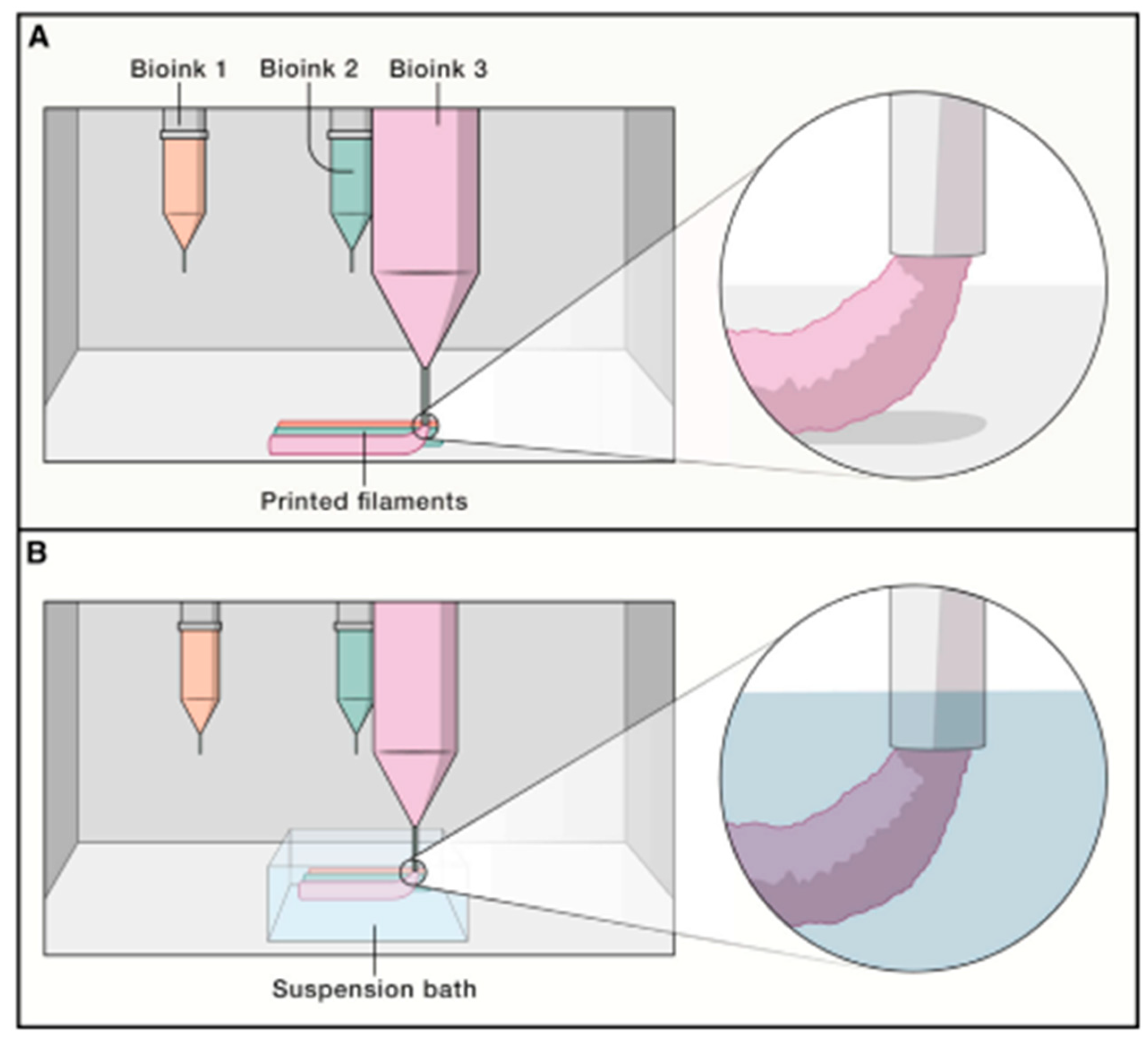
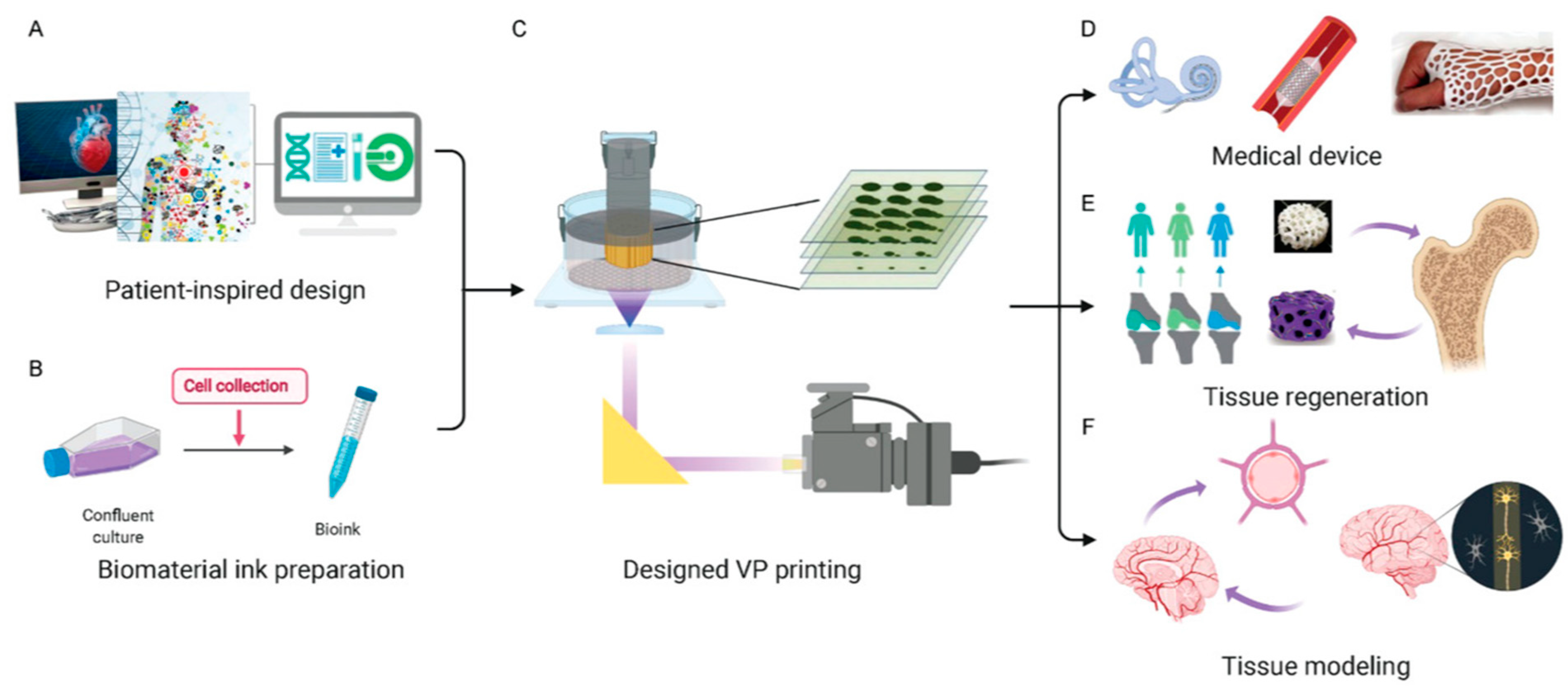
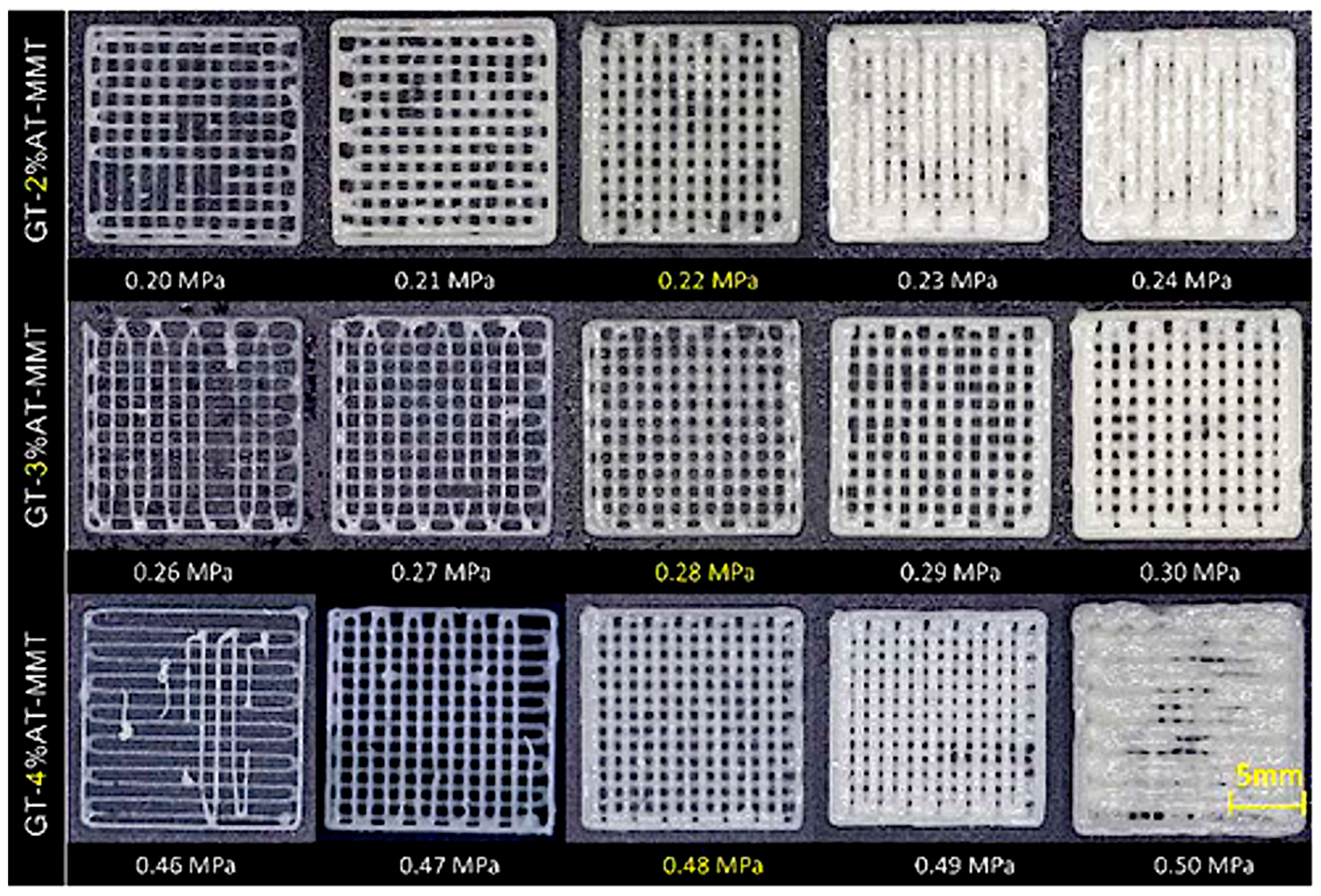
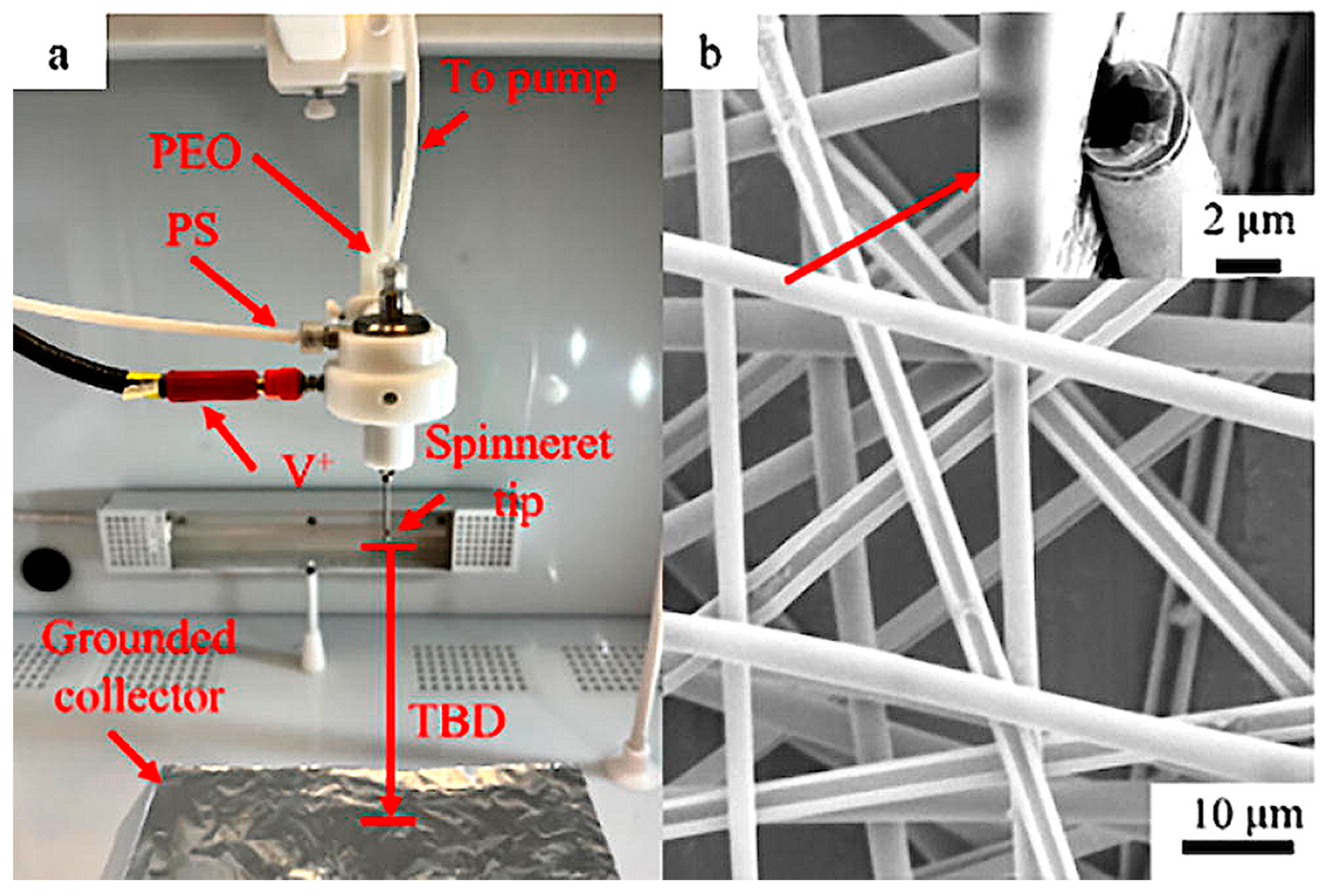
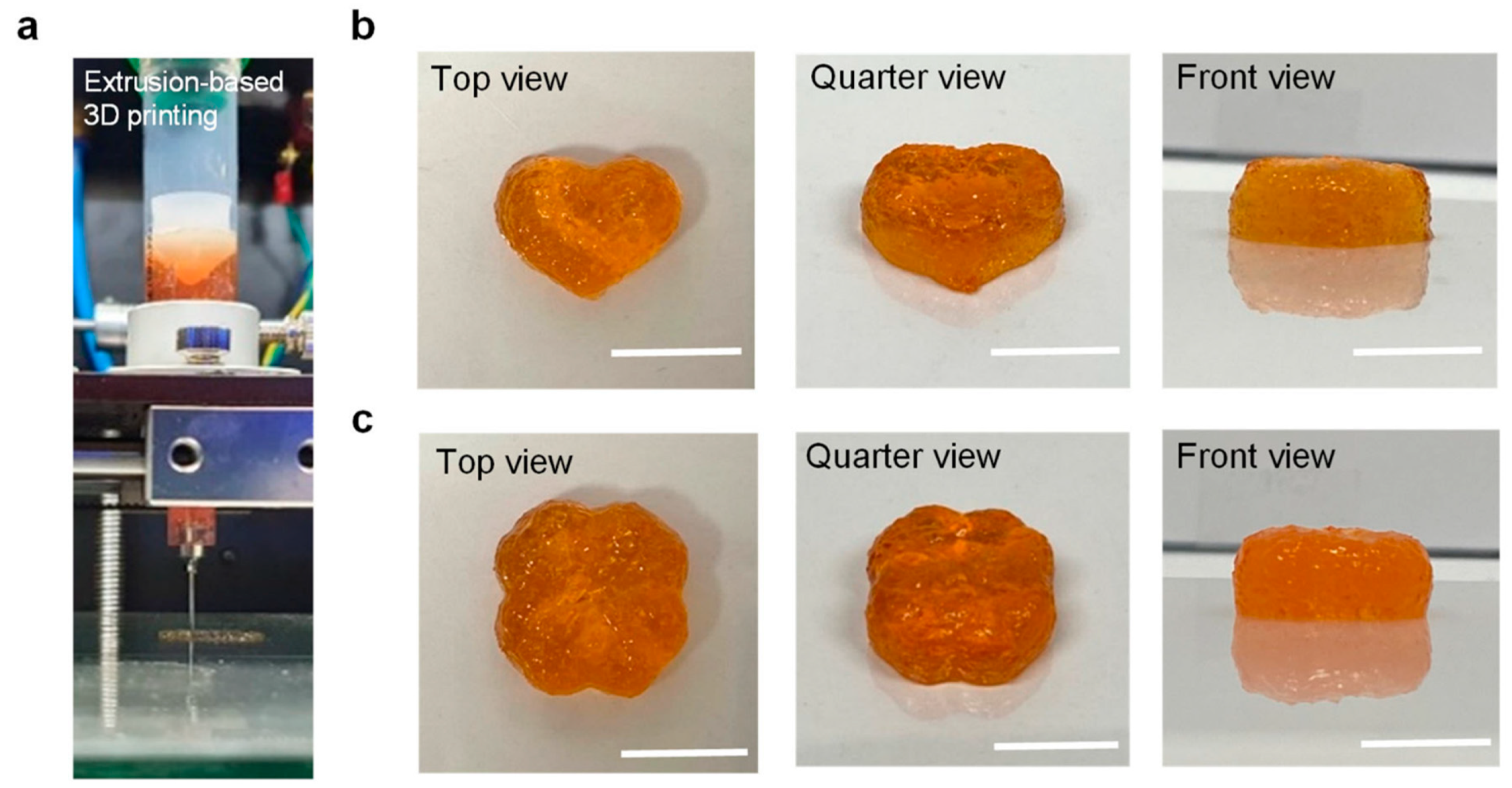
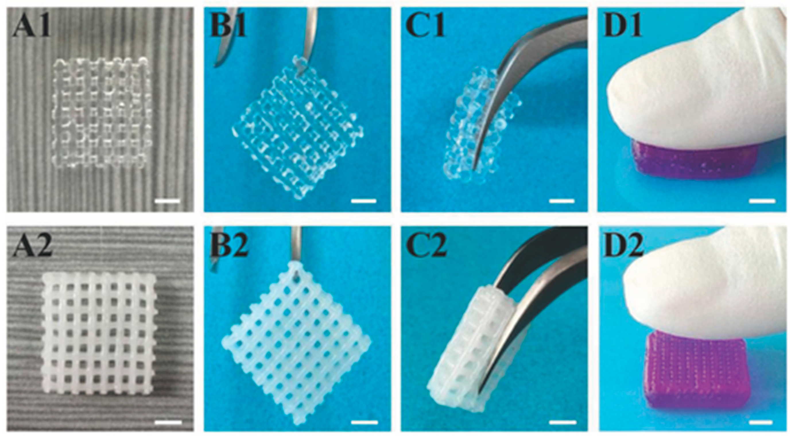
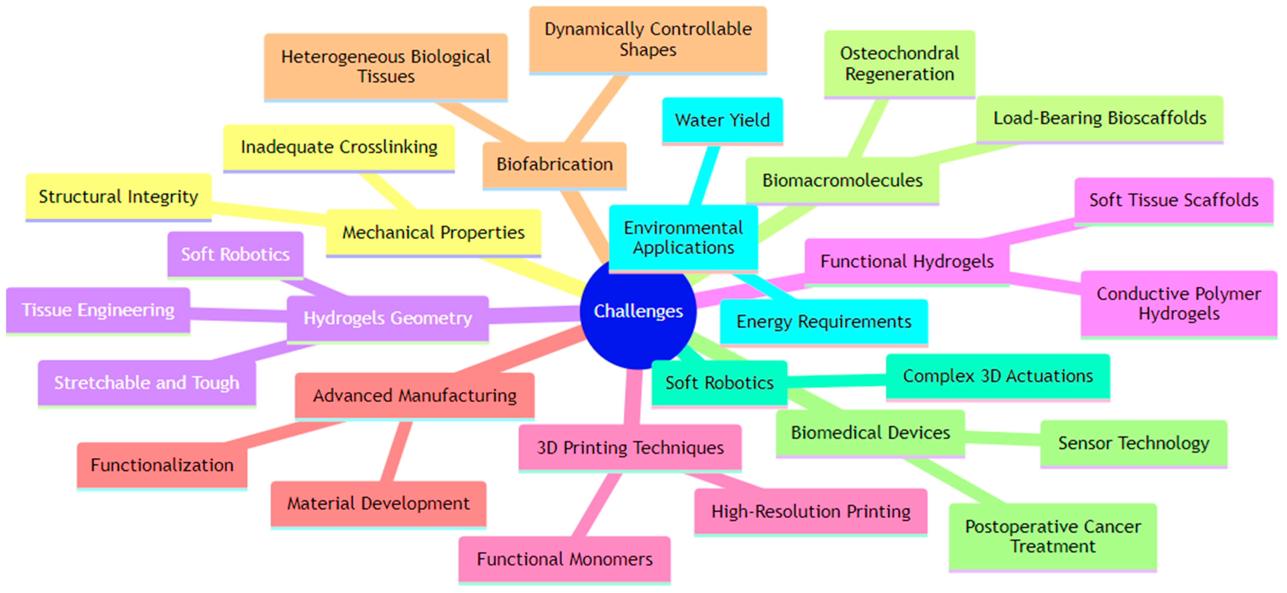
Table 1.
Three-dimensional printing technologies, material, and process optimization.
| Printing Technology | Material and Printable Concentrations | Printing Parameters and Resolution | Cell Viability (If Applicable) and Application | Ref. # |
|---|---|---|---|---|
| 3D printing | Sodium alginate, gelatin, and cellulose nanocrystals; glutaraldehyde and calcium chloride as crosslinkers | Optimized for uniform and gradient pore structures, pore sizes of 80–2125 µm | Biomedical scaffolds for tissue repair and regeneration | [1] |
| 3D bioprinting | Gelatin–alginate–montmorillonite (GT-AT-MMT) | Optimized with variable AT content | Tissue engineering, drug delivery, regeneration medicine | [2] |
| 3D printing (Direct Ink Writing) | Lanthanide-ion-coordinated supramolecular hydrogel | Multimaterial direct ink writing, controlled shape, luminescence, and opacity | Soft actuators with tunable luminescence and opacity | [3] |
| 3D printing | Hydrogel soft actuator | Air pressure-controlled contraction, mimics jellyfish contraction ratio | Jellyfish-mimic soft robots | [4] |
| Continuous Fiber Extruder (CFE) for FRESH | Long fibers embedded in hydrogels | Compatible with desktop 3D printers | Long-fiber embedded hydrogel 3D printing for enhanced mechanical properties | [5] |
| Hybrid Laser Printing (HLP) | PEGDA, GelMA hydrogels | Sequential additive and subtractive modes, microscale resolution | Multiscale, multimaterial 3D structures for optics, photonics, biomedical sciences | [6] |
| Two-Photon 3D printing | hydrogel for “hydrogel-in-hydrogel” | Precisely controlled channel-like hydrogel confinements, high-resolution hydrogel microstructures | Breast cancer cells migrate faster in stiffer confinements, studying cancer cell migration in confined 3D environments | [7] |
| 3D printing with sacrificial templates | GelMA, PVA for sacrificial templates | PVA templates dissolved to form channel networks, channels promote oxygen/nutrient supply and cell growth | Hydrogel constructs with channel networks for 3D cell culture, tissue repair | [8] |
| Two-Photon 3D printing and Scaffold Molding | Gelatin-based ink for printing, fibrin hydrogel | Optimized printing parameters and fibrin composition, perfusable multi-hydrogel vasculature | Supports growth and proliferation of human lung fibroblasts and endothelial cells, engineering perfusable vasculature models on-chip for tissue models | [9] |
| 3D Inkjet Printing | Alginate | Optimized reaction at the single-droplet level, tissue-like microvasculature | Live cells can be patterned, structuring hydrogels into tissue-like geometries | [10] |
| 3D printing with Photoinduced Self-crosslinking | Silk fibroin, tyramine-modified CMC-Na, XG, Ru(bpy)3Cl2, KPS | Extrusion-based printing with post photocrosslinking, ~550 µm × 1000 µm pores | Silk-fibroin-based composite hydrogel scaffolds for tissue engineering | [11] |
| 3D-printed PCL-Blended Scaffold | PCL, PVA(-)PAA hydrogel, sodium indomethacin | Evaluation of mechanical, thermal, and porosimetric properties | Load-bearing tissue damage support with anti-inflammatory drug delivery | [12] |
| 3D printing | CNCs, PCLA copolymers, DOX, lysozyme | In situ-forming thermosensitive, injectable, 3D-printable, stable 3D structures up to 10 layers | Controlled biodegradation, in vivo antitumor effect, controlled release, breast cancer treatment | [13] |
| 3D printing | Fmoc-FF hydrogel, silver or gold nanoparticles | Sensitive and reproducible SERS templates, low-concentration detection (as low as 100 pM) | SERS-based sensing | [24] |
| 3D printing | Low-melting-point agarose, sodium alginate | Solution flow rate (250–500 µL/min), stir speed (500–1250 rpm), bead diameters: 5–150 µm depending on conditions | MC3T3-E1 cells can survive and proliferate, preparation of hydrogel beads for cell therapy and tissue engineering | [44] |
| 3D printing | GelMA/beta-TCP/sodium alginate (Sr(2+))/MXene | MXene composite for photothermal antibacterial and osteogenic abilities | Biocompatibility and bone formation ability shown, bone regeneration in infected bone defects | [45] |
| FRESH 3D printing | Calcium-alginate hydrogels from brown seaweed | Thin-wall structures that are water-tight and pressurizable, small-scale actuators | Biodegradable, safely edible by marine organisms, biodegradable hydrogel actuators for marine robotics | [46] |
| Melt electrowriting (MEW) | Poly(ε-caprolactone), polyacrylamide, pHEMA hydrogels | Sinusoidal fiber deposition, radial-architecture for stabilizing fibers, diameter = 13.3 ± 0.3 µm, height = 330 µm | Improving shear properties of hydrogel–fiber composites | [47] |
| 3D printing | Electrolyte-filled hydrogel pen | Localized electrochemical reaction, small sizes (110 nm in diameter), tall heights (up to 30 µm) | Electrochemical applications, 3D printing in 3 dimensions | [48] |
| Extrusion-based bioprinting | Alginate, gelatin, methyl cellulose | Flow behavior and mechanical properties analyzed, examined pore size, strand diameter | 3D hydrogel scaffolds for bioprinting | [49] |
| Stereolithography | Hydrogel for double molding; PDMS | High-resolution stereolithographic prints, high-resolution features replicated in PDMS | Engineered tissue design, microheart muscles | [50] |
| 3D-printing-enabled microfluidics | GelMA, perfluorodecalin-oil-coated | Varying GelMA concentrations (7–15% w/v) for different sizes (35–250 µm diameters), cell-laden hydrogel microspheres | High long-term viability for human osteosarcoma Saos-2 cells, cell-laden hydrogel microspheres for tissue engineering | [51] |
| 3D printing by solvent exchange | N-heptyl-D-galactonamide in DMSO/water | Continuous extrusion | Biocompatible structures, temporary supports or sacrificial ink | [52] |
| 3D-printed protein hydrogel cages | Protein hydrogel | pH-stimulated reversible shape changes, microscale device dimensions | Shaping giant membrane vesicles for studying cell division and polarity | [53] |
| 3D printing | dECM, MSC-derived exosomes | Tissue-specific bioinspired scaffolds | Promotes MSC attachment, spread, migration, proliferation, differentiation, cartilage, and subchondral bone regeneration | [54] |
| 3D printing | UV-responsive CSMA, CaCO3, porous titanium | UV-responsive chitosan hydrogel mineralized with CO2 diffusion | Promoted adhesion and proliferation of BMSCs, cell proliferation and osteogenesis in vitro | [55] |
Table 2.
Three-dimensional hydrogel compositions for scaffold mechanical enhancement and biocompatibility.
Table 2.
Three-dimensional hydrogel compositions for scaffold mechanical enhancement and biocompatibility.
| Printing Technology | Material and Printable Concentrations | Printing Parameters and Resolution | Cell Viability (If Applicable) and Application | Ref. # |
|---|---|---|---|---|
| 3D printing | Poly(vinyl alcohol) (PVA) and chitosan (CS) with dual-physical crosslinking | Cyclic freezing–thawing followed by sodium citrate solution soaking, tensile strength of 12.71 MPa, Young’s modulus of 14.01 MPa | High-performance hydrogels for engineering, intelligent machines, and soft robotics | [14] |
| 3D printing | Hydrogel with titin-like domains | Multiple nozzles for topological design, enhanced extensibility, and toughness, mimicking titin toughening principle | Ultratough hydrogel structures for materials/structures with desired properties | [15] |
| Extrusion printing | Polyacrylamide hydrogel and poly(dimethylsiloxane) (PDMS) | Lithium-chloride-containing hydrogel printed onto treated PDMS, scaling resistance under tension and fatigue | Transparent and conductive integrated device fabrication | [16] |
| Digital Light Processing 3D printing | Poly(3,4-ethylenedioxythiophene) polystyrene sulfonate (PEDOT:PSS) and poly(2-hydroxyethyl acrylate) hydrogel–elastomer hybrids | Submillimeter-scale device resolution, high conductivity and superior electrical stability | Conductive hydrogel–elastomer hybrids for stretchable electronics | [17] |
| Photo-curing 3D printing | PHEA-PSS hydrogels with in situ-polymerized 3,4-ethylenedioxythiophene (EDOT) | Double network structure for complex spatial structures, tensile strength close to 8 MPa, electrical conductivity of 1.2 S cm−1) | High-strength conductive polymer hydrogels for flexible sensors | [18] |
| Vat photopolymerization | UV-curable hydrogel with polymerizable micelles, glycerol, and plant cellulose | Rapid layer-by-layer printing, 6 s per layer, enhanced mechanical toughness, 4.55 MJ/m3 | Strain sensing for flexible electronic devices | [19] |
| Fused Filament Fabrication (FFF) | PLA surfaces with atmospheric-pressure plasma jet (APPJ) treated and UV-grafted composite hydrogel (HEMA, PEGMA, HAp) | APPJ treatment and UV photo-grafting | Improved biocompatibility with osteoblast-like MG63 cells, bone scaffolds | [22] |
| 3D printing | Carboxymethyl cellulose/sodium alginate hydrogel with polystyrene microtubes or poly(ethylene oxide) | Viscosity, filament size, swelling, tensile, and compressive tests | Soft tissue scaffolds | [23] |
| Direct Ink Writing | Sodium alginate and agarose composite hydrogel | Submicron pores | Tissue engineering scaffolds with enhanced mechanical properties and conducive for cell growth | [56] |
| 3D printing | Hydroxybutyl chitosan (HBC) affected by ion effects | Temperature and salt concentration control | Cytocompatible, cartilage tissue repair with elaborate structure and good mechanical properties | [57] |
| Electron Beam Melting (EBM) | Ti-6Al-4V foam with hydrogel matrix containing deferoxamine mesylate (DFM) | 60% porosity, tensile strength of 18 GPa | Pre-osteoblasts and HUVECs show increased survival and proliferation, biomedical bone replacement implants with vascularization | [58] |
| Direct Ink Writing (DIW) / Direct Laser Writing (DLW) | Photopolymerizable copolypeptides (hydroxy ethyl-L-glutamine, vinylbenzyl-L-cysteine) | Polymer topology and/or molecular weight control, tailored for high-resolution macroscale or microscale 3D structures | Multipurpose hydrogel resins for 3D printing with adjustable mechanical properties | [59] |
| 3D Printing | PEEK implants with surface modifications of drug-loaded photocured hydrogel (acrylic acid-acrylamide with simvastatin-cefepime) | Surface sulfonation and hydrogel modification, improved hydrophilicity, reduced surface roughness | Enhanced biocompatibility and osseointegration capacity, orthopedic and dental implants | [60] |
| Direct Ink Writing | Poly(2-hydroxyethyl methacrylate) (pHEMA)/pyrrole ink | Printing through nozzles as small as 1 μm, good biocompatibility, ~94.5% viability with Aplysia californica neurons | High cell viability, conductive hydrogel platforms for neuron growth and electrophysiological recording | [61] |
| Materials extrusion | Biodegradable cellulose hydrogel inks with metallic powders (316 L stainless steel, copper) | Sintering at 1050 °C for 316 L and 985 °C for Cu, 31.92–32.07% porosity | Biomedical and energy applications with optimized porous metallic structures | [62] |
| DLP micro-3D printing | Electroactive hydrogel microstructures | Digital light processing technique for precise dimensional control, large deformation response | Soft robotic manipulation and locomotion | [63] |
| 3D printing | Carboxymethyl cellulose hydrogel for soybean sprouting substrate | Printed into different dimensions for soybean sprouting, influence of bean orientation on sprouting process | In-space seed nursery with hydrogel-based seed planter | [64] |
| 3D printing | Swollen chitosan microspheres in various functional monomer inks | Acid-driven tunable swelling ratio, strength (0.4–1.01 MPa), dissipated energy (0.11–3.25 MJ/m3), elongation at break (47–626%) | Soft device production including 4D printing robots and wearable strain sensors | [65] |
| 3D printing | Porous hydrogel scaffolds with medium pore sizes (100–1000 μm) | Medium pore sizes (100–1000 μm) | High activity and osteogenic differentiation of BMSCs, promotes viability and osteogenic differentiation of BMSCs in hydrogel scaffolds | [66] |
| 3D printing | Dialdehyde cellulose nanocrystals (DAC) and gelatin (GEL) | Optimal printing conditions including pressure and nozzle speed, additional crosslinking time, and temperature | Confirmed biocompatibility, tissue repair scaffolds | [67] |
| 3D printing | Cellulose nanocrystal (CNC), hard phenyl acrylate (PA), soft acrylamide (AAm) for interpenetrated polymer network | UV irradiation for crosslinking, tensile strength up to 16.5 MPa, toughness up to 6 MJ m−3) | Artificial meniscus with equivalent or superior mechanical performance to human meniscus | [68] |
| Photopolymerization | Zeolite/polymer-based hydrogel composite, FAU-13X zeolite (~40 wt%) | Visible LED light irradiation at room temperature, elastic modulus increased by more than 100% with 10% FAU-13X | Water absorption materials and 3D printing applications | [69] |
| Extrusion 3D printing | Cellulose solution in dimethyl sulfoxide (DMSO) and tetrabutylammonium hydroxide (TBAH) | Controlled extrusion and solvent-exchange with deionized water, compression strength of 96 kPa | Preparation of natural cellulose hydrogel scaffolds | [70] |
| 3D printing | Nanocellulose/polyethyleneglycol diacrylate (PEGDA) hydrogel | Light curing, 40 s of light curing for 1.8% nanocellulose mixture, maximum compression modulus of 0.91 MPa | Rapid prototyping of nanocellulose structures with controlled architecture | [71] |
| Direct Ink Writing (DIW) 3D Printing | Photocurable hydrogel with hydroxyethyl acrylate, gelatin, crosslinker, and photoinitiator | Cured with 405 nm visible light, adjustable mechanical properties from soft and fragile to rigid and tough | Printing of 3D objects with adjustable mechanical properties | [72] |
| 3D printing | Poly(3-sulfopropyl acrylate, potassium salt) (PSPA) with glycidyl methacrylated hyaluronic acid (GMHA) as a crosslinker | Stretchability increased to 49%, fracture toughness to 40 J/m2 | Electroactive hydrogels for soft robotics | [73] |
| Extrusion-based gradient printing | Tough alginate/poly(acrylamide) hydrogel and acrylated urethane-based UV curable adhesive material | Custom software to control dispensing rates for material gradients, continuous gradient of modulus | Bioinspired structures like artificial tendons | [74] |
| Embedded 3D printing | Various hydrogel inks in supportive baths (gelatin slurry, agarose fluid gel, Carbopol, oil-based baths) | Supportive bath matching for shapeability, dominated by diffusion-driven or charge-driven mechanisms | Tissue engineering to soft robotics | [75] |
| 3D printing and hydrogel integration | Soft polymeric materials for structure and different hydrogels for active components | Integration of hydrogel into 3D-printed constructs | Plant-inspired actuators | [76] |
| 3D Printing | Hydrogel with carbon nanotubes (PEGDA/MWCNT) for swelling-behavior-based detection | Swelling behavior for liquid detection, effective identification of liquid leakage position and volume | Flexible liquid sensors for biomedical engineering and environmental science | [77] |
| 3D printing | Poly(N-isopropyl acrylamide) with clay and phytic acid crosslinked polyaniline | Rheological properties modified for printing, high porosity, electrical conductivity, thermal response sensitivity | Thermo-responsive electroconductive hydrogel for motion sensors | [78] |
| 3D printing | Alginate-methylcellulose hydrogel with magnetic nanoparticles (MNPs) | Rheological properties influenced by MNPs, good mechanical stability | Magnetic hydrogel actuators for soft electronics, robotics, biomedical devices | [79] |
| 3D printing | High-strength supramolecular polymer hydrogel with poly(e-caprolactone) (PCL) and poly(N-acryloyl glycinamide) (PNAGA) | Radial and circumferential orientation to imitate collagen fibers, Young’s moduli of 20.15 MPa circumferentially and 10.43 MPa radially | Implanted in rabbit knee joints for 12 weeks, meniscus substitute with anisotropic microarchitecture | [80] |
| Microstereolithography | PEGDA photopolymerized hydrogels | Influence of curing parameters and ink formulation on structure and swelling, graded profile of curing and submicron pores | Impact on hydrogel structure and swelling behavior | [81] |
| Extrusion-Based 3D printing | DEAE-cellulose, alginate, gelatin hydrogel composites | Varying concentrations for optimal properties, improved shear thinning, viscosity, and mechanical integrity | Excellent hemocompatibility and cytocompatibility, tissue engineering biomaterial inks | [82] |
| Extrusion-based 3D printing | Chitosan methacrylate (CHMA) and polyvinyl alcohol (PVA) hybrid hydrogel microparticles | Self-healable pre-crosslinked hydrogel microparticles for high fidelity and biocompatibility, self-supportive yield strength (540 Pa) for printed construct fidelity | Supports growth of bone-marrow-derived mesenchymal stem cells, biomimetic scaffolds for tissue engineering | [83] |
| 3D printing | Chitosan methacrylate with various drugs for burn treatment | Customizable wound dressings via 3D printing | Biodegradable and biocompatible, personalized hydrogel wound dressings for thermal burns | [84] |
| 3D printing | Supramolecular polymer hydrogel with poly(e-caprolactone) and poly(N-acryloyl glycinamide) | Radial and circumferential orientation for structural integrity, Young’s moduli of 20.15 MPa circumferentially | Meniscus substitute with biomimetic structure | [85] |
| 3D printing | Thermo-responsive p(NIPAm-co-PEGDA) hydrogel with iron oxide nanoparticles | Additive fabrication of anisotropic hybrid 3D structure, superior light absorption properties | Direct solar vapor generation (SVG) device | [86] |
| 3D printing | SnS2/SnS nanocomposite hydrogel | Utilized as a photoenhanced triboelectric nanogenerator (PTNG), enhanced performance under light illumination | Energy harvesting and self-powered devices | [87] |
| 3D printing | Composite hydrogel with alginate and rice husk biochar | Designed for organic contaminants adsorption in tap water, significant adsorption capacities for IBU and MB | Water and wastewater treatment for organic contaminant removal | [88] |
Table 3.
Stretchable and conductive 3D-printed hydrogel compositions.
| Printing Technology | Material and Printable Concentrations | Printing Parameters and Resolution | Cell Viability (If Applicable) and Application | Ref. # |
|---|---|---|---|---|
| Suspension 3D printing | Liquid metal in self-healing hydrogel | Gel concentration, nozzle inner diameter, flow rate, printing speed, customizable droplet size | 3D macrostructures and stereo-electronic systems of liquid metal for flexible electronics | [21] |
| Optical Projection Lithography | DN hydrogels (covalent and ionic) | Hydrogel composition, crosslinker and photoinitiator concentrations, laser light intensity, high resolution | Soft robotics, tissue engineering | [89] |
| 3D printing | Kappa-carrageenan/PAAm DN hydrogel | UV exposure | Demonstrated excellent recoverability and self-healing capability, strain sensors for robotics and human motion detection | [90] |
| Cryogenic 3D printing | Chitosan (CS) and edge-functionalised expanded graphene (EFXG) | Custom cryogenic extrusion, control over ink and surface temperatures, as small as 200 µm | Supports NSC-34 mouse motor neuron-like cell viability, attachment, and proliferation, engineering of 3D-structured excitable cells | [91] |
| 3D printing/Interfacial Polymerization | Poly(ethylene glycol)diacrylate, pyrrole to polypyrrole (PPY) | Digital light processing system, complex and defined geometry | Conductive hydrogel creation | [92] |
| 3D printing | PEDOT:PSS, PVA, ionic macromolecular dopants | Composite engineering, freeze–thawing for EBI, high resolution | EBI-based skin electrodes for ECG and EMG signal recording | [93] |
| 3D-printed Stamp | Hydrogel–liquid metal composite | Molds for electrical pathway, liquid metal filling, customizable | Self-healable electrodes for multimodular sensor systems, biosignal detection | [94] |
| Direct Ink Writing (DIW) | PEDOT:PSS | Superior 3D printability, tissue-like mechanical compliance, instant tough bioadhesion | Implantable bioelectronics for electrophysiological monitoring and modulation | [95] |
| Digital Light Processing 3D printing | Acrylamide/acrylic acid, magnesium chloride (DN hydrogel) | Biomimetic fingerprint MH film, mechanical property tunability, 150 µm | Iontronic pressure sensor for tactile sensing and human motion detection | [96] |
| Continuous 3D printing | Conductive hydrogel with TPMS architecture | Self-powered DC energy generation upon mechanical stimulation, high flexibility with over 50% compressive strain | Self-powered electromechanical sensing for wearables, robotics, AI, environmental monitoring | [97] |
| 3D printing | Acrylamide and alginate DN hydrogel, MOF ligands | Prepolymer mixing, shear-thinning agent, copper ions for in situ MOF growth | Wearables, implantable and flexible sensors, chemical separations, soft robotics | [98] |
| Projection microstereolithography (PμSL) | Conductive crosslinked hydrogel | Temperature, pH, and chemical composition adjustments, ultra-fast programmable | Degradable, conductive wearable devices, human–machine interface | [99] |
| In Situ 3D printing | Liquid metal (LM)–hydrogel hybrids | Direct fabrication with mechanical compliance to soft tissues | Multifunctional soft bioelectronics and devices | [100] |
Table 4.
Self-healing and responsive 3D-printed hydrogel compositions.
| Printing Technology | Material and Printable Concentrations | Printing Parameters and Resolution | Cell Viability (If Applicable) and Application | Ref. # |
|---|---|---|---|---|
| Extrusion-based 3D printing | Poly(acrylic acid)-based hydrogel with 1.0 mol% covalent crosslinker and 2.0 mol% dynamic crosslinker | G’~1075 Pa and tan delta~0.12 | Self-healing hydrogel for 3D shape fabrication | [20] |
| Digital Light Processing (DLP) 3D printing | High-water-content hydrogel with water-soluble photo-absorber nanoparticles | Water-soluble photo-absorbers for high resolution, lateral resolution: 5 µm, vertical resolution: 20 µm | High-resolution, high-water-content hydrogel structures | [101] |
| Stereolithography 3D printing | Multi-responsive hydrogel-based actuators with functionally graded structures | Layers with different volume expansion properties, crosslinking density, chemical composition, high resolution | Hydrogel actuators with anisotropic swelling for rapid, controllable motion | [102] |
| 3D printing via ultraviolet light polymerization | Poly(N-isopropyl acrylamide)/graphene oxide (PNIPAm/GO) nanocomposite hydrogel | Optimal shear thinning properties adjusted by nanoclay, graphene oxide as infrared light absorber | Photothermally responsive actuators and functional programming | [103] |
| Direct-Ink-Writing Printing process | Graphene oxide (GO) flakes in sodium alginate (SA) matrix | Spontaneous alignment of GO flakes by shear force, locally controlled orientation | Multi-stimuli-responsive actuation with site-specific anisotropy | [104] |
| 3D printing | Thermo-responsive hydrogel | Capacitor circuit incorporation | Skin-like sensors for AI, wearable devices, human/machine interaction | [105] |
| Fused Deposition Modeling (FDM) 3D printing | Fish swim bladder hydrogel and wood flour-filled polylactic acid (WPLA) | Bio-hygromorph construction | Environment actuators, meteorosensitive architectures | [106] |
| Projection microstereolithography 3D printing | N-isopropylacrylamide-based photothermal responsive hydrogel | Grayscale variation for adjustable deformation, rapid prototyping of complex 3D structures | Ultra-fast photothermal responsive shape memory hydrogel for microrobots | [107] |
| Combining 3D printing with electrospinning | Stimuli-responsive hydrogels on electrospun membranes | Patterns printed for morphing behaviors guided by swelling/shrinkage mismatch | Rapid deformed hydrogel actuators with enhanced designability | [108] |
| Direct Ink Writing (DIW) | Hydrogel–fiber composites | Controlled fiber distribution by printing direction and nozzle size | Soft active materials, shape-morphing structures | [109] |
| Stereolithography-based 3D printing | Single-material hydrogel with secondary microstructures | Programmable microstructures for asymmetrical swelling | Soft robotics, tissue engineering, actuators | [110] |
| Additive manufacturing | Poly(electrolyte) complex, BZ reactions | Varied shapes, sizes, angles for spatiotemporal patterns | Smart structures, biological oscillators | [111] |
| Various 3D printing techniques | Stimuli-responsive hydrogels (SRHs) | Focus on printing techniques, stimuli mechanisms, shape-morphing behaviors | Tissue engineering, drug delivery, soft robots, sensors | [112] |
| 3D-printed Hydrogel–elastomer systems | Soft elastomer and hydrogel for anisotropic swelling | Star-like to circular geometry mimicking cactus | Soft robotics, controlled movements mimicking natural organisms | [113] |
| Hierarchical Filament 3D printing | Poly(ethylene glycol) diacrylate, poly(acrylic acid) microgels | Laminar flow for crosslink density organization | Soft actuators, sensors with programmed shape transformations | [114] |
| Extrusion-based 3D printing | Magnetic hydrogel (PNIPAm embedding Fe3O4 nanoparticles) and elastomer composite | Shape transformation under alternating magnetic field | ~50% cancer cell killing via magnetic hyperthermia during deformation, hyperthermia cancer therapy | [115] |
| Microreactive Inkjet Printing (MRIJP) | Alginate hydrogel | Controlled droplet collision and gelation dynamics, <40 cP viscosity for reactive inks | Tissue-mimicking structures, 3D microstructures | [116] |
| 3D printing | Hydrogel with photoactive titanium dioxide nanoparticles, alginate, gelatin, and dyes | Optimization of viscosity and ink composition for mechanical properties | UV exposure sensors, wearable sensors | [117] |
| 3D printing | Iron platinum (FePt) nanoparticle-embedded pentaerythritol triacrylate (PETA), poly N-isopropylacrylamide (pNIPAM), and poly N-isopropylacrylamide acrylic acid (pNIPAM-AAc) | Pollen-grain-inspired microrobot | Multifunctional microrobots for biomedical applications | [118] |
Table 5.
Three-dimensionally printed hydrogel compositions in osteochondral and bone tissue engineering.
Table 5.
Three-dimensionally printed hydrogel compositions in osteochondral and bone tissue engineering.
| Printing Technology | Material and Printable Concentrations | Printing Parameters and Resolution | Cell Viability (If Applicable) and Application | Ref. # |
|---|---|---|---|---|
| 3D printing with Lyophilization | Photocurable hydrogel of gelatin and hyaluronic acid | Methacrylic anhydride and a photoinitiator used, followed by lyophilization | Scaffolds supported mature cartilage regeneration | [25] |
| Direct 3D printing | Thermoresponsive supramolecular copolymer hydrogel (N-acryloyl glycinamide and N-[tris(hydroxymethyl)methyl] acrylamide) | Shear thinning, immediate gelation on cooled substrate | Facilitates attachment, spreading, and chondrogenic and osteogenic differentiation of hBMSCs for repair of osteochondral defect | [119] |
| 3D printing | PACG-GelMA (Poly(N-acryloyl 2-glycine) and methacrylated gelatin) | Photo-initiated polymerization | Supports cell attachment and spreading, enhances gene expression of chondrogenic and osteogenic differentiation for osteochondral Regeneration | [120] |
| DLP 3D printing | GelMA-SFMA (methacrylated gelatin and methacrylated silk fibroin) with ethanol treatment | Digital Light Processing (DLP) | Supports cell attachment and spreading, facilitates osteogenic differentiation for osteo-chondral tissue regeneration | [121] |
| Extrusion-based 3D printing | SF-SA (silk fibroin and sodium alginate) with 2.5% MgP (magnesium phosphate) | Extrusion-based 3D printing | Promoted adhesion of rat mesenchymal stem cells, improved osteogenesis for bone tissue engineering | [122] |
| Hydrogel 3D-printing | Gelatin/hydroxyapatite/3D-graphene | Hydrothermal autoclave, hydrogen gas injection | [123] | |
| 3D bioprinting | Hydrogel with encapsulated valve and mesenchymal stem cells | Photoencapsulation, cell-hydrogel printing | High cell viability for tissue-engineered heart valves | [124] |
| 3D printing | Alginate/Gelatin hydrogel with Bioactive Glass 45S5 and ZIF-8 nanoparticles | Shear-thinning behavior for extrusion | Higher MG-63 cell viability (>90%) than Alg/Gel alone for bone tissue regeneration | [125] |
| 3D printing with hydrogel integration | Alginate or alginate–bioglass composite hydrogels with 3D-printed poly(lactic acid) | Surface treated with polyacrylic acid (PAA) | Good cell viability and proliferation, promotes osteogenic differentiation and calcium mineralization for bone tissue engineering | [126] |
| 3D printing | Alginate/gelatin-based hydrogel with CaCO3 microspheres | Pre-crosslinking with Ca2+ for increased printability, pore area: 4.43 +/− 0.14 μm², line diameter: 184 +/− 25 μm | Improved NIH-3T3 cell proliferation for diabetic wound healing | [127] |
| 3D printing | ECM-enriched hydrogel in a PCL scaffold | Excellent viability and osteogenic activity of BM-MSCs for Vascularized Bone Regeneration | [128] | |
| 3D printing | Hydrogel with nanohydroxyapatite gradient (pure hydrogel, 40% nHA/60% hydrogel, 70% nHA/30% hydrogel) | Superior repair results with BMSC-loaded gradient scaffolds for repair of osteochondral defects | [129] | |
| 3D printing | Cellulase-laden cellulose nanofiber/chitosan hydrogel composites | Enzyme-mediated biodegradation | Excellent cytocompatibility (supports fibroblast cell attachment, proliferation, and growth) for tissue engineering | [130] |
| 3D printing | Titanium with tri-calcium-phosphate-loaded demineralized bone matrix hydrogel | Biocompatible, promotes osteogenesis for bone regeneration | [131] | |
| FDM and Photo-curable Hydrogel Printing | PLA microstructure with gelatin hydrogel and bioactive gold nanoparticles (GNPs) | Enhanced osteogenic differentiation of ADSCs for bone tissue regeneration | [132] | |
| Direct vs. indirect 3D printing | Methacrylamide-functionalized gelatin | Direct extrusion and indirect printing with PLA mold | Supports adipose stem cell adhesion and differentiation for adipose tissue regeneration | [133] |
| 3D printing with hydrogel coating | PCL/PU scaffolds with PDA-coated decellularized matrix hydrogel | Promotes cell survival, proliferation, and ECM production for temporomandibular joint disc repair | [134] | |
| 3D printing with magnetic hydrogel | Chitosan hydrogel with embedded iron oxide nanoparticles | Use of external magnetic force, nanoparticles modified with citrate | Increased osteoblast proliferation and differentiation for bone tissue regeneration | [135] |
| 3D printing | Sodium alginate/hydroxyapatite hydrogel ink, pre-crosslinked | Further crosslinking post printing to enhance mechanical properties, macro-porous structure, >80% porosity | Enhanced mBMSC adhesion for bone tissue engineering | [136] |
| 3D bioprinting | GelMA/nHAp and THA hydrogels | Optimal printing temperature of 20 °C | High cell viability in both GelMA/nHAp and THA hydrogels for osteochondral tissue modeling | [137] |
Table 6.
Three-dimensionally printed hydrogel compositions in drug delivery and therapeutics.
| Printing Technology | Material and Printable Concentrations | Printing Parameters and Resolution | Cell Viability (If Applicable) and Application | Ref. # |
|---|---|---|---|---|
| 3D printing | Pluronic F127 and sodium alginate, DOX, and copper ions (F127-SA/Cu-DOX hydrogel scaffold) | Postoperative chemo/chemodynamic therapy for cancer | [138] | |
| 3D printing | PHEMA hydrogel, Avastin-loaded | Simple, cost-effective design, comparable to commercial lens | Ocular drug delivery for diabetic retinopathy | [139] |
| 3D printing | DNA from salmon sperm, DNA-induced biosilica, hydrogel inks | Optimized for effective exudate and blood absorption, mechanical tunability | Diabetic wound healing | [140] |
Disclaimer/Publisher’s Note: The statements, opinions and data contained in all publications are solely those of the individual author(s) and contributor(s) and not of MDPI and/or the editor(s). MDPI and/or the editor(s) disclaim responsibility for any injury to people or property resulting from any ideas, methods, instructions or products referred to in the content. |
© 2024 by the authors. Licensee MDPI, Basel, Switzerland. This article is an open access article distributed under the terms and conditions of the Creative Commons Attribution (CC BY) license (https://creativecommons.org/licenses/by/4.0/).
Share and Cite
MDPI and ACS Style
Omidian, H.; Mfoafo, K. Three-Dimensional Printing Strategies for Enhanced Hydrogel Applications. Gels 2024, 10, 220. https://doi.org/10.3390/gels10040220
AMA Style
Omidian H, Mfoafo K. Three-Dimensional Printing Strategies for Enhanced Hydrogel Applications. Gels. 2024; 10(4):220. https://doi.org/10.3390/gels10040220
Chicago/Turabian StyleOmidian, Hossein, and Kwadwo Mfoafo. 2024. "Three-Dimensional Printing Strategies for Enhanced Hydrogel Applications" Gels 10, no. 4: 220. https://doi.org/10.3390/gels10040220
Note that from the first issue of 2016, this journal uses article numbers instead of page numbers. See further details here.