
Nanogels Based on N,N-Dimethylacrylamide and β-Cyclodextrin Triacrylate for Enhanced Solubility and Therapeutic Efficacy of Aripiprazole
 , ,
, ,  and
and
Abstract
:1. Introduction
2. Results and Discussion
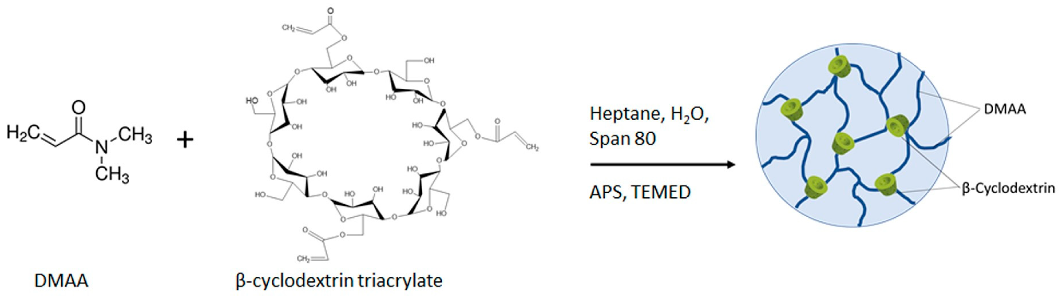
2.1. Synthesis of Nanogels
2.2. Fourier-Transform Infrared Spectroscopy
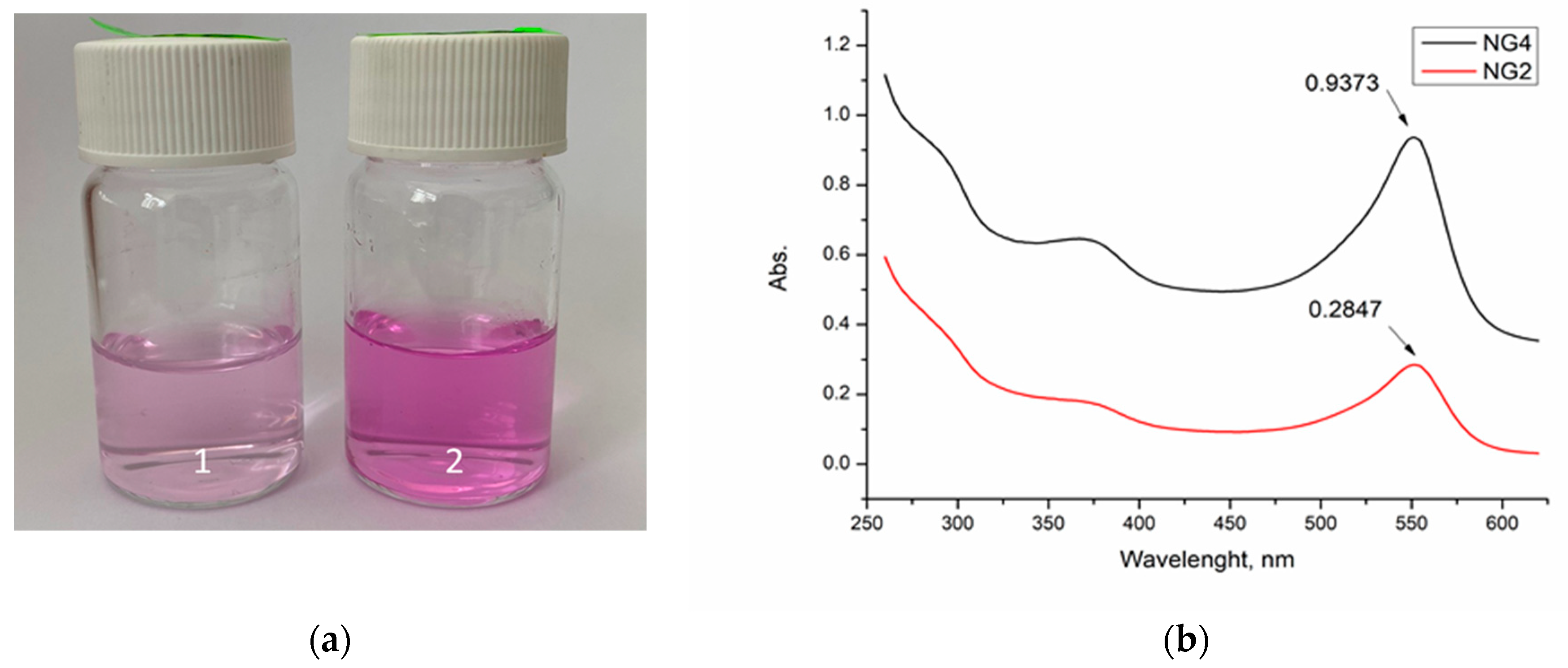
2.3. UV–Vis Spectroscopy
2.4. Drug Loading
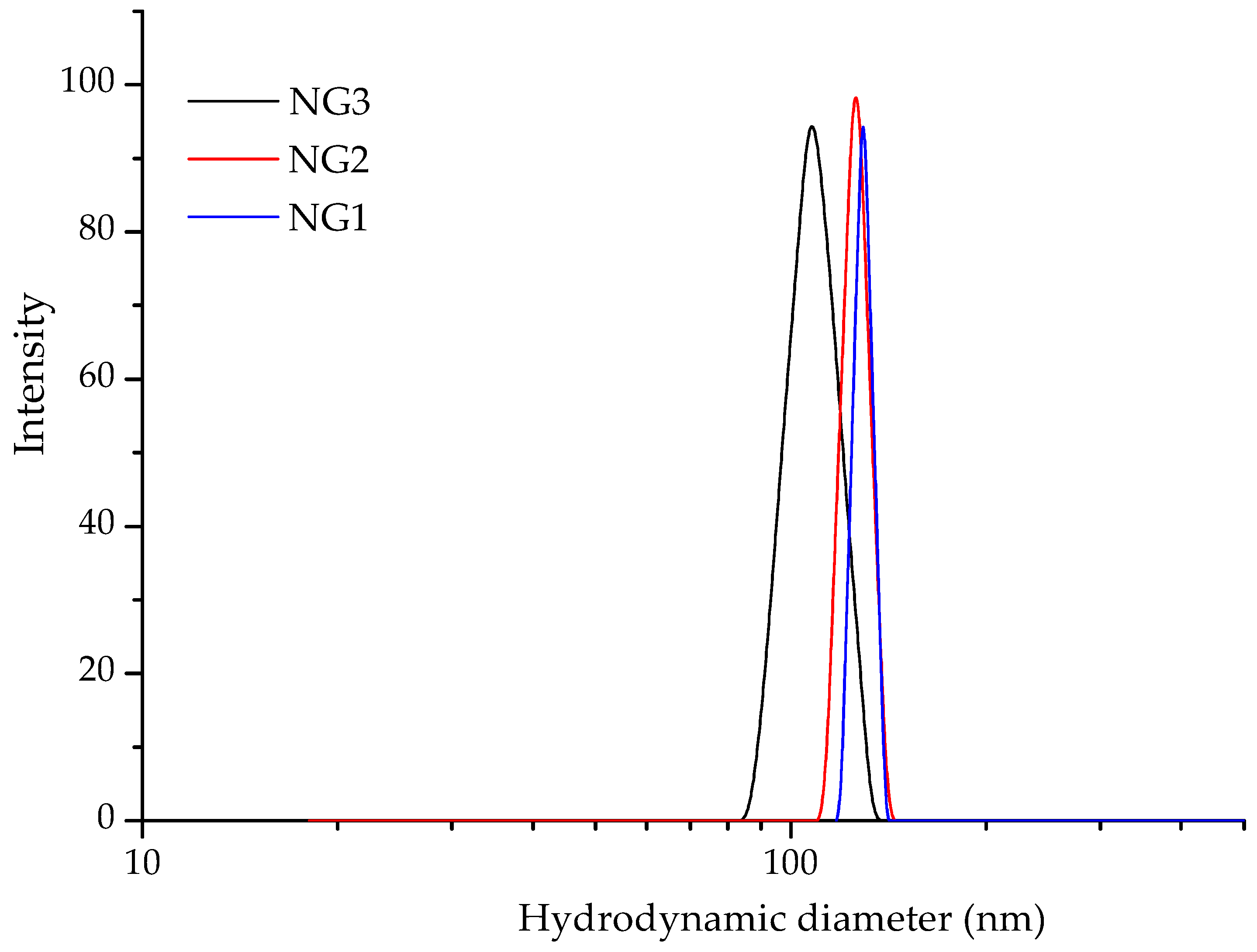
2.5. Dynamic Light Scattering and ζ-Potential Measurements
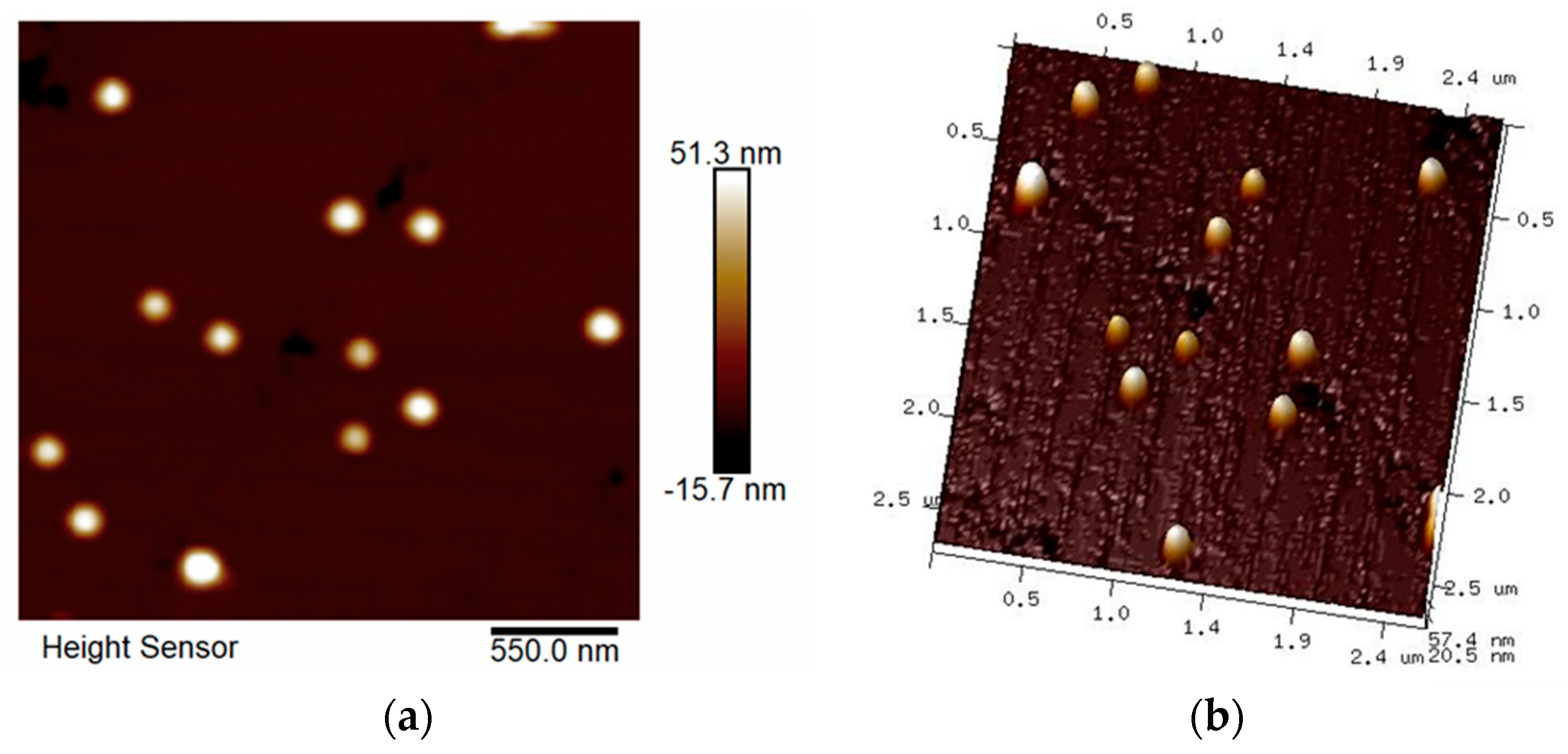
2.6. Atomic Force Microscopy (AFM)
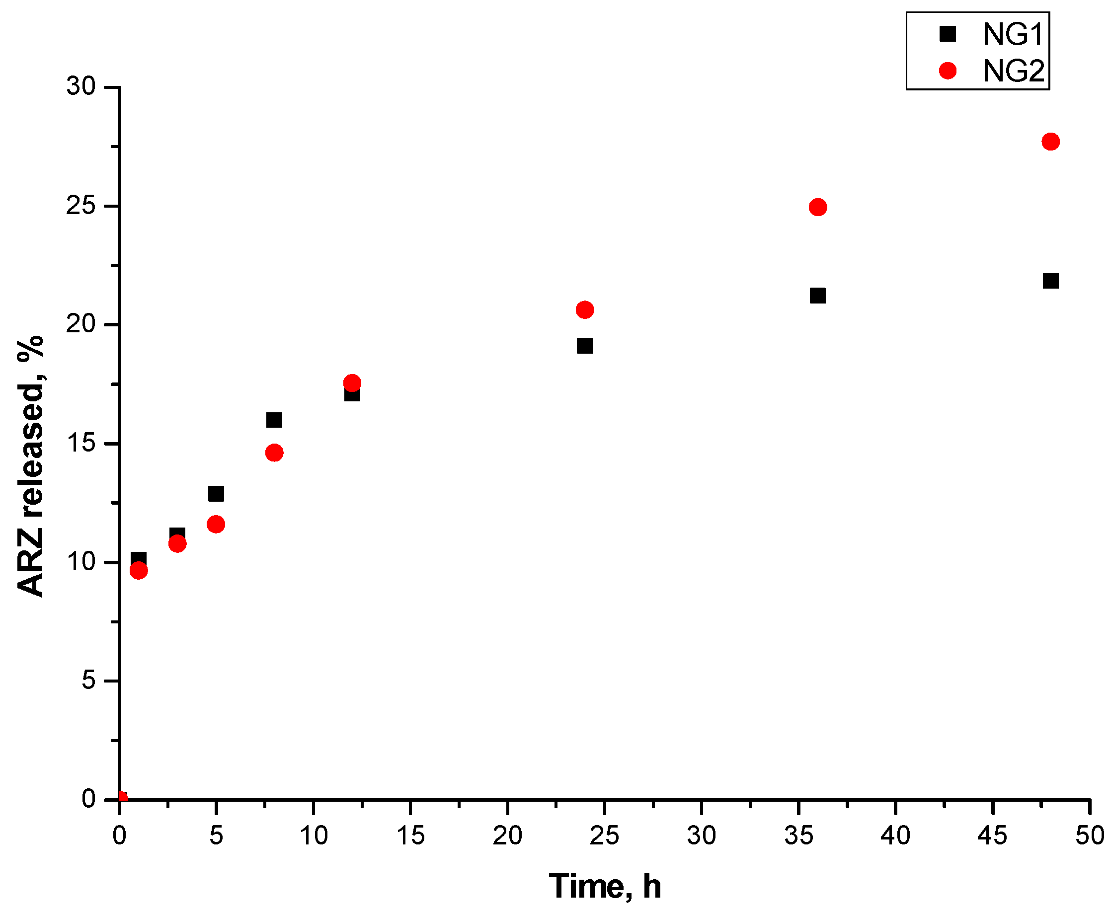
2.7. Drug Release Studies
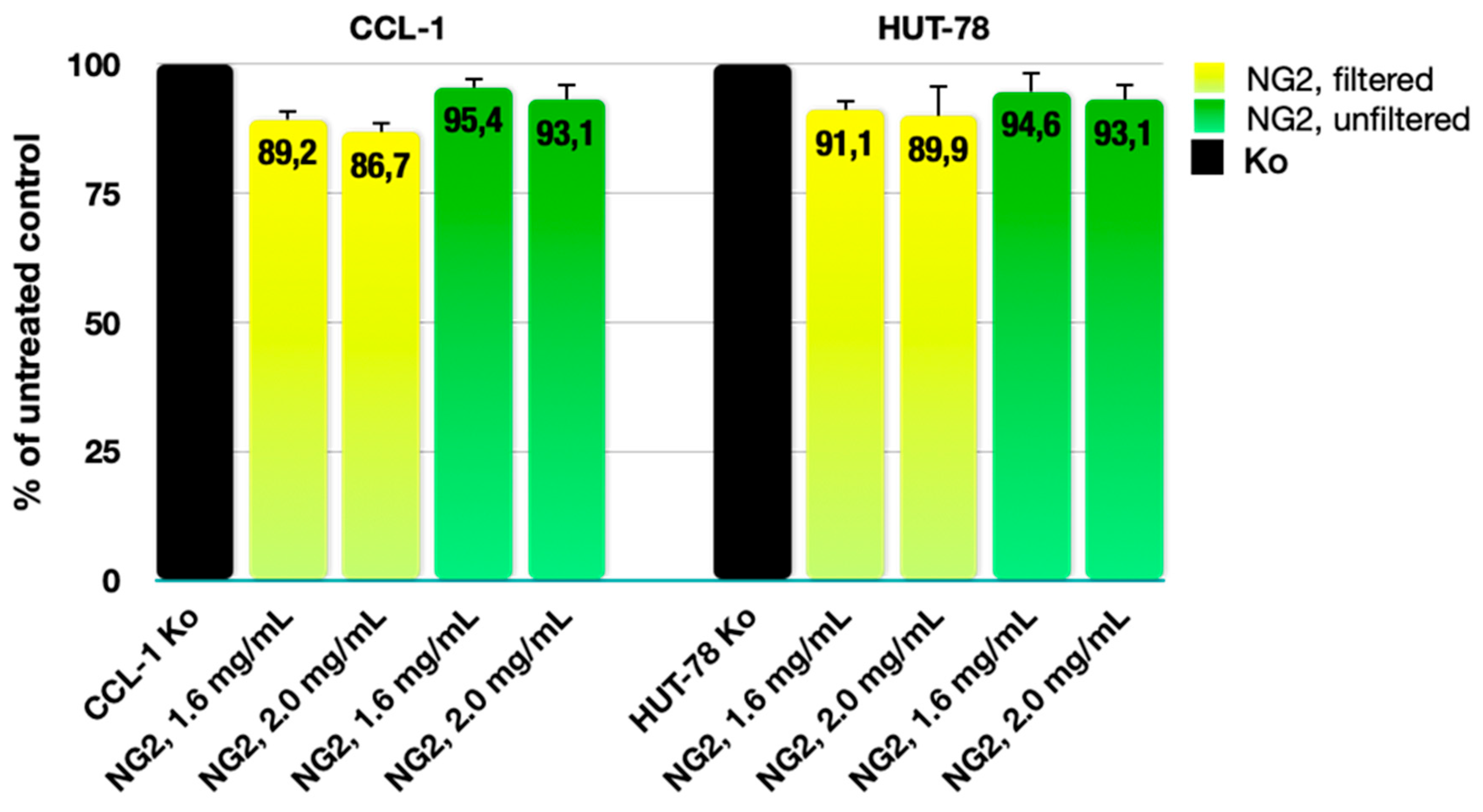
2.8. Cytotoxicity Assessment
3. Conclusions
4. Materials and Methods
4.1. Materials
4.2. Methods
4.2.1. Synthesis of Nanogels
4.2.2. Fourier-Transform Infrared Spectroscopy
4.2.3. UV–Vis Spectroscopy
4.2.4. Drug Loading
4.2.5. Dynamic Light Scattering and ζ-Potential Measurements
4.2.6. Atomic Force Microscopy
4.2.7. Drug Release Studies
4.2.8. Cytotoxicity Assessment
Cell Lines and Culture Conditions
In Vitro MTT Colorimetric Assay
Author Contributions
Funding
Institutional Review Board Statement
Informed Consent Statement
Data Availability Statement
Acknowledgments
Conflicts of Interest
References
- Casey, A.B.; Canal, C.E. Classics in Chemical Neuroscience: Aripiprazole. ACS Chem. Neurosci. 2017, 8, 1135–1146. [Google Scholar] [CrossRef] [PubMed]
- Madhav, N.V.S.; Ojha, A.; Jaiswal, V. A smart approach for delivery of aripiprazole via oro-soft palatal mucosal route for improved therapeutic efficacy. Braz. J. Pharm. Sci. 2018, 54, e17382. [Google Scholar] [CrossRef]
- Kirino, E. Efficacy and safety of aripiprazole in child and adolescent patients. Eur. Child Adolesc. Psychiatry 2012, 21, 361–368. [Google Scholar] [CrossRef] [PubMed]
- Awais, S.; Sultana, K.; Ansari, M.T.; Islam, N.; Afridi, M. Improved solubility and stability of aripiprazole in binary and ternary inclusion complexes using methyl-β-cyclodextrin and L-arginine. Pak. J. Pharm. Sci. 2022, 35, 1415–1422. [Google Scholar] [PubMed]
- Chennuri, A.; Prasanthi, D. Solubility enhancement of aripiprazole by solid-self emulsifying drug delivery systems. Int. J. Pharm. Sci. Drug Res. 2018, 10, 233–245. [Google Scholar] [CrossRef]
- Devangan, P.; Saini, A.; Patel, D.; Kolhe, U. Solubility Enhancement of Aripiprazole via Mesoporous Silica: Preparation, Characterization, In vitro Drug Release, and Solubility Determination. J. Pharm. Innov. 2023, 18, 1316–1327. [Google Scholar] [CrossRef]
- Hedayati, A.; Yazdi, S.G.; Dehghankelishadi, P.; Javan, N.B.; Akbari, H.; Dorkoosh, F.A. Preparation, Optimization and Physicochemical Characterization of Aripiprazole Loaded Nano-porous in situ Forming Implant. Pharm. Nanotechnol. 2017, 5, 138–147. [Google Scholar] [CrossRef]
- Chen, L.F.; Chen, Y.; Duan, Y.Y.; Zhang, M.M.; Xu, P.Y.; Kankala, R.K.; Wang, S.B.; Chen, A.Z. Preparation of aripiprazole-poly(methyl vinyl ether-co-maleic anhydride) nanocomposites via supercritical antisolvent process for improved antidepression therapy. Regen. Biomater. 2022, 9, rbac080. [Google Scholar] [CrossRef]
- Łyszczarz, E.; Hofmanová, J.; Szafraniec-Szczęsny, J.; Jachowicz, R. Orodispersible films containing ball milled aripiprazole-poloxamer®407 solid dispersions. Int. J. Pharm. 2020, 575, 118955. [Google Scholar] [CrossRef]
- Mihajlovic, T.; Kachrimanis, K.; Graovac, A.; Djuric, Z.; Ibric, S. Improvement of aripiprazole solubility by complexation with (2-hydroxy)propyl-β-cyclodextrin using spray drying technique. AAPS. PharmSciTech 2012, 13, 623–631. [Google Scholar] [CrossRef]
- Awais, S.; Farooq, N.; Muhammad, S.A.; El-Serehy, H.A.; Ishtiaq, F.; Afridi, M.; Ahsan, H.; Ullah, A.; Nadeem, T.; Sultana, K. Enhanced Solubility and Stability of Aripiprazole in Binary and Ternary Inclusion Complexes Using Hydroxy Propyl Beta Cyclodextrin (HPβCD) and L-Arginine. Molecules 2023, 28, 3860. [Google Scholar] [CrossRef] [PubMed]
- Kumbhar, S.A.; Kokare, C.R.; Shrivastava, B.; Gorain, B.; Choudhury, H. Antipsychotic Potential and Safety Profile of TPGS-Based Mucoadhesive Aripiprazole Nanoemulsion: Development and Optimization for Nose-To-Brain Delivery. J. Pharm. Sci. 2021, 110, 1761–1778. [Google Scholar] [CrossRef] [PubMed]
- Sawant, K.; Pandey, A.; Patel, S. Aripiprazole loaded poly(caprolactone) nanoparticles: Optimization and in vivo pharmacokinetics. Mater. Sci. Eng. C Mater. Biol. Appl. 2016, 66, 230–243. [Google Scholar] [CrossRef] [PubMed]
- Butreddy, A.; Sarabu, S.; Bandari, S.; Dumpa, N.; Zhang, F.; Repka, M.A. Polymer-Assisted Aripiprazole-Adipic Acid Cocrystals Produced by Hot Melt Extrusion Techniques. Cryst. Growth Des. 2020, 20, 4335–4345. [Google Scholar] [CrossRef] [PubMed]
- Patil, P.H.; Wankhede, P.R.; Mahajan, H.S.; Zawar, L.R. Aripiprazole-Loaded Polymeric Micelles: Fabrication, Optimization and Evaluation using Response Surface Method. Recent Pat. Drug Deliv. Formul. 2018, 12, 53–64. [Google Scholar] [CrossRef] [PubMed]
- Nahata, T.; Saini, T.R. Formulation optimization of long-acting depot injection of aripiprazole by using D-optimal mixture design. PDA J. Pharm. Sci. Technol. 2009, 63, 113–122. [Google Scholar] [PubMed]
- McFall, H.; Sarabu, S.; Shankar, V.; Bandari, S.; Murthy, S.N.; Kolter, K.; Langley, N.; Kim, D.W.; Repka, M.A. Formulation of aripiprazole-loaded pH-modulated solid dispersions via hot-melt extrusion technology: In vitro and in vivo studies. Int. J. Pharm. 2019, 554, 302–311. [Google Scholar] [CrossRef]
- Savjani, K.T.; Gajjar, A.K.; Savjani, J.K. Drug Solubility: Importance and Enhancement Techniques. ISRN Pharm. 2012, 2012, 195727. [Google Scholar] [CrossRef]
- Carneiro, S.B.; Costa Duarte, F.Í.; Heimfarth, L.; Siqueira Quintans, J.d.S.; Quintans-Júnior, L.J.; Veiga Júnior, V.F.d.; Neves de Lima, Á.A. Cyclodextrin–Drug Inclusion Complexes: In Vivo and In Vitro Approaches. Int. J. Mol. Sci. 2019, 20, 642. [Google Scholar] [CrossRef]
- Salústio, P.J.; Pontes, P.; Conduto, C.; Sanches, I.; Carvalho, C.; Arrais, J.; Marques, H.M.C. Mini-Review Advanced Technologies for Oral Controlled Release: Cyclodextrins for Oral Controlled Release. AAPS Pharm. Sci. Tech. 2011, 12, 1276–1292. [Google Scholar] [CrossRef]
- Conceição, J.; Adeoye, O.; Cabral-Marques, H.M.; Lobo, J.M.S. Cyclodextrins as drug carriers in pharmaceutical technology: The State of the Art. Curr. Pharm. Des. 2018, 24, 1–29. [Google Scholar] [CrossRef] [PubMed]
- Conceição, J.; Adeoye, O.; Cabral-Marques, H.M.; Lobo, J.M.S. Cyclodextrins as excipients in tablet formulations. Drug Discov. Today 2018, 23, 1274–1284. [Google Scholar] [CrossRef] [PubMed]
- Davis, E.M.; Brewster, E.M. Cyclodextrin-based pharmaceutics: Past, present and future. Nat. Rev. Drug Discov. 2004, 3, 1023–1035. [Google Scholar] [CrossRef] [PubMed]
- Helena, M.; Marques, C. A review on cyclodextrin encapsulation of essential oils and volatiles. Flavour Fragr. J. 2019, 25, 313–326. [Google Scholar] [CrossRef]
- Majd, M.; Yazdanpanah, M.; Bayatloo, M.R.; Nojavan, S. Recent advances and applications of cyclodextrins in magnetic solid phase extraction. Talanta 2021, 229, 122296. [Google Scholar] [CrossRef] [PubMed]
- Szejtli, J. Introduction and General Overview of Cyclodextrin Chemistry. Chem. Rev. 1998, 98, 1743–1753. [Google Scholar] [CrossRef] [PubMed]
- Gregório, C. Review: A History of Cyclodextrins. Chem. Rev. 2014, 114, 10940–10975. [Google Scholar] [CrossRef]
- Kayaci, F.; Uyar, T. Encapsulation of vanillin/cyclodextrin inclusion complex in electrospun polyvinyl alcohol (PVA) nanowebs: Prolonged shelf-life and high temperature stability of vanillin. Food Chem. 2012, 133, 641–649. [Google Scholar] [CrossRef]
- Tzankova, V.; Doneva, N.; Frosini, M.; Valoti, M.; Kostova, B.; Rachev, D.; Todorova, L.; Christova, D. In vitro cytotoxicity evaluation of functional peg-pdma block copolymer in liver hepg2 cells. Pharmacia 2016, 63, 9–13. [Google Scholar]
- Danov, Y.; Georgieva, D.; Mihaylova, R.; Kostova, B.; Petrov, P. Cryogel Carriers Comprising β-Cyclodextrin Moieties for Improved Solubilization and Delivery of Aripiprazole. Macromol. Chem. Phys. 2021, 222, 2100004. [Google Scholar] [CrossRef]
- Attama, A.A.; Nnamani, P.O.; Onokala, O.B.; Ugwu, A.A.; Onugwu, A.L. Nanogels as target drug delivery systems in cancer therapy: A review of the last decade. Front. Pharmacol. 2022, 13, 874510. [Google Scholar] [CrossRef] [PubMed]
- Srivastava, S.; Saha, S.; Jakhmola, V. Nanogel: Types, Methods of Preparation, Limitation, Evaluation and Application—A Systematic Review. Int. J. Drug Deliv. Technol. 2023, 13, 1631–1639. [Google Scholar] [CrossRef]
- Antonia-Nancy, H.; Iwatsuki, Y.; Yabuuchi, K.; Aso, S.; Katsumata, T.; Fukumoto, K.; Tanaka, Y.; Nakai, T.; Shimoboji, T.; Matsumoto, M.; et al. Self-assembled nanogels based on hyaluronic acid for antibody protection from heat denaturation. Biochem. Eng. J. 2023, 196, 108955. [Google Scholar] [CrossRef]
- Yu, H.; Wu, W.; Lin, X.; Feng, Y. Polysaccharide-Based Nanomaterials for Ocular Drug Delivery: A Perspective. Front. Bioeng. Biotechnol. 2020, 8, 601246. [Google Scholar] [CrossRef] [PubMed]
- Yang, H.Y.; Jang, M.S.; Sun, X.S.; Liu, C.L.; Lee, J.H.; Li, Y.; Fu, Y. CD44-mediated tumor homing of hyaluronic acid nanogels for hypoxia-activated photodynamic therapy against tumor. Colloids Surf. B Biointerfaces 2023, 228, 113395. [Google Scholar] [CrossRef] [PubMed]
- Pillarisetti, S.; Vijayan, V.; Rangasamy, J.; Bardhan, R.; Uthaman, S.; Park, I.-K. A multi-stimuli responsive alginate nanogel for anticancer chemo-photodynamic therapy. J. Ind. Eng. Chem. 2023, 123, 361–370. [Google Scholar] [CrossRef]
- Sarac, A.S. Redox polymerization. Prog. Polym. Sci. 1999, 24, 1149–1204. [Google Scholar] [CrossRef]
- Yu, H.; Fang, Y.; Chen, L.; Chen, S. Investigation of redox initiators for free radical frontal polymerization. Polym. Int. 2009, 58, 851–857. [Google Scholar] [CrossRef]
- Kolya, H.; Tripathy, T. Grafted polysaccharides based on acrylamide and N,N-dimethylacrylamide: Preparation and investigation of their flocculation performances. J. App. Polym. Sci. 2012, 127, 2786–2795. [Google Scholar] [CrossRef]
- Wang, F.; Yong, X.; Deng, J.; Wu, Y. Poly(N,N-dimethylacrylamide-octadecyl acrylate)-clay hydrogels with high mechanical properties and shape memory ability. RSC Adv. 2018, 8, 16773–16780. [Google Scholar] [CrossRef]
- Abarca, R.L.; Rodríguez, F.J.; Guarda, A.; Galotto, M.J.; Bruna, J.E. Characterization of beta-cyclodextrin inclusion complexes containing an essential oil component. Food Chem. 2016, 196, 968–975. [Google Scholar] [CrossRef]
- Basappa, C.; Rao, P.; Rao, D.N.; Divakar. A modified colorimetric method for the estimation of β-cyclodextrin using phenolphthalein. Int. J. Food Sci. Technol. 1998, 33, 517–520. [Google Scholar] [CrossRef]
- Mahajan, S.; Singh, D.; Sharma, R.; Singh, G.; Bedi, N. pH-Independent Dissolution and Enhanced Oral Bioavailability of Aripiprazole-Loaded Solid Self-microemulsifying Drug Delivery System. AAPS PharmSciTech 2021, 22, 24. [Google Scholar] [CrossRef]


| Sample Code | Composition DMAA/β-CD-Ac3 (BAA) Feed Ratio Calculated | Yield % | Dh nm | Dispersity Index | ζ-Potential mV | LE % | |
|---|---|---|---|---|---|---|---|
| NG1 | 1:1 | 1.86:1 | 72 ± 2 | 129 ± 2 | 0.16 ± 0.03 | −18.3 ± 0.5 | - |
| NG2 | 2.5:1 | 3.15:1 | 73 ± 2 | 125 ± 2 | 0.16 ± 0.04 | −21.8 ± 0.6 | - |
| NG3 | 5:1 | 7.11:1 | 94 ± 3 | 107 ± 2 | 0.24 ± 0.04 | −23.1 ± 0.6 | - |
| NG4 | 1:1 * | n.a. ** | 85 ± 2 | 110 ± 2 | 0.17 ± 0.03 | −8.5 ± 0.9 | - |
| NG1-ARZ | 1:1 | 1.86:1 | - | 169 ± 2 | 0.14 ± 0.02 | −8.7 ± 0.8 | 36.8 |
| NG2-ARZ | 2.5:1 | 3.15:1 | - | 166 ± 2 | 0.13 ± 0.02 | −7.2 ± 0.8 | 38.5 |
Disclaimer/Publisher’s Note: The statements, opinions and data contained in all publications are solely those of the individual author(s) and contributor(s) and not of MDPI and/or the editor(s). MDPI and/or the editor(s) disclaim responsibility for any injury to people or property resulting from any ideas, methods, instructions or products referred to in the content. |
© 2024 by the authors. Licensee MDPI, Basel, Switzerland. This article is an open access article distributed under the terms and conditions of the Creative Commons Attribution (CC BY) license (https://creativecommons.org/licenses/by/4.0/).
Share and Cite
Stoilova, S.; Georgieva, D.; Mihaylova, R.; Petrov, P.D.; Kostova, B. Nanogels Based on N,N-Dimethylacrylamide and β-Cyclodextrin Triacrylate for Enhanced Solubility and Therapeutic Efficacy of Aripiprazole. Gels 2024, 10, 217. https://doi.org/10.3390/gels10040217
Stoilova S, Georgieva D, Mihaylova R, Petrov PD, Kostova B. Nanogels Based on N,N-Dimethylacrylamide and β-Cyclodextrin Triacrylate for Enhanced Solubility and Therapeutic Efficacy of Aripiprazole. Gels. 2024; 10(4):217. https://doi.org/10.3390/gels10040217
Chicago/Turabian StyleStoilova, Siyka, Dilyana Georgieva, Rositsa Mihaylova, Petar D. Petrov, and Bistra Kostova. 2024. "Nanogels Based on N,N-Dimethylacrylamide and β-Cyclodextrin Triacrylate for Enhanced Solubility and Therapeutic Efficacy of Aripiprazole" Gels 10, no. 4: 217. https://doi.org/10.3390/gels10040217