Review

Post-Acute Sequelae of COVID-19 and Cardiovascular Autonomic Dysfunction: What Do We Know?

by
Giandomenico Bisaccia
1,†,
Fabrizio Ricci
1,2,3,*,†,
Vittoria Recce
1,
Antonio Serio
1,
Giovanni Iannetti
4,
Anwar A. Chahal
5,6,7,8,
Marcus Ståhlberg
9,
Mohammed Yunus Khanji
8,10,11,
Artur Fedorowski
2,9,† and
Sabina Gallina
1,†
1
Department of Neuroscience, Imaging and Clinical Sciences, Institute for Advanced Biomedical Technologies, “G. d’Annunzio” University of Chieti-Pescara, Via Luigi Polacchi, 66100 Chieti, Italy
2
Department of Clinical Sciences, Lund University, 20502 Malmö, Sweden
3
Casa di Cura Villa Serena, Città Sant’Angelo, 66013 Pescara, Italy
4
Department of Translational and Precision Medicine, Sapienza University of Rome, 00185 Rome, Italy
5
Center for Inherited Cardiovascular Diseases, WellSpan Health, 140 North Pointe Boulevard, Lancaster, PA 17601, USA
6
Division of Cardiology, Department of Medicine, University of Pennsylvania, 3400 Spruce Street, Philadelphia, PA 19104, USA
7
Department of Cardiovascular Diseases, Mayo Clinic, 200 First St. SW, Rochester, MN 55905, USA
8
Barts Heart Centre, Barts Health NHS Trust, London EC1A 7BE, UK
9
Cardiology, Heart, Vascular and Neuro Theme, Karolinska University Hospital, Department of Medicine, Solna, Karolinska Institutet, 17177 Stockholm, Sweden
10
Department of Cardiology, Newham University Hospital, Barts Health NHS Trust, London E13 8SL, UK
11
NIHR Barts Biomedical Research Centre, William Harvey Research Institute, Queen Mary University of London, London EC1A 7BE, UK
*
Author to whom correspondence should be addressed.
These authors equally contributed to this work.
J. Cardiovasc. Dev. Dis. 2021, 8(11), 156; https://doi.org/10.3390/jcdd8110156
Submission received: 26 October 2021 / Accepted: 10 November 2021 / Published: 15 November 2021
(This article belongs to the Section Acquired Cardiovascular Disease)

Abstract

:
Post-acute sequelae of SARS-CoV-2 (PASC), or long COVID syndrome, is emerging as a major health issue in patients with previous SARS-CoV-2 infection. Symptoms commonly experienced by patients include fatigue, palpitations, chest pain, dyspnea, reduced exercise tolerance, and “brain fog”. Additionally, symptoms of orthostatic intolerance and syncope suggest the involvement of the autonomic nervous system. Signs of cardiovascular autonomic dysfunction appear to be common in PASC and are similar to those observed in postural orthostatic tachycardia syndrome and inappropriate sinus tachycardia. In this review, we report on the epidemiology of PASC, discuss current evidence and possible mechanisms underpinning the dysregulation of the autonomic nervous system, and suggest nonpharmacological and pharmacological interventions to treat and relieve symptoms of PASC-associated dysautonomia.

1. Introduction

The coronavirus disease 2019 (COVID-19) pandemic was declared a global health emergency in early 2020 and has since affected over 217 million people worldwide, causing the death of more than 4.5 million by 1 September 2021 [1]. The long-term effects of severe acute respiratory syndrome coronavirus 2 (SARS-CoV-2) infection are largely unknown, yet beyond the first 30 days of illness, COVID-19 survivors are reportedly at higher risk of death, health care resource utilization, and exhibit a broad array of pulmonary and extrapulmonary clinical manifestations, including neurologic and cardiologic disorders, and a spectrum of symptoms related to poor general wellbeing, even after the resolution of the acute illness [2]. Since the publication of preliminary reports concerning post-COVID syndrome, a plethora of definitions have been proposed. The term “post-acute sequelae of SARS-CoV-2 infection” (PASC), otherwise referred to as “long COVID” or “long-haul COVID”, refers to persistent and prolonged effects after acute COVID-19 [3,4] and has been officially accepted by the National Institutes of Health [5] to describe the persistence of symptoms or development of sequelae beyond 3 weeks from the onset of acute symptoms of SARS-CoV-2 infection. The term “chronic COVID”, on the other hand, has been proposed [6] for patients presenting with ongoing symptoms beyond 12 weeks. Symptoms characterizing PASC include fatigue, dyspnea, post-exertional malaise, chest pain, and palpitations [7,8]. Concerns have been raised that PASC might be the clinical expression of COVID-related cardiovascular autonomic dysfunction [9], with some overlapping features suggestive of postural orthostatic tachycardia syndrome (POTS) [10]. In this review, we (1) discuss the most recent evidence on PASC-related dysautonomia and (2) provide possible pathways leading to cardiovascular autonomic dysfunction (CVAD) after SARS-CoV-2 infection.

2. Post-Acute Sequelae of COVID-19

The idea that SARS-CoV-2 infection could be associated with long-term health outcomes first appeared in the literature in mid-2020 [11], along with previous evidence of long-haul symptoms following the Ebola [12] virus outbreak and infections from previous coronaviruses [13,14]. COVID-19 survivors have been shown to experience a constellation of symptoms such as fatigue, dyspnea or breathlessness [8,15], palpitations [16,17], brain fog, lack of concentration [18,19], sleep disturbances (i.e., insomnia) [20], headache [17,21,22,23,24,25], orthostatic intolerance [25], anxiety and post-traumatic stress disorder [20,23,26,27,28], chest pain, joint pain [8], sore throat [17], and hair loss [15] persisting >4 weeks after recovery. These clinical sequelae share similarities with post-acute symptoms reported after severe acute respiratory syndrome (SARS) and Middle East Respiratory Syndrome (MERS) epidemics, caused by previous coronaviruses [14]. Such heterogeneous postmorbid clinical presentation, with a plethora of specific signs and symptoms often being interpreted as non-physiologic or related to mental-health-related conditions, lends itself to the potential marginalization of patients [28].
Reports on the persistence of symptoms after COVID-19 appeared in the literature as early as April 2020. Notably, patients in good general health before infection, with minimal or no comorbidities, started experiencing prolonged symptoms and cyclic recurrences. In a cohort of 143 Italian COVID-19 survivors, 55% still experienced ≥3 symptoms two months after disease onset, and 44% reported their quality of life was significantly worsened [8]. The most frequent symptoms described in this cohort included fatigue, dyspnea, joint pain, and chest pain. Similar findings were observed in another cohort of COVID-19 survivors demonstrating that the presence of severe fatigue was not related to disease severity in the acute phase of the illness [27]. In a longitudinal study conducted in the Faroe Islands, of 180 COVID-19 outpatients, 53% reported the persistence of ≥1 symptom four months after the disease onset [29]. In another cohort of 227 Spanish COVID-19 survivors, the persistence of symptoms was still recorded in >50% of patients three months after their illness [30]. Similarly, about 70% of patients enrolled in the post-hospitalization COVID-19 study (PHOSP-COVID) reported ongoing symptoms five months after discharge [31].
Fatigue is by far the most commonly reported symptom among subjects with previous COVID-19 infection. Severe fatigue is more frequently reported by female and younger patients [32], and was associated with a high prevalence of anxiety [33] and post-traumatic stress disorder. Notably, fatigue described by PASC patients shows some distinct features from chronic fatigue syndrome (CFS). Studies on CFS patients showed impaired cerebral blood flow [34] and reduced heart rate variability [35,36], although these findings could not be confirmed in PASC patients [33]. Furthermore, less than 50% of patients with PASC-related fatigue met the diagnostic criteria for CFS [37]. This suggests that PASC is not just a COVID-19-related manifestation of CFS but a specific pathophysiological entity with a specific CVAD phenotype.

How Long Will Symptoms Last?

This is currently unknown, although results of ongoing prospective studies should provide some answers. Contemporary reports show the high prevalence of symptoms several months after disease onset [38]. When reviewing the literature that explores the long-term effects of previous coronavirus epidemics, such as SARS, an overall reduction in quality of life was noted up to 1 year after infection, with the most commonly persisting symptoms being fatigue and exercise limitation [14].
There is growing concern that children and young people may also develop PASC [17,39]. Current evidence indicates that symptom patterns are similar to those seen in adults [40], although they occur less frequently in pediatric and younger adult populations, with the exception of fatigue [41] and insomnia [39]. Data from the Children and young people with Long COVID (CLoCk) UK study, collected at a time when the Alpha variant was predominant, indicate that young COVID-19 survivors reported higher rates of persistent symptoms, such as tiredness and headache, compared with matched individuals, regardless of symptoms experienced in the acute phase of infection [42].

3. COVID-19 Infection and Dysautonomia

A growing body of evidence links PASC to CVAD, with a number of reports suggesting the possibility of previously infected COVID-19 patients experiencing symptoms suggestive of POTS [10,13,16,43,44,45,46]. This is also consistent with previous evidence hinting at the presence of CVAD in SARS survivors during the 2003 epidemic [47].
It should be emphasized that POTS is a rare medical condition, and the onset of tachycardia following SARS-CoV-2 or any other viral infection can be more frequently explained as a physiological response of a perfectly intact autonomic nervous system. It has been recently suggested that post-COVID-19 tachycardia syndrome would represent a distinct disease entity, different from classical POTS [48].
Possible causes of tachycardia in COVID-19 survivors may include systemic conditions, deconditioning, anemia, hypoxia, anxiety, persisting fever, lung or cardiac disease, including sinus node dysfunction, myocarditis, and heart failure. Accordingly, comprehensive clinical workup and the appropriate use of cardiovascular and autonomic tests are mandatory to ensure accurate diagnosis (for more information, see Section 7, “Diagnosis of post-COVID dysautonomia”).
Estimation of the true prevalence and assessment of clinical features of CVAD in PASC patients can be challenging for many reasons: firstly, prognostic studies designed to measure clinical outcomes in COVID-19 survivors do not generally allow for the assessment of prevalent or incident dysautonomia, as this would require the rapid implementation of specific autonomic tests that are not part of clinical routine and/or not generally available [49], particularly during the pandemic [11,46]; secondly, heterogeneity in the timing of clinical evaluation may hide time-specific disease features (Table 1 and Table 2); third, current reports focused on post-COVID CVAD cannot be compared to previous literature because data on autonomic dysfunction following SARS and MERS-CoV infections are scarce.
There is some convincing evidence about the presence of CVAD-related symptoms in PASC patients. In a cohort of 179 COVID-19 survivors, palpitations and dizziness were reported in 20% of cases after eight-month follow-up, while headache and concentration problems were reported by 40% of participants [23]. Palpitations were reported in 7% of cases two weeks after acute COVID-19 in Nigerian survivors [22], in 9% of cases from a Chinese cohort of 1733 patients followed-up at 6 months from disease onset [53], and in 23% of cases from a multi-center UK cohort [38]. In the latter study, fatigue was reported by 82% of respondents, shortness of breath by 54%, sleep disturbances by 46%, headache by 39%, and dizziness or lightheadedness by 36%.
Fatigue is a relatively common feature in POTS [49] and is usually associated with other signs of dysautonomia, as seen in PASC patients [13]. Recent evidence showed the significant burden of fatigue, symptoms of autonomic dysfunction, and mood disorders in the aftermath of COVID-19 infection, but reassuringly did not demonstrate pathological findings during investigations for autonomic function [33]. Fatigue is linked to the higher prevalence of anxiety and post-traumatic stress disorder in COVID-19 survivors [23,27,33,54], while somatic anxiety in patients with chronic fatigue has been directly associated with autonomic nervous system dysfunction [55].
Table 2. Case series of patients diagnosed with CVAD following COVID-19.
Authors, YearDani et al. [16], 2021Johansson et al. [10], 2021Blitshteyn et al. [56], 2021Shouman et al. [25], 2021Wallukat et al. [57], 2021Goodman et al. [58], 2021Overall
Sample size5320207661
Age43364244454542
Female sex100%33%70%70%57%67%69%
CVAD (n)OI (4)
rTC (1)
POTS (3)POTS (15),
NCS (3), OH (3)
OH (14)
POTS (6)
POTS (7)POTS (4), OH (1), PHTN (3)POTS 69% of cases
Palpitations, tachycardia60%67%80%15%57%67%58%
Fatigue60%67%60%55%43%100%64%
Vertigo a40%67%25%100%0%100%55%
Dyspnea20%67%45%45%0%83%43%
Presyncope20%67%15%5%0%83%32%
Chest pain20%67%15%25%0%50%30%
Headache0%67%15%40%0%67%31%
Brain fog b0%67%5%25%14%0%19%
Sleep disturbances0%67%0%20%0%0%15%
CVAD, cardiovascular autonomic dysfunction; NCS, non-cardiogenic syncope; OH, orthostatic hypotension; OI, Orthostatic intolerance; PHTN, postural hypertension; POTS, postural orthostatic tachycardia syndrome; rTC, reactive tachycardia. All patients were assessed by clinical evaluation, except for one (Dani et al.) who was interviewed by phone call. Exact assessment timing was not reported in Dani et al., Wallukat et al. Among all studies that reported it, mean time at assessment was 4–5 months after acute COVID-19. Among all studies, POTS was diagnosed in 69% of patients. The study by Townsend et al. [33] was not included in this table for lack of information concerning the patients’ symptoms and diagnoses. a includes all patients reporting vertigo, lightheadedness, and/or dizziness. b includes all patients reporting brain fog and/or difficulty concentrating.

When Does CVAD Occur in COVID-19 Patients?

Current evidence seems to indicate that signs of CVAD, including relative bradycardia, first appear during the acute phase of illness. A study of 63 COVID-19 patients and 43 matched healthy controls showed increased parasympathetic tone during acute infection, with no significant difference between symptomatic and asymptomatic patients [59]. Moreover, dysautonomia has been reported as the main neurological manifestation in approximately 2% of acute COVID-19 cases [60].
The first two case reports on CVAD onset during SARS-CoV-2 infection concerned elderly patients. One case was of a 70-year-old lady hospitalized due to recurrent syncope and non-epileptic seizures, who tested positive for SARS-CoV-2 on the same day [61]. The second case involved a 72-year-old male with COVID-19 who presented with a syndrome characterized by highly labile blood pressure readings, with hypertensive crises alternating with hypotensive episodes explained by acute dysautonomia due to afferent baroreflex failure [62]. Other reports concerned COVID-19 patients presenting with orthostatic hypotension, syncope, and other CVAD clinical manifestations [43,45].
There is current uncertainty regarding PASC-related dysautonomia in the pediatric population. In a case series, five children (80% females) aged between 9 and 15 years old had been complaining of severe fatigue and palpitations for eight months after acute infection [17] but did not undergo any autonomic investigation. In another case series of three children with confirmed or probable COVID-19, the early development of symptoms of orthostatic intolerance led to a subsequent diagnosis of POTS during a passive standing test performed 5–7 months after the onset of the infection [41].

4. Postural Orthostatic Tachycardia Syndrome after COVID-19

Beyond appropriate sinus tachycardia, which comprises both physiological and pathologic causes of increased heart rate, a possible explanation for palpitations and fatigue in COVID-19 survivors is POTS, which is a dysautonomia characterized by an excess in heart rate increase on standing and orthostatic intolerance [49]; it is more frequent in females (80% of cases), and the young (especially 15–45 years old). POTS is misdiagnosed in up to 75% of patients and viral infection is recognized as a trigger in up to 41% of cases [63].
In March 2021, the American Autonomic Society (AAS) issued a statement concerning the clinical overlap of PASC with POTS [4]. The AAS stated that most PASC symptoms can be suggestive of POTS when the patient also suffers from excessive orthostatic tachycardia, and that further, in-depth studies are needed to elucidate the duration of post-COVID CVAD, its underlying pathophysiology, and optimum treatment.
There is increasing evidence of incident cases of postural tachycardia following COVID-19. In an international online survey on 802 COVID-19 survivors who received a post-acute diagnosis, 19% reported receiving a POTS diagnosis [54]. In this cohort, POTS was the second most common reported diagnosis in the aftermath of COVID-19, following migraine (27%).
The first case of POTS following acute SARS-CoV-2 infection concerned a 26-year-old lady with no history of CVAD [50]. The patient showed excessive postural tachycardia and was diagnosed with POTS during hospitalization; while respiratory symptoms resolved after about 3 weeks, hyperadrenergic symptoms linked to sympathetic over-compensation lasted for several months after the initial infection.
Following this, a number of cases were published describing CVAD occurring weeks or months after COVID-19. A 36-year-old woman who recovered from symptomatic SARS-CoV-2 infection in self-isolation started experiencing fatigue, headache, dizziness, chest pain, and orthostatic palpitations one month after the onset of acute infection [44] and was later diagnosed with POTS upon head-up tilt testing. Table 1 summarizes a list of cases reporting on persisting signs and symptoms of CVAD following COVID-19.

5. How Does SARS-CoV-2 Infection Cause Long-Term Dysautonomia?

Infections are known triggers of dysautonomia, particularly in POTS. Among various mechanisms by which SARS-CoV-2 may induce dysautonomia, three possible conditions have been proposed and gained support from preliminary evidence: hypovolemia, brainstem involvement, and autoimmunity [64].
Hypovolemia has been widely recognized in PASC patients with dysautonomic features [25,52]. Hypovolemia may trigger hyperadrenergic POTS and lead to cerebral hypoperfusion and the impairment of central autonomic networks [56]. Preliminary evidence on post-COVID POTS supports the correction of hypovolemia by the liberal intake of water and salt [16] and other nonpharmacological interventions aimed at intravascular volume expansion and increasing venous return [65] (Figure 1).
Brainstem dysfunction can be associated with any clinical manifestation of PASC [66], including autonomic failure [67]. Proposed mechanisms for brainstem dysfunction following SARS-CoV-2 infection include direct viral invasion, neuroinflammation, vascular activation [66], and brainstem compression [56]. The nucleus tractus solitarii and reticular activation system have been involved in pathogenic hypotheses. Notably, a mild grade of brainstem dysfunction has been consistently demonstrated via magnetic resonance imaging in some conditions overlapping and comorbid to POTS, such as chronic fatigue syndrome [68]. Brainstem dysfunction following COVID-19 has been found to be similar to that observed in takotsubo syndrome (TTS) [64], and interestingly, a case for COVID-19-related TTS has recently been described [69]. Although excessive endogenous stress hormone responses in the general population could likely explain the increased incidence of TTS during the pandemic [70,71], lesions of the brainstem, which has autonomic centers, have also been hypothesized as neural substrates [72].
There is evidence suggesting a major role for autoimmunity in the pathophysiology of post-viral POTS [73]. Patients with POTS have been shown to have a higher prevalence of specific autoantibodies (AAbs), including G-Protein coupled receptor (GPCR) antibodies, which could explain an increase in sympathetic tone by activating adrenergic receptors and eliciting a negative allosteric effect on muscarinic GPCRs [74,75]. These serum patterns have been confirmed in COVID-19 survivors with dysautonomia [57] and are comparable to those seen in patients with CFS and small-fiber neuropathy [56]. A severe orthostatic intolerance syndrome was reported in a young lady with previous COVID-19 infection who was later diagnosed with small-fiber neuropathy and who responded well to the administration of intravenous immunoglobulins, thus suggesting an autoimmune trait likely caused her symptoms [51]. AAbs may trigger autonomic symptoms by activating adrenergic and cholinergic receptors and/or inducing peripheral vasodilation [49]. In a case series of 31 patients suffering from different long COVID-19 symptoms, an unusually high number of functionally active AAbs targeting GPCR with chronotropic and vasoactive functions was detected in the sera of these patients [57]; the specific AAbs pattern identified has previously been observed in other neurological and cardiac conditions and might explain the development of PACS. Other recognized AAbs in POTS include circulating anti-nuclear, anti-thyroid, anti-cardiac protein, anti-phospholipid, and Sjögren’s antibodies. Notably, antiphospholipid antibodies were detected in a patient with PASC-related POTS [60] who developed clinical signs and symptoms of antiphospholipid syndrome and mast cell activation syndrome following SARS-CoV-2 infection.
The autoimmune hypothesis can also explain the higher prevalence of symptoms in women, as prolonged estrogen exposure and infections are known to synergistically trigger autoimmunity [76,77].
Another possible explanation of coronavirus-induced CVAD is the direct damage of extracardiac postganglionic sympathetic neurons by the virus [64]. However, there is currently no convincing evidence to support this hypothesis.
Finally, the role of excessive mast cell activation in the pathophysiology of POTS has been proposed. In mast cell activation disorder, the inappropriate release of histamine and other mast cell mediators in response to physical activity or orthostatic stress may lead to orthostatic tachycardia [49]. Flushing, headaches, and gastrointestinal symptoms are more prevalent in these individuals, and a recent study showed markedly elevated prostaglandins and histamine markers in patients with gastrointestinal complaints, reinforcing the case for a mast cell activation disorder in a subset of POTS patients [78]. A case of mast cell activation syndrome was described in a 50-year-old female with previous COVID-19 with signs and symptoms of POTS [52]. Pharmacological treatment and non-pharmacological measures against POTS and mast cell activation syndrome gradually improved her symptoms, despite the patient continuing to test positive for POTS by active testing more than one year after onset of COVID-19 infection.

6. Is PASC Gender-Specific?

Data from a large retrospective cohort study in the UK showed that patients admitted to the hospital for COVID-19 were more frequently male [79]. Males also showed higher mortality [15]. Conversely, COVID-19-related multi-organ dysfunction occurred in a gender-neutral fashion, with both male and female COVID-19 survivors showing a similar risk of respiratory and CV complications [79]. In a cross-sectional study, survivors experiencing symptoms such as fatigue six weeks after the onset of infection were typically females [27]. At five months from disease onset, among 1077 survivors in the PHOSP-COVID study, middle-aged females were more likely to experience long-haul symptoms [31,80]. In another study from the UK, it was shown that women aged below 50 years were 5 times less likely to be hospitalized for acute COVID-19, but had twice the risk of experiencing long term symptoms after seven months of follow-up [38]. According to a report from Nigeria, male subjects were at a lower risk of persistent symptoms following SARS-CoV-2 infection [22], whereas a study from Saudi Arabia did not confirm these findings [81]. In the CLoCK study that enrolled a cohort of children and young people aged 11–17, females more frequently reported multiple symptoms compared with males both at PCR-testing and 3-month follow-up [42].

Why Would Women Be at Higher Risk of Developing Post-COVID-19 Symptoms?

A single explanation is not currently available. The role of hormonal dysregulation has been proposed, but PASC can also be the result of autoimmunity [39], and, although less susceptible to infectious disease compared with men, women are more often prone to autoimmune diseases [39,76]. Male patients showed higher COVID-19-related mortality, while the female sex is overrepresented among symptomatic COVID-19 survivors [38]. Nevertheless, disease severity has not been shown to predict subsequent long COVID syndrome [27].
In a case series of 20 long COVID patients (70% women), 75% presented with POTS, followed by orthostatic hypotension and neurocardiogenic syncope [82]. From the analysis of case series currently available in the literature, the female sex accounts for roughly 70% of PASC cases with various dysautonomic features (Table 2). In particular, middle-aged women experience the most severe, long-lasting symptoms after being treated in hospital for COVID-19. Consistently, all published case reports, with the exception of one, concerned women (Table 1). This trend can be also observed in the pediatric population [17].

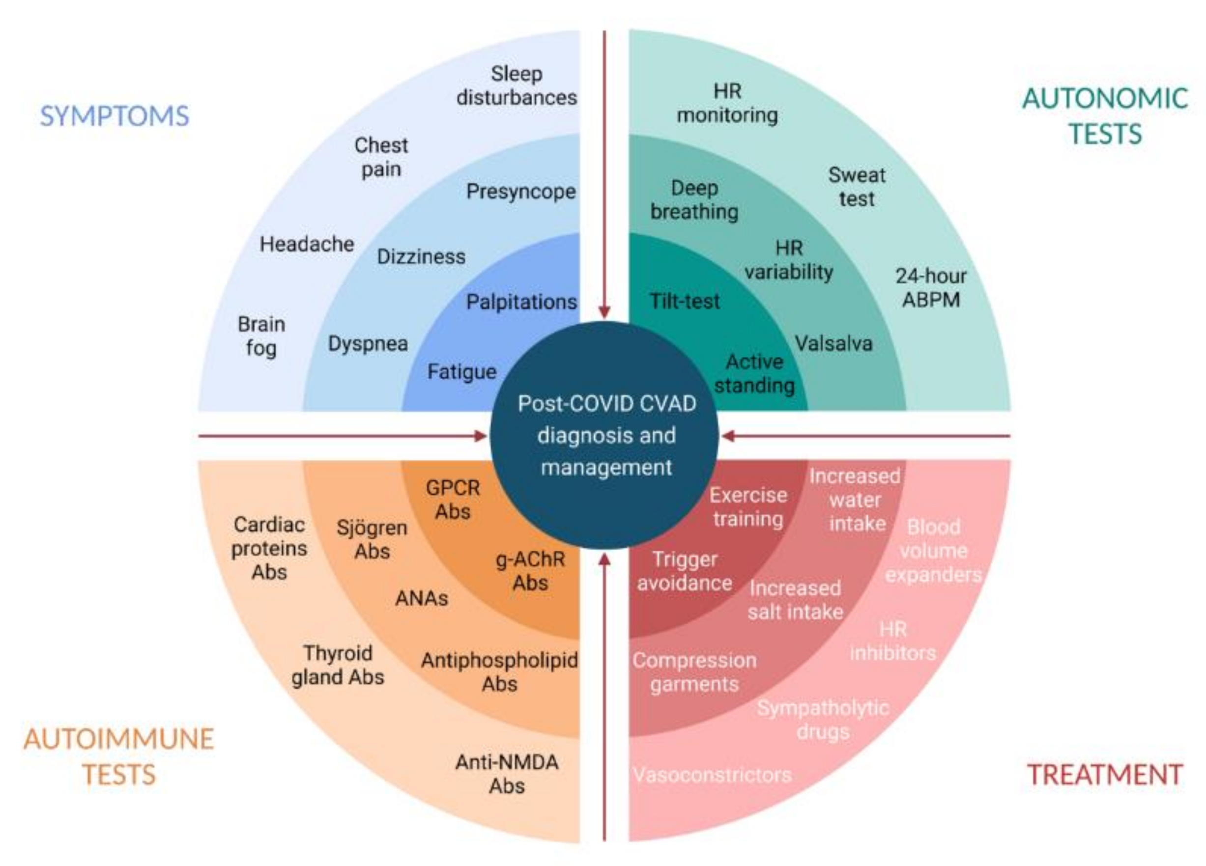
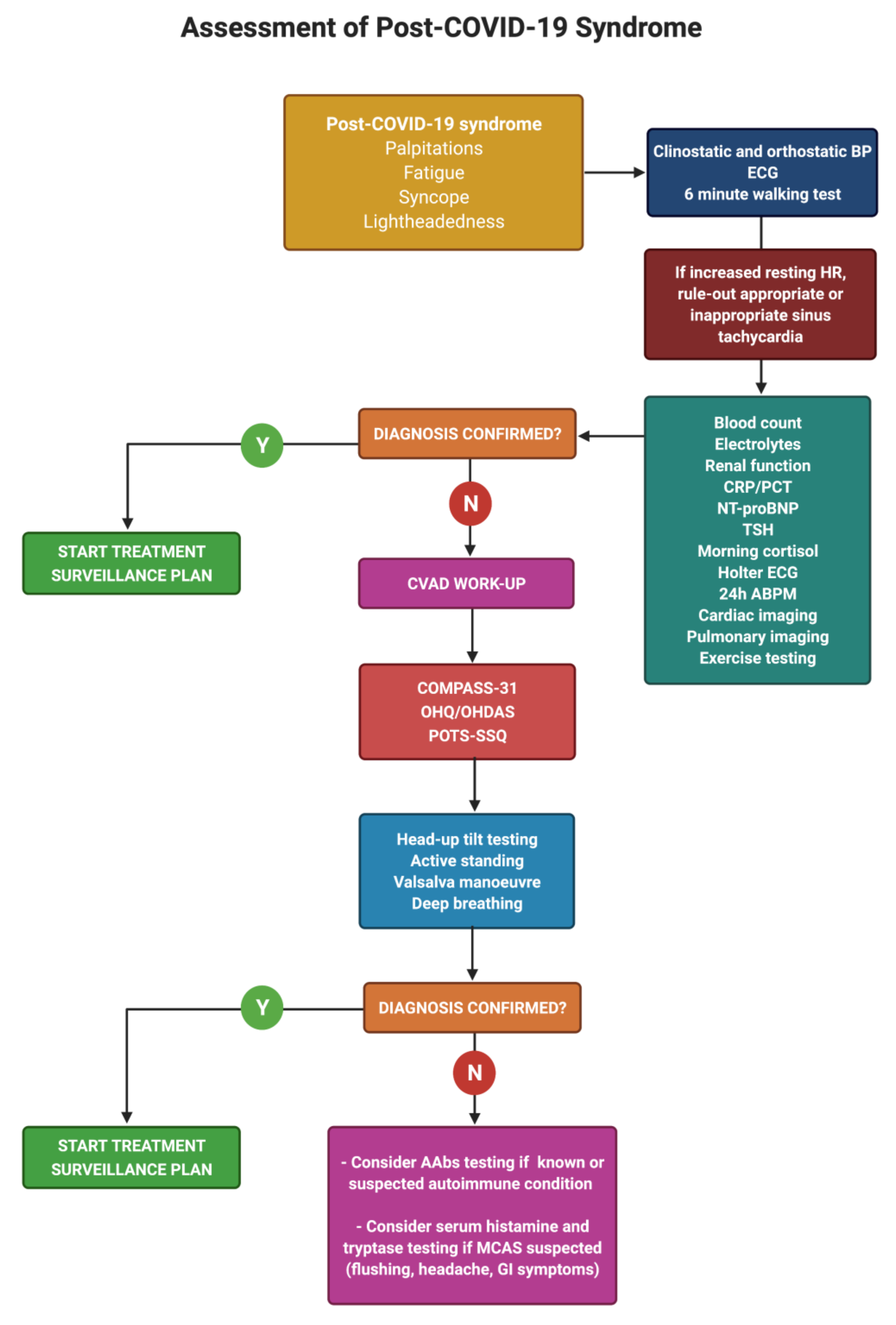
7. Diagnosis of Post-COVID Dysautonomia

The diagnosis of post-COVID dysautonomia requires the comprehensive assessment of previous medical history, the careful evaluation and characterization of symptoms, and expert interpretation of neurological and cardiovascular autonomic investigations [4,16,83].
Patients’ medical histories and their recollection of symptoms are essential in detecting the presence of dysautonomia [84]. The histories of SARS-CoV-2 survivors with persistent autonomic dysfunction may reveal frequent fainting episodes, dizziness, lightheadedness, and/or palpitations [63] prior to SARS-CoV-2 infection, revealing underlying hypotensive susceptibility or previous orthostatic intolerance syndromes. Frequent comorbidities include migraine, irritable bowel syndrome, chronic fatigue syndrome, and autoimmune diseases [73,77]. Lightheadedness, tachycardia, dyspnea, headache, and poor concentration are the most frequent symptoms in PASC patients. Fatigue is the single most frequent symptom, although it is not specific for dysautonomia [33]. Severe symptoms may last for weeks or even months after infection and negatively affect overall quality of life [23].
In post-COVID patients with suspected dysautonomia, the Composite Autonomic Symptom Scale 31 (COMPASS-31) questionnaire can be a sensitive tool to test the likelihood of autonomic dysfunction [85]. This questionnaire has been previously implemented in COVID-19 survivors, showing significantly higher scores than controls [86] with an optimal cut-point for rule-out of CVAD of 13.25 [86,87].
The differential diagnosis of fatigue, dyspnea, palpitations and sinus tachycardia should be carefully scrutinized in order to identify and treat common medical conditions overlapping with long COVID illness. Blood tests (including complete blood count, renal function, B-type natriuretic peptide, electrolytes, thyroid stimulating hormone, and morning cortisol), resting 12-lead ECG, supine and upright blood pressure, and the 6-min walking test should be routinely evaluated [88]. Cardiothoracic imaging (chest X-ray, chest CT, echocardiography, and cardiac magnetic resonance) and exercise testing are invaluable diagnostic tools in investigating PACS and COVID-19 complications.
When cardiovascular autonomic dysfunction is suspected, autonomic tests can be indicated to confirm the diagnosis (Figure 2).
Active standing and/or head-up tilt tests can be very useful in evaluating PASC patients (Table 3), especially among individuals with inappropriate/orthostatic tachycardia, unexplained syncope, or syndromes of orthostatic intolerance [88,89]. Other autonomic investigations include 24 h ambulatory blood pressure monitoring and ECG monitoring [16,33], heart rate variability, Valsalva maneuver, deep breathing [58], and sweat function testing [25,90].
PASC patients with POTS-like symptoms feature a high prevalence of circulating AAbs, at times with peculiar patterns [57] (Figure 1). Although neither sensitive nor specific, AAbs testing can be helpful in selected cases. Specific tests for mast cell activation syndrome can be also considered in PASC patients with flushing episodes, frequent headaches, and persistent gastrointestinal symptoms.

8. Management of Post-COVID Dysautonomia

Once a diagnosis of CVAD is obtained, several treatment options are available, although specific disease-modifying therapies are still limited and represent an unmet need [49].
Non-pharmacological measures should be considered as first-line treatment options and include physical reconditioning with aerobic progressive exercise training programs, the use of compression garments, the liberal intake of water and salt, drinking water before getting up in the morning, sleeping with the head of the bed elevated, the careful avoidance of situations that can exacerbate symptoms (sleep deprivation, heat exposure, alcohol intake, or large or heavy meals) [65,91]. Physical maneuvers such as leg crossing, muscle tensing, and squatting have been shown to be effective in delaying/preventing vasovagal syncope if used at the onset of prodromal symptoms.
Pharmacological therapies have been frequently used in PASC patients (Table 3). These should be reserved for patients which do not respond to nonpharmacological therapies and are complementary to nonpharmacological measures in patients with severe, refractory symptoms. Recommended drugs have also been extensively used where symptoms persist [10,16]. These include volume expanders (fludrocortisone, desmopressin, and intravenous saline), heart rate inhibitors (propranolol, ivabradine, and pyridostigmine), vasoconstrictors (midodrine, octreotide, methylphenidate, and droxidopa) and sympatholytic drugs (clonidine and methyldopa) [10,16].
Decisions regarding which treatment to initiate should be guided by specific symptoms and hemodynamic patterns, i.e., tachycardic vs. hypotensive phenotypes.
The tachycardic phenotype can be treated with beta-blockers, i.e., metoprolol, or ivabradine [88]. Recently, intravenous metoprolol has been tested for use in the treatment of acute respiratory distress in acute COVID-19 and was found to improve oxygenation and reduce alveolar inflammation, shortening the duration of invasive mechanical ventilation overall [92]. Large-scale randomized evidence is eagerly awaited to confirm alleged pleiotropic anti-inflammatory effects of metoprolol [93] beyond its established safety, efficacy, and indications [94].
In patients with the hypotensive phenotype, droxidopa, midodrine, or pyridostigmine may be considered. In hypovolemic patients, intravenous saline infusion and intravascular volume expansion would be highly desirable, while the use of fludrocortisone and desmopressin should be reserved to patients with severe refractory symptoms. Sympatholytic drugs, such as clonidine and methyldopa, can be proposed to patients with hyperadrenergic features, including hyperhidrosis and tachycardia [46].
Beyond these drugs, immunological therapy with intravenous immunoglobulins has been proposed for compassionate use in a patient with autoimmune features [51], leading to the regression of brain fog and the improvement of fatigue and headache. However, further studies are needed to test the rationale, safety, and efficacy of specific immunomodulators/immunosuppressant agents in PASC patients with refractory CVAD-related symptoms.

Author Contributions

Conceptualization, S.G., F.R. and A.F.; methodology, G.I., V.R., A.S. and F.R.; writing—original draft preparation, G.B. and F.R.; writing—review and editing, F.R., M.S., G.I., A.S., A.A.C. and M.Y.K.; supervision, A.F., M.S., S.G., F.R., A.A.C. and M.Y.K. All authors have read and agreed to the published version of the manuscript.

Funding

This research received no external funding.

Institutional Review Board Statement

Not applicable.

Informed Consent Statement

Not applicable.

Data Availability Statement

Not applicable.

Conflicts of Interest

The authors declare no conflict of interest.

References

  1. WHO. WHO COVID-19 Dashboard. 2020. Available online: https://covid19.who.int/ (accessed on 2 September 2021).
  2. Al-Aly, Z.; Xie, Y.; Bowe, B. High-dimensional characterization of post-acute sequalae of COVID-19. Nature 2021. [Google Scholar] [CrossRef]
  3. Rando, H.M.; Bennett, T.D.; Byrd, J.B.; Bramante, C.; Callahan, T.J.; Chute, C.G.; Davis, H.E.; Deer, R.; Gagnier, J.; Koraishy, F.M.; et al. Challenges in defining Long COVID: Striking differences across literature, Electronic Health Records, and patient-reported information. medRxiv 2021. [Google Scholar] [CrossRef]
  4. Raj, S.R.; Arnold, A.C.; Barboi, A.; Claydon, V.E.; Limberg, J.K.; Lucci, V.M.; Numan, M.; Peltier, A.; Snapper, H.; Vernino, S.; et al. Long-COVID postural tachycardia syndrome: An American Autonomic Society statement. Clin. Auton. Res. 2021. [Google Scholar] [CrossRef]
  5. Kalter, L.; WebMD Health News. Fauci Introduces New Acronym for Long COVID at White House Briefing. Medscape. 2021. Available online: https://www.medscape.com/viewarticle/946419 (accessed on 1 June 2021).
  6. Greenhalgh, T.; Knight, M.; A’Court, C.; Buxton, M.; Husain, L. Management of post-acute covid-19 in primary care. BMJ 2020, 370, m3026. [Google Scholar] [CrossRef]
  7. Nalbandian, A.; Sehgal, K.; Gupta, A.; Madhavan, M.V.; McGroder, C.; Stevens, J.S.; Cook, J.R.; Nordvig, A.S.; Shalev, D.; Sehrawat, T.S.; et al. Post-acute COVID-19 syndrome. Nat. Med. 2021, 27, 601–615. [Google Scholar] [CrossRef] [PubMed]
  8. Carfi, A.; Bernabei, R.; Landi, F. Persistent Symptoms in Patients After Acute COVID-19. JAMA 2020, 324, 603–605. [Google Scholar] [CrossRef]
  9. Davido, B.; Seang, S.; Tubiana, R.; de Truchis, P. Post-COVID-19 chronic symptoms: A postinfectious entity? Clin. Microbiol. Infect. 2020, 26, 1448–1449. [Google Scholar] [CrossRef] [PubMed]
  10. Johansson, M.; Stahlberg, M.; Runold, M.; Nygren-Bonnier, M.; Nilsson, J.; Olshansky, B.; Bruchfeld, J.; Fedorowski, A. Long-Haul Post-COVID-19 Symptoms Presenting as a Variant of Postural Orthostatic Tachycardia Syndrome: The Swedish Experience. JACC Case Rep. 2021. [Google Scholar] [CrossRef] [PubMed]
  11. Nath, A. Long-Haul COVID. Neurology 2020, 95, 559–560. [Google Scholar] [CrossRef]
  12. Clark, D.V.; Kibuuka, H.; Millard, M.; Wakabi, S.; Lukwago, L.; Taylor, A.; Eller, M.A.; Eller, L.A.; Michael, N.L.; Honko, A.N.; et al. Long-term sequelae after Ebola virus disease in Bundibugyo, Uganda: A retrospective cohort study. Lancet Infect. Dis. 2015, 15, 905–912. [Google Scholar] [CrossRef] [Green Version]
  13. Lo, Y.L. COVID-19, fatigue, and dysautonomia. J. Med. Virol. 2021, 93, 1213. [Google Scholar] [CrossRef]
  14. O’Sullivan, O. Long-term sequelae following previous coronavirus epidemics. Clin. Med. 2021, 21, e68–e70. [Google Scholar] [CrossRef]
  15. Cheng, D.; Calderwood, C.; Skyllberg, E.; Ainley, A. Clinical characteristics and outcomes of adult patients admitted with COVID-19 in East London: A retrospective cohort analysis. BMJ Open Respir. Res. 2021, 8, e000813. [Google Scholar] [CrossRef]
  16. Dani, M.; Dirksen, A.; Taraborrelli, P.; Torocastro, M.; Panagopoulos, D.; Sutton, R.; Lim, P.B. Autonomic dysfunction in ‘long COVID’: Rationale, physiology and management strategies. Clin. Med. 2021, 21, e63–e67. [Google Scholar] [CrossRef] [PubMed]
  17. Ludvigsson, J.F. Case report and systematic review suggest that children may experience similar long-term effects to adults after clinical COVID-19. Acta Paediatr. 2021, 110, 914–921. [Google Scholar] [CrossRef] [PubMed]
  18. Ostergaard, L. SARS CoV-2 related microvascular damage and symptoms during and after COVID-19: Consequences of capillary transit-time changes, tissue hypoxia and inflammation. Physiol. Rep. 2021, 9, e14726. [Google Scholar] [CrossRef] [PubMed]
  19. Theoharides, T.C.; Cholevas, C.; Polyzoidis, K.; Politis, A. Long-COVID syndrome-associated brain fog and chemofog: Luteolin to the rescue. Biofactors 2021, 47, 232–241. [Google Scholar] [CrossRef] [PubMed]
  20. Mazza, M.G.; De Lorenzo, R.; Conte, C.; Poletti, S.; Vai, B.; Bollettini, I.; Melloni, E.M.T.; Furlan, R.; Ciceri, F.; Rovere-Querini, P.; et al. Anxiety and depression in COVID-19 survivors: Role of inflammatory and clinical predictors. Brain Behav. Immun. 2020, 89, 594–600. [Google Scholar] [CrossRef] [PubMed]
  21. Dennis, A.; Wamil, M.; Alberts, J.; Oben, J.; Cuthbertson, D.J.; Wootton, D.; Crooks, M.; Gabbay, M.; Brady, M.; Hishmeh, L.; et al. Multiorgan impairment in low-risk individuals with post-COVID-19 syndrome: A prospective, community-based study. BMJ Open 2021, 11, e048391. [Google Scholar] [CrossRef]
  22. Osikomaiya, B.; Erinoso, O.; Wright, K.O.; Odusola, A.O.; Thomas, B.; Adeyemi, O.; Bowale, A.; Adejumo, O.; Falana, A.; Abdus-Salam, I.; et al. ‘Long COVID’: Persistent COVID-19 symptoms in survivors managed in Lagos State, Nigeria. BMC Infect. Dis. 2021, 21, 304. [Google Scholar] [CrossRef]
  23. Peluso, M.J.; Kelly, J.D.; Lu, S.; Goldberg, S.A.; Davidson, M.C.; Mathur, S.; Durstenfeld, M.S.; Spinelli, M.A.; Hoh, R.; Tai, V.; et al. Rapid implementation of a cohort for the study of post-acute sequelae of SARS-CoV-2 infection/COVID-19. medRxiv 2021. [Google Scholar] [CrossRef]
  24. Perlis, R.H.; Green, J.; Santillana, M.; Lazer, D.; Ognyanova, K.; Simonson, M.; Baum, M.A.; Quintana, A.; Chwe, H.; Druckman, J.; et al. Persistence of symptoms up to 10 months following acute COVID-19 illness. medRxiv 2021. [Google Scholar] [CrossRef]
  25. Shouman, K.; Vanichkachorn, G.; Cheshire, W.P.; Suarez, M.D.; Shelly, S.; Lamotte, G.J.; Sandroni, P.; Benarroch, E.E.; Berini, S.E.; Cutsforth-Gregory, J.K.; et al. Autonomic dysfunction following COVID-19 infection: An early experience. Clin. Auton. Res. 2021. [Google Scholar] [CrossRef]
  26. Gaber, T.A.K.; Ashish, A.; Unsworth, A. Persistent post-covid symptoms in healthcare workers. Occup. Med. 2021. [Google Scholar] [CrossRef] [PubMed]
  27. Townsend, L.; Dyer, A.H.; Jones, K.; Dunne, J.; Mooney, A.; Gaffney, F.; O’Connor, L.; Leavy, D.; O’Brien, K.; Dowds, J.; et al. Persistent fatigue following SARS-CoV-2 infection is common and independent of severity of initial infection. PLoS ONE 2020, 15, e0240784. [Google Scholar] [CrossRef]
  28. Auwaerter, P.G. The Race to Understand Post–COVID-19 Conditions. Ann. Intern. Med. 2021. [Google Scholar] [CrossRef]
  29. Petersen, M.S.; Kristiansen, M.F.; Hanusson, K.D.; Danielsen, M.E.; Gaini, S.; Strom, M.; Weihe, P. Long COVID in the Faroe Islands—a longitudinal study among non-hospitalized patients. Clin. Infect. Dis. 2020. [Google Scholar] [CrossRef]
  30. Moreno-Perez, O.; Merino, E.; Leon-Ramirez, J.M.; Andres, M.; Ramos, J.M.; Arenas-Jimenez, J.; Asensio, S.; Sanchez, R.; Ruiz-Torregrosa, P.; Galan, I.; et al. Post-acute COVID-19 syndrome. Incidence and risk factors: A Mediterranean cohort study. J. Infect. 2021, 82, 378–383. [Google Scholar] [CrossRef]
  31. Evans, R.A.; McAuley, H.; Harrison, E.M.; Shikotra, A.; Singapuri, A.; Sereno, M.; Elneima, O.; Docherty, A.B.; Lone, N.I.; Leavy, O.C.; et al. Physical, cognitive and mental health impacts of COVID-19 following hospitalisation—A multi-centre prospective cohort study. medRxiv 2021. [Google Scholar] [CrossRef]
  32. Halpin, S.J.; McIvor, C.; Whyatt, G.; Adams, A.; Harvey, O.; McLean, L.; Walshaw, C.; Kemp, S.; Corrado, J.; Singh, R.; et al. Postdischarge symptoms and rehabilitation needs in survivors of COVID-19 infection: A cross-sectional evaluation. J. Med. Virol. 2021, 93, 1013–1022. [Google Scholar] [CrossRef]
  33. Townsend, L.; Moloney, D.; Finucane, C.; McCarthy, K.; Bergin, C.; Bannan, C.; Kenny, R.-A. Fatigue following COVID-19 infection is not associated with autonomic dysfunction. PLoS ONE 2021, 16, e0247280. [Google Scholar] [CrossRef] [PubMed]
  34. van Campen, C.; Verheugt, F.W.A.; Rowe, P.C.; Visser, F.C. Cerebral blood flow is reduced in ME/CFS during head-up tilt testing even in the absence of hypotension or tachycardia: A quantitative, controlled study using Doppler echography. Clin. Neurophysiol. Pract. 2020, 5, 50–58. [Google Scholar] [CrossRef]
  35. Escorihuela, R.M.; Capdevila, L.; Castro, J.R.; Zaragozà, M.C.; Maurel, S.; Alegre, J.; Castro-Marrero, J. Reduced heart rate variability predicts fatigue severity in individuals with chronic fatigue syndrome/myalgic encephalomyelitis. J. Transl. Med. 2020, 18, 4. [Google Scholar] [CrossRef]
  36. Nelson, M.J.; Bahl, J.S.; Buckley, J.D.; Thomson, R.L.; Davison, K. Evidence of altered cardiac autonomic regulation in myalgic encephalomyelitis/chronic fatigue syndrome: A systematic review and meta-analysis. Medicine 2019, 98, e17600. [Google Scholar] [CrossRef]
  37. Kedor, C.; Freitag, H.; Meyer-Arndt, L.; Wittke, K.; Zoller, T.; Steinbeis, F.; Haffke, M.; Rudolf, G.; Heidecker, B.; Volk, H.; et al. Chronic COVID-19 Syndrome and Chronic Fatigue Syndrome (ME/CFS) following the first pandemic wave in Germany—a first analysis of a prospective observational study. medRxiv 2021. [Google Scholar] [CrossRef]
  38. Sigfrid, L.; Drake, T.M.; Pauley, E.; Jesudason, E.C.; Olliaro, P.; Lim, W.S.; Gillesen, A.; Berry, C.; Lowe, D.J.; McPeake, J.; et al. Long Covid in adults discharged from UK hospitals after COVID-19: A prospective, multicentre cohort study using the ISARIC WHO Clinical Characterisation Protocol. medRxiv 2021. [Google Scholar] [CrossRef]
  39. Ortona, E.; Buonsenso, D.; Carfi, A.; Malorni, W.; The Long Covid Kids study group. Long COVID: An estrogen-associated autoimmune disease? Cell Death Discov. 2021, 7, 77. [Google Scholar] [CrossRef] [PubMed]
  40. Funk, A.L.; Florin, T.A.; Dalziel, S.R.; Mintegi, S.; Salvadori, M.I.; Tancredi, D.J.; Neuman, M.I.; Payne, D.C.; Plint, A.C.; Klassen, T.P.; et al. Prospective cohort study of children with suspected SARS-CoV-2 infection presenting to paediatric emergency departments: A Paediatric Emergency Research Networks (PERN) Study Protocol. BMJ Open 2021, 11, e042121. [Google Scholar] [CrossRef] [PubMed]
  41. Petracek, L.S.; Suskauer, S.J.; Vickers, R.F.; Patel, N.R.; Violand, R.L.; Swope, R.L.; Rowe, P.C. Adolescent and Young Adult ME/CFS After Confirmed or Probable COVID-19. Front. Med. 2021, 8, 525. [Google Scholar] [CrossRef]
  42. Stephenson, T.; Stephenson, T.; Pereira, S.P.; Shafran, R.; De Stavola, B.; Rojas, N.; McOwat, K.; Simmons, R.; Zavala, M.; O’Mahoney, L.; et al. Long COVID—The physical and mental health of children and non-hospitalised young people 3 months after SARS-CoV-2 infection; a national matched cohort study (The CLoCk) Study. Nat. Portf. 2021. [Google Scholar] [CrossRef]
  43. Ghosh, R.; Roy, D.; Sengupta, S.; Benito-Leon, J. Autonomic dysfunction heralding acute motor axonal neuropathy in COVID-19. J. Neurovirol. 2020, 26, 964–966. [Google Scholar] [CrossRef] [PubMed]
  44. Kanjwal, K.; Jamal, S.; Kichloo, A.; Grubb, B.P. New-onset Postural Orthostatic Tachycardia Syndrome Following Coronavirus Disease 2019 Infection. J. Innov. Card Rhythm. Manag. 2020, 11, 4302–4304. [Google Scholar] [CrossRef] [PubMed]
  45. Moreno-Escobar, M.C.; Kataria, S.; Khan, E.; Subedi, R.; Tandon, M.; Peshwe, K.; Kramer, J.; Niaze, F.; Sriwastava, S. Acute transverse myelitis with Dysautonomia following SARS-CoV-2 infection: A case report and review of literature. J. Neuroimmunol. 2021, 353, 577523. [Google Scholar] [CrossRef]
  46. Umapathi, T.; Poh, M.Q.W.; Fan, B.E.; Li, K.F.C.; George, J.; Tan, J.Y.L. Acute hyperhidrosis and postural tachycardia in a COVID-19 patient. Clin. Auton. Res. 2020, 30, 571–573. [Google Scholar] [CrossRef] [PubMed]
  47. Loa, Y.L.; Leonga, H.N.; Hsua, L.Y.; Tana, T.T.; Kurupa, A.; Fook-Chonga, S.; Tana, B.H. Autonomic Dysfunction in Recovered Severe Acute Respiratory Syndrome Patients. Can. J. Neurol. Sci. 2005, 32, 264. [Google Scholar] [CrossRef] [Green Version]
  48. Ståhlberg, M.; Reistam, U.; Fedorowski, A.; Villacorta, H.; Horiuchi, Y.; Bax, J.; Pitt, B.; Matskeplishvili, S.; Lüscher, T.F.; Weichert, I.; et al. Post-COVID-19 Tachycardia Syndrome: A distinct phenotype of Post-acute COVID-19 Syndrome. Am. J. Med. 2021. [Google Scholar] [CrossRef]
  49. Fedorowski, A. Postural orthostatic tachycardia syndrome: Clinical presentation, aetiology and management. J. Intern. Med. 2019, 285, 352–366. [Google Scholar] [CrossRef]
  50. Miglis, M.G.; Prieto, T.; Shaik, R.; Muppidi, S.; Sinn, D.I.; Jaradeh, S. A case report of postural tachycardia syndrome after COVID-19. Clin. Auton. Res. 2020, 30, 449–451. [Google Scholar] [CrossRef]
  51. Novak, P. Post COVID-19 syndrome associated with orthostatic cerebral hypoperfusion syndrome, small fiber neuropathy and benefit of immunotherapy: A case report. eNeurologicalSci 2020, 21, 100276. [Google Scholar] [CrossRef]
  52. Schofield, J.R. Persistent Antiphospholipid Antibodies, Mast Cell Activation Syndrome, Postural Orthostatic Tachycardia Syndrome and Post-COVID Syndrome: 1 Year On. Eur. J. Case Rep. Intern. Med. 2021, 8, 002378. [Google Scholar] [CrossRef]
  53. Huang, C.; Huang, L.; Wang, Y.; Li, X.; Ren, L.; Gu, X.; Kang, L.; Guo, L.; Liu, M.; Zhou, X.; et al. 6-month consequences of COVID-19 in patients discharged from hospital: A cohort study. Lancet 2021, 397, 220–232. [Google Scholar] [CrossRef]
  54. Davis, H.E.; Assaf, G.S.; McCorkell, L.; Wei, H.; Low, R.J.; Re’em, Y.; Redfield, S.; Austin, J.P.; Akrami, A. Characterizing Long COVID in an International Cohort: 7 Months of Symptoms and Their Impact. medRxiv 2021. [Google Scholar] [CrossRef]
  55. Owens, A.P.; Low, D.A.; Iodice, V.; Critchley, H.D.; Mathias, C.J. The genesis and presentation of anxiety in disorders of autonomic overexcitation. Auton. Neurosci. 2017, 203, 81–87. [Google Scholar] [CrossRef]
  56. Blitshteyn, S. Is postural orthostatic tachycardia syndrome (POTS) a central nervous system disorder? J. Neurol. 2021, 1–8. [Google Scholar] [CrossRef] [PubMed]
  57. Wallukat, G.; Hohberger, B.; Wenzel, K.; Fürst, J.; Schulze-Rothe, S.; Wallukat, A.; Hönicke, A.S.; Müller, J. Functional autoantibodies against G-protein coupled receptors in patients with persistent post-COVID-19 symptoms. J. Transl. Autoimmun. 2021, 4, 100100. [Google Scholar] [CrossRef] [PubMed]
  58. Goodman, B.P.; Khoury, J.A.; Blair, J.E.; Grill, M.F. COVID-19 Dysautonomia. Front. Neurol. 2021, 12, 624968. [Google Scholar] [CrossRef]
  59. Kaliyaperumal, D.; Rk, K.; Alagesan, M.; Ramalingam, S. Characterization of cardiac autonomic function in COVID-19 using heart rate variability: A hospital based preliminary observational study. J. Basic Clin. Physiol. Pharmacol. 2021, 32, 247–253. [Google Scholar] [CrossRef]
  60. Romero-Sanchez, C.M.; Diaz-Maroto, I.; Fernandez-Diaz, E.; Sanchez-Larsen, A.; Layos-Romero, A.; Garcia-Garcia, J.; Gonzalez, E.; Redondo-Penas, I.; Perona-Moratalla, A.B.; Del Valle-Perez, J.A.; et al. Neurologic manifestations in hospitalized patients with COVID-19: The ALBACOVID registry. Neurology 2020, 95, e1060–e1070. [Google Scholar] [CrossRef]
  61. Logmin, K.; Karam, M.; Schichel, T.; Harmel, J.; Wojtecki, L. Non-epileptic seizures in autonomic dysfunction as the initial symptom of COVID-19. J. Neurol. 2020, 267, 2490–2491. [Google Scholar] [CrossRef]
  62. Eshak, N.; Abdelnabi, M.; Ball, S.; Elgwairi, E.; Creed, K.; Test, V.; Nugent, K. Dysautonomia: An Overlooked Neurological Manifestation in a Critically ill COVID-19 Patient. Am. J. Med. Sci. 2020, 360, 427–429. [Google Scholar] [CrossRef]
  63. Shaw, B.H.; Stiles, L.E.; Bourne, K.; Green, E.A.; Shibao, C.A.; Okamoto, L.E.; Garland, E.M.; Gamboa, A.; Diedrich, A.; Raj, V.; et al. The face of postural tachycardia syndrome—Insights from a large cross-sectional online community-based survey. J. Intern. Med. 2019, 286, 438–448. [Google Scholar] [CrossRef] [Green Version]
  64. Goldstein, D.S. The possible association between COVID-19 and postural tachycardia syndrome. Heart Rhythm 2021, 18, 508–509. [Google Scholar] [CrossRef] [PubMed]
  65. Bryarly, M.; Phillips, L.T.; Fu, Q.; Vernino, S.; Levine, B.D. Postural Orthostatic Tachycardia Syndrome: JACC Focus Seminar. J. Am. Coll. Cardiol. 2019, 73, 1207–1228. [Google Scholar] [CrossRef] [PubMed]
  66. Yong, S.J. Persistent Brainstem Dysfunction in Long-COVID: A Hypothesis. ACS Chem. Neurosci. 2021, 12, 573–580. [Google Scholar] [CrossRef]
  67. Lionetti, V.; Bollini, S.; Coppini, R.; Gerbino, A.; Ghigo, A.; Iaccarino, G.; Madonna, R.; Mangiacapra, F.; Miragoli, M.; Moccia, F.; et al. Understanding the heart-brain axis response in COVID-19 patients: A suggestive perspective for therapeutic development. Pharmacol. Res. 2021, 168, 105581. [Google Scholar] [CrossRef]
  68. Barnden, L.R.; Crouch, B.; Kwiatek, R.; Burnet, R.; Mernone, A.; Chryssidis, S.; Scroop, G.; Del Fante, P. A brain MRI study of chronic fatigue syndrome: Evidence of brainstem dysfunction and altered homeostasis. NMR Biomed. 2011, 24, 1302–1312. [Google Scholar] [CrossRef] [PubMed]
  69. Saeed, S.; Tadic, M.; Larsen, T.H.; Grassi, G.; Mancia, G. Coronavirus disease 2019 and cardiovascular complications: Focused clinical review. J. Hypertens. 2021, 39, 1282–1292. [Google Scholar] [CrossRef]
  70. Barbieri, L.; Galli, F.; Conconi, B.; Gregorini, T.; Lucreziotti, S.; Mafrici, A.; Pravettoni, G.; Sommaruga, M.; Carugo, S. Takotsubo syndrome in COVID-19 era: Is psychological distress the key? J. Psychosom. Res. 2021, 140, 110297. [Google Scholar] [CrossRef]
  71. Kir, D.; Beer, N.; De Marchena, E.J. Takotsubo cardiomyopathy caused by emotional stressors in the coronavirus disease 2019 (COVID-19) pandemic era. J. Card. Surg. 2021, 36, 764–769. [Google Scholar] [CrossRef]
  72. Suzuki, H.; Yasuda, S.; Shimokawa, H. Brain-heart connection in Takotsubo syndrome before onset. Eur. Heart J. 2021, 42, 1909–1911. [Google Scholar] [CrossRef]
  73. Vernino, S.; Stiles, L.E. Autoimmunity in postural orthostatic tachycardia syndrome: Current understanding. Auton. Neurosci. 2018, 215, 78–82. [Google Scholar] [CrossRef] [PubMed] [Green Version]
  74. Kharraziha, I.; Axelsson, J.; Ricci, F.; Martino, G.D.; Persson, M.; Sutton, R.; Fedorowski, A.; Hamrefors, V. Serum Activity Against G Protein-Coupled Receptors and Severity of Orthostatic Symptoms in Postural Orthostatic Tachycardia Syndrome. J. Am. Heart Assoc. 2020, 9, e015989. [Google Scholar] [CrossRef]
  75. Li, H.; Zhang, G.; Forsythe, E.; Okamoto, L.E.; Yu, X. Implications of Antimuscarinic Autoantibodies in Postural Tachycardia Syndrome. J. Cardiovasc. Transl. Res. 2021. [Google Scholar] [CrossRef] [PubMed]
  76. Molina, V.; Shoenfeld, Y. Infection, vaccines and other environmental triggers of autoimmunity. Autoimmunity 2005, 38, 235–245. [Google Scholar] [CrossRef] [PubMed]
  77. Dotan, A.; Muller, S.; Kanduc, D.; David, P.; Halpert, G.; Shoenfeld, Y. The SARS-CoV-2 as an instrumental trigger of autoimmunity. Autoimmun. Rev. 2021, 20, 102792. [Google Scholar] [CrossRef] [PubMed]
  78. Kohno, R.; Cannom, D.S.; Olshansky, B.; Xi, S.C.; Krishnappa, D.; Adkisson, W.O.; Norby, F.L.; Fedorowski, A.; Benditt, D.G. Mast Cell Activation Disorder and Postural Orthostatic Tachycardia Syndrome: A Clinical Association. J. Am. Heart Assoc. 2021, e021002. [Google Scholar] [CrossRef]
  79. Ayoubkhani, D.; Khunti, K.; Nafilyan, V.; Maddox, T.; Humberstone, B.; Diamond, I.; Banerjee, A. Post-covid syndrome in individuals admitted to hospital with covid-19: Retrospective cohort study. BMJ 2021, 372, n693. [Google Scholar] [CrossRef]
  80. Torjesen, I. COVID-19: Middle aged women face greater risk of debilitating long term symptoms. BMJ 2021, 372, n829. [Google Scholar] [CrossRef] [PubMed]
  81. El Sayed, S.; Shokry, D.; Gomaa, S.M. Post-COVID-19 fatigue and anhedonia: A cross-sectional study and their correlation to post-recovery period. Neuropsychopharmacol. Rep. 2021, 41, 50–55. [Google Scholar] [CrossRef]
  82. Blitshteyn, S.; Whitelaw, S. Postural orthostatic tachycardia syndrome (POTS) and other autonomic disorders after COVID-19 infection: A case series of 20 patients. Immunol. Res. 2021, 69, 205–211. [Google Scholar] [CrossRef] [PubMed]
  83. Figueroa, J.J.; Cheshire, W.P.; Claydon, V.E.; Norcliffe-Kaufmann, L.; Peltier, A.; Singer, W.; Snapper, H.; Vernino, S.; Raj, S.R. Autonomic function testing in the COVID-19 pandemic: An American Autonomic Society position statement. Clin. Auton. Res. 2020, 30, 295–297. [Google Scholar] [CrossRef] [PubMed]
  84. Brignole, M.; Moya, A.; de Lange, F.J.; Deharo, J.-C.; Elliott, P.M.; Fanciulli, A.; Fedorowski, A.; Furlan, R.; Kenny, R.A.; Martín, A.; et al. Practical Instructions for the 2018 ESC Guidelines for the diagnosis and management of syncope. Eur. Heart J. 2018, 39, e43–e80. [Google Scholar] [CrossRef]
  85. Sletten, D.M.; Suarez, G.A.; Low, P.A.; Mandrekar, J.; Singer, W. COMPASS 31: A refined and abbreviated Composite Autonomic Symptom Score. Mayo Clin. Proc. 2012, 87, 1196–1201. [Google Scholar] [CrossRef] [PubMed]
  86. Buoite Stella, A.; Furlanis, G.; Frezza, N.A.; Valentinotti, R.; Ajcevic, M.; Manganotti, P. Autonomic dysfunction in post-COVID patients with and witfhout neurological symptoms: A prospective multidomain observational study. J. Neurol. 2021. [Google Scholar] [CrossRef]
  87. Kim, Y.; Seok, J.; Park, J.; Kim, K.-H.; Min, J.-H.; Cho, J.W.; Park, S.; Kim, H.-J.; Kim, B.; Youn, J. The composite autonomic symptom scale 31 is a useful screening tool for patients with Parkinsonism. PLoS ONE 2017, 12, e0180744. [Google Scholar] [CrossRef] [Green Version]
  88. Raj, S.R.; Guzman, J.C.; Harvey, P.; Richer, L.; Schondorf, R.; Seifer, C.; Thibodeau-Jarry, N.; Sheldon, R.S. Canadian Cardiovascular Society Position Statement on Postural Orthostatic Tachycardia Syndrome (POTS) and Related Disorders of Chronic Orthostatic Intolerance. Can. J. Cardiol. 2020, 36, 357–372. [Google Scholar] [CrossRef] [Green Version]
  89. Thijs, R.D.; Brignole, M.; Falup-Pecurariu, C.; Fanciulli, A.; Freeman, R.; Guaraldi, P.; Jordan, J.; Habek, M.; Hilz, M.; Traon, A.P.; et al. Recommendations for tilt table testing and other provocative cardiovascular autonomic tests in conditions that may cause transient loss of consciousness: Consensus statement of the European Federation of Autonomic Societies (EFAS) endorsed by the American Autonomic Society (AAS) and the European Academy of Neurology (EAN). Clin. Auton. Res. 2021. [Google Scholar] [CrossRef]
  90. Hinduja, A.; Moutairou, A.; Calvet, J.H. Sudomotor dysfunction in patients recovered from COVID-19. Neurophysiol. Clin. 2021, 51, 193–196. [Google Scholar] [CrossRef]
  91. Ricci, F.; De Caterina, R.; Fedorowski, A. Orthostatic Hypotension: Epidemiology, Prognosis, and Treatment. J. Am. Coll. Cardiol. 2015, 66, 848–860. [Google Scholar] [CrossRef] [PubMed] [Green Version]
  92. Clemente-Moragón, A.; Martínez-Milla, J.; Oliver, E.; Santos, A.; Flandes, J.; Fernández, I.; Rodríguez-González, L.; Serrano del Castillo, C.; Ioan, A.-M.; López-Álvarez, M.; et al. Metoprolol in Critically Ill Patients With COVID-19. J. Am. Coll. Cardiol. 2021, 78, 1001–1011. [Google Scholar] [CrossRef] [PubMed]
  93. Clemente-Moragon, A.; Gomez, M.; Villena-Gutierrez, R.; Lalama, D.V.; Garcia-Prieto, J.; Martinez, F.; Sanchez-Cabo, F.; Fuster, V.; Oliver, E.; Ibanez, B. Metoprolol exerts a non-class effect against ischaemia-reperfusion injury by abrogating exacerbated inflammation. Eur. Heart J. 2020, 41, 4425–4440. [Google Scholar] [CrossRef] [PubMed]
  94. Deng, X.; Zhang, Y.; Liao, Y.; Du, J. Efficacy of β-Blockers on Postural Tachycardia Syndrome in Children and Adolescents: A Systematic Review and Meta-Analysis. Front. Pediatr. 2019, 7, 460. [Google Scholar] [CrossRef] [PubMed] [Green Version]
Figure 1. Post-COVID cardiovascular autonomic dysfunction: diagnosis and management. Abs, antibodies; ANAs, antinuclear antibodies; CVAD, cardiovascular autonomic dysfunction; g-AChR, ganglionic neuronal nicotinic acetylcholine receptor; GPCR, G-Protein coupled receptor; HR, heart rate. Created with BioRender.com.
Jcdd 08 00156 g001
Figure 2. Assessment of post-COVID-19 Syndrome. AAbs, autoantibodies; ABPM, ambulatory blood pressure monitoring; BP, blood pressure; COMPASS-31, Composite Autonomic Symptom Scale 31; CRP, C-reactive protein; CVAD, cardiovascular autonomic dysfunction; ECG, electrocardiogram; GI, gastrointestinal; MCAS, mast cell activation syndrome; NT-proBNP, N-terminal prohormone of brain natriuretic peptide; OHDAS, orthostatic hypotension daily activity scale; OHQ, orthostatic hypotension questionnaire; PCT, procalcitonin; POTS-SSQ, postural orthostatic tachycardia syn-drome symptom scoring questionnaire, as in Johannsson and colleagues [10]; TSH, thyroid stimu-lating hormone. Created with BioRender.com.
Jcdd 08 00156 g002
Table 1. Case reports of patients diagnosed with CVAD following COVID-19.
Authors, YearMiglis et al. [50],
2020
Kanjwal et al. [44],
2020
Novak et al. [51],
2020
Umapathi et al. [46], 2020Schofield et al. [52], 2021
Follow-up (months)5.5132.512
Mean age (years)2636643950
SexFFFMF
DiagnosisPOTS, OHPOTSOCHOSPOTSPOTS
Palpitations,
tachycardia
YesYes-Yes-
FatigueYesYesYes--
Vertigo aYesYes---
DyspneaYes---Yes
PresyncopeYes---Yes
Chest painYesYes--Yes
Headache-YesYes--
Brain fog bYes-Yes--
Sleep disturbancesYes----
OCHOS, orthostatic cerebral hypoperfusion syndrome; OH, orthostatic hypotension; POTS, postural orthostatic tachycardia syndrome. All patients were subject to clinical observation. Patients’ previous medical history included exercise-induced asthma, eczema, diffuse urticaria, gastroesophageal reflux disease, hypertension, thyroid disease, diabetes mellitus, headache and/or episodic migraine, Lyme disease, obsessive compulsive disorder, small fiber neuropathy and orthostatic cerebral hypoperfusion syndrome. One patient not reporting significant medical history was described by Kanjwal et al. [44]. a includes all patients reporting vertigo, lightheadedness, and/or dizziness. b includes all patients reporting brain fog and/or difficulty concentrating.
Table 3. COVID-19 related CVAD: overview of diagnostic modalities and strategies of treatment.
Study FeaturesAutonomic Testing ModalityTreatment Strategy
Authors, yearStudy typeHUTActive standingDeep breathingValsalvaSudomotor testHolter
monitoring
NPT aVolume expanders bHR
Inhibitors c
Sympatholytic
Drugs d
IT e
Miglis
et al. [61], 2020
ReportYes--Yes----YesYes-
Kanjwal
et al. [42], 2020
ReportYes-----Yes-Yes--
Novak
et al. [73], 2020
ReportYes-YesYesYes-----Yes
Umapathi
et al. [44], 2020
ReportYesYes----YesYesYes--
Schofield
et al. [63], 2021
Report-Yes----N.A.
Dani
et al. [16], 2021
SeriesYesYes---YesN.A.
Townsend
et al. [32], 2021
Series-YesYesYesYesYesN.A.
Johansson
et al. [10], 2021
SeriesYesYes-Yes--Yes-Yes--
Blitshteyn
et al. [77], 2021
SeriesYesYes----YesYes---
Shouman
et al. [25], 2021
SeriesYes--YesYes-Yes-Yes--
Goodman
et al. [80], 2021
SeriesYes-YesYesYes-N.A.
HR, heart rate; HUT, head-up tilt test; IT, immunotherapy; NFT, non-pharmacological therapy. The case series by Wallukat et al. (#69) is not included in this table, since neither specific testing nor therapy for patients with a diagnosis of dysautonomia were reported. Schofield et al., Dani et al., Townsend et al., and Goodman et al. did not report specific treatment. a Non-pharmacological therapy includes exercise training, withdrawal of exacerbating drugs, increased water intake, increased salt intake, and use of compressive garments; b volume expanders include fludrocortisone, desmopressin, and acute or chronic use of intravenous saline solution; c HR inhibitors include propranolol, ivabradine, and pyridostigmine; d sympatholytic drugs include clonidine and methyldopa; e immunotherapy consists of intravenous administration of immunoglobulins.
Publisher’s Note: MDPI stays neutral with regard to jurisdictional claims in published maps and institutional affiliations.

