Review

The Anatomy, Development, and Evolution of the Atrioventricular Conduction Axis

1
Institute of Genetic Medicine, Newcastle University, Newcastle upon Tyne NE1 4EP, UK
2
Division of Cardiovascular Medicine, Department of Internal Medicine, Kobe University Graduate School of Medicine, 7-5-1 Kusunoki-cho, Chuo-ku, Kobe 650-0017, Hyogo, Japan
3
Department of Pediatric Cardiology, University of Florida, Gainesville, FL 32610, USA
4
Department of Anatomy and Cell Biology, Faculty of Medicine, University of Extremadura, 06006 Badajoz, Spain
5
University of Amsterdam, Amsterdam UMC, Department of Medical Biology, Amsterdam Cardiovascular Sciences, Meibergdreef 15, 1105AZ Amsterdam, The Netherlands
*
Author to whom correspondence should be addressed.
J. Cardiovasc. Dev. Dis. 2018, 5(3), 44; https://doi.org/10.3390/jcdd5030044
Submission received: 10 August 2018 / Revised: 16 August 2018 / Accepted: 19 August 2018 / Published: 22 August 2018

Abstract

:
It is now well over 100 years since Sunao Tawara clarified the location of the axis of the specialised myocardium responsible for producing coordinated ventricular activation. Prior to that stellar publication, controversies had raged as to how many bundles crossed the place of the atrioventricular insulation as found in mammalian hearts, as well as the very existence of the bundle initially described by Wilhelm His Junior. It is, perhaps surprising that controversies continue, despite the multiple investigations that have taken place since the publication of Tawara’s monograph. For example, we are still unsure as to the precise substrates for the so-called slow and fast pathways into the atrioventricular node. Much has been done, nonetheless, to characterise the molecular make-up of the specialised pathways, and to clarify their mechanisms of development. Of this work itself, a significant part has emanated from the laboratory coordinated for a quarter of a century by Antoon FM Moorman. In this review, which joins the others in recognising the value of his contributions and collaborations, we review our current understanding of the anatomy, development, and evolution of the atrioventricular conduction axis.

1. Introduction

The identification of the atrioventricular conduction axis by Sunao Tawara, in 1906 [1] ushered in the correct understanding of cardiac conduction [2,3]. As was stated by Keith, stimulated by the findings, and who together with Flack was shortly after to identify the sinus node [4], “with the discovery of the conduction system of Tawara, heart research entered a new epoch” [5]. Prior to Tawara’s publication, His had already described a connecting muscular bundle crossing the plane of atrioventricular insulation [6]. The situation had been clouded, however, by the counterclaim of Kent that multiple myocardial pathways crossed the atrioventricular junctions [7]. Prior to these studies, furthermore, which were carried out in the last decade of the 19th century, it had generally been believed that atrioventricular conduction was neurogenic rather than myogenic, although the experiments of Gaskell had already provided evidence in favor of the myogenic concept [8]. It was the study of Tawara that clarified once and for all the morphology of the solitary myocardial pathway responsible for normal atrioventricular conduction. It is surprising, however, that Keith was unable to identify the conduction axis on the basis of the illustration provided by His (Figure 1A).
Tawara studied multiple mammalian species and showed in greater detail how the pathway commenced in the atrioventricular node (Figure 1B), which is located in the base of the atrial septum. It then extended through the insulating place, and branched on the crest of the muscular ventricular septum. The branches were insulated by fibrous sheaths, which surrounded the fascicles before they ramified at the ventricular apices as the so-called “Purkinje” network [1]. It is surprising, given the excellence of these initial descriptions, that uncertainty continues to exist regarding the precise correlation between the anatomy and function of the atrioventricular node. We still do not know, for example, whether there are dual or multiple anatomical pathways through the node. If there are dual pathways, we have yet to identify the precise location and substrates for the so-called “fast” and “slow” options. With regard to development, the studies of Wessels and colleagues showed that the core of the node was derived from a ring of histologically and immunocytochemically specialized cells surrounding the embryonic interventricular foramen [9]. The significance of the derivatives of other parts of this ring, such as the so-called “retroaortic node”, has still to be established [10]. In particular, we remain uncertain regarding the mode of development of the atrial connections of the node. The potential influence of the vestibular spine, described by the father of the discoverer of the atrioventricular bundle [11] remains unexplored. Evidence is also now emerging of the evolutionary origin of the atrioventricular bundle in reptiles and other ectothermic vertebrates [12]. Taking advantage of our experiences in these various fields, therefore, we review here the anatomy, development, and evolution of the atrioventricular conduction axis.

2. The Location and Architecture of the Normal Atrioventricular Conduction Axis

2.1. The Structure of the Normal Atrioventricular Junctions

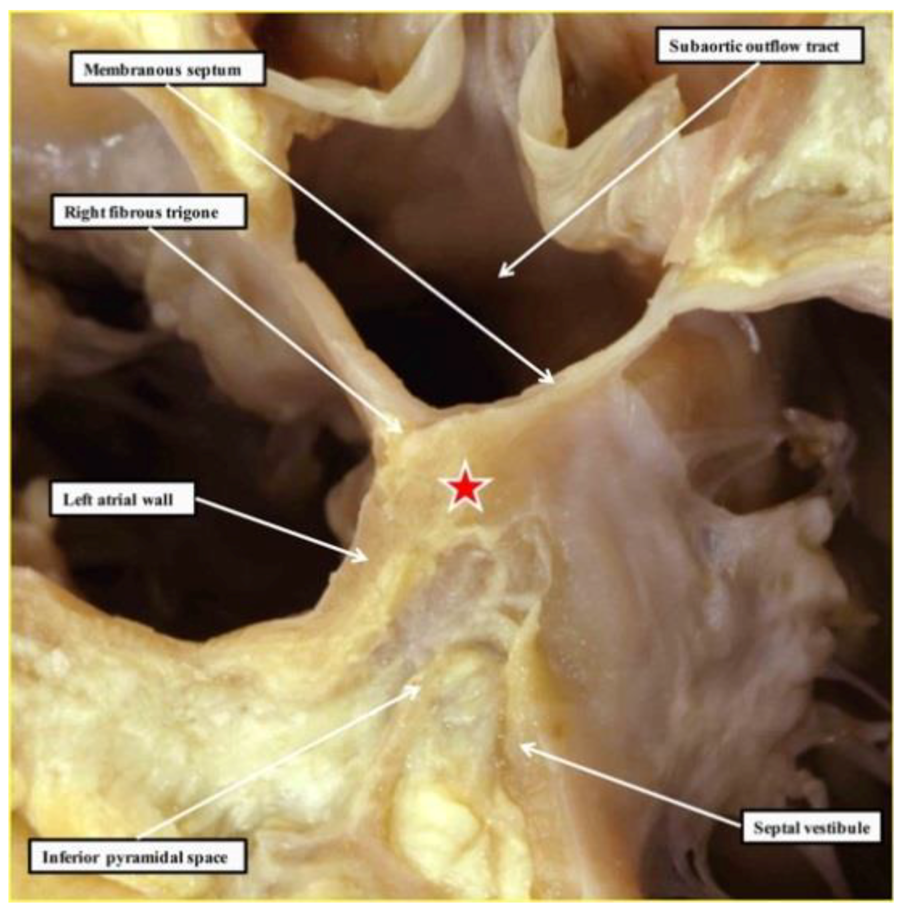
As already shown (Figure 1), Tawara had provided an excellent guide to the anatomical location of the axis [1]. His had demonstrated the location of its penetrating component, now justifiably usually described as the bundle of His [6]. Fully to appreciate the location of these various components of the axis in the human heart, it is also necessary to understand the structure of the atrioventricular junctions. Of particular importance is their relationship to the aortic root. The atrioventricular node, located at the apex of the triangle of Koch, is centrally located within the cardiac base. Despite this central location, the node itself is directly related to the area of fibroadipose tissue interposing between the septal component of the right atrial vestibule and the crest of the muscular ventricular septum. This is because a cranial continuation of the inferior atrioventricular groove extends to the inferior aspect of the central fibrous body. The central fibrous body itself is made up of the rightward extent of the area of fibrous continuity between the leaflets of the aortic and mitral valves in the roof of the left ventricle, an area usually described as the right fibrous trigone, and the membranous septum. The membranous septum itself forms the rightward margin of the subaortic outflow tract. The atrioventricular component of the membranous septum, furthermore, is the only atrioventricular septal structure to be found in the human heart. This is because the floor of the triangle of Koch is a sandwich rather than a true muscular septum (Figure 2).

2.2. Location of the Atrioventricular Node

The anatomical arrangement is such that the base of the atrioventricular node lies directly adjacent to the tissue plane interposed between the right atrial septal vestibule and the crest of the muscular ventricular septum. The surface of the ventricular septum itself is sloping, such that the body of the node forms a compact half-circle set with its base against the septal crest (Figure 3B).
The superficial atrial cardiomyocytes form overlaying fascicles that extend down from the atrial septum and insert into the hinge of the septal leaflet of the tricuspid valve. The cardiomyocytes of the atrial septum themselves form short transitional zones to the leftward half of the nodal half-oval. When the compact half oval itself is traced inferiorly, the columns of nodal cells branch to form short tracts that extend, on the left side, towards the hinge of the mural leaflet of the mitral valve. On the right side, they enter the right atrial septal vestibule, forming the inferior nodal extension (Figure 3A). When traced cranially, the cells of the compact node bunch together. Having collected together, they enter the substance of the central fibrous body, where the axis becomes the penetrating atrioventricular bundle (compare Figure 3C,D). The point at which the axis becomes insulated from the atrial cardiomyocytes is the best anatomical marker of the transition from the compact node to the penetrating atrioventricular bundle, or the bundle of His [1].

2.3. Penetration and Branching of the Conduction Axis

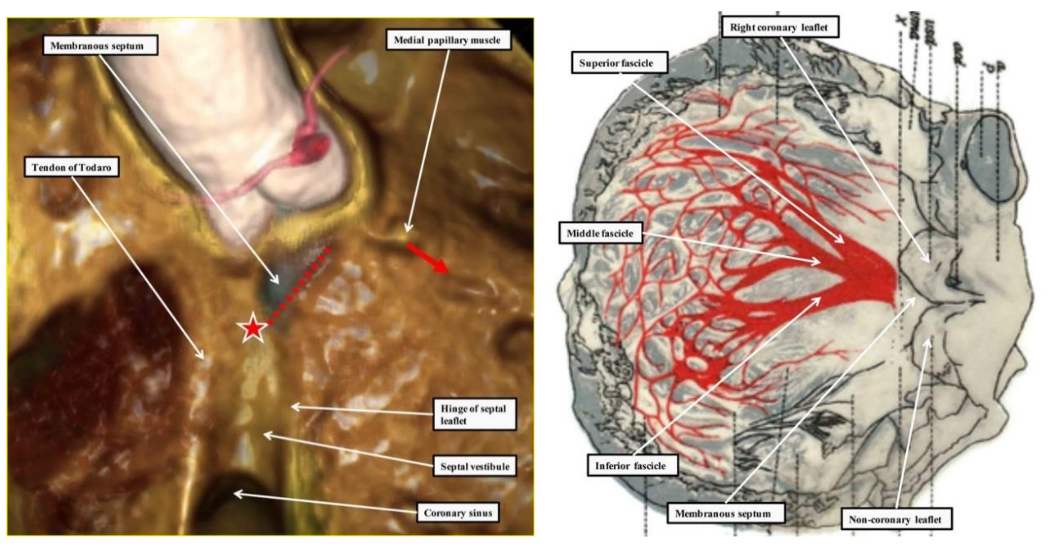
The atrioventricular conduction axis penetrates from the apex of the triangle of Koch through the atrioventricular component of the membranous septum. Having penetrated, the axis is positioned on the crest of the muscular ventricular septum. It has a non-branching component, which runs for a short distance along the septal crest before beginning to give rise to the fascicles of the left bundle branch. As was shown with exquisite accuracy by the reconstructions provided by Tawara [1], the fascicles of the left bundle branch, which are insulated by a fibrous sheath from the underlying septal musculature, cascade in a trifascicular fashion down the smooth left ventricular septal surface (Figure 4—right-hand panel). The most superior fascicle extends towards the supero-lateral papillary muscle of the mitral valve, while the inferior fascicle runs towards the infero-septal muscle. The middle fascicle descends between the two, forming a mesh-like network on the septal surface. The right bundle branch takes off from the bundle at the level of origin of the superior fascicle of the left bundle branch, passing through the substance of the ventricular septum to emerge beneath the medial papillary muscle of the tricuspid valve (Figure 4—left-hand panel). The axis itself, however, continues beyond the origin of the right bundle branch, extending into the subaortic outflow tract on the septal crest as the so-called “dead-end tract”. The extent of the terminal ramifications of the ventricular conduction system varies substantially between species, such that, for instance, the papillary muscles of the bovine heart is rich in Purkinje cells, whereas in other mammal hearts the ventricular myocardium harbors few Purkinje cells, or the Purkinje cells are poorly differentiated [13,14].
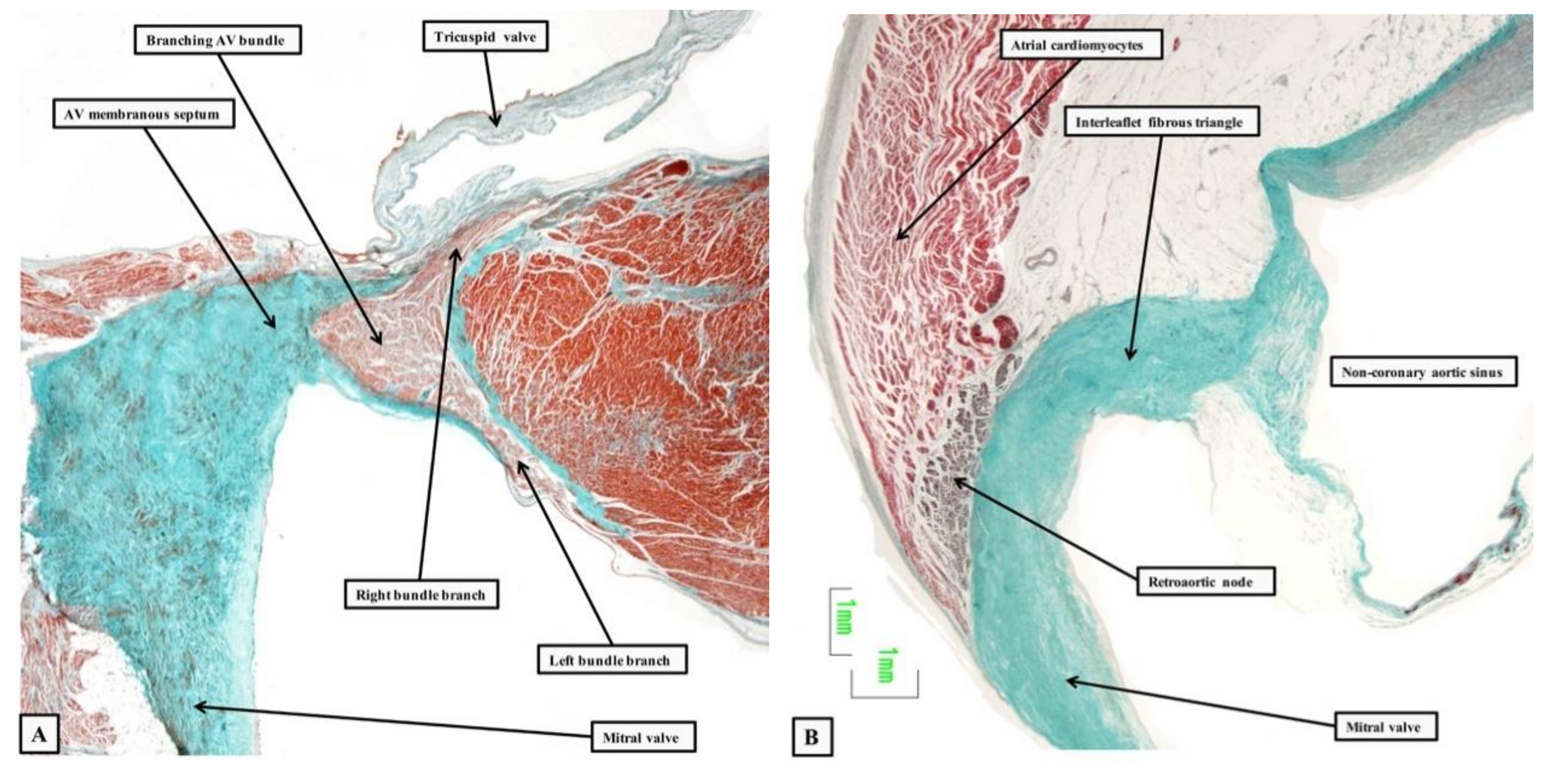
The bundle branches themselves continue to be insulated by fibrous sheaths from the substance of the muscular ventricular septum (Figure 5—left-hand panel).
The site of branching is usually directly astride the crest of the muscular septum. As already discussed, the left bundle branch fans out in trifascicular fashion, as demonstrated in the original monograph of Tawara (Figure 4—right-hand panel). The right bundle branch passes through the substance of the ventricular septum, emerging beneath the medial papillary muscle. It then extends through the substance of the septum as a small muscular strand before branching at the ventricular apex, usually with a prominent branch passing within the moderator band to the parietal ventricular wall. In a minority of cases, the central component of the axis branches on the left ventricular aspect of the septum, away from the septal crest [15]. The axis itself, nonetheless, continues beyond the branching component, extending into the aortic root as the so-called “dead-end tract” [16]. This component is part of the initial ring of primary myocardium that surrounded the embryonic interventricular communication. It is the remnant of this ring, lying with the vestibule of the right atrium, that were mistakenly taken by Kent to represent multiple pathways for atrioventricular conduction in the normal heart [7]. The remnants do exist, but are sequestrated within the vestibular myocardium on the atrial aspect of the insulating plane in individuals with normal atrioventricular conduction [17]. It is these remnants that produce the anomalous atrioventricular nodes found in situations when the heart is congenitally malformed, such as congenitally-corrected transposition or straddling tricuspid valve [18,19].

2.4. The Retroaortic Node

A particularly prominent remnant of the ring is to be found in the atrial myocardium at the junction of the vestibules of the tricuspid and mitral valves. This is the retroaortic node (Figure 5—right-hand panel), which has recently been implicated as a possible substrate for arrhythmias ablated from the non-coronary sinus of the aortic valve [20]. As yet, we do not have sufficient information to offer precise details regarding the size and full significance of this structure. Our current knowledge of cardiac development, however, now permits us to provide a detailed account of the formation of these various parts of the conduction axis.

3. Development of the Cardiac Chambers

In the embryo, the beating heart is initially a myocardial tube. In the chambered heart, as was noted by Gaskell, the developing chambers are separated by junctions themselves comprised of myocardium with a phenotype comparable to that of the initial tube [8]. Keith had observed that the crest is the earliest part of the ventricular septum to be formed [4]. The junctions between the chambers and the ventricular septal crest, however, constitute a miniscule part of the total cardiac mass. The disparity in mass between the junctions and chambers is a testimony to the manner of cardiogenesis [21,22].
Cardiac chambers form locally within the heart tube, by virtue of greater rates of growth, in a process now commonly referred to as ballooning [23,24,25]. Molecular assessments of proliferation have validated the greater growth rates of chambers [26,27,28,29,30]. The transcription factors that promote formation and growth of the chambers, such as GATA4, TBX5, and NKX2.5, are expressed throughout the embryonic heart tube [29,31,32,33]. The recognition of the junctions between chambers, and, consequently, formation of the distinct chambers themselves, is made possible by repression of transcription factors. Repressors like TBX2 and TBX3 are particularly important [21,34,35,36,37,38,39]. These insights were much facilitated by the detection of mRNA by non-radioactive means on tissue sections [40]. Developing chambers undergo a gestational increment in the speed of electrical propagation [41,42]. This is reflected by the presence of gap junctions with low resistance between the cardiomyocytes making up the walls of the chambers, such as CX40 and CX43. The cardiomyocytes making up the atrioventricular junctional myocardium, in contrast, express the high resistance CX45, and at relatively low densities [13,43].
When traced from the venous to the arterial pole, the alternation between chambers and junctions corresponds to alternation between fast and slow propagation of the electrical activity. An adult-like electrocardiogram with atrial and ventricular activation separated by a substantial atrioventricular delay can be recorded from the embryonic heart soon after the chambers have started to form [41,42,44,45,46]. In the initial stages, as the ventricles begin their maturation [47,48,49], activation is initially detected near the forming septum. In the fetal stages, the first epicardial activation is deep at the ventricular apexes, presumably at some terminal parts of the left and right bundle branches [47,48,49,50]. The ventricular septum nonetheless remains the earliest activated part of the ventricles [51,52,53]. The patterns of activation therefore suggest that a pathway of preferential conduction has already been established at the site of the future atrioventricular bundle. Later development of the atrioventricular conduction axis sees an incremental regulatory role of nervous tissues and, as recently shown, resident macrophages, but the basic properties of conduction still arise from the patterning of the myocardium [25,54].

4. The Development of the Atrioventricular Conduction Axis

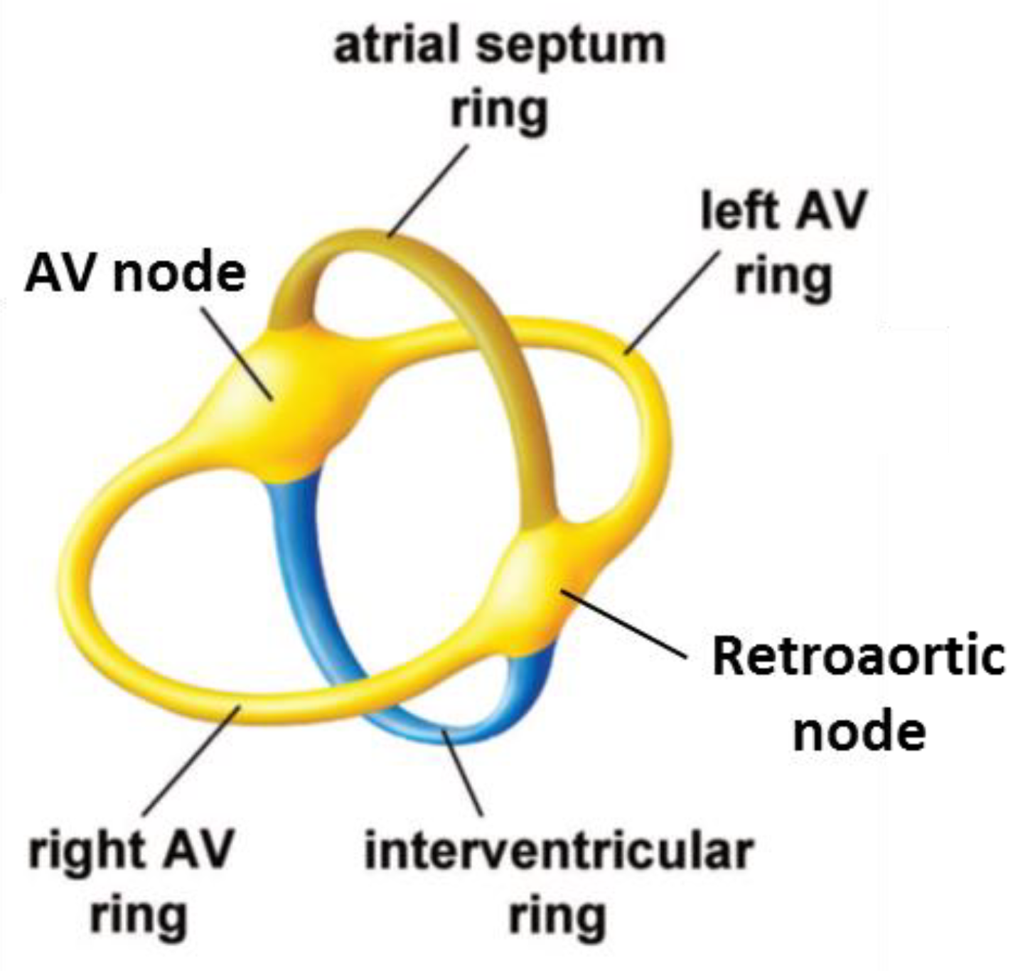
As the ventricular and atrial chambers begin to balloon out from the primary heart tube, the repressing transcription factors TBX2 and TBX3 start to be expressed in a circular domain between the chambers [25,55]. This area will become the atrioventricular canal [21,56]. The crest of the developing ventricular septum, formed between the ballooning cavities of the left and right ventricles, also expresses TBX3. This produces a domain that reaches the atrioventricular canal, which can be called the primary ring. The early electrical activation near the forming septum is likely due to preferential conduction along the primary ring [47,57]. The leading edge of the atrial septum also expresses TBX3 [21,56]. Collectively, the septal crests form a circular domain in a plane that is perpendicular to, and intersecting, the domain of the atrioventricular canal (Figure 6). The dorsal intersection of these rings marks the site of formation of the atrioventricular node. The ventral intersection will become the retroaortic node (Figure 6).
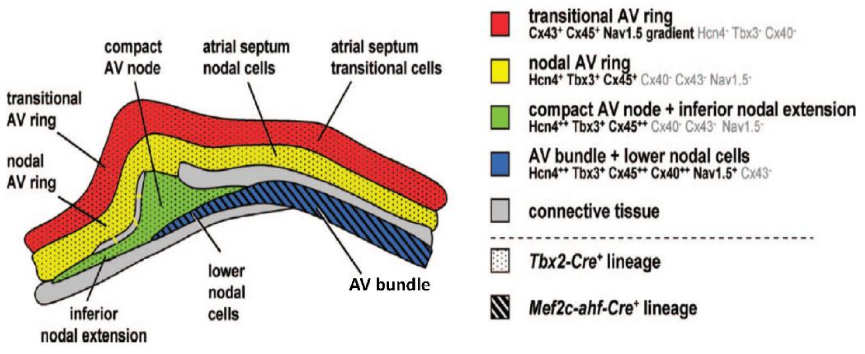
Around the time of transition from the embryonic to the fetal stages of development, concomitant with growth of the muscular ventricular septum, TBX3 will start to be expressed on the left and right of the septal crest. This delineates the areas of formation of the proximal parts of the left and right bundle branches [21,56]. Later stages see the development of an insulating plane of fibro-fatty tissue around the atrioventricular junctions. This disrupts the myocardial continuity between the atrial and ventricular components, save at the site of penetration of the axis; in other words, the His bundle [59]. As part of this process, the myocardium of the atrioventricular canal becomes confined to the atrial side as the atrial vestibules (Figure 7). Subsequent to the development of the insulating plane, the electrical impulse generated by the sinus node passes through the atrial myocardium. If running dorsally, it reaches the myocardium of the atrioventricular canal, and then the atrioventricular node and, finally, the His bundle. If the impulse takes this course, then much of atrioventricular delay comes from the relatively slow propagation through the atrioventricular canal myocardium [60,61]. This is the so-called “slow pathway”. An alternative route is provided, however, subsequent to the completion of atrial septation. This route extends ventral to the oval fossa, and corresponds with the so-called “fast pathway” into the atrioventricular node. The development of this pathway remains to be clarified. Once the electrical impulse reaches the His bundle, it can activate the crest of the ventricular septum directly, as in mice [52], or propagate along insulated bundle branches such that early activation is closer to the apex, as in humans [51].

5. Molecular Characterization of the Atrioventricular Conduction Axis

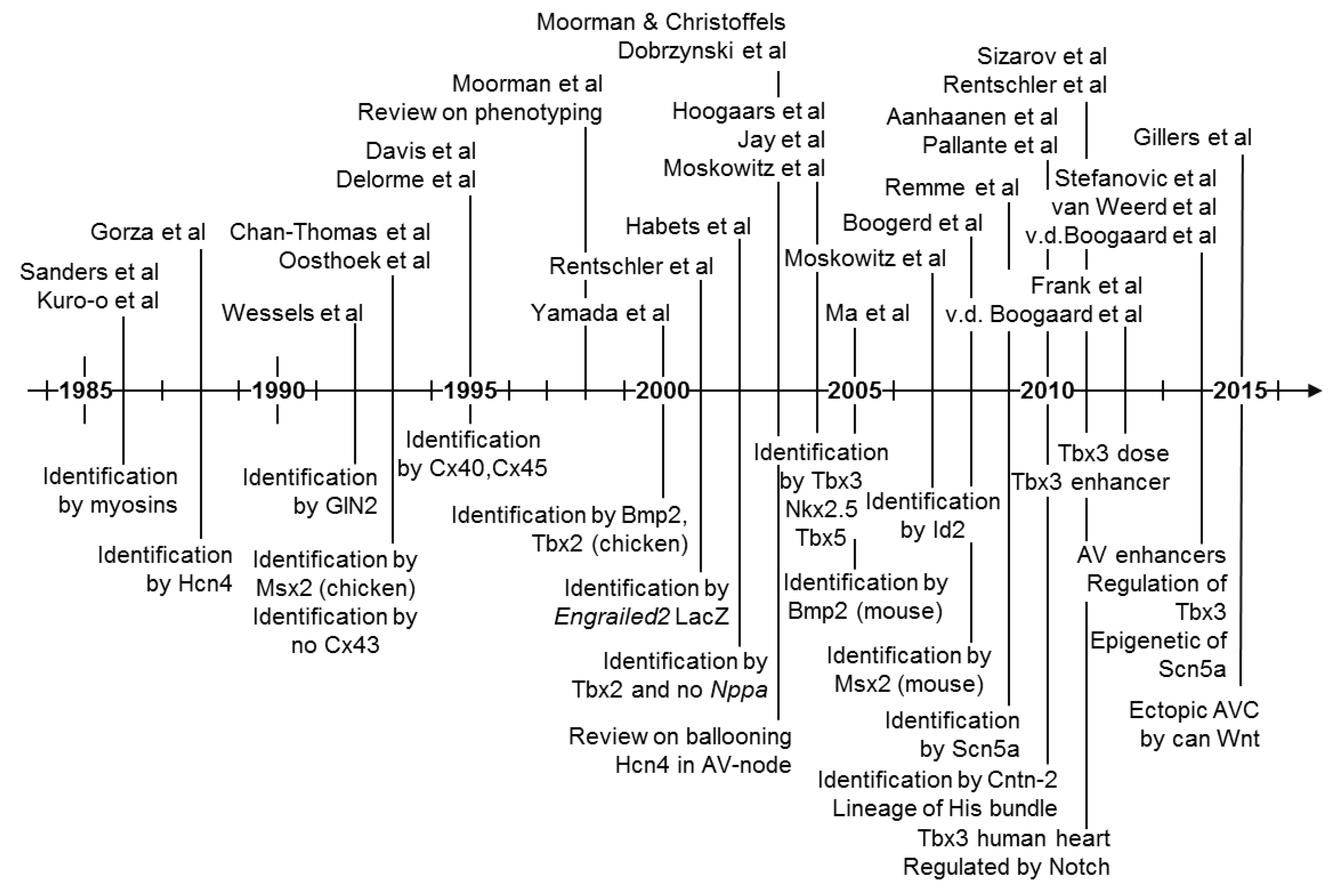
The pale staining characteristics as seen in classic histological sections stained with hematoxylin-eosin or Masson’s trichrome reflect a low level of expression of cytoplasmic proteins, in particular proteins of the contractile apparatus. Such staining characteristics do not give gene or protein-specific information. Gene-specific information is typically obtained by quantitative PCR, which is dependent on skilled micro-dissection for crude spatial information. High-resolution spatial information requires histology or whole mount stains. This typically relies on in situ hybridization, or immunohistochemistry, for transcript- and protein-specific signals, respectively. Key studies in the molecular characterization and understanding of the atrioventricular conduction axis are shown in Figure 8. Identification of important factors has been made on multiple levels of organization. These include epigenetic regulation and differentiation by transcription factors such as Bmp2, Tbx2/3, Tbx5, Nkx2.5, Notch, Wnt, and identification of proteins, and their transcripts, which underscore physiological functions, such as the gap junctional proteins Cx30.2, Cx40, Cx43, Cx45, and the ion channels Hcn4 and Scn5a.

6. Evolutionary Origin of the Atrioventricular Conduction Axis

The key components of the atrioventricular conduction axis are the atrioventricular node and the His bundle. They are distinct from each other, and the surrounding myocardium, on the basis of their anatomical, molecular, and electrophysiological characteristics [58,80]. The presence of each character can, in principle, be assessed in living vertebrates. By plotting the prevalence of each character on the phylogenetic tree of vertebrates, which itself is derived predominantly from fossil and genetic data, we can infer the most likely evolution of each character. As yet, however, there is no clear-cut synchrony in the appearance of these characteristics [12].
A cardinal feature of the atrioventricular node is the delay it establishes between atrial and ventricular contraction. All vertebrates have an atrioventricular delay, from hagfish to humans. A histologically discrete atrioventricular node, however, is present only in birds and mammals [24,81,82,83]. Irrespective of the presence or absence of an atrioventricular node, the atrioventricular delay is regulated by vagal stimulation to the point that an atrioventricular block can be induced [84,85]. Accordingly, Cranefield proposed that all vertebrates have the functional equivalent of an atrioventricular node [84]. We have recently shown that the atrioventricular delay of mammals and birds is some four times shorter, not longer, compared to reptiles. The latter animals are without an anatomically identifiable atrioventricular node (Boukens et al., in press). This permits the perhaps surprising conjecture that the evolution of the anatomically distinct atrioventricular node associates with a reduction, rather than an extension, of the atrioventricular delay. It is not yet known whether the difference in the duration of the delay is due to differences in expression, for instance, of gap junction proteins.
The mammalian atrioventricular conduction axis is dorsal in the atrioventricular junction and it is the only route from the atrial myocardium to the ventricular myocardial mass. In turtle hearts, however, Mines showed that both ventral and dorsal parts of the atrioventricular junction can propagate the electrical impulse [86]. The mammalian atrioventricular junctions are different from the arrangements found in ectothermic vertebrates by virtue of the extensive disruption of the atrioventricular junctional myocardium by the plane of atrioventricular insulation [13,81,82,83,87,88]. The comparative analysis suggests that the development of the insulating plane was sufficient, during evolution, to produce a mammalian-like atrioventricular conduction axis. A possible negation of this conjecture comes from findings in crocodilians. Crocodiles, alligators, and the gharials, are the only vertebrates, together with mammals and birds, which have a full ventricular septum [89,90]. Crocodilians and birds are phylogenetically grouped in the archosaur clade. This is about 100 million years younger than the earliest branches of the mammalian lineage. The evolution of the full septum in archosaurs and mammals, therefore, is widely regarded as a case of convergent evolution [91].
The crocodilians are the only ectotherms with considerable development of a plane of atrioventricular insulation. In the American alligator, the ventral half of the atrioventricular junctional myocardium is interrupted by an insulating plane [92]. Accordingly, cuts to this myocardium do not affect the propagation of the atrioventricular impulse [12]. It had been noted over 100 years ago that the dorsal myocardium of the atrioventricular canal of crocodilians projected to the ventricular septum [93,94]. We now know that cuts to the dorsal atrioventricular junctional myocardium induce atrioventricular block [12]. The dorsal atrioventricular propagation occurs through a narrow subset of the atrioventricular junctional myocardium that can be considered to constitute a specialised pathway. This specialised pathway, however, is not insulated by connective tissues to form a bundle of His. Instead, it is found within a broad area of myocardial continuity between the atrial and ventricular walls in the dorsal part of the atrioventricular junction. The specialised pathway, nonetheless, can be identified by its expression of Tbx3 [12], as is the case for the bundle of His of mammals and birds [21,95]. The Tbx3-defined bundle of the alligator heart, furthermore, is juxtaposed to the crux, which is the intersection of the atrioventricular groove and the dorsal interventricular coronary artery. This is again comparable with the bundle of His of mammals and birds [12]. Consistent with the presence of a specialised pathway in the alligator, the crest of the ventricular septum shows the earliest activation [12,50]. Early electrical activation also occurs deep in the ventricles in crocodilians, as in mammals and birds. This is in contrast to other reptiles, where early ventricular activation is in the vicinity of the atrioventricular canal [12,96,97]. The identification of the Tbx3-defined bundle corroborates some of the early anatomical works on crocodilians [93,94]. Other anatomical studies claimed to have found an atrioventricular conduction axis in non-crocodilian reptiles, but Prakash [98], for example, mistook an atrioventricular valve for the His bundle. The majority view is that non-crocodilian reptiles are without a specialised atrioventricular conduction axis [12]. Accordingly, the presence of Tbx2/Tbx3 does not reveal a slender pathway in non-crocodilian ectotherms [87,99]. Findings in the crocodilians, therefore, offer an alternative view on the evolution and development of the atrioventricular conduction axis, namely that the atrioventricular conduction axis can evolve without being completely insulated by fibro-fatty tissues. Its formation may depend on little more than the presence of a full ventricular septum.

7. Conclusions

Generations of scientists have built an ever-larger body of knowledge on the foundational observations on the atrioventricular conduction axis by Wilhelm His Jr. and Sunao Tawara. Much of the anatomy of the atrioventricular conduction axis is well understood. Our knowledge is now being deepened by significant advances in electrophysiology and in particular molecular biology. These advances include major contributions from Antoon F.M. Moorman, who sought the complementary insights of anatomy, development, evolution, electrophysiology, and molecular biology [100]. The branch of knowledge generated by Wilhelm His Jr. and Sunao Tawara keeps on sprouting and evolving! It still leaves areas requiring clarification, most notably the anatomic substrates for the slow, as opposed to the fast, pathways into the atrioventricular node.

Author Contributions

R.H.A. wrote the initial draft, provided several of the illustrations, and was involved in the subsequent finalisation of the manuscript. S.M. provided the virtual dissections of the living heart, and contributed to the finalisation of the manuscript. D.E.S. produced dissections to illustrate the location of the conduction axis, and again was involved in the finalisation of the manuscript, as was D.S.-Q., who produced the histological sections showing the location of the conduction axis. B.J. contributed to the initial concept, produced the sections on evolution and molecular identify of the conduction axis, and oversaw the final production of the work.

Funding

This research received no external funding.

Acknowledgments

All the authors acknowledge the huge contributions made by Antoon Moorman to the understanding of the overall development of the heart, not least the conduction system. In the case of R.H.A., acknowledgement is made of the fruitful collaborations enjoyed over a period of more than 25 years. For B.J., Antoon has served as mentor and ideal role model. We all wish him the very best in his retirement.

Conflicts of Interest

The authors declare no conflict of interest.

References

  1. Tawara, S. Das Reizleitungssystem des Säugetierherzens; Gustav Fischer: Jena, Germany, 1906. [Google Scholar]
  2. Anderson, R.H.; Ho, S.Y. The Morphology of the Specialized Atrioventricular Junctional Area: The Evolution of Understanding. Pacing Clin. Electrophysiol. 2022, 25, 957–966. [Google Scholar] [CrossRef]
  3. Silverman, M.E.; Grove, D.; Upshaw, C.B., Jr. Why does the heart beat? The discovery of the electrical system of the heart. Circulation 2006, 113, 2775–2781. [Google Scholar] [CrossRef] [PubMed]
  4. Keith, A.; Flack, M. The Form and Nature of the Muscular Connections between the Primary Divisions of the Vertebrate Heart. J. Anat. Physiol. 1907, 41, 172–189. [Google Scholar] [PubMed]
  5. Keith, A. An Autobiography; Watts & Co: London, UK, 1950; p. 255. [Google Scholar]
  6. His, W. Die Tätigkeit des embryonalen Herzens und deren Bedeutung für die Lehre von der Herzbewegung beim Menschen. Arb Med. Klin. (Leipzig) 1893, 14–49. [Google Scholar]
  7. Kent, A.F. Researches on the Structure and Function of the Mammalian Heart. J. Physiol. 1983, 14, 233–254. [Google Scholar] [CrossRef]
  8. Gaskell, W.H. On the Innervation of the Heart, with especial reference to the Heart of the Tortoise. J. Physiol. 1883, 4, 43–230. [Google Scholar] [CrossRef] [PubMed] [Green Version]
  9. Wessels, A.; Vermeulen, J.L.M.; Verbeek, F.J.; Viragh, S.Z.; Kalman, F.; Lamers, W.H.; Moorman, A.F.M. Spatial distribution of “tissue-specific” antigens in the developing human heart and skeletal muscle III. An immunohistochemical analysis of the distribution of the neural tissue antigen G1N2 in the embryonic heart; implications for the development of the atrioventricular conduction system. Anat. Rec. 1992, 232, 97–111. [Google Scholar] [PubMed]
  10. Yanni, J.; Boyett, M.R.; Anderson, R.H.; Dobrzynski, H. The extent of the specialized atrioventricular ring tissues. Heart Rhythm 2009, 6, 672–680. [Google Scholar] [CrossRef] [PubMed]
  11. Anderson, R.H.; Mori, S. Wilhelm His Junior and his bundle. J. Electrocardiol. 2006, 49, 637–643. [Google Scholar] [CrossRef] [PubMed]
  12. Jensen, B.; Boukens, B.J.; Crossley, D.A., II; Conner, J.; Mohan, R.A.; van Duijvenboden, K.; Postma, A.V.; Gloschat, C.R.; Elsey, R.M.; Sedmera, D.; et al. Specialized impulse conduction pathway in the alligator heart. eLife 2008, 7, e32120. [Google Scholar] [CrossRef] [PubMed]
  13. Moorman, A.F.M.; de Jong, F.; Denyn, M.M.; Lamers, W.H. Development of the Cardiac Conduction System. Circ. Res. 1998, 82, 629–644. [Google Scholar] [CrossRef] [PubMed] [Green Version]
  14. Stephenson, R.S.; Boyett, M.R.; Hart, G.; Nikolaidou, T.; Cai, X.; Corno, A.F.; Alphonso, N.; Jeffery, N.; Jarvis, J.C. Contrast enhanced micro-computed tomography resolves the 3-dimensional morphology of the cardiac conduction system in mammalian hearts. PLoS ONE 2012, 7, e35299. [Google Scholar] [CrossRef]
  15. Massing, G.K.; James, T.N. Anatomical configuration of the His bundle and bundle branches in the human heart. Circulation 1976, 53, 609–621. [Google Scholar] [CrossRef] [PubMed]
  16. Kurosawa, H.; Becker, A.E. Dead-end tract of the conduction axis. Int. J. Cardiol. 1985, 7, 13–20. [Google Scholar] [CrossRef]
  17. Anderson, R.H.; Davies, M.J.; Becker, A.E. Atrioventricular ring specialized tissue in the normal heart. Eur. J. Cardiol. 1974, 2, 219. [Google Scholar]
  18. Anderson, R.H.; Becker, A.E.; Arnold, R.; Wilkinson, J.L. The conducting tissues in congenitally corrected transposition. Circulation 1974, 50, 911–923. [Google Scholar] [CrossRef] [PubMed]
  19. Milo, S.; Ho, S.Y.; Macartney, F.J.; Wilkinson, J.L.; Becker, A.E.; Wenink, A.C.; De Groot, A.C.G.; Anderson, R.H. Straddling and overriding atrioventricular valves: Morphology and classification. Am. J. Cardiol. 1979, 44, 1122–1134. [Google Scholar] [CrossRef]
  20. Bohora, S.; Lokhandwala, Y.; Sternick, E.B.; Anderson, R.H.; Wellens, H.J.J. Reappraisal and new observations on atrial tachycardia ablated from the non-coronary aortic sinus of Valsalva: Authors’ reply. Europace 2018, 20, 214–215. [Google Scholar] [CrossRef] [PubMed]
  21. Hoogaars, W.M.; Tessari, A.; Moorman, A.F.; De Boer, P.A.; Hagoort, J.; Soufan, A.T.; Campione, M.; Christoffels, V.M. The transcriptional repressor Tbx3 delineates the developing central conduction system of the heart. Cardiovasc. Res. 2004, 62, 489–499. [Google Scholar] [CrossRef] [PubMed]
  22. Jensen, B.; Wang, T.; Christoffels, V.M.; Moorman, A.F. Evolution and development of the building plan of the vertebrate heart. Biochim. Biophys. Acta 2013, 1833, 783–794. [Google Scholar] [CrossRef] [PubMed] [Green Version]
  23. Moorman, A.F.; Vermeulen, J.L.; Koban, M.U.; Schwartz, K.; Lamers, W.H.; Boheler, K.R. Patterns of expression of sarcoplasmic reticulum Ca2+-ATPase and phospholamban mRNAs during rat heart development. Circ. Res. 1995, 76, 616–625. [Google Scholar] [CrossRef] [PubMed]
  24. Moorman, A.F.; Christoffels, V.M. Cardiac chamber formation: Development, genes, and evolution. Physiol. Rev. 2003, 83, 1223–1267. [Google Scholar] [CrossRef] [PubMed]
  25. Van Eif, V.W.W.; Devalla, H.D.; Boink, G.J.J.; Christoffels, V.M. Transcriptional regulation of the cardiac conduction system. Nat. Rev. Cardiol. 2018. [Google Scholar] [CrossRef] [PubMed]
  26. Sedmera, D.; Reckova, M.; DeAlmeida, A.; Coppen, S.R.; Kubalak, S.W.; Gourdie, R.G.; Thompson, R.P. Spatiotemporal pattern of commitment to slowed proliferation in the embryonic mouse heart indicates progressive differentiation of the cardiac conduction system. Anat. Rec. A Discov. Mol. Cell. Evol. Biol. 2003, 274, 773–777. [Google Scholar] [CrossRef] [PubMed] [Green Version]
  27. Soufan, A.T.; Van den Berg, G.; Ruijter, J.M.; De Boer, P.A.; Van den Hoff, M.J.; Moorman, A.F. Regionalized sequence of myocardial cell growth and proliferation characterizes early chamber formation. Circ. Res. 2006, 99, 545–552. [Google Scholar] [CrossRef] [PubMed]
  28. De Boer, B.A.; Van den Berg, G.; De Boer, P.A.; Moorman, A.F.; Ruijter, J.M. Growth of the developing mouse heart: An interactive qualitative and quantitative 3D atlas. Dev. Biol. 2012, 368, 203–213. [Google Scholar] [CrossRef] [PubMed] [Green Version]
  29. Sizarov, A.; Ya, J.; De Boer, B.A.; Lamers, W.H.; Christoffels, V.M.; Moorman, A.F. Formation of the building plan of the human heart: Morphogenesis, growth, and differentiation. Circulation 2011, 123, 1125–1135. [Google Scholar] [CrossRef] [PubMed]
  30. Luxán, G.; Casanova, J.C.; Martínez-Poveda, B.; Prados, B.; D’amato, G.; MacGrogan, D.; Gonzalez-Rajal, A.; Dobarro, D.; Torroja, C.; Martinez, F.; et al. Mutations in the NOTCH pathway regulator MIB1 cause left ventricular noncompaction cardiomyopathy. Nat. Med. 2013, 19, 193–201. [Google Scholar] [CrossRef] [PubMed]
  31. Sylva, M.; Van den Hoff, M.J.; Moorman, A.F. Development of the human heart. Am. J. Med. Genet. A 2014, 164A, 1347–1371. [Google Scholar] [CrossRef] [PubMed]
  32. Moskowitz, I.P.; Pizard, A.; Patel, V.V.; Bruneau, B.G.; Kim, J.B.; Kupershmidt, S.; Roden, D.; Berul, C.I.; Seidman, C.E.; Seidman, J.G. The T-Box transcription factor Tbx5 is required for the patterning and maturation of the murine cardiac conduction system. Development 2004, 131, 4107–4116. [Google Scholar] [CrossRef] [PubMed]
  33. Jay, P.Y.; Harris, B.S.; Maguire, C.T.; Buerger, A.; Wakimoto, H.; Tanaka, M.; Kupershmidt, S.; Roden, D.M.; Schultheiss, T.M.; O’Brien, T.X.; et al. Nkx2-5 mutation causes anatomic hypoplasia of the cardiac conduction system. J. Clin. Investig. 2004, 113, 1130–1137. [Google Scholar] [CrossRef] [PubMed] [Green Version]
  34. Habets, P.E.; Moorman, A.F.; Clout, D.E.; Van Roon, M.A.; Lingbeek, M.; Van Lohuizen, M.; Campione, M.; Christoffels, V.M. Cooperative action of Tbx2 and Nkx2.5 inhibits ANF expression in the atrioventricular canal: Implications for cardiac chamber formation. Genes Dev. 2002, 16, 1234–1246. [Google Scholar] [CrossRef] [PubMed]
  35. Singh, R.; Hoogaars, W.M.; Barnett, P.; Grieskamp, T.; Rana, M.S.; Buermans, H.; Farin, H.F.; Petry, M.; Heallen, T.; Martin, J.F.; et al. Tbx2 and Tbx3 induce atrioventricular myocardial development and endocardial cushion formation. Cell. Mol. Life Sci. 2012, 69, 1377–1389. [Google Scholar] [CrossRef] [PubMed]
  36. Frank, D.U.; Carter, K.L.; Thomas, K.R.; Burr, R.M.; Bakker, M.L.; Coetzee, W.A.; Tristani-Firouzi, M.; Bamshad, M.J.; Christoffels, V.M.; Moon, A.M. Lethal arrhythmias in Tbx3-deficient mice reveal extreme dosage sensitivity of cardiac conduction system function and homeostasis. Proc. Natl. Acad. Sci. USA 2012, 109, E154–E163. [Google Scholar] [CrossRef] [PubMed]
  37. Aanhaanen, W.T.; Boukens, B.J.; Sizarov, A.; Wakker, V.; de Gier-de Vries, C.; van Ginneken, A.C.; Moorman, A.F.; Coronel, R.; Christoffels, V.M. Defective Tbx2-dependent patterning of the atrioventricular canal myocardium causes accessory pathway formation in mice. J. Clin. Investig. 2011, 121, 534–544. [Google Scholar] [CrossRef] [PubMed]
  38. Gillers, B.S.; Chiplunkar, A.; Aly, H.; Valenta, T.; Basler, K.; Christoffels, V.M.; Efimov, I.R.; Boukens, B.J.; Rentschler, S. Canonical Wnt Signaling Regulates Atrioventricular Junction Programming and Electrophysiological Properties. Circ. Res. 2015, 116, 398–406. [Google Scholar] [CrossRef] [PubMed] [Green Version]
  39. Mohan, R.A.; Mommersteeg, M.T.; Domínguez, J.N.; Choquet, C.; Wakker, V.; De Gier-de Vries, C.; Boink, G.J.; Boukens, B.J.; Miquerol, L.; Verkerk, A.O.; et al. Embryonic Tbx3+ cardiomyocytes form the mature cardiac conduction system by progressive fate restriction. Development 2018. [Google Scholar] [CrossRef] [PubMed]
  40. Moorman, A.F.; Houweling, A.C.; De Boer, P.A.; Christoffels, V.M. Sensitive nonradioactive detection of mRNA in tissue sections: Novel application of the whole-mount in situ hybridization protocol. J. Histochem. Cytochem. 2001, 49, 1–8. [Google Scholar] [CrossRef] [PubMed]
  41. De Jong, F.; Opthof, T.A.A.M.W.; Wilde, A.A.M.; Janse, M.J.; Charles, R.; Lamers, W.H.; Moorman, A.F.M. Persisting zones of slow impulse conduction in developing chicken hearts. Circ. Res. 1992, 71, 240–250. [Google Scholar] [CrossRef] [PubMed]
  42. Arguello, C.; Alanis, J.; Pantoja, O.; Valenzuela, B. Electrophysiological and ultrastructural study of the atrioventricular canal during the development of the chick embryo. J. Mol. Cell. Cardiol. 1986, 18, 499–510. [Google Scholar] [CrossRef]
  43. Dobrzynski, H.; Anderson, R.H.; Atkinson, A.; Borbas, Z.; D’souza, A.; Fraser, J.F.; Inada, S.; Logantha, S.J.; Monfredi, O.; Morris, G.M.; et al. Structure, function and clinical relevance of the cardiac conduction system, including the atrioventricular ring and outflow tract tissues. Pharmacol. Ther. 2013, 139, 260–288. [Google Scholar] [CrossRef] [PubMed]
  44. Bogue, J.Y. The Electrocardiogram of the Developing Chick. J. Exp. Biol. 1993, 10, 286–292. [Google Scholar]
  45. Bogue, J.Y. The Heart Rate of the Developing Chick. J. Exp. Biol. 1932, 9, 351–358. [Google Scholar]
  46. Van Mierop, L.H. Location of pacemaker in chick embryo heart at the time of initiation of heartbeat. Am. J. Physiol. 1967, 212, 407–415. [Google Scholar] [CrossRef] [PubMed] [Green Version]
  47. Rentschler, S.; Vaidya, D.M.; Tamaddon, H.; Degenhardt, K.; Sassoon, D.; Morley, G.E.; Jalife, J.; Fishman, G.I. Visualization and functional characterization of the developing murine cardiac conduction system. Development 2001, 128, 1785–1792. [Google Scholar] [PubMed]
  48. Chuck, E.T.; Meyers, K.; France, D.; Creazzo, T.L.; Morley, G.E. Transitions in ventricular activation revealed by two-dimensional optical mapping. Anat. Rec. A Discov. Mol. Cell. Evol. Biol. 2004, 280, 990–1000. [Google Scholar] [CrossRef] [PubMed] [Green Version]
  49. Sankova, B.; Benes, J., Jr.; Krejci, E.; Dupays, L.; Theveniau-Ruissy, M.; Miquerol, L.; Sedmera, D. The effect of connexin40 deficiency on ventricular conduction system function during development. Cardiovasc. Res. 2012, 95, 469–479. [Google Scholar] [CrossRef] [PubMed] [Green Version]
  50. Thomas, K.; Goudy, J.; Henley, T.; Bressan, M. Optical Electrophysiology in the Developing Heart. J. Cardiovasc. Dev. Dis. 2018, 5, 28. [Google Scholar] [CrossRef] [PubMed]
  51. Durrer, D.; Van Dam, R.T.; Freud, G.E.; Janse, M.J.; Meijler, F.L.; Arzbaecher, R.C. Total excitation of the isolated human heart. Circulation 1970, 41, 899–912. [Google Scholar] [CrossRef] [PubMed]
  52. Van Rijen, H.V.; Van Veen, T.A.; Van Kempen, M.J.; Wilms-Schopman, F.J.; Potse, M.; Krueger, O.; Willecke, K.; Opthof, T.; Jongsma, H.J.; De Bakker, J.M. Impaired conduction in the bundle branches of mouse hearts lacking the gap junction protein connexin40. Circulation 2001, 103, 1591–1598. [Google Scholar] [CrossRef] [PubMed]
  53. Miquerol, L.; Meysen, S.; Mangoni, M.; Bois, P.; Van Rijen, H.V.; Abran, P.; Jongsma, H.; Nargeot, J.; Gros, D. Architectural and functional asymmetry of the His-Purkinje system of the murine heart. Cardiovasc. Res. 2004, 63, 77–86. [Google Scholar] [CrossRef] [PubMed]
  54. Hulsmans, M.; Clauss, S.; Xiao, L.; Aguirre, A.D.; King, K.R.; Hanley, A.; Hucker, W.J.; Wülfers, E.M.; Seemann, G.; Courties, G.; et al. Macrophages Facilitate Electrical Conduction in the Heart. Cell 2017, 169, 510–522. [Google Scholar] [CrossRef] [PubMed]
  55. Christoffels, V.M.; Habets, P.E.; Franco, D.; Campione, M.; De Jong, F.; Lamers, W.H.; Bao, Z.Z.; Palmer, S.; Biben, C.; Harvey, R.P.; et al. Chamber formation and morphogenesis in the developing mammalian heart. Dev. Biol. 2000, 223, 266–278. [Google Scholar] [CrossRef] [PubMed]
  56. Sizarov, A.; Devalla, H.D.; Anderson, R.H.; Passier, R.; Christoffels, V.M.; Moorman, A.F. Molecular Analysis of the Patterning of the Conduction Tissues in the Developing Human Heart. Circ. Arrhythm. Electrophysiol. 2011, 4, 532–542. [Google Scholar] [CrossRef] [PubMed]
  57. Sedmera, D. Function and form in the developing cardiovascular system. Cardiovasc. Res. 2011, 91, 252–259. [Google Scholar] [CrossRef] [PubMed] [Green Version]
  58. Aanhaanen, W.T.; Mommersteeg, M.T.; Norden, J.; Wakker, V.; De Gier-de Vries, C.; Anderson, R.H.; Kispert, A.; Moorman, A.F.; Christoffels, V.M. Developmental origin, growth, and three-dimensional architecture of the atrioventricular conduction axis of the mouse heart. Circ. Res. 2010, 107, 728–736. [Google Scholar] [CrossRef] [PubMed]
  59. Wessels, A.; Van den Hoff, M.J.; Adamo, R.F.; Phelps, A.L.; Lockhart, M.M.; Sauls, K.; Briggs, L.E.; Norris, R.A.; Van Wijk, B.; Perez-Pomares, J.M.; et al. Epicardially derived fibroblasts preferentially contribute to the parietal leaflets of the atrioventricular valves in the murine heart. Dev. Biol. 2012, 366, 111–124. [Google Scholar] [CrossRef] [PubMed] [Green Version]
  60. Anderson, R.H.; Durrer, D.; Janse, M.J.; Van Capelle, F.J.; Billete, J.; Becker, A.E.; Durrer, D. A combined morphological and electrophysiological study of the atrioventricular node of the rabbit heart. Circ. Res. 1974, 35, 909–922. [Google Scholar] [CrossRef] [PubMed]
  61. McGuire, M.A.; De Bakker, J.M.; Vermeulen, J.T.; Moorman, A.F.; Loh, P.; Thibault, B.; Vermeulen, J.L.; Becker, A.E.; Janse, M.J. Atrioventricular junctional tissue. Discrepancy between histological and electrophysiological characteristics. Circulation 1996, 94, 571–577. [Google Scholar] [CrossRef] [PubMed]
  62. Sanders, E.; De Groot, I.J.M.; Geerts, W.J.C.; De Jong, F.; Van Horssen, A.A.; Los, J.A.; Moorman, A.F.M. The local expression of adult chicken heart myosins during development. II. Ventricular conducting tissue. Anat. Embryol. 1986, 174, 187–193. [Google Scholar] [CrossRef] [PubMed]
  63. Kuro-o, M.; Tsuchimochi, H.; Ueda, S.; Takaku, F.; Yazaki, Y. Distribution of cardiac myosin isozymes in human conduction system. Immunohistochemical study using monoclonal antibodies. J. Clin. Investig. 1986, 77, 340–347. [Google Scholar] [CrossRef] [PubMed]
  64. Gorza, L.; Schiaffino, S.; Vitadello, M. Heart conduction system: A neural crest derivative? Brain Res. 1988, 457, 360–366. [Google Scholar] [CrossRef]
  65. Chan-Thomas, P.S.; Thompson, R.P.; Robert, B.; Yacoub, M.H.; Barton, P.J. Expression of homeobox genes Msx-1 (Hox-7) and Msx-2 (Hox-8) during cardiac development in the chick. Dev. Dyn. 1993, 197, 203–216. [Google Scholar] [CrossRef] [PubMed] [Green Version]
  66. Oosthoek, P.W.; Viragh, S.; Lamers, W.H.; Moorman, A.F. Immunohistochemical delineation of the conduction system II: The atrioventricular node and Purkinje fibers. Circ. Res. 1993, 73, 482–491. [Google Scholar] [CrossRef] [PubMed]
  67. Davis, L.M.; Rodefeld, M.E.; Green, K.; Beyer, E.C.; Saffitz, J.E. Gap junction protein phenotypes of the human heart and conduction system. J. Cardiovasc. Electrophysiol. 1995, 6, 813–822. [Google Scholar] [CrossRef] [PubMed]
  68. Delorme, B.; Dahl, E.; Jarry-guichard, T.; Marics, I.; Briand, J.P.; Willecke, K.; Gros, D.; Théveniau-Ruissy, M. Developmental regulation of connexin 40 gene expression in mouse heart correlates with the differentiation of the conduction system. Dev. Dyn. 1995, 204, 358–371. [Google Scholar] [CrossRef] [PubMed] [Green Version]
  69. Yamada, M.; Revelli, J.P.; Eichele, G.; Barron, M.; Schwartz, R.J. Expression of chick Tbx-2, Tbx-3, and Tbx-5 genes during early heart development: Evidence for BMP2 induction of Tbx2. Dev. Biol. 2000, 228, 95–105. [Google Scholar] [CrossRef] [PubMed]
  70. Dobrzynski, H.; Nikolski, V.P.; Sambelashvili, A.T.; Greener, I.D.; Yamamoto, M.; Boyett, M.R.; Efimov, I.R. Site of origin and molecular substrate of atrioventricular junctional rhythm in the rabbit heart. Circ. Res. 2003, 93, 1102–1110. [Google Scholar] [CrossRef] [PubMed]
  71. Ma, L.; Lu, M.F.; Schwartz, R.J.; Martin, J.F. Bmp2 is essential for cardiac cushion epithelial-mesenchymal transition and myocardial patterning. Development 2005, 132, 5601–5611. [Google Scholar] [CrossRef] [PubMed] [Green Version]
  72. Moskowitz, I.P.; Kim, J.B.; Moore, M.L.; Wolf, C.M.; Peterson, M.A.; Shendure, J.; Nobrega, M.A.; Yokota, Y.; Berul, C.; Izumo, S.; et al. A molecular pathway including Id2, Tbx5, and Nkx2-5 required for cardiac conduction system development. Cell 2007, 129, 1365–1376. [Google Scholar] [CrossRef] [PubMed]
  73. Boogerd, K.J.; Wong, L.E.; Christoffels, V.M.; Klarenbeek, M.; Ruijter, J.M.; Moorman, A.F.; Barnett, P. Msx1 and Msx2 are functional interacting partners of T-box factors in the regulation of Connexin43. Cardiovasc. Res. 2008, 78, 485–493. [Google Scholar] [CrossRef] [PubMed] [Green Version]
  74. Remme, C.A.; Verkerk, A.O.; Hoogaars, W.M.H.; Aanhaanen, W.T.J.; Scicluna, B.P.; Annink, C.; Van den Hoff, M.J.B.; Wilde, A.A.M.; Van Veen, T.A.B.; Veldkamp, M.W.; et al. The cardiac sodium channel displays differential distribution in the conduction system and transmural heterogeneity in the murine ventricular myocardium. Basic Res. Cardiol. 2009, 104, 511–522. [Google Scholar] [CrossRef] [PubMed] [Green Version]
  75. Pallante, B.A.; Giovannone, S.; Fang-Yu, L.; Zhang, J.; Liu, N.; Kang, G.; Dun, W.; Boyden, P.A.; Fishman, G.I. Contactin-2 expression in the cardiac Purkinje fiber network. Circ. Arrhythm. Electrophysiol. 2010, 3, 186–194. [Google Scholar] [CrossRef] [PubMed]
  76. Rentschler, S.; Harris, B.S.; Kuznekoff, L.; Jain, R.; Manderfield, L.; Lu, M.M.; Morley, G.E.; Patel, V.V.; Epstein, J.A. Notch signaling regulates murine atrioventricular conduction and the formation of accessory pathways. J. Clin. Investig. 2011, 121, 525–533. [Google Scholar] [CrossRef] [PubMed] [Green Version]
  77. van den Boogaard, M.; Wong, L.E.; Tessadori, F.; Bakker, M.L.; Dreizehnter, L.K.; Wakker, V.; Bezzina, C.R.; AC‘t Hoen, P.; Bakkers, J.; Barnett, P.; et al. Genetic variation in T-box binding element functionally affects SCN5A/SCN10A enhancer. J. Clin. Investig. 2012, 122, 2519–2530. [Google Scholar] [CrossRef] [PubMed] [Green Version]
  78. Van den Boogaard, M.; Smemo, S.; Burnicka-Turek, O.; Arnolds, D.E.; van de Werken, H.J.; Klous, P.; McKean, D.; Muehlschlegel, J.D.; Moosmann, J.; Toka, O.; et al. A common genetic variant within SCN10A modulates cardiac SCN5A expression. J. Clin. Investig. 2014, 124, 1844–1852. [Google Scholar] [CrossRef] [PubMed] [Green Version]
  79. Stefanovic, S.; Barnett, P.; Van Duijvenboden, K.; Weber, D.; Gessler, M.; Christoffels, V.M. GATA-dependent regulatory switches establish atrioventricular canal specificity during heart development. Nat. Commun. 2014, 5, 3680. [Google Scholar] [CrossRef] [PubMed] [Green Version]
  80. Park, D.S.; Fishman, G.I. Development and Function of the Cardiac Conduction System in Health and Disease. J. Cardiovasc. Dev. Dis. 2017, 4, 7. [Google Scholar] [CrossRef] [PubMed]
  81. Davies, F. The conducting system of the vertebrate heart. Br. Heart J. 1942, 4, 66–76. [Google Scholar] [CrossRef] [PubMed]
  82. Truex, R.C.; Smythe, M.G. Comparative morphology of the cardiac conduction tissue in animals. Ann. N. Y. Acad. Sci. 1965, 127, 19–33. [Google Scholar] [CrossRef] [PubMed]
  83. Chiodi, V.; Bortolami, R. The Conducting System of the Vertebrate Heart; Edizioni Calderini: Bologna, Italy, 1967. [Google Scholar]
  84. Cranefield, P.F. The atrioventricular node and the ventricular conducting system in the nonmammalian vertebrate heart. Ann. N. Y. Acad. Sci. 1965, 127, 145–150. [Google Scholar] [CrossRef] [PubMed]
  85. Valentinuzzi, M.E.; Hoff, H.E. The sinus venosus-atrial Wenckebach-Luciani phenomenon. J. Electrocardiol. 1972, 5, 1–14. [Google Scholar] [CrossRef]
  86. Mines, G.R. On dynamic equilibrium in the heart. J. Physiol. 1913, 46, 349–383. [Google Scholar] [CrossRef] [PubMed] [Green Version]
  87. Jensen, B.; Boukens, B.J.; Postma, A.V.; Gunst, Q.D.; van den Hoff, M.J.; Moorman, A.F.; Wang, T.; Christoffels, V.M. Identifying the evolutionary building blocks of the cardiac conduction system. PLoS ONE 2012, 7, e44231. [Google Scholar] [CrossRef] [PubMed]
  88. Anderson, R.H.; Spicer, D.E.; Brown, N.A.; Mohun, T.J. The development of septation in the four-chambered heart. Anat. Rec. 2014, 297, 1414–1429. [Google Scholar] [CrossRef] [PubMed]
  89. Cook, A.C.; Tran, V.H.; Spicer, D.E.; Rob, J.M.; Sridharan, S.; Taylor, A.; Anderson, R.H.; Jensen, B. Sequential segmental analysis of the crocodilian heart. J. Anat. 2017, 231, 484–499. [Google Scholar] [CrossRef] [PubMed]
  90. Webb, G.J.W. Comparative cardiac anatomy of the reptilia III. The heart of crocodilians and an hypothesis on the completion of the interventricular septum of crocodilians and birds. J. Morph. 1979, 161, 221–240. [Google Scholar] [CrossRef]
  91. Poelmann, R.E.; Gittenberger-de Groot, A.C.; Vicente-Steijn, R.; Wisse, L.J.; Bartelings, M.M.; Everts, S.; Hoppenbrouwers, T.; Kruithof, B.P.; Jensen, B.; de Bruin, P.W.; et al. Evolution and development of ventricular septation in the amniote heart. PLoS ONE 2014, 9, e106569. [Google Scholar] [CrossRef] [PubMed]
  92. Davies, F.; Francis, E.T.; King, T.S. The conducting (connecting) system of the crocodilian heart. J. Anat. 1952, 86, 152–161. [Google Scholar] [PubMed]
  93. Greil, A. Beitrage zur vergelichenden anatomie und entwicklungsgeschichte des herzens und des trauncus arteriosus der wirbelthiere. Morph. Jahrb. 1903, 31, 123–310. [Google Scholar]
  94. Swett, F.H. The connecting systems of the reptile heart—Alligator. Anat. Rec. 1923, 26, 129–140. [Google Scholar] [CrossRef]
  95. Bakker, M.L.; Boukens, B.J.; Mommersteeg, M.T.; Brons, J.F.; Wakker, V.; Moorman, A.F.; Christoffels, V.M. Transcription factor Tbx3 is required for the specification of the atrioventricular conduction system. Circ. Res. 2008, 102, 1340–1349. [Google Scholar] [CrossRef] [PubMed]
  96. Christian, E.; Grigg, G.C. Electrical activation of the ventricular myocardium of the crocodile Crocodylus johnstoni: A combined microscopic and electrophysiological study. Comp. Biochem. Physiol. A Mol. Integr. Physiol. 1999, 123, 17–23. [Google Scholar] [CrossRef]
  97. Gregorovicova, M.; Sedmera, D.; Jensen, B. Relative position of the atrioventricular canal determines the electrical activation of developing reptile ventricles. J. Exp. Biol. 2018, 221. [Google Scholar] [CrossRef] [PubMed]
  98. Prakash, R. The Heart and Its Conducting System in the Lizard Calotes versicolor (Daudin). Anat. Rec. 1960, 136, 469–475. [Google Scholar] [CrossRef] [PubMed]
  99. Kokubo, N.; Matsuura, M.; Onimaru, K.; Tiecke, E.; Kuraku, S.; Kuratani, S.; Tanaka, M. Mechanisms of heart development in the Japanese lamprey, Lethenteron japonicum. Evol. Dev. 2010, 12, 34–44. [Google Scholar] [CrossRef] [PubMed]
  100. Moorman, A.F. Pioneer who Gave Shape to Cardiac Morphology: Antoon FM Moorman, PhD, FESC (vol 126, pg f43, 2012). Circulation 2012, 126. [Google Scholar]
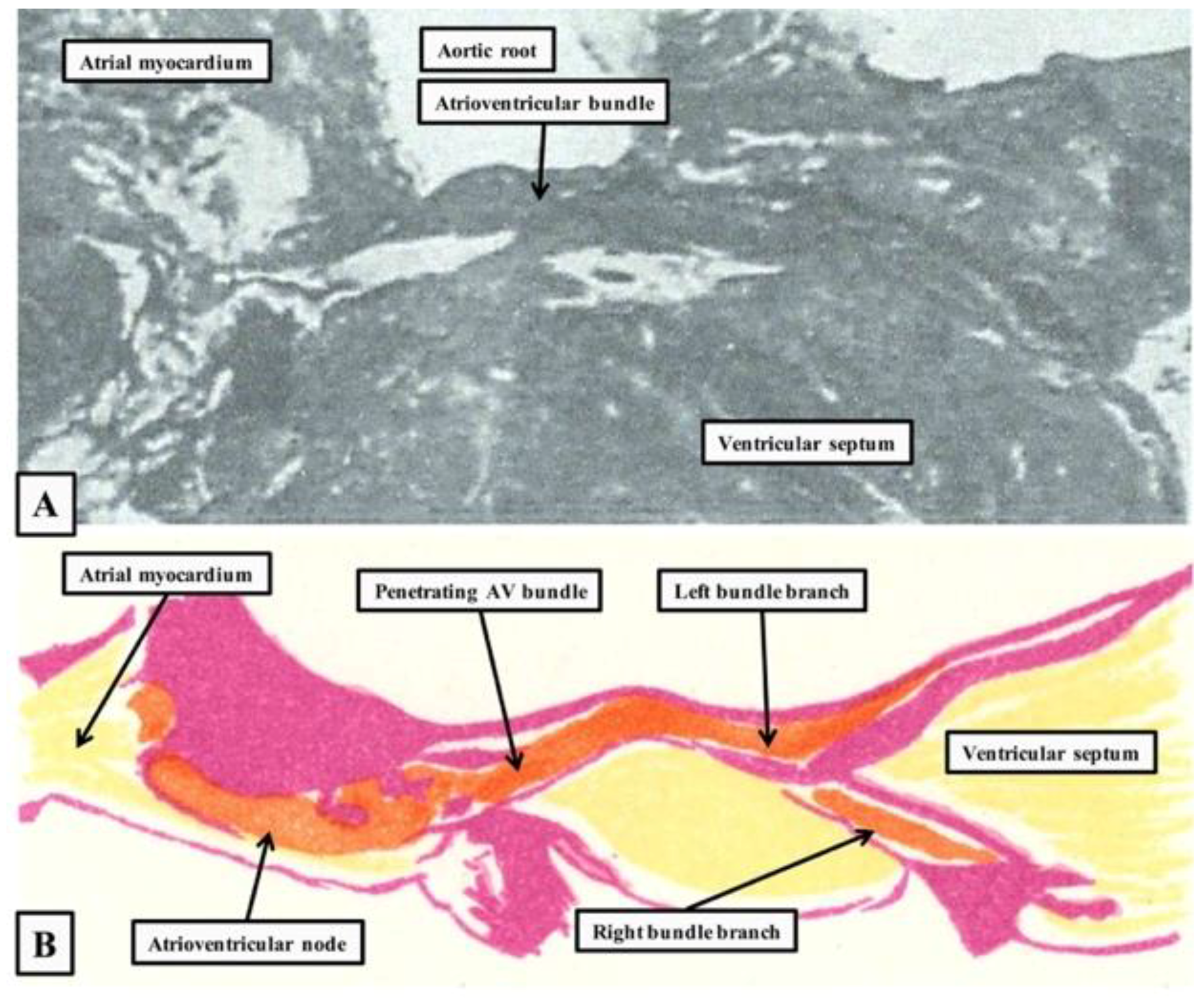
Figure 1. The figure is prepared by scanning the figures from the original publication of His [6] (A) and the monograph of Tawara [1] (B). His’s figure is shown as seen from the right side, with the heart in “Valentine” orientation. Tawara’s figure is re-orientated to show the arrangement as seen in attitudinally appropriate fashion, with the endocardial surface of the right-sided chambers to the bottom of the picture. His’ image shows the penetrating portion of the atrioventricular (AV) conduction axis, whereas the figure of Tawara shows its overall extent.
Jcdd 05 00044 g001
Figure 2. The top panel shows the opened right atrium, revealing the landmarks of the triangle of Koch, namely the tendon of Todaro and the hinge of the septal leaflet of the tricuspid valve. The atrioventricular node (red star with white borders), located at the apex of the triangle, is more-or-less centrally positioned within the cardiac base as viewed from the internal aspect of the right-sided chambers. As is shown in the bottom panel, which is a dissection made by removing the floor of the triangle, the node lies directly adjacent to a cranial continuation of the inferior atrioventricular groove. This fibroadipose area forms the inferior pyramidal space. The cranial extent of the space is bounded by the atrioventricular component of the membranous septum. It is this area that is penetrated by the bundle of His.
Jcdd 05 00044 g002aJcdd 05 00044 g002b
Figure 3. The sections are from a series prepared from a human heart, showing the atrial components of the conduction axis, and its transition to the bundle of His. They are orientated in attitudinally appropriate fashion, but with the chambers of the right heart shown to the top of the images. Panel (A) shows the rightward and leftward inferior extensions from the compact node, which is seen in Panel (B). Panels (C,D) show how the axis becomes the penetrating bundle once it is insulated by the fibrous tissue of the central fibrous body from the atrial cardiomyocytes.
Jcdd 05 00044 g003
Figure 4. The left-hand image, prepared from a computed tomographic dataset obtained from an individual undergoing investigation for coronary arterial disease, shows how the location of the atrioventricular conduction axis can be predicted based on knowledge of the landmarks of the right atrial and right ventricular septal surfaces. The star shows the location of the atrioventricular node at the apex of the triangle of Koch. The red dotted line shows the location of the non-branching and branching components of the axis, which are carried on the crest of the muscular ventricular septum. The red arrow shows the site of emergence of the right bundle branch, marked by the medial papillary muscle of the tricuspid valve. The right-hand image is from the original monograph of Tawara [1]. It has been scanned and rotated through 90 degrees relative to the original, placing the image in more attitudinally appropriate orientation. The reconstructions made by Tawara show the origin and distribution of the left bundle branch within the left ventricle.
Jcdd 05 00044 g004
Figure 5. The images are from the same series as shown in Figure 4. Panel (A) shows the cranial continuation of the conduction axis, which has divided into the right and left bundle branches of the crest of the muscular ventricular septum. Panel (B) shows a section through the vestibule of the left atrium directly adjacent to the non-coronary sinus of the aortic root. The retroaortic node is seen on the atrial aspect of the insulating plane.
Jcdd 05 00044 g005
Figure 6. The domain of Tbx3-expressing myocardium partakes in dividing the heart in atrial and ventricular compartments, and also in the division of the atrial and ventricular compartment, respectively, by expression on the crests of the atrial and ventricular septum. Adapted from [58].
Jcdd 05 00044 g006
Figure 7. The identities of the myocardium of the atrioventricular junction of the formed murine heart. Adapted from [58].
Jcdd 05 00044 g007
Figure 8. Chronology of key studies in the molecular identification of the atrioventricular conduction axis. Cited studies: [9,13,21,24,32,33,34,36,37,38,47,56,58,62,63,64,65,66,67,68,69,70,71,72,73,74,75,76,77,78,79]. There are recent and excellent reviews on the regulatory and transcriptional networks behind the formation of the cardiac conduction system [25,80].
Jcdd 05 00044 g008