Communication

miR-27b and miR-23b Modulate Cardiomyocyte Differentiation from Mouse Embryonic Stem Cells

by
José Manuel Vilches
,
Antonio Pulido
,
Francisco Hernández-Torres
,
Diego Franco
and
Amelia Aránega
*
Cardiovascular Development Group, Department of Experimental Biology, University of Jaén, 23071 Jaén, Spain
*
Author to whom correspondence should be addressed.
J. Cardiovasc. Dev. Dis. 2014, 1(1), 41-51; https://doi.org/10.3390/jcdd1010041
Submission received: 1 February 2014 / Revised: 21 March 2014 / Accepted: 24 March 2014 / Published: 31 March 2014

Abstract

:
Diverse types of stem cells represent a potentially attractive source of cardiac cells for the treatment of cardiovascular diseases. However, most of the functional benefits reported for stem cell have been modest and mainly due to paracrine effects rather than differentiation into cardiomyocytes of the applied cells. Therefore, new tools need to be developed in order to improve the efficiency of stem cell differentiation towards specific cardiovascular lineages. Here we show that microRNAs that display early differential expression during ventricular maturation, such as miR-27b, inhibits cardiac differentiation from mouse embryonic stem cells whereas miRNAs that display late differential expression, such as miR-23b, regulates the beating phenotype during in vitro cardiac differentiation from Embryonic Stem Cells (ESCs). This study could have an impact on regenerative medicine since we showed that miR-27b and miR-23b overexpression differentially modify the ESC cell fate towards the cardiac lineage.

1. Introduction

Heart development takes place through a complex series of steps, including the induction of cardiac mesoderm, formation of the cardiovascular progenitor cells and the commitment of cells to the cardiovascular lineage. Each step is precisely regulated at various levels, including transcriptional, posttranscriptional and posttranslational mechanisms [1].
microRNAs (miRNAs) have been implicated as critical regulators in cardiovascular development and pathological conditions at posttranscriptional level. The essential role of miRNA in early heart development was first revealed by a knockout study of Dicer, a miRNA-processing enzyme. The early lethality in Dicer mutant indicated the crucial role of miRNAs in early heart development [2]. Moreover, conditional Dicer deletion in the adult mouse heart causes various cardiac defects and postnatal lethality [3].
Additional studies led to the discovery of functional miRNAs such as miR-1, miR-15, miR-133, miR-138, miR-218 in regulating different step of vertebrate cardiogenesis [4,5,6,7,8]. Therefore, miRNAs now constitute an important component of the regulatory circuits that govern heart development and this knowledge has revolutionized our understanding of the diverse roles of non-protein-coding RNA in cardiac biology. However, with the human and mouse genome encoding over 4000 miRNAs, only a handful of which have been studied in the heart, much remains to be learned about this new class of regulatory RNA.
Recently we have reported a comprehensive profile of microRNA expression during cardiac maturation [9]. This analysis showed that only a relatively small set of microRNAs display increased expression levels, whereas none display decreased expression levels, supporting the notion that increasing microRNA expression levels provides a means to block and/or inhibit certain transcriptional programs over time. Here we show that microRNAs that display early differential expression profiles during cardiac maturation, such as miR-27b, inhibits cardiac differentiation from mouse embryonic stem cells whereas miRNAs that display late differential expression profiles, such as miR-23b, regulates the beating phenotype during in vitro cardiac differentiation from ESCs. These data support the notion that increasing expression levels of microRNAs in discrete temporal developmental windows during in vitro cardiac differentiation progressively modulate particular signaling pathways, which can alter the cardiogenic lineage differentiation.

2. Material and Methods

2.1. ESC Culture and microRNA Transfection Assays

R1-ESC line, derived from the 129 S1/SvImJ strain, was kindly provided by Andras Nagy (Samuel Lunenfeld Research Institute, Mount Sinai Hospital, Toronto, Canada). R1-ESCs were cultured and differentiated as previously described [10]. We designated day 0 as when embryonic bodies (EBs), obtained from hanging drops, were plated. In general, we identified contracting regions within the EBs between 3–4 days after plating (10–11 total days).
microRNA transfection assays were performed when EBs were plated (day 0) as previously described [10]. Pre-miR-27b and pre-miR-23b and negative controls (a scrambled oligonucleotide, scrambled-pre-miR) were obtained from Ambion (Life Technologies). In order to maintain miRNA over-expression levels during EB culture, miRNA-transfections were repeatedly carried out every two days of culturing, as the EBs-differentiation medium was changed. EBs or dissected beating areas respectively were collected at 7 days of transfection.

2.2. RNA Isolation, Reverse Transcriptase Reaction and Quantitative Real Time PCR

Because spontaneously contracting CMs constitute only a small fraction of EBs, parts of EBs containing beating areas were dissected under a binocular microscope. EBs as well as dissected specimens were used to perform reverse transcriptase reactions (RT). Total RNA isolation, RT reactions and quantitative real time PCR was performed as previously described [10]. Specific primers for each gene analyzed, annealing temperature and amplicon size are shown in the Table 1.
Table 1. Gene-specific primers, annealing temperatures, and amplicon sizes for quantitative Real-Time PCR.
PrimersSequenceAnnealing TemperatureAmplicon size
Wnt3aSense: 5'-TTCTGCAGGAACTACGTGGA-3'60 °C179 bp
Antisense:5'-AGATAGCAGCTGATCCCTCTG-3'
BrachyurySense: 5'- CGACCACAAAGATGTAATGGAG-3'59.5 °C120 bp
Antisense: 5'-AACTGAGGGTGGGAGCTG-3'
Kcnk3Sense: 5'-TGTGCACCTTCACCTACCTG-3'59.7 °C203 bp
Antisense: 5'-GGTGATGGCGAAGTAGAAGG-3'
Kcnh1Sense: 5'-ACGCCCTTCAGAAAGTGCTA-3'62 °C182 bp
Antisense: 5'-GTGGTCAGGAGGCAGGATAA-3'
c-Troponin TSense: 5'-TTCGACCTGCAGGAAAAGTT-3'64 °C133bp
Antisense: 5'-GCACAGCTTTGACGAGAACA-3'
Hcn4Sense: 5'-CAGCGTCAGAGCGGATACTT-3'60 °C158bp
Antisense: 5'-TGTGGAGGAGGATGGAGTTC-3'
Cx40Sense: 5'-CAGAGCCTGAAGAAGCCAAC-3'60 °C178bp
Antisense: 5'-ATGCGGAAAATGAACAGGAC-3'
Cx43Sense: 5'-GAGAGCCCGAACTCTCCTTT-3'60 °C158bp
Antisense 5'-TGGAGTAGGCTTGGACCTTG-3'
miR 27b5'-UUCACAGUGGCUAAGUUCUGC-3'60 °C21 bp
miR 23b5'-AUCACAUUGCCAGGGAUUACC-3'60 °C21 bp

3. Results

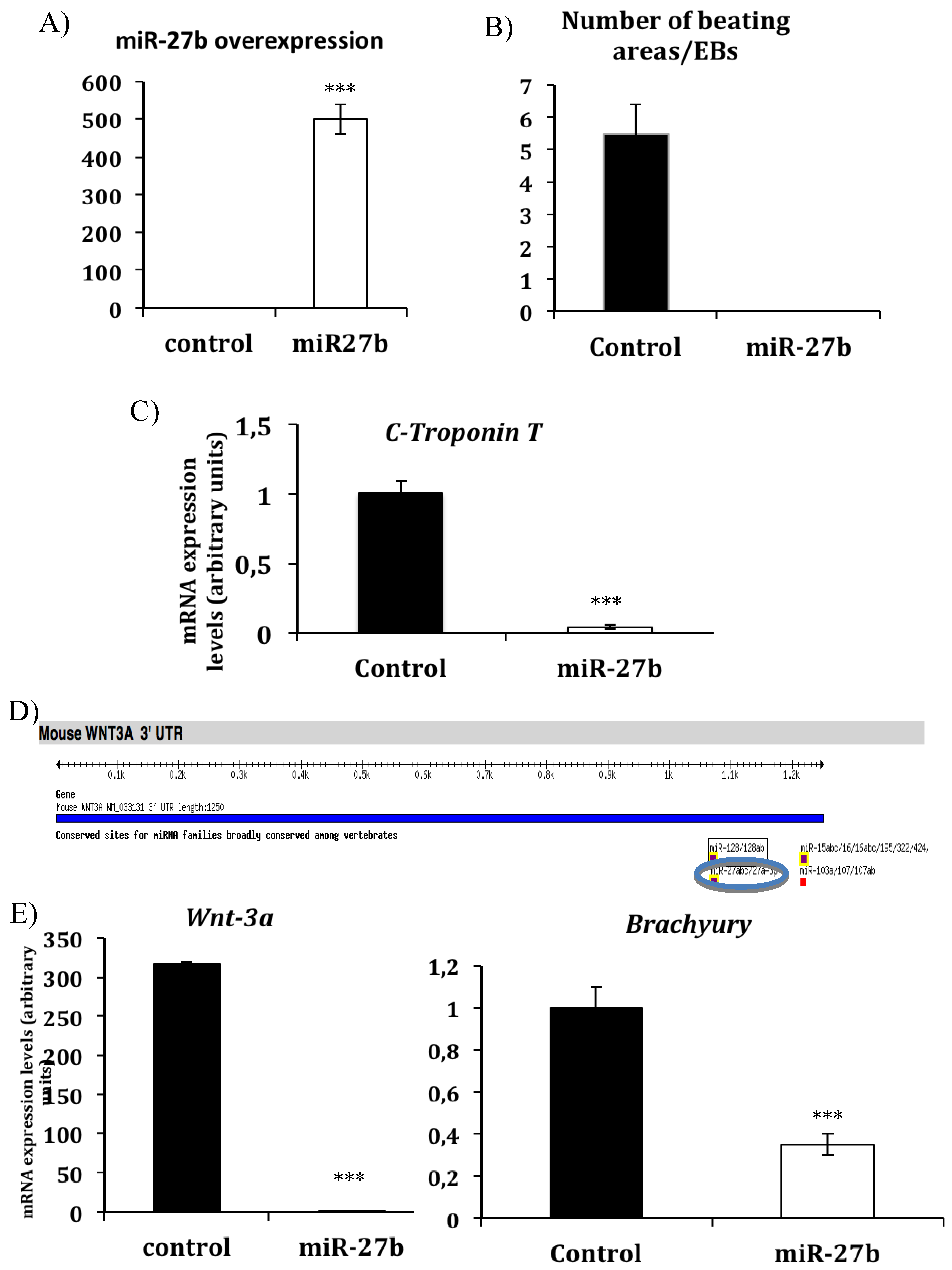
3.1. miR-27b Inhibits in Vitro Cardiac Differentiation by Modulating Wnt-Beta Catenin Canonical Pathway

Our previous microarray analyses of the microRNA expression profile during mouse cardiac chamber formation at three different developmental stages (E12.5, E15.5, and E18.5) revealed that only a cluster, representing the 11% of the analyzed miRNAs, display increasing expression levels during this developmental window [9]. To further explore the role of this cluster of miRNAs on cardiomyocyte differentiation we have used the in vitro system to cardiomyocyte differentiation from murine embryonic stem cells. We decided to analyze first the effects of those microRNAs that display early differential expression profiles, such as miR-27b. For this purpose, embryos bodies formed from mouse embryonic stem cell R1 cell line were transfected when platted with pre-miR-27b and cultured for 7 days on differentiating medium. Cardiac phenotype was analyzed at this step. Isolated beating areas and embryos bodies were collected for RT-PCR analyses. Surprisingly we observed that miR-27b-transfected EBs fail to form beating areas and the expression of cardiac markers such as c-Troponin T was clearly diminished, indicating that cardiac differentiation was inhibited in the EBs overexpressing miR-27b (Figure 1A–C, Supplementary Material Figures 1 and 2).
We have previously demonstrated that miR-27b regulates Mef2c in differentiated myogenic cells [9]. However, Mef2 transcription factor family members are dispensable for cardiac lineage specification and determination [11]. Therefore, we have searched for novel miR27 targets, which might mediate early cardiac specification. Using bioinformatics analyses (Targetscan software) we identify Wnt3a as a miR-27b predicted target (Figure 1C). RT-PCR analyses showed that Wnt3a is clearly down regulated on EBs-overexpressing miR-27b (Figure 1D) indicating that this effective activator of the Wnt canonical pathway is regulated by miR27b in our system. Given that in murine ES cells, the early inhibition of the canonical Wnt3a pathway blocks the expression of mesoderm specific marker genes such as Brachyury [12], we have looked for Brachyury expression in our system. As showed in Figure 1D, the expression levels of Brachyury was clearly decreased on EBs-overexpressing miR-27b indicating that the lost of mesoderm induction mediated by Brachyury may underlay the inhibition of cardiac differentiation in this system. These results point out a role of miR-27b regulating mesoderm specification and as a consequence cardiac differentiation from cultured EBs.

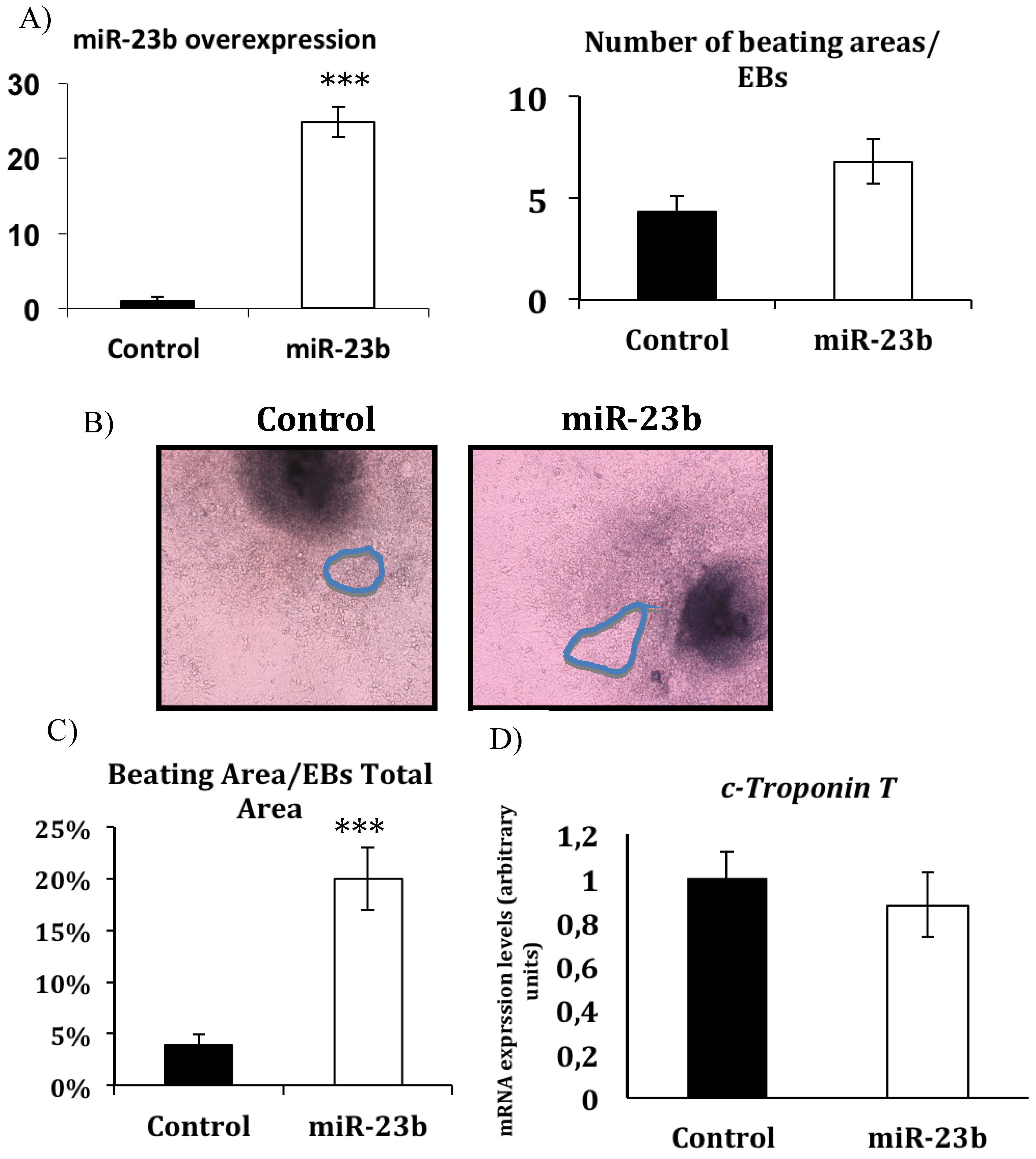
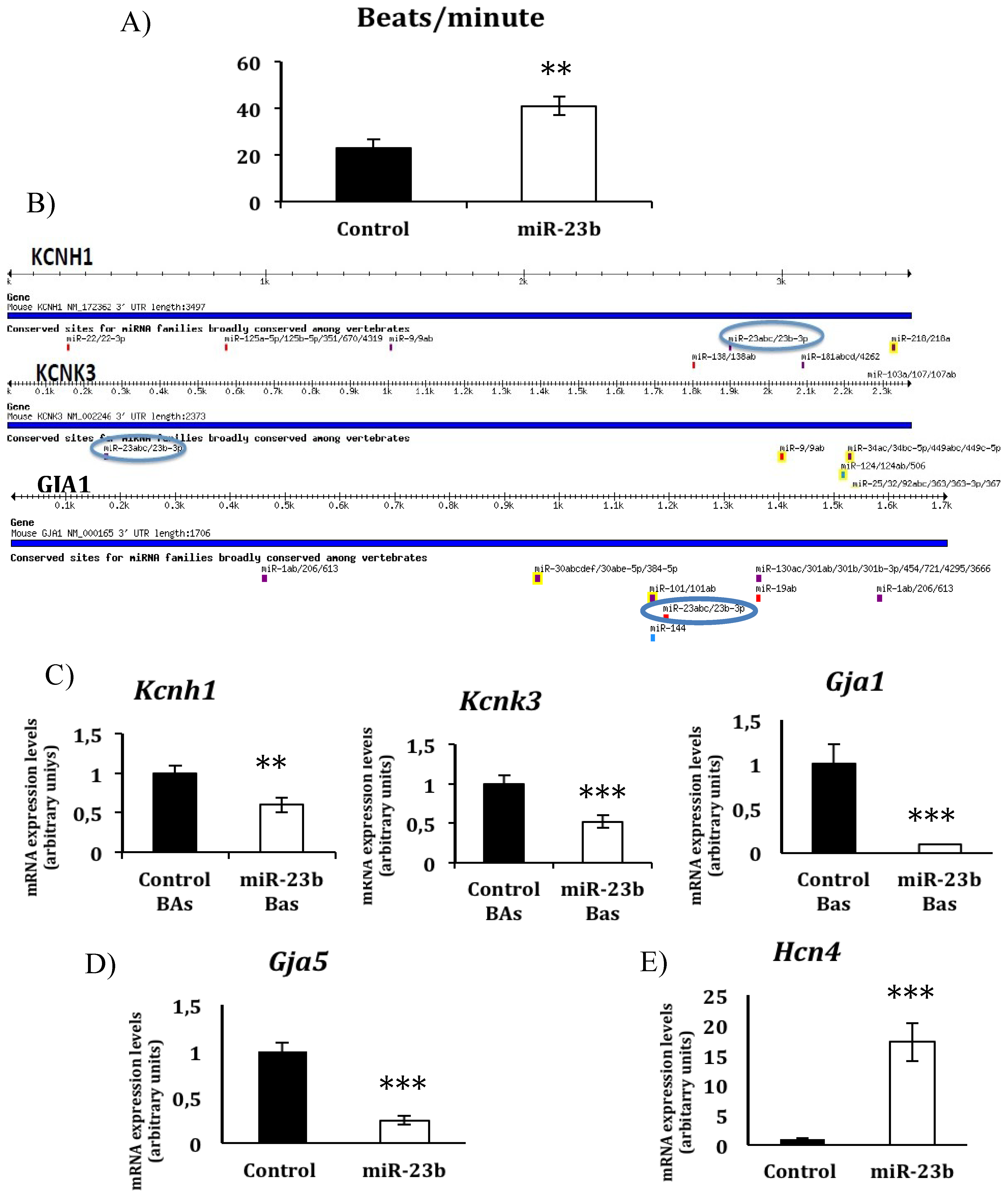
3.2. miR-23b Regulates the Beating Phenotype During in Vitro Cardiac Differentiation from ESCs

Next, we tested the effects on in vitro cardiomyocyte differentiation of the microRNAs that display late differential expression profiles during cardiac maturation, such as miR-23b. In contrast to miR-27b, EBs overexpressing miR-23b normally develop beating areas in a similar ratio as to control EBs, indicating that miR-23b overexpression does not alter beating areas formation (Figure 2A). However the size of beating areas from EBs ovesrexpressing miR-23b was significantly larger than the size of control beating areas (Figure 2B,C) whereas no changes in the expression levels for terminally differentiated genes such as c-troponin T were detected (Figure 2D). Moreover, we found that the number of beats per minute was significantly higher in beating areas-overexpressing miR-23b (Figure 3A, Supplementary Material Figures 2 and 3, suggesting that miR-23b overexpression instead to increase cardiac differentiation, might be modifying the beating activity of cardiomyocytes derived from ESCs.
Figure 1. (A) Overexpression of miR-27b in embryonic bodies (EBs) derived from murine Embryonic Stem Cells (ESCs) analyzed by RT-PCR. (B) EBs-overexpressing miR-27b fail to form beating areas (n = 22). (C) c-Troponin T expression profiles on EBs overexpressing miR-27b. (D) Schematic representation of the putative microRNA binding sites in Wnt3a 3'-UTR region as revealed by TargetScan althgoritm (www.targetscan.org). (E) Wnt3a and Brachyury expression profiles on EBs overexpressing miR-27b.
Jcdd 01 00041 g001
Figure 2. (A) Overexpression of miR-23b in EBs derived from murine ESCs analyzed by RT-PCR and number of beating areas in EBs overexpressing miR-27b respect to control. (B) Representative image showing the size of beating areas in EBs overexpressing miR-23b respect to control. (C) Percentage of the size of beating areas respect to total area of EBs (n = 20). (D) c-Troponin T expression profiles on EBs overexpressing miR-27b.
Jcdd 01 00041 g002
Figure 3. (A) Quantification of the number of beats per minute in beating areas from EBs overexpressing miR-23b (n = 15) in comparison with beating areas derived from EBs control (n = 13). (B) Schematic representation of the putative microRNA binding sites in Hcnh1 and Kcnk3 3'-UTR regions as revealed by TargetScan algorithm (www.targetscan.org). (C) Hcnh1, Kcnk3 and Gja1 expression profiles on EBs overexpressing miR-27b. (D) Gja5 expression profile on EBs overexpressing miR-27b. (E) Hcn4 expression profile on EBs overexpressing miR-27b.
Jcdd 01 00041 g003
Interestingly, using bioinformatics algorisms as previously mentioned, we identified Kcnh1, Kcnk3 and Gja1 as predicted targets for miR-23b (Figure 3B). Kcnh1 and Kcnk3, encode different potassium channels, which distinctly modify the action potential of excitable cells [13,14] and Gja1 encodes for connexin 43 (Cx43), one of the fast conductance gap junctions more extensively distributed in the myocardium [15]. RT-PCR analysis for Kcnh1, Kcnk3 and Gja1 revealed that Kcnh1, Kcnk3 and more significantly Gja1 were down-regulated in beating areas overexpressing miR-23b (Figure 3C). Additionally, connexin 40 (Cx40, Gja5); which together with Cx43 is abundantly expressed in the myocardium and the conduction system but sparsely expressed in the sinoatrial and atrio-ventricular nodes [15], was found to be also down regulated (Figure 3D). However, an up-regulation for the pacemaker specific marker Hcn4 was detected in beating areas overexpressing miR-23b (Figure 3E). Taken together, these findings reveal that miR-23b plays a role in modulating the electrophysiological features of differentiating cardiomyocytes from embryonic stem cells.

4. Discussion

Since the functional benefits reported for stem cell have been modest and mainly due to paracrine effects rather than differentiation of the applied cells into cardiomyocytes [16]; new tools are necessary in order to improve stem cell differentiation towards specific cardiovascular lineages. In the last decade microRNAs have been found to play important roles in the regulation of multiple biologic functions, including the control of stem cell and tissue differentiation [16]. Here, we report the role of miR-27b and miR-23b in cardiomyogenesis using an overexpression strategy in murine embryonic stem cells.
Early in development cardiac progenitors cells are specified in the mesodermal germ layer and many studies have shown that the formation of mesoderm is dependent on canonical Wnt signaling. In fact, loss of Wnt3a function is accompanied by the absence of mesoderm-specific marker genes [12,17]. Here we have shown that miR-27b regulates cardiac differentiation from murine embryonic stem cells by modulating Wnt canonical pathway leading to down-regulation of mesoderm-specific genes such as Brachyury. These finding reveal previous unknown functions of miR-27b on early cardiac specification.
Cardiomyocyte maturation from embryonic stem cells requires the acquisition of specific electrophysiological properties that lead to the formation of specific cardiac phenotype such as atrial, nodal or ventricular cardiomyocytes [18] and it has been previously reported that miRNAs such as miR-1 and miR-133 can post-transcriptionally regulate the electrophysiological characteristics on cardiomyocytes [19]. Here we report that miR-23b overexpression modify the beating during in vitro cardiac differentiation from ESCs. We have shown that miR-23b overexpression down-regulates the expression of its putative targets Kcnh1, Kcnk3 and Gja1. The Kcnh1 gene encodes a voltage-gated potassium channel highly expressed in neurons and is involved in tumor cell proliferation [20]; however, its physiological on the heart remain largely unclear. Kcnk3 encodes for a member of the two-pore-domain potassium channel family, the TASK-1 channels which has been considered to have a role in determining the action potential duration [14]. Gja1 (Cx43) and Gja5 (Cx40) down-regulation together with Hcn4 up-regulation has been recently reported to be crucial for activation of the pacemaker gene program, and suppression of the working myocardial gene program in EBs [21]. Besides Gja1 down-regulation we have found that the expression levels for Gja5 were lower and the expression levels for Hcn4 higher in beating areas overexpressing miR-23b suggesting that miR-23b overexpression leads to a more defined pacemarker phenotype. Therefore, our findings point out a role of miR-23b defining the electrophysiological characteristics of cardiomyocytes derived from ESCs.

5. Conclusions

In conclusion, this study could have a relevant impact in regenerative medicine since we showed that miR-27b and miR-23b overexpression modify the cell fate though cardiac lineage from ECSs. Additional experiments and further analyses could leads us to purpose these miRNAs as excellent tools to obtain specific cardiac cell types from ESCs.

Acknowledgments

This work was partially supported by grants BFU2012-38111 (Ministerio de Economía y Competitividad, Gobierno de España), CTS-1614, P08-CTS-03878, BIO-302 (Junta de Andalucia) and AFM2012-16074 (AFM).

Author Contributions

Jose Manel Vilches: Collection and/or assembly of data; Antonio Pulido: Collection and/or assembly of data; Francisco Hernández-Torres: Collection and/or assembly of data; Diego Franco: Data analysis and interpretation; Manuscript writing; Amelia Aránega: Conception and design; Data analysis and interpretation, Financial support, Manuscript writing.

Conflicts of Interest

The authors declare no conflict of interest.

References

  1. Srivastava, D. Making or breaking the heart: From lineage determination to morphogenesis. Cell 2006, 126, 1037–1048. [Google Scholar] [CrossRef]
  2. Bernstein, E.; Kim, S.Y.; Carmell, M.A.; Murchison, E.P.; Alcorn, H.; Li, M.Z.; Mills, A.A.; Elledge, S.J.; Anderson, K.V.; Hannon, G.J. Dicer is essential for mouse development. Nat. Genet. 2003, 35, 215–217. [Google Scholar] [CrossRef]
  3. da Costa Martins, P.A.; Bourajjaj, M.; Gladka, M.; Kortland, M.; van Oort, R.J.; Pinto, Y.M.; Molkentin, J.D.; De Windt, L.J. Conditional dicer gene deletion in the postnatalmyocardium provokes spontaneous cardiac remodeling. Circulation 2008, 118, 1567–1576. [Google Scholar] [CrossRef]
  4. Zhao, Y.; Ransom, J.F.; Li, A.; Vedantham, V.; von Drehle, M.; Muth, A.N.; Tsuchihashi, T.; McManus, M.T.; Schwartz, R.J.; Srivastava, D. Dysregulation of cardiogenesis, cardiac conduction, and cell cycle in mice lacking miRNA-1–2. Cell 2007, 129, 303–317. [Google Scholar] [CrossRef]
  5. Liu, N.; Bezprozvannaya, S.; Williams, A.H.; Qi, X.; Richardson, J.A.; Bassel-Duby, R.; Olson, E.N. microRNA-133a regulates cardiomyocyte proliferation and suppresses smooth muscle gene expression in the heart. Genes Dev. 2008, 22, 3242–3254. [Google Scholar] [CrossRef]
  6. Wilson, K.D.; Hu, S.; Venkatasubrahmanyam, S.; Fu, J.D.; Sun, N.; Abilez, O.J.; Baugh, J.J.; Jia, F.; Ghosh, Z.; Li, R.A.; Butte, A.J.; Wu, J.C. Dynamic microRNA expression programs during cardiac differentiation of human embryonic stem cells: Role for miR-499. Circ. Cardiovasc. Genet. 2010, 3, 426–435. [Google Scholar] [CrossRef]
  7. Cordes, K.R.; Sheehy, N.T.; White, M.P.; Berry, E.C.; Morton, S.U.; Muth, A.N.; Lee, T.H.; Miano, J.M.; Ivey, K.N.; Srivastava, D. miR-145 and miR-143 regulate smooth muscle cell fate and plasticity. Nature 2009, 460, 705–710. [Google Scholar]
  8. Ivey, K.N.; Muth, A.; Arnold, J.; King, F.W.; Yeh, R.F.; Fish, J.E.; Hsiao, E.C.; Schwartz, R.J.; Conklin, B.R.; Bernstein, H.S.; Srivastava, D. MicroRNA regulation of cell lineages in mouse and humanem bryonic stem cells. Cell. Stem Cell. 2008, 6, 219–229. [Google Scholar]
  9. Chinchilla, A.; Lozano, E.; Daimi, H.; Esteban, F.J.; Crist, C.; Aranega, A.E.; Franco, D. MicroRNA profiling during mouse ventricular maturation: A role for miR-27 modulating Mef2c expression. Cardiovasc. Res. 2011, 89, 98–108. [Google Scholar] [CrossRef]
  10. Lozano, E.; Chinchilla, A.; Martínez, S.; Hernández, F.; Navarro, F.; Lyons, G.E.; Franco, D.; Aránega, A.E. Pitx2c modulates cardiac-specific transcription factors networks in differentiating cardiomyocytes from murine embryonic stem cells. Cells Tissues Organs 2011, 194, 349–362. [Google Scholar] [CrossRef]
  11. Black, B.L.; Olson, E.N. Transcriptional control of muscle development by myocyte enhancer factor-2 (MEF2) proteins. Annu. Rev. Cell Dev. Biol. 1998, 14, 167–196. [Google Scholar] [CrossRef]
  12. Lindsley, R.C.; Gill, J.G.; Kyba, M.; Murphy, T.L.; Murphy, K.M. Canonical Wnt signaling is required for development of embryonic stem cell-derived mesoderm. Development 2006, 133, 3787–3796. [Google Scholar] [CrossRef]
  13. Stengel, R.; Rivera-Milla, E.; Sahoo, N.; Ebert, C.; Bollig, F.; Heinemann, S.H.; Schönherr, R.; Englert, C. Kcnh1 voltage-gated potassium channels are essential for early zebrafish development. J. Biol. Chem. 2012, 287, 35565–35575. [Google Scholar] [CrossRef]
  14. Putzke, C.; Wemhöner, K.; Sachse, F.B.; Rinné, S.; Schlichthörl, G.; Li, X.T.; Jaé, L.; Eckhardt, I.; Wischmeyer, E.; Wulf, H.; Preisig-Müller, R.; Daut, J.; Decher, N. The acid-sensitive potassium channel TASK-1 in rat cardiac muscle. Cardiovasc. Res. 2007, 75, 59–68. [Google Scholar] [CrossRef]
  15. Boyett, M.R.; Honjo, H.; Kodama, I. The sinoatrial node, a heterogeneous pacemaker structure. Cardiovasc. Res. 2000, 47, 658–687. [Google Scholar] [CrossRef]
  16. Porrello, E.R. microRNAs in cardiac development and regeneration. Clin. Sci. 2013, 125, 151–166. [Google Scholar] [CrossRef]
  17. Huelsken, J.; Vogel, R.; Brinkmann, V.; Erdmann, B.; Birchmeier, C.; Birchmeier, W. Requirement for beta-catenin in anterior-posterior axis formation in mice. J. Cell Biol. 2000, 148, 567–578. [Google Scholar] [CrossRef]
  18. van den Heuvel, N.H.; van Veen, T.A.; Lim, B.; Jonsson, M.K. Lessons from the heart: Mirroring electrophysiological characteristics during cardiac development to in vitro differentiation of stem cell derived cardiomyocytes. J. Mol. Cell. Cardiol. 2013, 67, 12–25. [Google Scholar] [CrossRef]
  19. Belevych, A.E.; Sansom, S.E.; Terentyeva, R.; Ho, H.T.; Nishijima, Y.; Martin, M.M.; Jindal, H.K.; Rochira, J.A.; Kunitomo, Y.; Abdellatif, M.; Carnes, C.A.; Elton, T.S.; Györke, S.; Terentyev, D. MicroRNA-1 and -133 increase arrhythmogenesis in heart failure by dissociating phosphatase activity from RyR2 complex. PLoS ONE 2011, 6. [Google Scholar] [CrossRef]
  20. Spitzner, M.; Ousingsawat, J.; Scheidt, K.; Kunzelmann, K.; Schreiber, R. Voltage-gated K+ channels support proliferation of colonic carcinoma cells. FASEB J. 2007, 21, 35–44. [Google Scholar]
  21. Hashem, S.I.; Lam, M.L.; Mihardja, S.S.; White, S.M.; Lee, R.J.; Claycomb, W.C. Shox2 regulates the pacemaker gene program in embryoid bodies. Stem Cells Dev. 2013, 22, 2915–2926. [Google Scholar] [CrossRef]

