Article

Global Regulator AdpA_1075 Regulates Morphological Differentiation and Ansamitocin Production in Actinosynnema pretiosum subsp. auranticum

1
State Key Laboratory of Bioreactor Engineering, East China University of Science and Technology, 130 Meilong Road, Shanghai 200237, China
2
Shanghai Collaborative Innovation Center for Biomanufacturing Technology, 130 Meilong Road, Shanghai 200237, China
*
Authors to whom correspondence should be addressed.
Bioengineering 2022, 9(11), 719; https://doi.org/10.3390/bioengineering9110719
Submission received: 30 October 2022 / Revised: 16 November 2022 / Accepted: 17 November 2022 / Published: 21 November 2022

Abstract

:
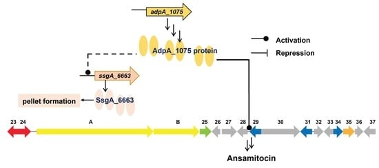
Actinosynnema pretiosum is a well-known producer of maytansinoid antibiotic ansamitocin P-3 (AP-3). Growth of A. pretiosum in submerged culture was characterized by the formation of complex mycelial particles strongly affecting AP-3 production. However, the genetic determinants involved in mycelial morphology are poorly understood in this genus. Herein a continuum of morphological types of a morphologically stable variant was observed during submerged cultures. Expression analysis revealed that the ssgA_6663 and ftsZ_5883 genes are involved in mycelial aggregation and entanglement. Combing morphology observation and morphology engineering, ssgA_6663 was identified to be responsible for the mycelial intertwining during liquid culture. However, down-regulation of ssgA_6663 transcription was caused by inactivation of adpA_1075, gene coding for an AdpA-like protein. Additionally, the overexpression of adpA_1075 led to an 85% increase in AP-3 production. Electrophoretic mobility shift assays (EMSA) revealed that AdpA_1075 may bind the promoter regions of asm28 gene in asm gene cluster as well as the promoter regions of ssgA_6663. These results confirm that adpA_1075 plays a positive role in AP-3 biosynthesis and morphological differentiation.

Graphical Abstract

1. Introduction

Ansamitocin P-3 (AP-3) exhibits antitumor activity against various cancer cell lines [1,2,3]. Its derivatives are commonly used as the ‘warhead’ molecule in antibody-conjugated drug (ADC) for the treatment of various solid tumors [4]. AP-3 is a member of various ansamitocin congeners produced by A. pretiosum. Ansamitocins are of limited industrial applicability because of their low production yields. In recent decades, considerable efforts have been made to further enhance AP-3 yield to satisfy the industrial demands with medium optimiztion and genetic modifications because of its great pharmaceutical value [5,6,7,8,9,10,11,12].
From an industrial point of view, liquid culture is favorable for large scale production of antibiotics. Actinomycetes are usually subjected to submerged fermentation. Unlike other bacteria, Actinobacteria remarkably exhibits complex morphology during submerged cultivation. In liquid culture, their mycelium shows filamentous growth, exhibiting dispersed mycelial form or compact mycelial network [13]. It is well studied that different morphological forms lead to various degrees of nutrient and oxygen transfer during the fermentation process [14]. Three types of morphologies-freely dispersed mycelia, open mycelial networks, and compact mycelial network are generally distinguished in submerged cultures [15]. The morphological phenotypes are genetically determined and differ considerably between strains. The formation of unfavorable morphology during liquid-grown cultures may be a major bottleneck hindering the industrial production of antibiotics [16]. Submerged culture studies for different antibiotic production have made efforts to select strains with better growth characteristics. Forming dispersed mycelium helps productivity in the bioreactor for Streptomyces hygroscopicusin producing rapamycin [17]. A similar situation was found in previous studies of tylosin and nystatin production in both Streptomyces fradiae and Streptomyces noursei [18,19]. However, for production of nikkomycin and erythromycin pelleted growth is preferred [20,21]. Additionally, forming dense pellets also contributed to the productivity optimization of Streptomyces lividans TK21 for a hybrid antibiotic production as well [22].
Therefore, in order to optimize mycelial morphology in a more targeted and flexible manner, several genetic determinants have been identified that play roles in the control of morphogenesis [23]. The genetic factors include cell-matrix proteins and extracellular polymers. Morphoproteins with specific roles in liquid-culture morphogenesis for apical growth and hypha branching include the cell wall remodeling protein SsgA-like proteins (SALPs) [24], and the cellulose synthase-like protein CslA [25]. Members of the family of SALPs are required to activate cell division in both solid and liquid culture sporulation [26,27,28]. SsgB is the archetypal SALP and functions by colocalizing with SsgA for recruiting FtsZ during aerial hyphae early division stage [29,30,31]. Morphology engineering strategies for ssgA gene modification were employed to obtain desirable morphologies and fast growth [32,33].
Interestingly, morphological differentiation caused by ssgA transcriptional variations are generally controlled by AdpA [28,34,35]. AdpA is universally present in Actinomycetes, as a global regulator of morphological differentiation and secondary metabolism [36,37]. In S. griseus, AdpA positively controls the expression of genes involved in spore formation and aerial mycelium formation, as well as activates the transcription of various genes related to secondary metabolism [38,39]. Whereas, overexpression of the adpAsx gene in S. xiamenensis 318 had negative effects on cell division genes, such as putative ssgA, ftsZ, ftsH, and whiB. Besides, it functions as a bidirectional regulator for the biosynthesis of xiamenmycin and PTMs [40]. To date, AdpA has been proven to contain a C-terminal domain with two helix-turn-helix (HTH) DNA binding motifs [41]. As a global transcription factor, the regulatory relationship between AdpA and target genes has been investigated in Streptomyces. AdpA and its orthologs activate or down-regulate genes, including repression of its own transcription, by directly binding to operator regions containing a consensus sequence [38,42,43].
Although A. pretiosum is being developed as a sustainable industrial production platform, the genes involved in cell division and morphological development are still poorly investigated. Gene APASM_4178 was identified as a subtilisin-like serine peptidase encoding gene responsible for mycelial fragmentation [44]. FtsZ protein from A. pretiosum as the analogue of β-tubulin, was demonstrated to be the AP-3 binding target. Overexpression of APASM_5716 gene that encodes FtsZ resulted in AP-3 resistance and overproduction in A. pretiosum ATCC 31280 [45]. A two-component signal transduction system, PhoPR homolog was identified in the genome of the A. pretiosum X47 strain. PhoP is the response regulator, negatively affecting morphological development and excluding its regulation on the biosynthesis of AP-3 in X47 strain [46]. However, for filamentous microorganisms, the importance of understanding the relationship between mycelial morphological development and antibiotic biosynthesis is nontrivial. In this study, a variant of A. pretiosum has been observed to form dense pellets, while the control strain formed loose clumps. In addition, excessive mycelial fragmentation of the control strain was observed early in the fermentation. We investigated several putative genes that may contribute to cell division and pellet architecture. Gene ssgA_6663 was identified as a key genetic determinant of compact mycelial network formation during solid and liquid cultures. We also characterized the roles of AdpA_1075 in controlling the morphological differentiation and AP-3 production. AdpA_1075 was determined to positively control the biosynthesis of ansamitocin by directly regulating the expression of asm28.

2. Materials and Methods

2.1. Bacterial Strains, Plasmids and Culture Conditions

All plasmids and strains used in this study are listed in Supplementary Table S1. A. pretiosum subsp. auranticum L40 was derived from A. pretiosum subsp. auranticum ATCC 31565 by atmospheric and room temperature plasma (ARTP) mutagenesis [12]. Strain L40 and its derivatives in this study were cultivated as described previously [47]. YMG agar plates (for solid culture) and TSBY broth (for liquid culture) were employed for strain culture. For fermentation experiments, strains were cultured in shake flasks at 28 °C for 8 days.
The stability of strain MD15 was tested following the method described by former study [10] with some modifications. In brief, strain MD15 was transferred at 24 h intervals for a total of twenty-five passages in YMG liquid medium. The original strain, fifteenth and twenty-fifth passages were selected to test the fermentation performances stability of strain MD15 in liquid fermentation.

2.2. Construction of Recombinant Strains

CRISPR-Cas9 mediated gene inactivation. Mutants with gene ssgA_6663, adpA_1075, or asm28 disruption were performed by pCRISPR-Cas9apre with a unified construction process [47]. As an example, the construction of mutant with gene ssgA_6663 deletion was described briefly. Two homologous arms (upstream 1.2 kb, downstream 1.3 kb) for ssgA_6663 deletion were amplified and together cloned to StuI-digested plasmid pCRISPR-Cas9apre by NEB DNA Assembly Master Mix (New England Biolabs, Ipswich, MA, USA) to give the pCRISPR-Cas9apreΔssgA. The ApE software (a plasmid editor, version 2.0.50. https://jorgensen.biology.utah.edu/wayned/ape/, accessed on 5 October 2016) was used to search N20 targeting sequences of sgRNAs. The sgRNA cassettes were cloned into the XmaJI/SnaBI site of pCRISPR-Cas9apreΔssgA. The amplification primers used to construct pCRISPR-Cas9apre series gene knockout plasmids are shown in Supplementary Table S2. E. coli ET12567 (pUZ8002) was employed to introduce the resulting plasmid into L40. According to protocol described elsewhere [47], the conjugants were induced and screened for the correct constructs by colony PCR and Sanger sequencing (Supplementary Figure S1).
Construction of plasmids for gene overexpression. pSETK derived from pSET152 was used to prepare overexpression plasmids of ssgA_6663, adpA_1075, ftsZ_5883 or asm28. More specifically, the kasOp*-rbs fragment was introduced to XbaI/EcoRV cloning site of pSET152. Aforementioned genes were amplified from A. pretiosum L40 chromosome. The amplicons were cloned into NdeI/EcoRV site of pSETK, respectively. The obtained recombinant plasmids pSETKssgA, pSETKftsZ, pSETKadpA, pSETKasm28, pSETKftsZ:ssgA, and control plasmid pSETK were individually transferred into E. coli ET12567(pUZ8002) and then integrated into the attB site of strain MD02 by intergeneric conjugation. The verification of these recombinant strains was performed by PCR (Supplementary Figure S2A–C). To construct the pSETKftsZ:ssgA plasmid, the fragment containing kasOp*-rbs-ssgA_6663 expression cassette cloned from pSETKssgA plasmid was inserted into pSETKftsZ digested by EcoRV to generate pSETKftsZ:ssgA. The verification of gene co-expression was double checked by PCR using two primer pairs 152yz-F/R and ftsZchk-f/ssgAchk-r (Supplementary Figure S2D–F).

2.3. RNA Isolation, cDNA Synthesis and Quantitative Real-Time PCR (qRT-PCR)

Total RNA was extracted using a bacterial RNA extraction kit (Jiangsu Cowin Biotech Co., Ltd., Taizhou, China). Isolated RNA was treated by DNase I before being reverse transcribed with cDNA Synthesis Kit (Jiangsu Cowin Biotech Co., Ltd., Taizhou, China). The cDNA templates were amplified in triplicate for each transcription analysis using MagicSYBR Mixture (Jiangsu Cowin Biotech Co., Ltd., Taizhou, China) with primers listed in Supplemental Table S2. The transcription of target genes was determined by RT-PCR using a CFX96 Real-Time System (Bio-Rad, Richmond, CA, USA). 16S rRNA gene was used for internal normalization. Relative transcript level of genes was quantified by the 2−ΔΔCt method [48].

2.4. Determination of AP-3 Production

AP-3 was extracted from the culture supernatant using a previously described method [12]. HPLC analysis of AP-3 was operated on Agilent series 1260 (Agilent Technologies, Inc., Santa Clara, CA, USA) equipped with a SinoChrom ODS-BP C18 column (4.6 mm × 250 mm, 5 μm, Elite, Dalian, China) coupled to UV detector at 254 nm. The column was eluted with 85% methanol and 15% water at 28 °C.

2.5. Mycelial Morphology Observation

Mycelial morphology was observed using an optical microscope (Olympus CX 31, Olympus Corporation, Tokyo, Japan). Culture broth (10 μL) was pipetted onto a standard glass slide (25 × 75 mm), dyed with crystal violet. Images were captured under an oil immersion lens (magnification, 100×).

2.6. Scanning Electron Microscope (SEM)

Mycelium was harvested by centrifugation, and washed with 0.1 M PBS. The mycelium was resuspended in 2.5% glutaraldehyde solution for 3 h. The fixed samples were then washed twice with 0.1 M PBS. Samples were subjected to gradient dehydration with ethanol solution (50%, 70%, 95% and 100%). Finally, the dehydrated samples analyses were carried out on a S3400-N scanning electron microscopy (Hitachi, Tokyo, Japan).

2.7. Heterologous Overexpression of AdpA-1075

Gene adpA_1075 was amplified with primers 28a1075-F/R. The adpA_1075 cassette was cloned in HindIII/NdeI-digested pET28a (+), generating plasmid pET-28a-adpA_1075. The plasmid was transformed into E. coli BL21(DE3) for protein overexpression. The generated strain BL21(DE3)/pET-28a-adpA_1075 was cultivated at 37 °C for 2–3 h in 100 mL LB medium containing 50 µg/mL kanamycin until OD600 reached about 0.6–0.8. Isopropyl-β-D-thiogalactoside (IPTG, 0.1 mM) was added after 30 min of cooling at 4 °C and further incubated overnight at 16 °C for AdpA_1075 expression. The cells were harvested and resuspended in 50 mM phosphate buffer solution (pH 7.5). His-tagged AdpA_1075 protein was released from cells by homogenization. Ni SepharoseTM 6 Fast Flow (GE Healthcare Life Sciences, Marlborough, MA, USA) was applied to proteins purification with elute buffer (50 mM phosphate buffer solution, 250 mM imidazole, 500 mM NaCl). The purified His-tagged AdpA-1075 was analyzed by 12.5% sodium dodecyl sulfate-polyacrylamide gel electrophoresis (SDS-PAGE).

2.8. Electrophoretic Mobility Shift Assays (EMSA)

EMSA was performed using a Chemiluminescent EMSA Kit (Beyotime Biotechnology, Shanghai, China) according to the manufacturer’s instructions. The two complementary oligonucleotides were annealed, labeled with biotin, and incubated with recombinant proteins in the absence or presence of excess amounts of unlabeled wildtype oligonucleotides. The protein-DNA complexes were separated on 5% polyacrylamide gels and the signals were captured with a Chemiluminescence Imaging System (BG-gdsAUTO 720, Baygene Biotechnol Co., Ltd., Shanghai, China). As a control, for each target gene, excessive unlabeled specific DNA fragments were added to the reactions, resulting in the appearance of free un-shifted probe and demonstrating that the binding was specific.

3. Results

3.1. Identification of ssgA in A. pretiosum subsp. auranticum

The previously constructed mutant MD15 with tandem deletion of two gene clusters (cluster T1PKS-15 and T1PKS/NRPS-5) [47] showed excellent fermentation stability (Figure 1A). To better understand the improvement of fermentation performance, the mycelia from culture broth were collected at 16, 24 and 72 h, and aerial hyphae were examined on YMG medium. Scanning electron microscopy (SEM) was used to demonstrate in detail the morphological differences between the mutant strain MD15 and the control strain L40. During the first 24 h of fermentation in both control strain L40 and mutant strain MD15, pellets were formed by aggregative hyphae. Mycelial fragmentation occurred in the control strain L40 at around 72 h, while in contrast, the mycelia of MD15 remained dense pellets with well-developed mycelial boundaries (Figure 1B). Compared with the loosely interwoven mycelial morphological characteristics of L40, the aerial mycelia of MD15 formed a tight mycelial network (Figure 1C). We speculate that gene expression may be altered in strain MD15. SsgA, FtsZ, SsgB, and CslA encoding genes were found, namely ssgA_6663, ftsZ_5883, ssgB_2072, and cslA_0512, respectively, in A. pretiosum subsp. auranticum ATCC 31565 genome. To better understand the role of these genes in mutant MD15 morphological development, the transcription levels of these target genes were measured by qRT-PCR on the third day of fermentation. In mutant MD15, transcription levels of ssgA_6663 and ftsZ_5883 were 124% and 160% higher than those in strain L40, respectively (Figure 1D). The transcription of gene ssgB_2072 was not detected, suggesting that the strain was defective in the initiation of sporulation, as the protein complexes SsgA and SsgB cannot colocalize in aerial hyphae. It has been supported by phenotypic observations. In strain L40 and its derivative, smooth and uncoiled aerial hyphae formed, but no spores (Figure 1C). Negligible changes were observed in cslA_0512, which encodes a cellulose synthase-like protein homologue essential for pellet formation [49]. This fact excludes the involvement of cslA_0512 in the formation of compact pellets in A. pretiosum.
Mycelial fragmentation has previously been reported to impede AP-3 production [12,44]. Pelleted growth is preferred for A. pretiosum producing AP-3 in submerged cultivation [12]. The sporulation-specific gene ssgA directly activates cell division in Streptomyces [27,50]. Moreover, the expression of ssgA_6663 also varies with strain morphology in non-spore producing A. pretiosum (Figure 1) [12]. We therefore assume that both ssgA_6663 and ftsZ_5883 play important roles in controlling the morphogenesis in A. pretiosum, especially in the formation of intertwined mycelial network. As reported earlier, filamentous growth and sporulation of actinobacteria require the bacterial tubulin homolog FtsZ [39,51,52]. Overexpression of ftsZ gene might also be used to alleviate AP-3 toxicity and improve resistance of A. pretiosum to AP-3. Since FtsZ was determined to be the intracellular binding target of AP-3 [45]. A high transcriptional level of ftsZ_5883 was observed in mutant with a denser mycelial interweaving pattern (Figure 1D). Whether ssgA_6663 and ftsZ_5883 are involved in specific cellular process and contribute to stable fermentation remains to be investigated.

3.2. Deletion of ssgA_6663 Affected the Morphological Differentiation of A. pretiosum

We then investigated the effects of ssgA_6663 deletion on mycelium development and AP-3 production. The length of aerial mycelia of ssgA_6663 disruption mutant was much shorter than that of strain L40 without intertwining (Figure 2A). In submerged cultivation, mycelium of ΔssgA_6663 mutant developed into dispersed mycelial form (Figure 2B). The short aerial mycelium probably prevented the strain from making biomass from nutrients (Figure 2C). Further HPLC analysis revealed that AP-3 was almost undetected in the fermentation culture of the ssgA_6663 disruption mutant, and the biomass in the fermentation was also reduced (Figure 2D). However, the introduction of multiple copies of gene ssgA may cause an excessive mycelial fragmentation in spore-producing Streptomyces [50]. To verify whether ssgA_6663 and ftsZ_5883 play a positive role in mycelial aggregation and entanglement, these two cell division genes were overexpressed in tandem under the drive of kasOp*. The co-overexpression of ftsZ_5883 and ssgA_6663 caused the long hyphae to intertwine closely with each other and form clumps, which was more visible than that of the strain overexpressing ftsZ_5883 alone (Figure 2B). This phenomenon may be consistent with previous reports that secondary metabolism and morphological differentiation can be regulated by global regulator, rather than morphology directly affecting secondary products biosynthesis [35]. The timing of SsgA expression in Streptomyces sporulation-specific cell division and morphogenesis can be regulated by global regulator AdpA [50]. As previously reported in A. pretiosum ATCC 31280, AdpA-like protein APASM_1021 was found [44]. Gene adpA_1075 was identified as an AdpA orthologue from three putative AraC family protein encoding genes in A. pretiosum subsp. auranticum for the high amino acid sequence identity (97.17%) between AdpA_1075 and APASM_1021 (Supplementary Figure S3). We therefore speculate that AdpA_1075 may also play a regulatory role in the L40 strain. Furthermore, the lawns of ssgA_6663-null mutant on YMG plate exhibited a bald phenotype distinct from L40, whereas deletion of adpA_1075 had little restriction on the arising of aerial hyphae (Figure 2C). Interestingly, deletion of adpA_1075 also resulted in a significant decrease in AP-3 production without inhibition of biomass accumulation, suggesting that adpA_1075 is essential for AP-3 biosynthesis as a pleiotropic transcriptional regulator (Figure 2D).

3.3. Overexpression of adpA_1075 Increased the Production of AP-3

As reported previously, ssgA is essential for septum formation in aerial hyphae, a late step in morphological differentiation. Therefore, ssgA mutation may not affect the production of secondary metabolites [26,27,28]. In this study, ssgA_6663 or adpA_1075 was individually overexpressed under the strong promoter kasOp* on the shuttle vector pSETK, to investigate the effects of the enhanced expression of these two proteins on ansamitocin biosynthesis. The fermentation experiments were performed for the recombinant strains overexpressing ssgA_6663, ftsZ_5883, or adpA_1075, and the control strain MD02::pSETK (MD02 integrated with the vector pSETK), respectively. The results showed that the overexpression of ssgA_6663 did not improve AP-3 production, which was consistent with what was observed in S. griseus [26,28]. As expected, enhanced expression of ftsZ_5883 improved strain resistance against AP-3 [45], alleviated cell toxicity, and increased AP-3 production by 45% (Figure 3A). Moreover, overexpression of adpA_1075 increased AP-3 production by 85% without affecting dry cell weight (DCW) at the end of fermentation (Figure 3A,B). Therefore, we may conclude that overexpression of ssgA_6663 only causes morphological changes but does not directly promote AP-3 production. Overexpression of adpA_1075 may not only regulate ansamitocins biosynthesis, but also regulate strain morphology by controlling the expression of ssgA_6663.

3.4. AdpA_1075 Is Involved in the Regulation of ssgA_6663 Transcription in A. pretiosum

The AraC family transcription factor known as AdpA is a global regulator of morphological differentiation and secondary metabolism [42,43,53,54]. Cell division genes are included in members of AdpA regulon, such as ssgA which requires AdpA to turn on transcription [28,50,55,56]. Since the fermentation morphology changes caused by ssgA_6663 inactivation did not directly affect the production of AP-3, we hypothesized that AdpA_1075 might be involved in both ssgA_6663 expression and ansamitocins production. Diametrically opposed transcriptional patterns of ssgA_6663 in adpA_1075 inactivation mutant seemed to further validate the hypothesis. In parent strain L40, the transcription of ssgA_6663 increased significantly on day 3 of fermentation, but decreased sharply when the strain switched to the stationary growth phase (Figure 4). In contrast, in mutant MD08, ssgA_6663 transcription was down-regulated due to the absence of AdpA_1075 and remained low from the third day of fermentation (Figure 4).

3.5. AdpA_1075 Binds to Promoters of ssgA_6663 and asm28

The above analyses demonstrated that AdpA_1075 regulated morphological development by controlling the expression of ssgA_6663. To determine whether AdpA_1075 directly regulated the gene transcription of ssgA_6663, EMSA was performed. As previously reported, a consensus AdpA-binding sequence such as 5′-TGGCSNGWWY-3′ (S: G or C; W: A or T; Y: T or C; N: any nucleotide) was identified in S. griseus (Figure 5A) [57]. Consequently, the upstream region of ssgA_6663 was analyzed by gene sequence alignment and manual correction, according to the reported AdpA-binding motifs in Streptomyces and A. pretiosum ATCC 31280 [44,57]. A conserved AdpA-binding motif, 5′-TGGCCGGAAC-3′ (in reversed orientation) was identified (Figure 5B). AdpA_1075-His6 protein was then purified, and biotin labeled probes were prepared as mentioned above. EMSA results showed that the complex AdpA_1075-PssgA was formed in a protein concentration-dependent manner, confirming that AdpA_1075 bound specifically to the promoter region of ssgA_6663 (Figure 5E). Surprisingly, we not only found an AdpA-binding sequence in the promoter region of ssgA_6663, but also observed a potential AdpA-binding site 5′-TGGCCCGAAC-3′ (in reversed orientation) in the upstream region of asm28 (Figure 5C). Compared to the transcriptional changes of ssgA_6663 and ftsZ_5883 in adpA_1075 overexpression mutant, transcriptional level of asm28 in the adpA_1075 overexpression strain was 3.5 times higher than that in the parent strain (Figure 5D). Unexpectedly, the overexpression of adpA_1075 did not affect the expression of ftsZ_5883. The slight up-regulation of ssgA_6663 indicated that another regulator may be also involved in the regulation of ssgA_6663 transcription. Results of EMSAs showed that Pasm28 probe was shifted when incubated with 1000 nM AdpA_1075-His6 (Figure 5E). The above results suggested that AdpA_1075 specifically binds to the promoters of ssgA_6663 and asm28, implying that AdpA_1075 controls the transcription of these genes directly. The upstream region of asm28 is the only potential AdpA binding site on the asm gene cluster, as it contains a 10-bp consensus sequence mentioned above. However, the function of asm28 has not been reported. Therefore, we further investigated the effect of asm28 gene on AP-3 production. Mutant with asm28 deletion was constructed by CRISPR-Cas9-mediated vector pCRISPR-Cas9apreΔasm28 (Supplementary Figure S4A,B). AP-3 production of the obtained mutant MD19 was only 69% of that of parent strain MD01. However, overexpression of asm28 promoted ansamitocin biosynthesis and resulted in an approximately 25% increase in AP-3 production, without any effect on DCW of the strain at the end of fermentation (Figure S4D,E). These results demonstrated that AdpA_1075 activated asm28 and ssgA_6663 on the third to fourth day of fermentation, consistent with the enhanced AP-3 production and mycelial pellet formation in strains.

4. Discussion

The mycelial fragmentation of A. pretiosum under submerged conditions has long attracted researchers’ attention [12,44,58]. However, in our previous work, pelleted growth was found to be more conducive to the production of ansamitocin [12]. Other studies have revealed that cell-wall remodeling protein SsgA may control the development of fragmentation and promote the growth rate of spore-forming Streptomyces strains [32,33]. The absence of SsgB in A. pretiosum makes it a non-spore-forming bacterium (Figure 1C,D) [59,60]. Additionally, both mycelial fragmentation and significant biomass enhancement was observed in ssgA_6663-overexpressing A. pretiosum (Figure 2B and Figure 3B). All these results indicate that ssgA_6663 is not responsible for mycelial fragmentation and AP-3 yield reduction in A. pretiosum.
Therefore, we hypothesized that, similar to that reported in S. limosus [32], A. pretiosum strain may produce large mycelial mat structures in the early growth phase, while the enhanced expression of ssgA_6663 leads to pellet formation during stationary phase. As expected, a significant increase in the transcriptional profile of ssgA_6663 was observed in the parent strain L40, followed by a noticeable decrease from day 2 to day 4 of fermentation (Figure 4). However, this transcriptional pattern was disrupted when gene adpA_1075 was inactivated (Figure 4).
AdpA as a pleiotropic transcriptional regulator can regulate both morphological differentiation and secondary metabolism [61]. We further investigated the adpA_1075 gene, encoding an AdpA-like protein. Specific regulation of AdpA-like protein on subtilisin-family serine peptidase encoding gene resulted in a delayed fragmentation, which has been demonstrated in A. pretiosum ATCC 31280 [44]. Moreover, the ssgA_6663 gene has also been identified as being primarily responsible for the formation of mycelial intertwining. Based on these findings, we hypothesized that AdpA_1075, as a global regulator, may comprehensively control the development of multicellular structures and AP-3 biosynthesis during strain fermentation.
Despite the presence of many regulatory genes in the asm gene cluster, few studies have been reported on the regulation of ansamitocin biosynthesis. The functions of these regulatory genes were characterized by gene inactivation, complementation, transcriptional analysis, and feeding experiments [62,63,64,65]. For example, negative regulatory gene asm2 and asm34, and positive regulatory gene asm8 and asm18 are the putative regulatory genes on the asm gene cluster. Whereas, direct evidence for these regulators directly controlled asm structural genes was absent. Most of them probably control the expression of genes involved in resistance to xenobiotics, regulation of efflux pumps, and response to stressors [41].
AdpA homologs were well-studied, and have been proven to contain a C-terminal domain with two helix-turn-helix (HTH) DNA binding motifs [37]. In this study, the adpA_1075 gene was identified as an ortholog of pleiotropic regulator AdpA, and its DNA-binding site is similar to the conserved sequence of the AdpA-binding motifs in many Streptomyces species [57]. Earlier work revealed that the expression of APASM_4178, which is responsible for mycelial fragmentation, is specifically regulated by an AdpA-like protein in A. pretiosum ATCC 31280 [44]. In this work, the EMSA results also demonstrated the interaction between AdpA_1075 and promoter region of the subtilisin-family serine peptidase encoding gene (data not shown). Our data showed that AdpA_1075 of the A. pretiosum L40 strain directly binds to the promoters of the ssgA_6663 and asm28. Therefore, these findings deepen insights into the regulatory roles of the AdpA-like protein in A. pretiosum, revealing a high functional similarity to the AdpA homolog in Streptomyces reported previously [35].
In this study, an intergenic region containing AdpA-binding motif was identified in the upstream region of asm28 (Figure 5). Our findings in vitro and in vivo experiments suggested that asm28 may be the target of AdpA_1075 for regulating ansamitocin biosynthesis (Figure 5 and Figure S4). Moreover, the global regulator bldA controls antibiotic production as well, by regulating sporulation and antibiotic production in Streptomyces [66]. Generally, the TTA codons are very rare in GC-rich genomes of Actinobacteria. TTA-containing genes are usually the cluster-situated regulators of secondary metabolite biosynthesis. In this study, we found that the asm28 open reading frame (ORF) also contains a TTA codon, indicating that asm28 might be an important regulatory target during ansamitocin biosynthesis. However, the function of asm28 gene and its encoded protein remain uncharacterized at present. Follow-up studies are needed to confirm this hypothesis and to elucidate the complex regulatory network in which asm28 is involved.

5. Conclusions

In this study, we have elucidated the function of ssgA_6663 in mycelial development. ssgA_6663 can dominate the mycelial intertwining and pellet formation. The silencing of the ssgB_2072 gene resulted in the absence of SsgB, which may explain the fact that the strain developed into mycelium without sporulation septa.
Additionally, we characterized the regulatory role of AdpA_1075 in A. pretiosum. AdpA_1075 acts as a global regulator, affecting morphological differentiation and promoting the biosynthesis of ansamitocin. Our findings provide additional useful evidence for the regulatory mechanism of ansamitocin biosynthesis.

Supplementary Materials

The following supporting information can be downloaded at: https://www.mdpi.com/article/10.3390/bioengineering9110719/s1, Table S1: Strains and plasmids used in this study; Table S2: Primers used in this study; Figure S1: Verification of ssgA_6663 and adpA_1075 gene deletion mutant strains; Figure S2: Identification of recombinant strains; Figure S3: Sequence alignment of AdpA-like protein APASM_1021 with AdpA_1075; Figure S4: Effects of asm28 gene deletion or overexpression.

Author Contributions

Conceptualization, Q.H. and F.L.; methodology, S.G. and T.L.; validation, S.G. and X.S.; investigation, S.G. and J.Z.; data curation, S.G., R.L., J.C. and F.H.; writing—original draft preparation, S.G.; writing—review and editing, Q.H. and F.L.; supervision, Q.H. and F.L.; funding acquisition, Q.H. and F.L. All authors have read and agreed to the published version of the manuscript.

Funding

This work was supported by the National Natural Science Fund for Young Scholars (32001035) and Shanghai Super postdoctoral program 2020.

Institutional Review Board Statement

Not applicable.

Informed Consent Statement

Not applicable.

Data Availability Statement

All data generated or analyzed during this study are included in this article.

Conflicts of Interest

The authors declare no competing interest.

References

  1. Prelog, V.; Oppolzer, W. Ansamycins, a novel class of microbial metabolites. Helv. Chim. Acta 1973, 56, 2279–2287. [Google Scholar] [CrossRef] [PubMed]
  2. Martin, K.; Müller, P.; Schreiner, J.; Prince, S.S.; Lardinois, D.; Heinzelmann-Schwarz, V.A.; Thommen, D.S.; Zippelius, A. The microtubule-depolymerizing agent ansamitocin P3 programs dendritic cells toward enhanced anti-tumor immunity. Cancer Immunol. Immunother 2014, 63, 925–938. [Google Scholar] [CrossRef] [PubMed]
  3. Kashyap, A.S.; Fernandez-Rodriguez, L.; Zhao, Y.; Monaco, G.; Trefny, M.P.; Yoshida, N.; Martin, K.; Sharma, A.; Olieric, N.; Shah, P.; et al. GEF-H1 signaling upon microtubule destabilization is required for dendritic cell activation and specific anti-tumor responses. Cell Rep. 2019, 28, 3367–3380.e8. [Google Scholar] [CrossRef] [PubMed] [Green Version]
  4. Barok, M.; Joensuu, H.; Isola, J. Trastuzumab emtansine: Mechanisms of action and drug resistance. Breast Cancer Res. 2014, 16, 209. [Google Scholar] [CrossRef] [PubMed] [Green Version]
  5. Fan, Y.; Gao, Y.; Zhou, J.; Wei, L.; Chen, J.; Hua, Q. Process optimization with alternative carbon sources and modulation of secondary metabolism for enhanced ansamitocin P-3 production in Actinosynnema pretiosum. J. Biotechnol. 2014, 192, 1–10. [Google Scholar] [CrossRef]
  6. Li, T.; Fan, Y.; Nambou, K.; Hu, F.; Imanaka, T.; Wei, L.; Hua, Q. Improvement of ansamitocin P-3 production by Actinosynnema mirum with fructose as the sole carbon source. Appl. Biochem. Biotechnol. 2015, 175, 2845–2856. [Google Scholar] [CrossRef]
  7. Fan, Y.; Hu, F.; Wei, L.; Bai, L.; Hua, Q. Effects of modulation of pentose-phosphate pathway on biosynthesis of ansamitocins in Actinosynnema pretiosum. J. Biotechnol. 2016, 230, 3–10. [Google Scholar] [CrossRef]
  8. Zhao, M.; Fan, Y.; Wei, L.; Hu, F.; Hua, Q. Effects of the methylmalonyl-CoA metabolic pathway on ansamitocin production in Actinosynnema pretiosum. Appl. Biochem. Biotechnol. 2017, 181, 1167–1178. [Google Scholar] [CrossRef]
  9. Ning, X.; Wang, X.; Wu, Y.; Kang, Q.; Bai, L. Identification and engineering of post-PKS modification bottlenecks for ansamitocin P-3 titer improvement in Actinosynnema pretiosum subsp. pretiosum ATCC 31280. Biotechnol. J. 2017, 12, 1700484. [Google Scholar] [CrossRef]
  10. Du, Z.Q.; Zhang, Y.; Qian, Z.G.; Xiao, H.; Zhong, J.J. Combination of traditional mutation and metabolic engineering to enhance ansamitocin P-3 production in Actinosynnema pretiosum. Biotechnol. Bioeng. 2017, 114, 2794–2806. [Google Scholar] [CrossRef] [PubMed]
  11. Du, Z.Q.; Zhong, J.J. Rational approach to improve ansamitocin P-3 production by integrating pathway engineering and substrate feeding in Actinosynnema pretiosum. Biotechnol. Bioeng. 2018, 115, 2456–2466. [Google Scholar] [CrossRef] [PubMed]
  12. Li, J.; Guo, S.; Hua, Q.; Hu, F. Improved AP-3 production through combined ARTP mutagenesis, fermentation optimization, and subsequent genome shuffling. Biotechnol. Lett. 2021, 43, 1143–1154. [Google Scholar] [CrossRef]
  13. Kumar, P.; Dubey, K.K. Mycelium transformation of Streptomyces toxytricini into pellet: Role of culture conditions and kinetics. Bioresour. Technol. 2017, 228, 339–347. [Google Scholar] [CrossRef]
  14. Celler, K.; Picioreanu, C.; van Loosdrecht, M.C.M.; van Wezel, G.P. Structured morphological modeling as a framework for rational strain design of Streptomyces species. Antonie Van Leeuwenhoek 2012, 102, 409–423. [Google Scholar] [CrossRef] [Green Version]
  15. Paul, G.C.; Thomas, C.R. Characterisation of mycelial morphology using image analysis. Adv. Biochem. Eng. Biotechnol. 1998, 60, 1–59. [Google Scholar] [CrossRef] [PubMed]
  16. Wang, H.; Zhao, G.; Ding, X. Morphology engineering of Streptomyces coelicolor M145 by sub-inhibitory concentrations of antibiotics. Sci. Rep. 2017, 7, 13226. [Google Scholar] [CrossRef] [PubMed] [Green Version]
  17. Fang, A.; Pierson, D.L.; Mishra, S.K.; Demain, A.L. Growth of Streptomyces hygroscopicus in rotating-wall bioreactor under simulated microgravity inhibits rapamycin production. Appl. Microbiol. Biotechnol. 2000, 54, 33–36. [Google Scholar] [CrossRef] [Green Version]
  18. Park, Y.; Tamura, S.; Koike, Y.; Toriyama, M.; Okabe, M. Mycelial pellet intrastructure visualization and viability prediction in a culture of Streptomyces fradiae using confocal scanning laser microscopy. J. Ferment. Bioeng. 1997, 84, 483–486. [Google Scholar] [CrossRef]
  19. Jonsbu, E.; McIntyre, M.; Nielsen, J. The influence of carbon sources and morphology on nystatin production by Streptomyces noursei. J. Biotechnol. 2002, 95, 133–144. [Google Scholar] [CrossRef]
  20. Vecht-Lifshitz, S.E.; Sasson, Y.; Braun, S. Nikkomycin production in pellets of Streptomyces tendae. J. Appl. Bacteriol. 1992, 72, 195–200. [Google Scholar] [CrossRef]
  21. Wardell, J.N.; Stocks, S.M.; Thomas, C.R.; Bushell, M.E. Decreasing the hyphal branching rate of Saccharopolyspora erythraea NRRL 2338 leads to increased resistance to breakage and increased antibiotic production. Biotechnol. Bioeng. 2002, 78, 141–146. [Google Scholar] [CrossRef] [PubMed]
  22. Sarrà, M.; Casas, C.; Poch, M.; Gòdia, F. A simple structured model for continuous production of a hybrid antibiotic by Streptomyces lividans pellets in a fluidized-bed bioreactor. Appl. Biochem. Biotechnol. 1999, 80, 39–50. [Google Scholar] [CrossRef]
  23. van Dissel, D.; Claessen, D.; van Wezel, G.P. Chapter One-Morphogenesis of Streptomyces in submerged cultures. Adv. Appl. Microbiol. 2014, 89, 1–45. [Google Scholar] [PubMed]
  24. Noens, E.E.; Mersinias, V.; Willemse, J.; Traag, B.A.; Laing, E.; Chater, K.F.; Smith, C.P.; Koerten, H.K.; Van Wezel, G.P. Loss of the controlled localization of growth stage-specific cell-wall synthesis pleiotropically affects developmental gene expression in an ssgA mutant of Streptomyces coelicolor. Mol. Microbiol. 2007, 64, 1244–1259. [Google Scholar] [CrossRef]
  25. Nothaft, H.; Dresel, D.; Willimek, A.; Mahr, K.; Niederweis, M.; Titgemeyer, F. The phosphotransferase system of Streptomyces coelicolor is biased for N-acetylglucosamine metabolism. J. Bacteriol. 2003, 185, 7019–7023. [Google Scholar] [CrossRef] [Green Version]
  26. Jiang, H.; Kendrick, K.E. Characterization of ssfR and ssgA, two genes involved in sporulation of Streptomyces griseus. J. Bacteriol. 2000, 182, 5521–5529. [Google Scholar] [CrossRef] [PubMed] [Green Version]
  27. van Wezel, G.P.; van der Meulen, J.; Kawamoto, S.; Luiten, R.G.M.; Koerten, H.K.; Kraal, B. SsgA Is Essential for Sporulation of Streptomyces Coelicolor A3(2) and Affects Hyphal Development by Stimulating Septum Formation. J. Bacteriol. 2000, 182, 5653–5662. [Google Scholar] [CrossRef] [PubMed] [Green Version]
  28. Yamazaki, H.; Ohnishi, Y.; Horinouchi, S. Transcriptional Switch on of SsgA by A-Factor, Which Is Essential for Spore Septum Formation in Streptomyces Griseus. J. Bacteriol. 2003, 185, 1273–1283. [Google Scholar] [CrossRef] [PubMed] [Green Version]
  29. Bi, E.; Lutkenhaus, J. FtsZ Ring Structure Associated with Division in Escherichia Coli. Nature 1991, 354, 161–164. [Google Scholar] [CrossRef]
  30. Xu, W.; Huang, J.; Lin, R.; Shi, J.; Cohen, S.N. Regulation of Morphological Differentiation in S. Coelicolor by RNase III (AbsB) Cleavage of MRNA Encoding the AdpA Transcription Factor: AbsB Regulates AdpA in S. Coelicolor. Mol. Microbiol. 2010, 75, 781–791. [Google Scholar] [CrossRef]
  31. Xiao, X.; Willemse, J.; Voskamp, P.; Li, X.; Prota, A.E.; Lamers, M.; Pannu, N.; Abrahams, J.P.; van Wezel, G.P. Ectopic Positioning of the Cell Division Plane Is Associated with Single Amino Acid Substitutions in the FtsZ-Recruiting SsgB in Streptomyces. Open. Biol. 2021, 11, 200409. [Google Scholar] [CrossRef] [PubMed]
  32. Van Wezel, G.P.; Krabben, P.; Traag, B.A.; Keijser, B.J.; Kerste, R.; Vijgenboom, E.; Heijnen, J.J.; Kraal, B. Unlocking Streptomyces spp. for use as sustainable industrial production platforms by morphological engineering. Appl. Environ. Microbiol. 2006, 72, 5283–5288. [Google Scholar] [CrossRef] [PubMed] [Green Version]
  33. Nguyen, H.T.; Pham, V.T.T.; Nguyen, C.T.; Pokhrel, A.R.; Kim, T.-S.; Kim, D.; Na, K.; Yamaguchi, T.; Sohng, J.K. Exploration of cryptic organic photosensitive compound as zincphyrin IV in Streptomyces venezuelae ATCC 15439. Appl. Microbiol. Biotechnol. 2020, 104, 713–724. [Google Scholar] [CrossRef]
  34. Traag, B.A.; Kelemen, G.H.; Van Wezel, G.P. Transcription of the sporulation gene ssgA is activated by the IclR-type regulator SsgR in a whi-independent manner in Streptomyces coelicolor A3(2). Mol. Microbiol. 2004, 53, 985–1000. [Google Scholar] [CrossRef] [Green Version]
  35. Horinouchi, S.; Beppu, T. Hormonal control by A-factor of morphological development and secondary metabolism in Streptomyces. Proc. Jpn. Acad. Ser. B 2007, 83, 277–295. [Google Scholar] [CrossRef] [Green Version]
  36. Ohnishi, Y.; Kameyama, S.; Onaka, H.; Horinouchi, S. The A-factor regulatory cascade leading to streptomycin biosynthesis in Streptomyces griseus: Identification of a target gene of the A-factor receptor. Mol. Microbiol. 1999, 34, 102–111. [Google Scholar] [CrossRef] [PubMed]
  37. Ohnishi, Y.; Yamazaki, H.; Kato, J.; Tomono, A.; Horinouchi, S. AdpA, a central transcriptional regulator in the A-factor regulatory cascade that leads to morphological development and secondary metabolism in Streptomyces griseus. Biosci. Biotechnol. Biochem. 2005, 69, 431–439. [Google Scholar] [CrossRef] [Green Version]
  38. Akanuma, G.; Hara, H.; Ohnishi, Y.; Horinouchi, S. Dynamic changes in the extracellular proteome caused by absence of a pleiotropic regulator AdpA in Streptomyces griseus. Mol. Microbiol. 2009, 73, 898–912. [Google Scholar] [CrossRef] [PubMed]
  39. Bush, M.J.; Tschowri, N.; Schlimpert, S.; Flärdh, K.; Buttner, M.J. c-di-GMP signalling and the regulation of developmental transitions in streptomycetes. Nat. Rev. Microbiol. 2015, 13, 749–760. [Google Scholar] [CrossRef]
  40. Bu, X.-L.; Weng, J.-Y.; He, B.-B.; Xu, M.-J.; Xu, J. A novel AdpA homologue negatively regulates morphological differentiation in Streptomyces xiamenensis 318. Appl. Environ. Microbiol. 2019, 85, e03107-18. [Google Scholar] [CrossRef]
  41. Romero-Rodríguez, A.; Robledo-Casados, I.; Sánchez, S. An overview on transcriptional regulators in Streptomyces. Biochim. Biophys. Acta 2015, 1849, 1017–1039. [Google Scholar] [CrossRef] [PubMed]
  42. Higo, A.; Hara, H.; Horinouchi, S.; Ohnishi, Y. Genome-wide distribution of AdpA, a global regulator for secondary metabolism and morphological differentiation in Streptomyces, revealed the extent and complexity of the AdpA regulatory network. DNA Res. 2012, 19, 259–273. [Google Scholar] [CrossRef]
  43. Rabyk, M.; Yushchuk, O.; Rokytskyy, I.; Anisimova, M.; Ostash, B. Genomic insights into evolution of AdpA family master regulators of morphological differentiation and secondary metabolism in Streptomyces. J. Mol. Evol. 2018, 86, 204–215. [Google Scholar] [CrossRef] [PubMed]
  44. Wu, Y.; Kang, Q.; Zhang, L.-L.; Bai, L. Subtilisin-involved morphology engineering for improved antibiotic production in actinomycetes. Biomolecules 2020, 10, 851. [Google Scholar] [CrossRef] [PubMed]
  45. Wang, X.; Wang, R.; Kang, Q.; Bai, L. The antitumor agent ansamitocin P-3 binds to cell division protein FtsZ in Actinosynnema pretiosum. Biomolecules 2020, 10, 699. [Google Scholar] [CrossRef]
  46. Zhang, P.; Zhang, K.; Liu, Y.; Fu, J.; Zong, G.; Ma, X.; Cao, G. Deletion of the response regulator PhoP accelerates the formation of aerial mycelium and spores in Actinosynnema pretiosum. Front. Microbiol. 2022, 13, 845620. [Google Scholar] [CrossRef] [PubMed]
  47. Guo, S.; Sun, X.; Li, R.; Zhang, T.; Hu, F.; Liu, F.; Hua, Q. Two strategies to improve the supply of pks extender units for ansamitocin P-3 biosynthesis by CRISPR–Cas9. Bioresour. Bioprocess. 2022, 9, 90. [Google Scholar] [CrossRef]
  48. Livak, K.J.; Schmittgen, T.D. Analysis of relative gene expression data using real-time quantitative PCR and the 2-ΔΔCT method. Methods 2001, 25, 402–408. [Google Scholar] [CrossRef]
  49. Xu, H.; Chater, K.F.; Deng, Z.; Tao, M. A cellulose synthase-like protein involved in hyphal tip growth and morphological differentiation in Streptomyces. J. Bacteriol. 2008, 190, 4971–4978. [Google Scholar] [CrossRef] [Green Version]
  50. Kawamoto, S.; Watanabe, H.; Hesketh, A.; Ensign, J.C.; Ochi, K. Expression analysis of the ssgA gene product, associated with sporulation and cell division in Streptomyces griseus. Microbiology 1997, 143, 1077–1086. [Google Scholar] [CrossRef]
  51. McCormick, J.R.; Su, E.P.; Driks, A.; Losick, R. Growth and viability of Streptomyces coelicolor mutant for the cell division gene ftsZ. Mol. Microbiol. 1994, 14, 243–254. [Google Scholar] [CrossRef] [PubMed]
  52. Santos-Beneit, F.; Roberts, D.M.; Cantlay, S.; McCormick, J.R.; Errington, J. A mechanism for FtsZ-independent proliferation in Streptomyces. Nat. Commun. 2017, 8, 1378. [Google Scholar] [CrossRef] [PubMed] [Green Version]
  53. Yushchuk, O.; Ostash, I.; Vlasiuk, I.; Gren, T.; Luzhetskyy, A.; Kalinowski, J.; Fedorenko, V.; Ostash, B. Heterologous AdpA transcription factors enhance landomycin production in Streptomyces cyanogenus S136 under a broad range of growth conditions. Appl. Microbiol. Biotechnol. 2018, 102, 8419–8428. [Google Scholar] [CrossRef] [PubMed]
  54. Kang, Y.; Wang, Y.; Hou, B.; Wang, R.; Ye, J.; Zhu, X.; Wu, H.; Zhang, H. AdpAlin, a pleiotropic transcriptional regulator, is involved in the cascade regulation of lincomycin biosynthesis in Streptomyces lincolnensis. Front. Microbiol. 2019, 10, 2428. [Google Scholar] [CrossRef] [PubMed]
  55. Higo, A.; Horinouchi, S.; Ohnishi, Y. Strict regulation of morphological differentiation and secondary metabolism by a positive feedback loop between two global regulators AdpA and BldA in Streptomyces griseus. Mol. Microbiol. 2011, 81, 1607–1622. [Google Scholar] [CrossRef] [PubMed]
  56. Guyet, A.; Benaroudj, N.; Proux, C.; Gominet, M.; Coppée, J.-Y.; Mazodier, P. Identified members of the Streptomyces lividans AdpA regulon involved in differentiation and secondary metabolism. BMC Microbiol. 2014, 14, 81. [Google Scholar] [CrossRef] [PubMed] [Green Version]
  57. Yamazaki, H.; Tomono, A.; Ohnishi, Y.; Horinouchi, S. DNA-binding specificity of AdpA, a transcriptional activator in the A-factor regulatory cascade in Streptomyces griseus: DNA-binding specificity of AdpA. Mol. Microbiol. 2004, 53, 555–572. [Google Scholar] [CrossRef]
  58. Watanabe, K.; Okuda, T.; Yokose, K.; Furumai, T.; Maruyama, H. Actinosynnema mirum, a new producer of nocardicin antibiotics. J. Antibiot. 1983, 36, 321–324. [Google Scholar] [CrossRef] [PubMed]
  59. Keijser, B.J.F.; Noens, E.E.E.; Kraal, B.; Koerten, H.K.; van Wezel, G.P. The Streptomyces coelicolor ssgB gene is required for early stages of sporulation. FEMS Microbiol. Lett. 2003, 225, 59–67. [Google Scholar] [CrossRef] [Green Version]
  60. Xu, Q.; Traag, B.A.; Willemse, J.; McMullan, D.; Miller, M.D.; Elsliger, M.-A.; Abdubek, P.; Astakhova, T.; Axelrod, H.L.; Bakolitsa, C.; et al. Structural and functional characterizations of SsgB, a conserved activator of developmental cell division in morphologically complex actinomycetes. J. Biol. Chem. 2009, 284, 25268–25279. [Google Scholar] [CrossRef] [PubMed]
  61. Makitrynskyy, R.; Ostash, B.; Tsypik, O.; Rebets, Y.; Doud, E.; Meredith, T.; Luzhetskyy, A.; Bechthold, A.; Walker, S.; Fedorenko, V. Pleiotropic regulatory genes bldA, adpA and absB are implicated in production of phosphoglycolipid antibiotic moenomycin. Open Biol. 2013, 3, 130121. [Google Scholar] [CrossRef] [PubMed] [Green Version]
  62. Bandi, S.; Kim, Y.; Chang, Y.K.; Shang, G.; Yu, T.W.; Floss, H.G. Construction of asm2 deletion mutant of Actinosynnema pretiosum and medium optimization for ansamitocin P-3 production using statistical approach. J. Microbiol. Biotechnol. 2006, 16, 1338–1346. [Google Scholar]
  63. Ng, D.; Chin, H.K.; Wong, V.V.T. Constitutive overexpression of asm2 and asm39 increases AP-3 production in the actinomycete Actinosynnema pretiosum. J. Ind. Microbiol. Biotechnol. 2009, 36, 1345–1351. [Google Scholar] [CrossRef] [PubMed]
  64. Pan, W.; Kang, Q.; Wang, L.; Bai, L.; Deng, Z. Asm8, a specific LAL-type activator of 3-amino-5-hydroxybenzoate biosynthesis in ansamitocin production. Sci. China Life Sci. 2013, 56, 601–608. [Google Scholar] [CrossRef] [Green Version]
  65. Li, S.; Lu, C.; Chang, X.; Shen, Y. Constitutive overexpression of asm18 increases the production and diversity of maytansinoids in Actinosynnema pretiosum. Appl. Microbiol. Biotechnol. 2016, 100, 2641–2649. [Google Scholar] [CrossRef]
  66. Hackl, S.; Bechthold, A. The gene bldA, a regulator of morphological differentiation and antibiotic production in Streptomyces. Arch. Pharm. 2015, 348, 455–462. [Google Scholar] [CrossRef]
Figure 1. Fermentation stability and morphological variation of mutant MD15. (A) Fermentation stability of strain MD15. The strains from original, fifteenth and twenty-fifth subculture were collected to perform liquid fermentation. Fermentation experiments were carried out in three biological replicates. The average AP-3 production of the original MD15 strain was set to 100% as the standard, and the values are means ± SD (standard deviations) of three independent experiments. (B) Mycelium morphology changes of strain L40 and mutant MD15 during fermentation. Magnification, 100×. (C) Scanning electron micrographs of strain L40 and mutant MD15 on day 3 of YMG solid culture. Scale bar: 5 μm, Magnification, 7500×. (D) Transcription levels of the cell division genes of mutant MD15 on the third day of fermentation. The average expression value of genes in the control strain L40 was set to 1 as the standard, and the values are means ± SD of triplicate analyses. Gene expression level was determined by 2−ΔΔCt method.
Bioengineering 09 00719 g001
Figure 2. Differences in morphology (AC) and AP-3 production (D) between mutant strains and L40 strain. (A) Scanning electron micrographs of ssgA_6663 deletion mutant and strain L40 on day 3 of YMG plate culture. Scale bar: 5 μm. Magnification, 7500×. (B) Scanning electron microscopic observation of mycelium of strain L40, ssgA_6663 deletion mutant, strain overexpressing ssgA_6663 and ftsZ_5883 in tandem, and mutant with ftsZ_5883 overexpression in liquid seed culture for 16 h. Scale bar: 5 μm. Magnification, 7500×. (C) Phenotypes of control strain L40, ssgA_6663 deletion mutant, and adpA_1075 deletion mutant grown on YMG plates at 28 °C (day 3). (D) AP-3 production and cell growth of strains L40, MD07, and MD08. MD07, mutant with ssgA_6663 deletion. MD08, mutant with adpA_1075 deletion. All fermentation experiments were performed in three biological replicates.
Bioengineering 09 00719 g002
Figure 3. Effects of overexpression of cell division genes on AP-3 production (A) and cell growth (B). Three biological replicates were performed in fermentation experiments. The values are means ± SD of triplicate analyses. Differences were analyzed by one-way ANOVA, **, p < 0.01, ***, p < 0.001.
Bioengineering 09 00719 g003
Figure 4. Relative transcription of ssgA_6663 gene in L40 and MD08 at different culture times. RNA sample data were obtained from days 2, 3, and 4 of fermentation. Gene expression level was determined by 2−ΔΔCt method, and the values are means ± SD of triplicate analyses. Transcription data on day 2 was were set as the baseline for comparison. 16s rRNA was used as the internal control. Three biological replicates were performed in fermentation experiments. Significant differences were analyzed by one-way ANOVA, *, p < 0.05, **, p< 0.01, ****, p < 0.0001, ns, no significant. MD08, mutant with adpA_1075 deletion.
Bioengineering 09 00719 g004
Figure 5. AdpA_1075 binds PssgA and Pasm28. (A) The Sequence of AdpA-binding motif. (B,C) Conserved (shown in red) AdpA-binding motifs in the upstream region of ssgA_6663 and asm28. The start codon of ssgA_6663 or asm28 are shown in bold black. *, stop codons. (D) Relative transcription of gene ssgA_6663, ftsZ_5883, asm28 in mutant with adpA_1075 overexpression on day 3 of fermentation. Gene expression level was determined by 2−ΔΔCt method. The average expression value of genes in the strain MD02 was set to 1 as the standard, and the values are means ± SD of triplicate analyses. Differences were analyzed by one-way ANOVA, ns, no significant, *, p < 0.05. (E) EMSAs of purified AdpA_1075-His6 binding to the probes PssgA and Pasm28 labeled with biotin in the upstream region of ssgA_6663 and asm28. 20 ng biotin-labeled probes were incubated for 10 min at room temperature. The experiment was repeated for three times.
Bioengineering 09 00719 g005
Publisher’s Note: MDPI stays neutral with regard to jurisdictional claims in published maps and institutional affiliations.