Determination of Pigments in Virgin and Extra-Virgin Olive Oils: A Comparison between Two Near UV-Vis Spectroscopic Techniques


Abstract
:
1. Introduction
2. Materials and Methods
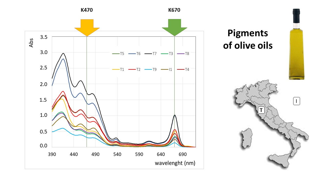
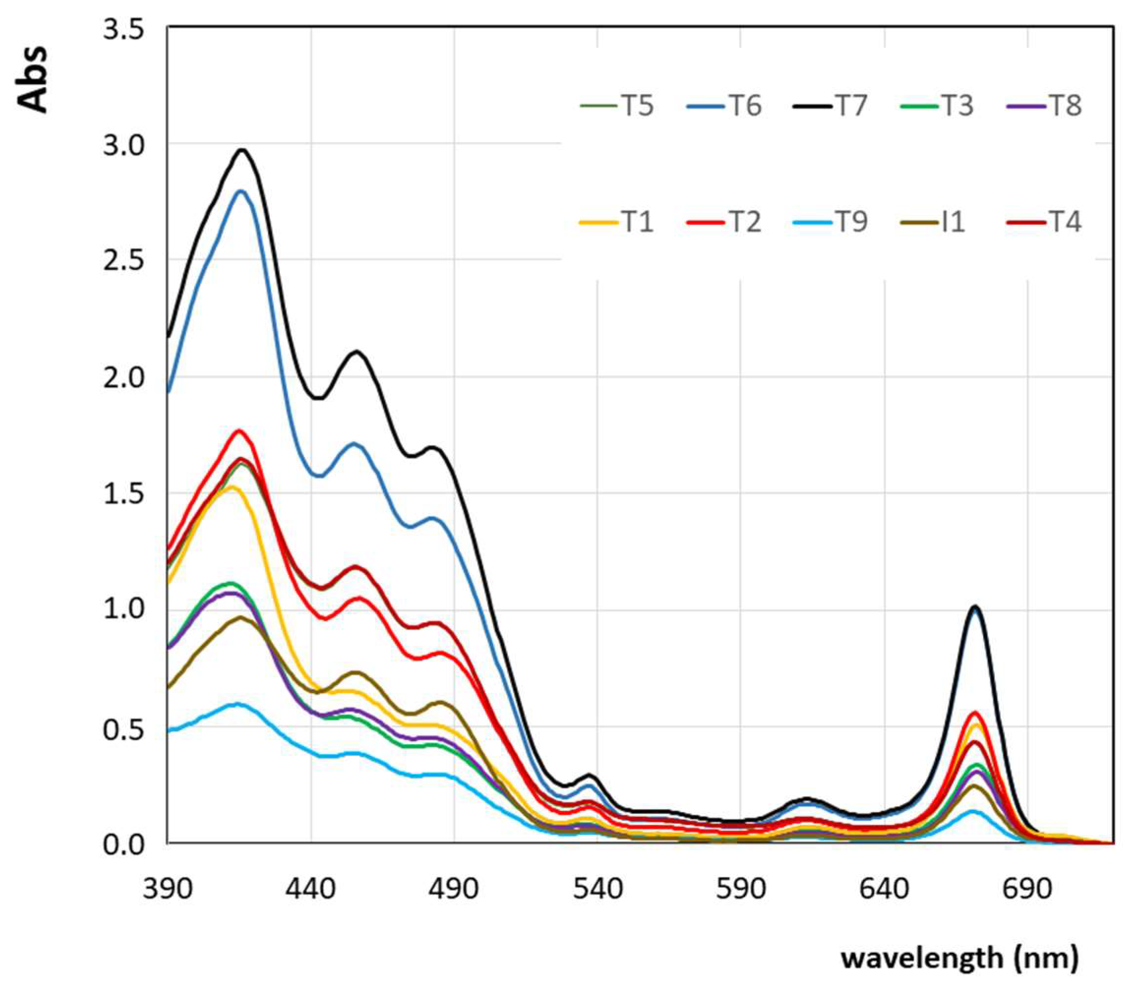
2.1. Samples
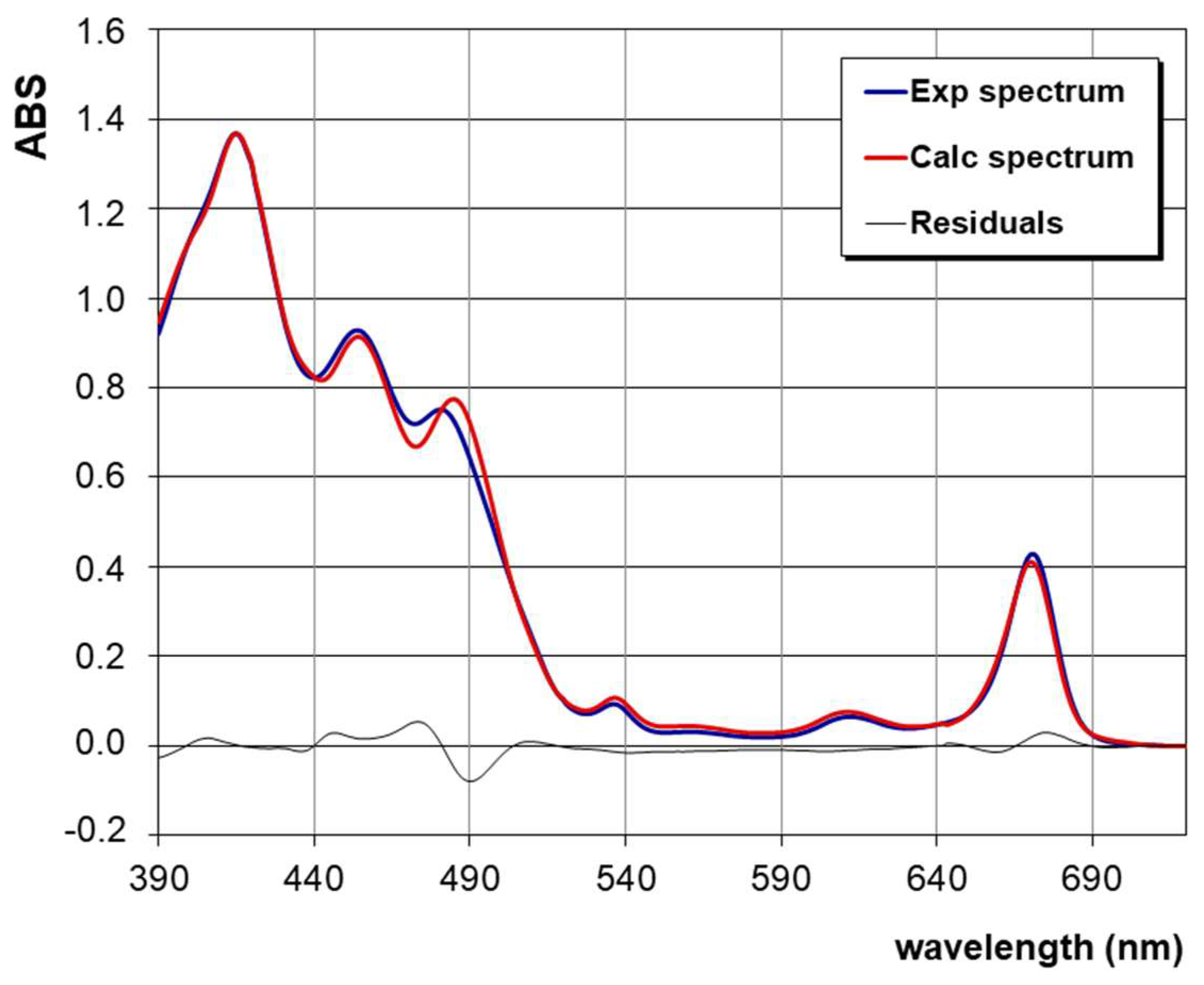
2.2. Methods
2.3. Statistical Analysis
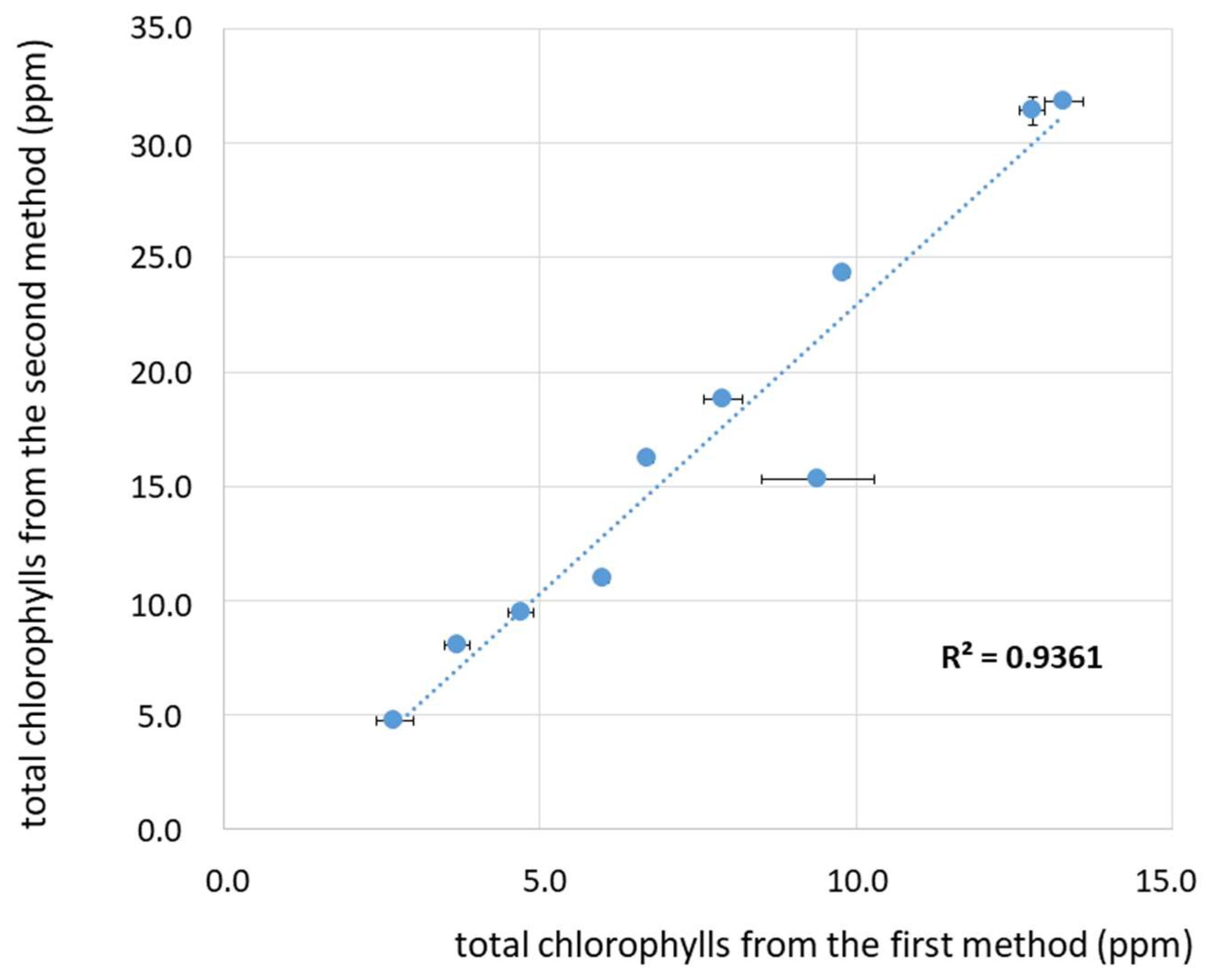
3. Results and Discussion
4. Conclusions
Author Contributions
Funding
Acknowledgments
Conflicts of Interest
References
- Boskou, D. Olive fruits, table olives, and olive oil bioactive constituents. In Olive and Olive Oil Bioactive Constituents, 1st ed.; Boskou, D., Ed.; AOCS Press: Urbana, IL, USA, 2015; pp. 1–30. [Google Scholar]
- Covas, M.-I.; Fito, M.; de la Torre, R. Minor Bioactive Olive Oil Components and Health: Key Data for Their Role in Providing Health Benefits in Humans. In Olive and Olive Oil Bioactive Constituents, 1st ed.; Boskou, D., Ed.; AOCS Press: Urbana, IL, USA, 2015; pp. 31–52. [Google Scholar]
- Angerosa, F.; Campestre, C.; Giansante, L. Analysis and Authentication. In Olive Oil Chemistry and Technology, 2nd ed.; Boskou, D., Ed.; AOCS Press: Urbana, IL, USA, 2006; Chapter 7; pp. 113–172. [Google Scholar]
- Crespo, M.C.; Tomé-Carneiro, J.; Dávalos, A.; Visioli, F. Pharma-Nutritional Properties of Olive Oil Phenols. Transfer of New Findings to Human Nutrition. Foods 2018, 7, 90. [Google Scholar] [CrossRef]
- Cicero, N.; Albergamo, A.; Salvo, A.; Bua, G.D.; Bartolomeo, G.; Mangano, V.; Rotondo, A.; Di Stefano, V.; Di Bella, G.; Dugo, G. Chemical characterization of a variety of cold-pressed foods available on the Brazilian market. Food Res. Int. 2018, 109, 517–525. [Google Scholar] [CrossRef] [PubMed]
- D’Imperio, M.; Mannina, L.; Capitani, D.; Bidet, O.; Rossi, E.; Bucarelli, F.M.; Quaglia, G.B.; Segre, A.L. NMR and statistical study of olive oils from Lazio: A geographical, ecological and agronomic characterization. Food Chem. 2007, 105, 1256–1267. [Google Scholar] [CrossRef]
- Salvo, A.; La Torre, G.; Rotondo, A.; Mangano, V.; Casale, K.E.; Pellizzeri, V.; Clodoveo, M.L.; Corbo, F.; Cicero, N.; Dugo, G. Determination of Squalene in Organic Extra Virgin Olive Oils (EVOOs) by UPLC/PDA Using a Single-Step SPE Sample Preparation. Food Anal. Methods 2017, 10, 1377–1385. [Google Scholar] [CrossRef]
- Rotondo, A.; Salvo, A.; Giuffrida, D.; Dugo, G.; Rotondo, E. NMR analysis of aldehydes in Sicilian extra-virgin olive oils by DPFGSE techniques. AAPP Phys. Math. Nat. Sci. 2011, 89. [Google Scholar] [CrossRef]
- Lazzerini, C.; Cifelli, M.; Domenici, V. Pigments in Extra-Virgin Olive Oil: Authenticity and Quality. In Production of Olive Tree; Boskou, D., Clodoveo, M.L., Eds.; InTech: Rijeka, Croatia, 2016; Chapter 6; pp. 95–114. [Google Scholar]
- Goodwin, T.W. The Biochemistry of the Carotenoids, Vol. II Animals; Springer: London, UK, 1984. [Google Scholar] [CrossRef]
- Mínguez-Mosquera, M.I.; Gandul-Rojas, B.; Garrido-Fernández, J.; Gallardo-Guerrero, L. Pigments Present in Virgin Olive Oil. J. Am. Oil Chem. Soc. 1990, 67, 192–196. [Google Scholar] [CrossRef]
- Gandul-Rojas, B.; Roca, M.; Gallardo-Guerrero, L. Chlorophylls and carotenoids in food products from olive tree. In Products from Olive Tree, 1st ed.; Boskou, D., Clodoveo, M.L., Eds.; InTech: Rijeka, Croatia, 2016; Chapter 5; pp. 67–98. [Google Scholar]
- Mínguez-Mosquera, M.I.; Rejavo-Navarro, L.; Gandul-Rojas, B.; Sanchez-Gomez, A.H.; Garrido-Fernández, J. Color-Pigment Correlation in Virgin Olive Oil. J. Am. Oil Chem. Soc. 1991, 68, 332–336. [Google Scholar] [CrossRef]
- Psomiadou, E.; Tsimidou, M. Simultaneous HPLC Determination of Tocopherols, Carotenoids, and Chlorophylls for Monitoring Their Effect on Virgin Olive Oil Oxidation. J. Agric. Food Chem. 1998, 46, 5132–5138. [Google Scholar] [CrossRef]
- Minguez-Mosquera, M.I.; Gandul-Rojas, B.; Gallardo-Guerrero, M.L. Rapid Method of Quantification of Chlorophylls and Carotenoids in Virgin Olive Oil by High-Performance Liquid Chromatography. J. Agric. Food Chem. 1992, 40, 60–63. [Google Scholar] [CrossRef]
- Cayuela, J.A.; Yousfi, K.; Martinez, M.C.; Garcia, J.M. Rapid Determination of Olive Oil Chlorophylls and carotenoids by Using Visible Spectroscopy. J. Am. Oil Chem. Soc. 2014, 91, 1677–1684. [Google Scholar] [CrossRef]
- Domenici, V.; Ancora, D.; Cifelli, M.; Serani, A.; Veracini, C.A.; Zandomeneghi, M. Extraction of Pigment Information from Near-UV Vis Absorption Spectra of Extra Virgin Olive Oils. J. Agric. Food Chem. 2014, 62, 9317–9325. [Google Scholar] [CrossRef] [PubMed]
- Lazzerini, C.; Cifelli, M.; Domenici, V. Pigments’ content in Extra Virgin Olive Oils from different Mediterranean Countries produced in 2014. Food Sci. Technol. 2017, 84, 586–594. [Google Scholar]
- Ancora, D. UV-Vis and 1H-NMR Spectroscopic Methods Applied to the Study of Extra-Virgin Olive Oil Produced in Tuscany and Apulia. Master’s Thesis, University of Pisa, Pisa, Italy, 15 June 2014. [Google Scholar]
- Lazzerini, C. Application of a New Spectrophotometric Method to Quantify Pigments in Extra-Virgin Olive Oils and Comparison with Chromatographic Techniques. Master’s Thesis, University of Pisa, Pisa, Italy, 16 July 2016. [Google Scholar]
- Lazzerini, C.; Buti, F.; Cifelli, M.; Zandomeneghi, M.; Domenici, V. Olio di oliva extravergine Toscano: Uno studio sul contenuto dei pigmenti e prospettive per un nuovo indice di qualità. In Codice Armonico. Sesto Congress di Scienze Naturali Ambiente Toscano, 6th ed.; Associazione Amici della Natura Rosignano, Ed.; Edizioni ETS: Pisa, Italy, 2016; pp. 155–165. [Google Scholar]
- Giuffrida, S.; Salvo, F.; Salvo, A.; La Pera, D.; Dugo, G. Pigments composition in monovarietal olive oils from various Sicilian olive varieties. Food Chem. 2007, 101, 833–837. [Google Scholar] [CrossRef]
- Giuffrida, D.; Salvo, F.; Salvo, A.; Cossignani, L.; Dugo, G. Pigments profile in monovarietal virgin olive oils from various Italian olive varieties. Food Chem. 2011, 124, 1119–1123. [Google Scholar] [CrossRef]
- Lazzerini, C.; Domenici, V. Pigments in Extra-Virgin Olive Oils Produced in Tuscany (Italy) in Different Years. Foods 2017, 6, 25. [Google Scholar] [CrossRef] [PubMed]
- Roca, M.; Minguez-Mosquera, M.I. Change in the natural ratio between chlorophylls and carotenoids in olive fruit during processing for virgin olive oil. J. Am. Oil Chem. Soc. 2001, 78, 133–138. [Google Scholar] [CrossRef]
- Criado, M.N.; Romero, M.P.; Casanovas, M.; Motilva, M.J. Pigment profile and colour of monovarietal virgin olive oils from Arbequina cultivar obtained during two consecutive crop seasons. Food Chem. 2008, 110, 873–880. [Google Scholar] [CrossRef] [PubMed]
- Gandul-Rojas, B.; Roca, M.; Minguez-Mosquera, M.I. Use of Chlorophyll and Carotenoid Pigment Composition to Determine Authenticity of Virgin Olive oil. J. Am. Oil Chem. Soc. 2000, 77, 853–858. [Google Scholar] [CrossRef]
- Ferreiro-Gonzalez, M.; Barbero, G.F.; Alvarez, J.A.; Ruiz, A.; Palma, M.; Ayuso, J. Authentication of virgin olive oil by a novel curve resolution approach combined with visible spectroscopy. Food Chem. 2017, 220, 331–336. [Google Scholar] [CrossRef]
- Aroca-Santos, R.; Cancila, J.C.; Pariente, E.S.; Torrecilla, J.S. Neural networks applied to characterize blends containing refined and extra-virgin olive oils. Talanta 2016, 161, 304–308. [Google Scholar] [CrossRef]
- Torrecilla, J.S.; Rojo, E.; Dominguez, C.J.; Rodriguez, F. A Novel Method to Quantify the Adulteration of Extra Virgin Olive Oil with Low-Grade Olive Oils by UV-Vis. J. Agric. Food Chem. 2010, 58, 1679–1684. [Google Scholar] [CrossRef] [PubMed]
- Carranco, N.; Farrés-Cebrián, M.; Saurina, J.; Núñez, O. Authentication and Quantitation of Fraud in Extra Virgin Olive Oils Based on HPLC-UV Fingerprinting and Multivariate Calibration. Foods 2018, 7, 44. [Google Scholar] [CrossRef]
- Rigane, G.; Ayadi, M.; Boukhris, M.; Sayadi, S.; Bouaziz, M. Characterization and phenolic profiles of two rare olive oils from southern Tunisia: Dhokar and Gemri-Dhokar cultivars. J. Sci. Food Agric. 2013, 93, 527–534. [Google Scholar] [CrossRef] [PubMed]
- Moyano, M.J.; Melendez-Martinez, A.J.; Alba, J.; Heredia, F.J. A comprehensive study on the colour of virgin olive oils and its relationship with their chlorophylls and carotenoids indexes (I): CIEXYZ non-uniform colour space. Food Res. Int. 2008, 41, 505–512. [Google Scholar] [CrossRef]
- Kruzlicova, D.; Mocak, J.; Katsoyannos, E.; Lankmayr, E. Classification and characterization of olive oils by UV-vis absorption spectrometry and sensorial analysis. J. Food Nutr. Res. 2008, 47, 181–188. [Google Scholar]
- Calabrese, I.; Merli, M.; Liveri, M.L.T. Deconvolution procedure of the UV-vis spectra. A powerful tool for the estimation of the blinding of a model drug to specific solubilisation loci of bio-compatible aqueous surfactant-forming micelle. Spectrochim. Acta Part A 2015, 142, 150–158. [Google Scholar] [CrossRef] [PubMed]
- Aroca-Santos, R.; Lastra-Mejias, M.; Cancila, J.C.; Torrecilla, J.S. Intelligent modelling to monitor the evolution of quality of extra virgin olive oil in simulated distribution conditions. Biosyst. Eng. 2018, 172, 49–56. [Google Scholar] [CrossRef]
- Gonçalves, T.R.; Rosa, L.N.; Gonçalves, R.P.; Torquato, A.S.; Março, P.H.; Marques Gomes, S.T.; Matsushita, M.; Valderrama, P. Monitoring the Oxidative Stability of Monovarietal Extra Virgin Olive Oils by UV–Vis Spectroscopy and MCR–ALS. Food Anal. Methods 2018, 11, 1936–1943. [Google Scholar] [CrossRef]
- Agriturismo Santa Annunziata Presso San Vincenzo (Toscana). Available online: http://www.ssannunziata.it/oliveto-a-san-vincenzo/ (accessed on 25 November 2018).
- Roncucci, D. Implementation of a Near UV-Vis Spectroscopic Method to Determine Pigments in Fresh Olive Oils. Bachelor’s Thesis, University of Pisa, Pisa, Italy, 2018. [Google Scholar]
- Borello, E.; Roncucci, D.; Lazzerini, C.; Cifelli, M.; Domenici, V. Dal colore al contenuto in pigmenti: Uno studio su campioni di olio di oliva extravergine ottenuti da olive di diverse cultivar in Toscana. In Settimo Congress di Scienze Naturali Ambiente Toscano, 7th ed.; Associazione Amici della Natura Rosignano, Ed.; Edizioni ETS: Pisa, Italy, 2018; pp. 258–267. [Google Scholar]
- Borello, E. Quantification of Main Pigments in Fresh and Old Olive Oils by Means of a Mathematical Deconvolution of Their Near-UV-Vis Absorption Spectrum and a Kinetic Study. Master’s Thesis, University of Pisa, Pisa, Italy, 2018. [Google Scholar]


| Label | Cultivar | Geographic Origin | Year of Harvesting | Storage Temperature | Classification 1 |
|---|---|---|---|---|---|
| I1 | blend | Italy 2 | 2015 | ≈22 °C | EVOO |
| T1 | Frantoio | Italy, Tuscany (LI) | 2015 | ≈22 °C | EVOO |
| T2 | Frantoio | Italy, Tuscany (LI) | 2015 | ≈4 °C | EVOO |
| T3 | Leccino | Italy, Tuscany (LI) | 2015 | ≈22 °C | EVOO |
| T4 | Moraiolo | Italy, Tuscany (LI) | 2015 | ≈22 °C | EVOO |
| T5 | Blend 3 | Italy, Tuscany (LI) | 2012 | ≈4 °C | EVOO |
| T6 | Blend 3 | Italy, Tuscany (LI) | 2015 | ≈4 °C | EVOO |
| T7 | Blend 3 | Italy, Tuscany (LI) | 2015 | ≈4 °C | EVOO |
| T8 | Pendolino | Italy, Tuscany (LI) | 2015 | ≈22 °C | EVOO |
| T9 | blend | Italy, Tuscany (FI) | 2012 | ≈22 °C | VOO |
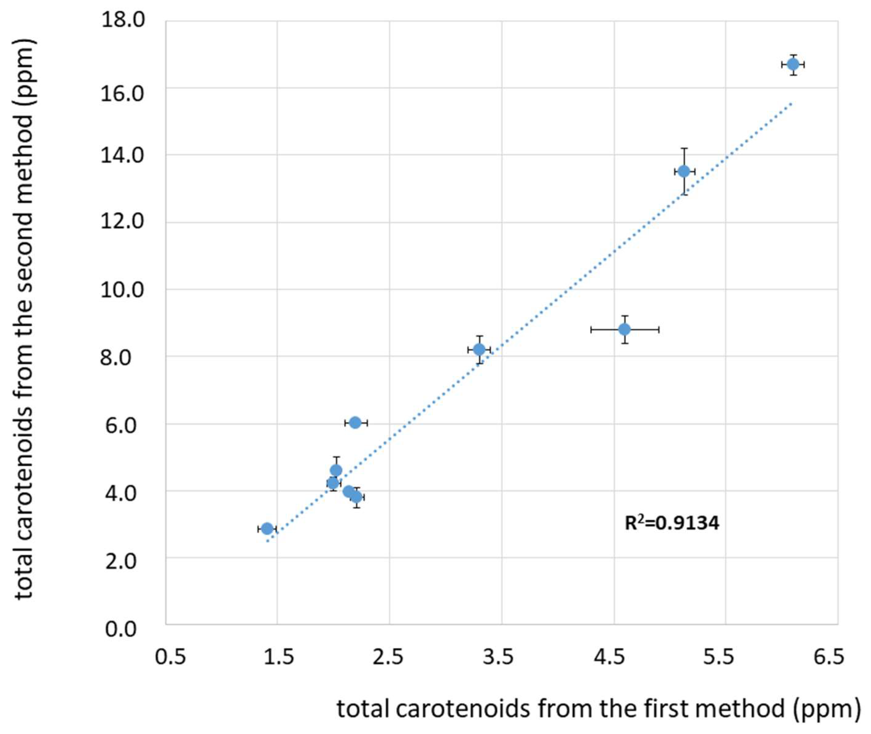
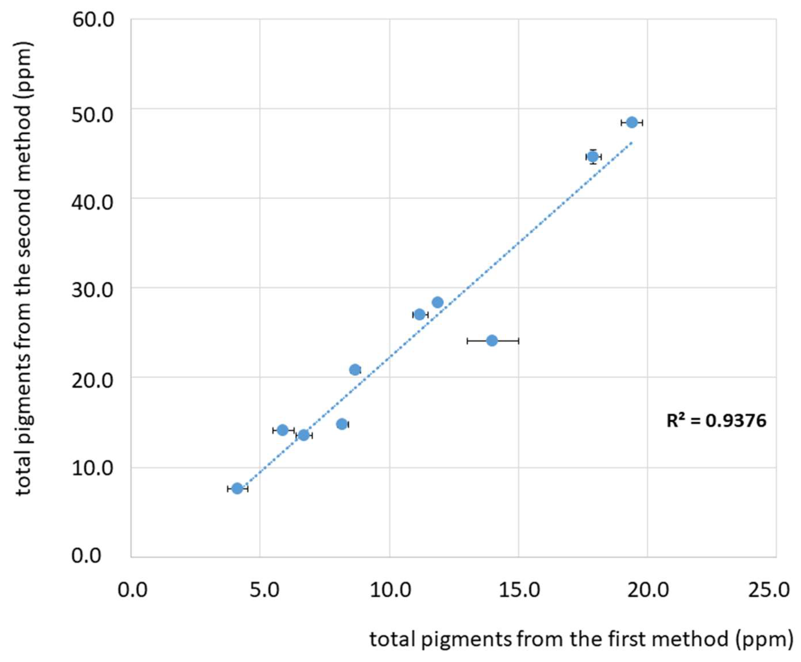
| Label | Total Chlorophylls’ Derivatives (ppm) | Total Carotenoids (ppm) | Sum of Pigments (ppm) |
|---|---|---|---|
| I1 | 3.7 ± 0.2 | 2.2 ± 0.1 | 5.9 ± 0.2 |
| T1 | 6.7 ± 0.1 | 2.03 ± 0.03 | 8.7 ± 0.1 |
| T2 | 7.9 ± 0.1 | 3.3 ± 0.1 | 11.2 ± 0.1 |
| T3 | 6.0 ± 0.1 | 2.2 ± 0.1 | 8.2 ± 0.1 |
| T4 | 9.8 ± 0.9 | 2.14 ± 0.02 | 11.9 ± 0.9 |
| T5 | 9.4 ± 0.3 | 4.6 ± 0.3 | 14.0 ± 0.3 |
| T6 | 12.8 ± 0.2 | 5.1 ± 0.1 | 17.9 ± 0.2 |
| T7 | 13.3 ± 0.3 | 6.1 ± 0.1 | 19.4 ± 0.3 |
| T8 | 4.7 ± 0.2 | 2.0 ± 0.1 | 6.7 ± 0.2 |
| T9 | 2.7 ± 0.3 | 2.4 ± 0.1 | 5.1 ± 0.3 |
| Label | Total Chlorophylls’ Derivatives (ppm) | Total Carotenoids (ppm) | Sum of Pigments (ppm) | R2 (Fitting Method) |
|---|---|---|---|---|
| I1 | 8.1 ± 0.1 | 6.0 ± 0.1 | 14.1 ± 0.1 | 0.9836 |
| T1 | 16.2 ± 0.2 | 4.6 ± 0.4 | 20.8 ± 0.2 | 0.9788 |
| T2 | 18.8 ± 0.1 | 8.2 ± 0.4 | 27.0 ± 0.1 | 0.9846 |
| T3 | 11.0 ± 0.1 | 3.8 ± 0.3 | 13.8 ± 0.1 | 0.9861 |
| T4 | 24.3 ± 0.2 | 3.97 ± 0.04 | 28.3 ± 0.2 | 0.9875 |
| T5 | 15.3 ± 0.1 | 8.8 ± 0.4 | 24.1 ± 0.4 | 0.9789 |
| T6 | 31.4 ± 0.6 | 13.5 ± 0.7 | 44.5 ± 0.6 | 0.9782 |
| T7 | 31.8 ± 0.1 | 16.7 ± 0.3 | 48.5 ± 0.3 | 0.9864 |
| T8 | 9.5 ± 0.1 | 4.2 ± 0.2 | 14.7 ± 0.2 | 0.9852 |
| T9 | 4.8 ± 0.1 | 2.9 ± 0.1 | 7.7 ± 0.1 | 0.9863 |
© 2019 by the authors. Licensee MDPI, Basel, Switzerland. This article is an open access article distributed under the terms and conditions of the Creative Commons Attribution (CC BY) license (http://creativecommons.org/licenses/by/4.0/).
Share and Cite
Borello, E.; Domenici, V. Determination of Pigments in Virgin and Extra-Virgin Olive Oils: A Comparison between Two Near UV-Vis Spectroscopic Techniques. Foods 2019, 8, 18. https://doi.org/10.3390/foods8010018
Borello E, Domenici V. Determination of Pigments in Virgin and Extra-Virgin Olive Oils: A Comparison between Two Near UV-Vis Spectroscopic Techniques. Foods. 2019; 8(1):18. https://doi.org/10.3390/foods8010018
Chicago/Turabian StyleBorello, Eleonora, and Valentina Domenici. 2019. "Determination of Pigments in Virgin and Extra-Virgin Olive Oils: A Comparison between Two Near UV-Vis Spectroscopic Techniques" Foods 8, no. 1: 18. https://doi.org/10.3390/foods8010018
APA StyleBorello, E., & Domenici, V. (2019). Determination of Pigments in Virgin and Extra-Virgin Olive Oils: A Comparison between Two Near UV-Vis Spectroscopic Techniques. Foods, 8(1), 18. https://doi.org/10.3390/foods8010018