
Metal Oxide Nanoparticles in Food Packaging and Their Influence on Human Health
by
 , ,
, ,
Mariana Stuparu-Cretu
1,*,
Gheorghe Braniste
2,
Gina-Aurora Necula
2,
Silvius Stanciu
3,
Dimitrie Stoica
4 and
Maricica Stoica
2,* 1
Faculty of Medicine and Pharmacy, “Dunarea de Jos” University of Galati, 35 Alexandru Ioan Cuza Street, 800010 Galati, Romania
2
Cross-Border Faculty, “Dunarea de Jos” University of Galati, 111 Domneasca Street, 800201 Galati, Romania
3
Faculty of Food Science, “Dunarea de Jos” University of Galati, 111 Domneasca Street, 800201 Galati, Romania
4
Faculty of Economics and Business Administration, “Dunarea de Jos” University of Galati, 59-61 Balcescu Street, 800001 Galati, Romania
*
Authors to whom correspondence should be addressed.
Foods 2023, 12(9), 1882; https://doi.org/10.3390/foods12091882
Submission received: 27 March 2023
/
Revised: 29 April 2023
/
Accepted: 1 May 2023
/
Published: 3 May 2023
(This article belongs to the Section Food Packaging and Preservation)
Abstract
:It is a matter of common knowledge in the literature that engineered metal oxide nanoparticles have properties that are efficient for the design of innovative food/beverage packages. Although nanopackages have many benefits, there are circumstances when these materials are able to release nanoparticles into the food/beverage matrix. Once dispersed into food, engineered metal oxide nanoparticles travel through the gastrointestinal tract and subsequently enter human cells, where they display various behaviors influencing human health or wellbeing. This review article provides an insight into the antimicrobial mechanisms of metal oxide nanoparticles as essential for their benefits in food/beverage packaging and provides a discussion on the oral route of these nanoparticles from nanopackages to the human body. This contribution also highlights the potential toxicity of metal oxide nanoparticles for human health. The fact that only a small number of studies address the issue of food packaging based on engineered metal oxide nanoparticles should be particularly noted.

1. Introduction
We are living in the nanobiotechnology era [1,2]. This newly emerging technology has an enormous potential in various multidimensional fields of modern day life, such as health sciences and engineering (medicine, pharma, sport, agri-food, and other industries) [2,3,4,5,6,7,8,9]. Nanomedicine refers particularly to biosensors, imaging, diagnosis, or drug delivery for therapy, while in sport, nanomaterials (a new class of engineering materials) offer smarter products, such as functional nanosportswear, self-cleaning and antibacterial sportswear and shoes, gadgets, therapeutic bands, rackets, etc. [2,8,10,11]. The agricultural field mainly uses nanomaterials as antimicrobials, larvicidals, insecticidals, nanofungicides and nanofertilizers for their superior efficacy over chemical fertilizers and pesticides [4,12,13]. In the food industry, numerous NPs (nanoparticles) are extensively incorporated into the host polymer/biopolymer matrix to provide strong food packaging characteristics (impactful antimicrobials, biosensors, gas barrier and mechanical strength enhancers, oxygen and water vapor scavengers, etc.) or are directly added into the food matrix to create new food functionalities (coloring, flavoring, safety/stability agents, etc.) [1,4,6,9,14,15,16,17,18,19,20]. Various NPs are used, including nanocellulose, nanoclays, Ag-NPs (nanosilver), Au-NPs (nanogold), nanoforms of some metal oxides (CuO-NPs: copper oxide NPs; Fe3O4-NPs: triiron tetraoxide NPs; MgO-NPs: magnesium oxide NPs; TiO2-NPs: titanium dioxide NPs; ZnO-NPs: zinc oxide NPs), food-grade biopolymers, nanoliposomes, etc. These nanoparticles display unique properties, such as surface effects (larger surface areas, larger surface-to-volume ratios) and pronounced quantum effects due to their nanoscale dimensions (range 1–100 nm), which are directly reflected in their reactivity [1,3,4,6,7,9,15,18,21,22]. The global market size of metal and metal oxide NPs is expected to grow from USD 2.65 billion in 2022 to USD 6.39 billion by 2030, owing to their growing use in various industries (medical sector, pharma, agricultural, food area, and other end-use industries) and increasing awareness of the benefits of using these NPs in different applications [23,24]. Nanoforms of some metal oxides, such as CuO-NPs, Fe3O4-NPs, MgO-NPs, TiO2-NPs, and ZnO-NPs, are used in food nanopackaging owing to their advantages over metal-based nanoparticles (e.g., NPs possess a crystalline structure with more edges and corners [25,26,27,28,29], as can be seen in Figure 1) and their benefits, highlighted in Table 1.
EMo-NPs are effective in enhancing food packaging properties and are excellent antimicrobials owing to their capacity to generate high levels of intracellular ROS (reactive oxygen species) through metal ion release [9,49]. EMo-NPs do not migrate from food/beverage packaging into the food matrix as they are completely encapsulated by the host polymer/biopolymer [16,45,50]. However, in some circumstances, nanostructures (polymer/biopolymer-based matrix) are able to release EMo-NPs into food/beverages, depending on certain factors (food storage conditions, food chemical nature, food processing, etc.) [9,16,45,50]. Once released into the food/beverage matrix, EMo-NPs pass through the human GI (gastrointestinal) tract, cross the GI mucosa and enter human cells, where they display various behaviors. EMo-NPs can be significantly bioaccumulated and exert potential toxicity on human health [1,9,15]. This article offers an insight into the antimicrobial mechanisms of engineered metal oxide nanoparticles as a key benefit in the food/beverage packaging and offers a discussion on the digestive pathway of nanoparticles from contact with food packaging material to the human body. This review also highlights potential toxicity of metal oxide nanoparticles to human health. The article is based on the synthesis of scientific data from the literature, creativity, and knowledge in the field of engineering and health sciences, and it may contribute to understanding the potential toxicity of metal oxide nanoparticles, inspiring researchers to be more concerned with the study of nanopackaging in the food industry.
2. Antimicrobial Mechanisms of EMo-NPs
EMo-NPs have been used in the food packaging industry for several years due to their benefits, particularly their excellent antimicrobial activity (as shown in Introduction, Table 1) due to ROS induction (extracellular/intracellular) through metal ion release. ROS consists of very cytotoxic radicals O2− (superoxide anion radical), 1O2 (singlet oxygen), OH (hydroxyl radical), and nonradical H2O2 (hydrogen peroxide) which attack the bacterial cell components (cell wall, DNA (deoxyribonucleic acid), proteins, mitochondria, etc.) [51] (Figure 2).
EMo-NPs can attack numerous microorganisms through ROS induction via Fenton-type and Haber–Weiss reactions, as well as via photocatalytical reactions and NP surface defects [52].
2.1. ROS Induction through Fenton-Type and Haber–Weiss Reactions
EMo-NPs release metal ion species which can start Fenton, Fenton-like, and Haber–Weiss Reactions. The Haber–Weiss and Fenton-like reactions can together generate an avalanche of ROS [9,52]. For instance, Cu-based NPs can attack numerous bacteria and yeasts (e.g., Bacillus, Escherichia, Pseudomonas, Salmonella, Staphylococcus, Streptococcus, Saccharomyces) due to ROS induction based on Cu+ ions’ release [53]. Cu+-based ROS induction takes place via Fenton-type (oxidized Cu+ interacts with H2O2, producing ·OH) and Haber–Weiss (induce oxidative stress by generating ·O2− and ·OH) mechanisms (Equations (1)–(3)) [54,55,56,57].
Fe3O4-NPs are also known to catalyze Haber–Weiss reactions [52]. These NPs are a special type of EMo-NP with superparamagnetic and redox properties, possessing very remarkable biocidal activity against various antibiotic-resistant microorganisms (e.g., Acinetobacter, Achromobacter, Klebsiella, Pseudomonas, Serratia, Shigella, Yersinia) due to the ROS induction based on Fe2+ ions’ release, which are essential for Fenton-like reactions and Haber–Weiss reactions (Equations (4)–(6)) [31,58,59,60,61,62].
Other EMo-NPs, such as MgO-NPs, also have remarkable antimicrobial potential against important foodborne pathogens (Escherichia, Klebsiella, Pseudomonas, Salmonella, Staphylococcus) by induction of superoxide radicals [63]. While the biocidal mechanism of these NPs is still not clear, it may be due to the following: the generation of more active superoxide radicals outside the microbial cells under light exposure; Mg2+ delivery; strong electrostatic interaction with bacterial cells (stronger in Gram-negative strains), causing the disfunctions of cell walls (which could be the crucial mechanism of bacterial destruction); pH change and alkaline effect [51,64,65,66,67,68,69,70]. To sum up, the Fenton and Fenton-like reactions oxidize the metal ions, while the Haber–Weiss reactions reduce the metal ions, inducing bacterial damage [52].
2.2. ROS Induction by EMo-NPs through Photocatalytical Reactions
TiO2-NPs and ZnO-NPs promote photochemical reactions as they are activated under illumination (visible light/UV light, TiO2 being more active in UV light) and many highly reactive pairs ((e−/h+) (e−: electrons, which can act as reductants; h+: holes, which can act as oxidants)) are produced (Equation (7)) [9,33,51,52,71,72,73,74]:
where hv is solar light with photonic energy, e−(cb) is a conduction band electron, and h+(vb) refers to energy for hydrogen bond formation. The (e−/h+) pairs have a high potential to generate ROS. The mechanism of photocatalytical microbial inactivation for TiO2 under solar light are shown in Equations (8)–(11) [75,76]:
TiO2-NPs do not deliver metal ions. Their biocidal potential might be attributed to interaction with intracellular biomolecules, adsorbed onto NPs, that possess cytotoxicity, phototoxicity, and ROS induction capacity [53,56,72,77]. In addition, TiO2-NPs can induce ROS under dark conditions as a consequence of catalytic reactions from O2 and NP surface defects (Equation (12)) [61].
ZnO-NPs are another photocatalytical, possessing high antibacterial action due to their distinctive electronic configuration [53]. ZnO-NPs damage microorganisms by electrostatic forces which damage the cell membrane, Zn2+ ions’ delivery, and ROS formation capacity (the most plausible mechanism). ZnO-NPs induce ROS owing to their photocatalytic and UV light photocorrosion (pitting of cell membrane due to ROS) properties [9]. The antimicrobial activity of ZnO-NPs is enhanced by exposure to visible light [1].
3. EMo-NPs’ Route from Food/Beverage Packaging into Human GI Tract
3.1. EMo-NPs Migration from the Package Matrix into Food/Beverage Matrix: Possible Mechanisms
This subsection of the article deals with the possible migration mechanisms of EMo-NPs. Innovatively engineered nanopackages, able to contribute to the quality and safety of food/beverages, are viewed as an excellent substitute for conventional packaging composites. Nevertheless, the intentional embedding of engineered nanoparticles, such as EMo-NPs, into the polymer/biopolymer matrix raises some issues, such as the EMo-NPs’ transfer from the package and subsequent release into food/beverages [37,45,78,79,80]. Of all the possible interactions which take place between the nanopackage and the food/beverage contained therein (permeation: diffusion of gases across the package wall; migration: bulk movement not only of EMo-NPs but also of monomers, antioxidant and coloring agents, printing inks, and antibacterials from the package; scalping: uptake of the food/beverage components by the package) (Figure 3), migration is crucial due to the concern that human health might be endangered by the leaching of migrants from the package into the food/beverages [50,81,82,83,84].
EMo-NPs’ release, a highly complex phenomenon, has four potential mechanisms: (desorption: extemporaneous release of NPs bound at the nanopackage interface; diffusion (migration): mass movement (entropically driven) of EMo-NPs from nanopackages and their subsequent release into food/beverage; dissolution: EMo-NPs’ dissolution into ionic species, followed by release of metal ions into food/beverage; EMo-NPs’ releasing as a consequence of any procedure which modifies the host matrix (e.g., photooxidation, abrasion of the host polymer/biopolymer) [6,16,45,50]. EMo-NPs’ (quasi-molecules with evident molecular volume) migration should be ruled by interrelated factors which cause the transfer of conventional molecular chemicals (migrant nature, temperature and contact time, contact type, food chemical nature, etc.), although embarrassing factors may come into play, giving rise to supplemental incertitudes [16,45,85,86,87,88,89]. Nanosize may introduce statistical complexity into migration kinetics; in addition, the rate of EMo-NPs moving through the host matrix is highly impacted when EMo-NPs’ surfaces are coated with surfactants. EMo-NPs take part in complex interactions with the host matrix, and the Fickian diffusion (which obeys Fick’s laws in the case of molecular-scale substances) model cannot be applied. EMo-NPs inserted into the host matrix diffuse from their starting point (dissolved ions, whole) to the interfacial boundary; then, they are released (a notably more complex phenomenon), whereas surface-bound EMo-NPs can be extemporaneously released into the food matrix [45]. Migration depends on properties of EMo-NPs such as the polymer/biopolymer matrix, contact time, contact type, temperature, pressure, and even the manner in which the package is opened. One of the most significant parameters of EMo-NPs is molecular volume [78,90,91]. EMo-NPs that are usually used in materials that come into contact with food are based on polymers larger than 5 nm in size. In addition, they are quasi-immobilized into the host matrix, demonstrating extremely low diffusion even at high levels of NP usage in the polymer/biopolymer. The completely embedded EMo-NPs (covered or encapsulated by the host matrix) do not travel through polymer layers, do not penetrate the outer layer, and do not diffuse into foods [16,45]. However, there is a risk of possible EMo-NP release at or close to the food contact surface in the case of mechanical impact (abrasion) and/or the senescence of the packaging matrix (chemically, mechanically, or thermally stress-induced) [16,45,50]. At a cut edge of food packaging, EMo-NPs will come into direct contact with the food matrix, and the food constituents’ scalping into the polymer/biopolymer package structure occurs more vigorously here. Consequently, EMo-NPs will be dissolved by food constituents or may even be physically released, again with the possibility of being dissolved [16,45,86]. The migration measurements of EMo-NPs or metal ions inside the food matrix are difficult to carry out (EMo-NPs are chemical chameleons, appearing in various size and shapes and sometimes dissolved into ions or reduced back into their constituents; in addition, food/beverage is a very complex matrix) but are essential to determine their possible health implications, as they are often considered to have toxic potential [6,16,53].
3.2. EMo-NPs’ Route from the Food/Beverage Matrix into the Human GI Tract
The human body is easily poly-exposed to EMo-NPs via oral (ingestion), dermal, and respiratory (inhalation) routes [4,6,92,93,94]. Food/beverage ingestion is the primary route of EMo-NPs’ absorption [94]. EMo-NPs enter into food/beverage formulation through transfer out of nanopackaging or nanopackaging headspace or as direct food additives (fortifying agents, dietary supplements, coloring agents, etc.) [1,9,15,92,95]. EMo-NPS dispersed into the food/beverage matrix enter the human GI tract after ingestion (Figure 4), where they display various behaviors.
The absorption of EMo-NPs could start in the oral cavity (1) through buccal and sublingual mucosa (vastly permeable), directly passing into the circulatory system and, subsequently, penetrating into cells through fine capillaries [92]. Unabsorbed EMo-NPs pass through the oral cavity (characterized by approximately neutral pH, electrolytes, metabolic enzymes, biopolymers, and mastication), move through the esophagus (2) to the stomach (3) (characterized by highly acidic pH, around 2–3; electrolytes; enzymes and biopolymers; peristalsis; and churning), and enter into the small intestine (4) (pH 5–7; electrolytes; bile salts; enzymes and biopolymers; and peristalsis). Once ingested, EMo-NPs can adhere to, travel through, or be adsorbed by the mucus layer of the GI tract. If EMo-NPs are not absorbed in the (2–4) segments of the GI tract, they are released into large intestine fluids (5) (pH 7–8) as the food matrix is disrupted and digested [15,96]. EMo-NPs adhere to enteric mucosa (the first barrier—a complex hydrogel consisting mainly of mucin proteins, negatively charged, influencing the adhesion of EMo-NPs through electrostatic interactions) and diffuse through it; then, they are absorbed by the chylomicron uptake mechanism of enterocytes and transported across the epithelium (the second barrier with the highest resistance against EMo-NPs’ passage) [92,96]. EMo-NPs are taken up by active (transcellular) or passive (paracellular, with a minor role in the passage of NPs) mechanisms into cells (or even on the subcellular level) and various organs (brain, heart, intestines, kidneys, liver, lungs, spleen, and stomach). The liver and spleen absorb and accumulate EMo-NPs significantly faster than others [15,92,97]. EMo-NPs are not metabolized in the GI tract and can be absorbed in their intact form (being bio-persistent) [1,6,15,94]. The large pH gradient, electrolytes, physical forces, and characteristics of EMo-NPs are significant parameters that may impact EMo-NP interactions with various biomolecules originating from the ingested food/beverage or GI tract. The significant modification of the interfacial properties of EMo-NPs that subsequently occurs could influence biological tissues and cellular response. EMo-NPs have a highly specific surface area, providing a large area for the adsorption of any surface-active components such as bile salts, metabolic enzymes (amylases, lipases, peptidases), proteins, or phospholipids (resulting in a reduction in their activity) [15,96]. EMo-NPs in high concentrations could decrease the digestion of lipids, proteins, and starch within the GI tract, alter the normal function of the epithelial cells of intestinal tissue and nutrients’ absorption, and stimulate an immune response with potential adverse effects on human health [1,15]. The properties of ingested EMo-NPs are also significantly modified by bacteria from the human GI tract through secreted enzymes and biopolymers. Conversely, ingested EMo-NPs demonstrate complex antimicrobial activity that could impact the colonic microbiota (which has an important role in the maintenance of the structural integrity of the mucosal barrier) and alter the host’s physiology, exerting a powerful influence on wellbeing and human health [6,15,98].
4. Influences of EMo-NPs on Human Health
4.1. Intracellular ROS Induction in Eukaryotic Cells
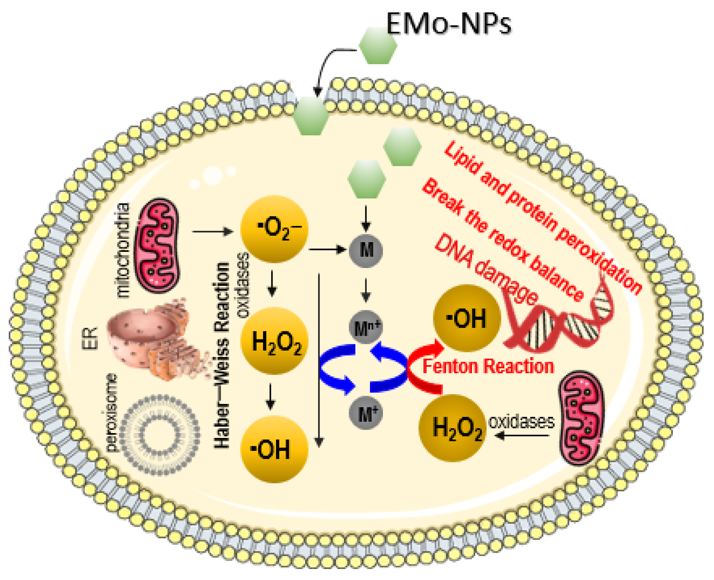
This subsection discusses the potential toxicity effects of EMo-NPs, especially those involved in subcellular ROS generation. ROS are produced by various biological entities such as cell membrane and subcellular compartments (oxidative organelles: ER (endoplasmic reticulum), mitochondria, peroxisome). These mitochondria generate approximately 90% of cytoplasmic ROS (with a favorable role in cell physiology at low or moderate levels). At the same time, the mitochondria maintain a balanced amount of ROS in the cell as soon as they are generated. Excessive ROS production results in oxidative stress (cell damage, cell death) [99,100,101]. EMo-NPs are extremely reactive; they generate excessive levels of intracellular ROS, which can have effects on the overall functionality of the cell, oxidizing/damaging the cellular biological macromolecules (DNA, lipids, proteins, and enzymes) and biological entities, including the cell wall and organelles [6,15,53,94,97,102] (Figure 5).
In the cell, ROS are natural byproducts of normal oxygen metabolism, being generated by mitochondria during physiological (or pathological) conditions [99,103]. The ·O2− radical generated inside the cell by mitochondria is changed by oxidases (superoxide dismutase) into H2O2, which is then changed into very reactive ·OH radical through Haber–Weiss or Fenton-type reactions. The endogenous ROS is kept in balance by numerous intracellular antioxidant mechanisms. Conversely, in the presence of metal ions released from internalized EMo-NPs, H2O2 can be rapidly converted to high a ·OH level through Fenton reaction [103]. Excessive generation of ROS by external input of EMo-NPs can break the redox balance and may lead to harmful effects, such as DNA damage, lipid and protein peroxidation, cellular apoptosis, mitochondria dysfunctions (via depolarization of mitochondrial membrane), as well as other related phenomena, such as cellular signaling fluctuations involved in cell differentiation and cell proliferation [99,104].
4.2. EMo-NPs: Main Concerns
The data on possible EMo-NP toxicity to humans are limited. Most studies related to EMo-NP toxicity were conducted on various in vivo models: animals, aquatic organisms, simulated body fluids (saliva, stomach, and intestinal fluids), and human blood plasma. A summary of the main concerns is shown in Table 2.
EMo-NPs’ Immunotoxicity
EMo-NPs’ immunotoxicity has been revealed (Table 2). Whether EMo-NPs are recognized by the immune system or not, they influence the human immune system (immune organs, cells, molecules) through different immune reactions such as: oxidative stress (as can be seen in Section 4.1), inflammatory/anti-inflammatory responses, and genotoxicity [154,155,156]. Inflammation, the immediate natural response of the body against external chemicals, is favorable for human health; however, uncontrolled inflammation can lead to severe disorders [155,157]. EMo-NPs can induce the release of cytokines/chemokines in tissues and organs (e.g., spleen, liver), which have an important role in controlling and promoting of inflammatory response [155]. The immune system activates phagocytic cells; for instance, polymorphonuclear neutrophils, which migrate to an inflammatory site and induce inflammatory mediators, recruiting more polymorphonuclear neutrophils and other immune cells such as macrophages and lymphocytes. Macrophage cells exert an initial reaction to EMo-NP exposure. They initiate and propagate an inflammatory response with their capacity to recognize/engulf/digest EMo-NPs. Macrophage cells’ interaction with EMo-NPs usually results in activation of NADPH oxidase, leading to ROS induction along with oxidative burst [158,159]. EMo-NPs can also produce oxidative organelle (ER, mitochondria, lysosome) damage or dysfunction in immune cells owing to direct NP accumulation or indirect subcellular changes [160]. ER, the largest organelle, is responsible not only for protein synthesis and lipid metabolism (mainly) but also for upregulating cell response to stress. EMo-NPs may lead to protein misfolding, which induces ER stress [161]. EMo-NPs can also interact with mitochondria, leading to impaired mitochondrial function (mitochondrial stress) after their internalization, which may induce some metabolic disorders and reduce overall cellular energy levels [162,163,164]. EMo-NPs can typically accumulate in lysosomes (endpoints of the endocytosis pathway which act as digestive organelles and are essential for maintaining cellular homeostasis) and lead to impaired lysosomal dysfunction (lysosome membrane permeabilization, resulting in mitochondrial outer membrane permeabilization that induces ROS generation and apoptosis; massive lysosome membrane permeabilization can produce cytosolic acidification and necrosis) [165,166]. EMo-NPs’ genotoxicity has also been revealed (Figure 5, Table 2). Its genotoxicity is primarily size dependent: the smaller the size, the higher the reactivity of the surface area and, therefore, the higher the ROS induction, genotoxic reactions, and DNA damage [9,167,168]. The degree of severity may be closely related to the oxidative stress caused, and it is also dependent on EMo-NPs’ concentration and their physicochemical features. However, owing to the influence of key factors related to the toxicity of EMo-NPs (size, shape, morphology, surface coating, surface reactivity, specific surface area, solubility, bonded surface species, oxidation status, agglomeration/aggregation degrees), the relevant immunotoxicity mechanism is not completely understood. Long-term systematic studies are required to explore this further and to clearly explain the interaction between EMo-NPs and human tissues [155].
5. Conclusions
Engineered metal oxide nanoparticles are dominant and promising in the food sector. They are employed in innovative food packaging as impactful antimicrobials, biosensors, and gas barrier and mechanical strength enhancers, and in the functional food field as coloring, flavoring, and safety/stability agents. This review offers an insight into the antimicrobial mechanism of engineered metal oxide nanoparticles as a key benefit in food/beverage packaging, with a focus on extracellular/intracellular ROS induction as a primary antimicrobial mechanism (via Fenton-type, Haber–Weiss, and photocatalytical reactions). The paper also highlighted the migration of engineered metal oxide nanoparticles from the package matrix into the food/beverage matrix and their route from the food/beverage matrix into the human GI tract. Engineered metal oxide nanoparticles for food packaging are completely embedded in the host matrix. They do not possess the potential to travel through the polymer layers to the food contact layer where transfer into the food could occur. However, in certain circumstances (chemical, mechanical, or thermal stress-based package) a series of interactions (such as migration) may occur between the food product and its package, permitting the nanopackaging to transfer the desorbed nanoparticles into the food product. Once migrated into foods, nanosized metal oxide particles can enter into the GI tract (via the oral route) and, from there, can readily enter human cells through the circulatory system. Then, the nanoparticles can be absorbed by biological tissues, causing adverse influences on human health due to excessive intracellular ROS induction. Excessive ROS induction by external input of engineered metal oxide nanoparticles results in negative effects on the overall functionality of the cells and may lead to DNA damage, lipid and protein peroxidation, cellular apoptosis, mitochondria dysfunctions, and cell proliferation. This article does not conclude that engineered metal oxide nanoparticles should not be used; rather, sufficient attention should be paid to their use for food packaging as they may produce undesirable consequences for human health. Furthermore, innovative tests are necessary to obtain a clear illustration of their migration, behavior, and toxicity, and to obtain a better comprehension of the eventual health hazards related to overexposure to nanoparticles. We hope that this article inspires life science researchers, helping them to identify some aspects related to metal oxide nanoparticles so that their use does not pose any risk to human health.
Author Contributions
Methodology, M.S.-C. and S.S., Supervision, M.S.-C.; Writing—original draft, M.S.-C. and M.S.; Writing—review & editing, M.S.-C., G.B., G.-A.N., S.S., D.S. and M.S.; Investigation, G.B., G.-A.N., S.S. and D.S.; Resources, G.B. and G.-A.N.; Formal analysis, S.S. and D.S.; Software, D.S.; Conceptualization, M.S.; Visualization, M.S.; Project administration, M.S. All authors have read and agreed to the published version of the manuscript.
Funding
This research received no external funding.
Data Availability Statement
No new data were created or analyzed in this study. Data sharing is not applicable to this article.
Conflicts of Interest
The authors declare no conflict of interest.
References
- Kumar, A.; Choudhary, A.; Kaur, H.; Mehta, S.; Husen, A. Metal-based nanoparticles, sensors, and their multifaceted application in food packaging. J. Nanobiotechnol. 2021, 19, 256. [Google Scholar] [CrossRef] [PubMed]
- Miteva, A. Nanotechnology in sport and security. Strateg. Policy Sci. Educ. 2021, 29, 46–53. [Google Scholar] [CrossRef]
- Ghebretatios, M.; Schaly, S.; Prakash, S. Nanoparticles in the Food Industry and Their Impact on Human Gut Microbiome and Diseases. Int. J. Mol. Sci. 2021, 22, 1942. [Google Scholar] [CrossRef]
- Joudeh, N.; Linke, D. Nanoparticle classification, physicochemical properties, characterization, and applications: A comprehensive review for biologists. J. Nanobiotechnol. 2022, 20, 262. [Google Scholar] [CrossRef]
- Pal, M. Nanotechnology: A New Approach in Food Packaging. J. Food. Microbiol. Saf. Hyg. 2017, 2, 1000121. [Google Scholar] [CrossRef]
- Onyeaka, H.; Passaretti, P.; Miri, T.; Al-Sharify, Z.T. The safety of nanomaterials in food production and packaging. Curr. Res. Food Sci. 2022, 5, 763–774. [Google Scholar] [CrossRef]
- Nile, S.H.; Baskar, V.; Selvaraj, D.; Nile, A.; Xiao, J.; Kai, G. Nanotechnologies in Food Science: Applications, Recent Trends, and Future Perspectives. Nano-Micro Lett. 2020, 12, 45. [Google Scholar] [CrossRef]
- Sim, S.; Wong, N.K. Nanotechnology and its use in imaging and drug delivery (Review). Biomed. Rep. 2021, 14, 42. [Google Scholar] [CrossRef]
- Stoica, M.; Stoica, D. Nanofillers for Food Packaging: Antimicrobial Potential of Metal-Based Nanoparticles. CNTP 2020, 1, 1–23. [Google Scholar] [CrossRef]
- Yadav, S.K.; Yadav, R.D.; Tabassum, H.; Aria, M. Recent Developments in Nanotechnology-Based Biosensors for the Diagnosis of Coronavirus. Plasmonics 2023. [Google Scholar] [CrossRef]
- Xie, X. Application of Nanomedicine in Diagnostic Technology. Highlights Sci. Eng. Technol. 2023, 40, 125–131. [Google Scholar] [CrossRef]
- Tsuzuki, T. Mechanochemical synthesis of metal oxide nanoparticles. Commun. Chem. 2021, 4, 143. [Google Scholar] [CrossRef] [PubMed]
- Yin, J.; Su, X.; Yan, S.; Shen, J. Multifunctional Nanoparticles and Nanopesticides in Agricultural Application. Nanomaterials 2023, 13, 1255. [Google Scholar] [CrossRef]
- Anvar, A.A.; Ahari, H.; Ataee, M. Antimicrobial Properties of Food Nanopackaging: A New Focus on Foodborne Pathogens. Front. Microbiol. 2021, 12, 690706. [Google Scholar] [CrossRef]
- McClements, D.J.; Xiao, H. Is nano safe in foods? Establishing the factors impacting the gastrointestinal fate and toxicity of organic and inorganic food-grade nanoparticles. NPJ Sci. Food. 2017, 1, 6. [Google Scholar] [CrossRef]
- Franz, R.; Bott, J.; Störmer, A. Considerations for and Guidance to Testing and Evaluating Migration/Release of Nanoparticles from Polymer Based Nanocomposites. J. Nanomater. 2020, 10, 1113. [Google Scholar] [CrossRef] [PubMed]
- Stoica, M. Biodegradable nanomaterials for drink packaging. In Nanotechnology in the Beverage Industry: Fundamentals and Applications, 1st ed.; Abdeltif, A., Ranjendran, S., Nguyen, T.A., Assadi, A., MahdySharoba, A., Eds.; Elsevier: Amsterdam, The Netherlands, 2020; pp. 609–632. [Google Scholar]
- Hasan, K.M.F.; Xiaoyi, L.; Shaoqin, Z.; Horvath, P.G.; Bak, M.; Bejo, L.; Sipos, G.; Alpar, T. Functional silver nanoparticles synthesis from sustainable point of view: 2000 to 2023—A review on game changing materials. Heliyon 2022, 8, e12322. [Google Scholar] [CrossRef]
- Lingle, R. Nanotechnology’s Favorable Future in Food Packaging. Available online: https://www.packagingdigest.com/smart-packaging/nanotechnologys-favorable-future-food-packaging (accessed on 20 April 2023).
- Sharma, A.; Ranjit, R.; Kumar, N.; Kumar, M.; Giri, B.S. Nanoparticles based nanosensors: Principles and their applications in active packaging for food quality and safety detection. Biochem. Eng. J. 2023, 193, 108861. [Google Scholar] [CrossRef]
- Perera, K.Y.; Hopkins, M.; Jaiswal, A.K.; Jaiswal, S. Nanoclays-containing bio-based packaging materials: Properties, applications, safety, and regulatory issues. J. Nanostruct. Chem. 2023. [Google Scholar] [CrossRef]
- El Gohary, H.G.; Alhagri, I.A.; Qahtan, T.F.; Al-Hkimi, A.N.; Saeed, A.; Abolaban, F.; Alshammari, E.M.; Asnag, G.M. Reinforcement of structural, thermal and electrical properties and antibacterial activity of PVA/SA blend filled with hybrid nanoparticles (Ag and TiO2NPs): Nanodielectric for energy storage and food packaging industries. Ceram. Int. 2023, in press. [Google Scholar] [CrossRef]
- Global Metal & Metal Oxide Nanoparticles Market by Type (Aluminium, Iron, Gold, Copper, Silver, Magnesium, Platinum, Zinc, Others), By Application (Chemical & Coatings, Pharma & Healthcare, Transportation, Personal Care & Cosmetics, Electrical & Electronics, Defence, Other) And By Region (North America, Latin America, Europe, Asia Pacific and Middle East & Africa), Forecast from 2022 to 2030. Available online: https://dataintelo.com/report/metal-metal-oxide-nanoparticles-market/ (accessed on 20 April 2023).
- Metal Nanoparticles Market Share, Size, Trends, Industry Analysis Report, By Metal (Platinum, Gold, Silver, Iron, Copper, Nickel); By Synthesis Method; By End-Use Industry; By Region; Segment Forecast, 2022–2030. Available online: https://www.polarismarketresearch.com/industry-analysis/metal-nanoparticles-market (accessed on 21 April 2023).
- Chen, S.; Mao, S.S. Titanium Dioxide Nanomaterials: Synthesis, Properties, Modifications, and Applications. Chem. Rev. 2007, 107, 2891–2959. [Google Scholar] [CrossRef] [PubMed]
- Singh, P.; Kumar, A.; Kaushal, A.; Kaur, D.; Pandey, A.; Goyal, R.N. In situ high temperature XRD studies of ZnO nanopowder prepared via cost effective ultrasonic mist chemical vapour deposition. Bull. Mater. Sci. 2008, 31, 573–577. [Google Scholar] [CrossRef]
- Annathurai, S.; Chidambaram, S.; Baskaran, B.; Venkatesan, G.P.P. Green Synthesis and Electrical Properties of p-CuO/n-ZnO Heterojunction Diodes. J. Inorg. Organomet. Polym. Mater. 2019, 29, 535–540. [Google Scholar] [CrossRef]
- Sharifpur, M.; Meyer, J.P.; Aybar, H.S. Nanofluids Opportunities and Challenges. In Proceedings of the 11th International Conference on Heat Transfer, Fluid Mechanics and Thermodynamics, Kruger National Park, South Africa, 20–23 July 2015. [Google Scholar]
- Shu, Z.; Wang, S. Synthesis and Characterization of Magnetic Nanosized Fe3O4/MnO2 Composite Particles. J. Nanomater. 2009, 2009, 340217. [Google Scholar] [CrossRef]
- Efatian, H.; Ahari, H.; Shahbazzadeh, D.; Nowruzi, B.; Yousefi, S. Fabrication and characterization of LDPE/silver-copper/titanium dioxide nanocomposite films for application in Nile Tilapia (Oreochromis niloticus) packaging. J. Food Meas. Charact. 2021, 15, 2430–2439. [Google Scholar] [CrossRef]
- Gabrielyan, L.; Hovhannisyan, A.; Gevorgyan, V.; Ananyan, M.; Trchounian, A. Antibacterial effects of iron oxide (Fe3O4) nanoparticles: Distinguishing concentration-dependent effects with different bacterial cells growth and membrane-associated mechanisms. Appl. Microbiol. Biotechnol. 2019, 103, 2773–2782. [Google Scholar] [CrossRef]
- Golabiazar, R.; Omar, Z.A.; Ahmad, R.N.; Hasan, S.A.; Sajadi, S. Mohammad Synthesis and characterization of antibacterial magnetite-activated carbon nanoparticles. J. Chem. Res. 2020, 44, 80–87. [Google Scholar] [CrossRef]
- Huang, Y.; Mei, L.; Chen, X.; Wang, Q. Recent Developments in Food Packaging Based on Nanomaterials. Nanomaterials 2018, 8, 830. [Google Scholar] [CrossRef]
- Xu, J.K.; Zhang, F.F.; Sun, J.J.; Sheng, J.; Wang, F.; Sun, M. Bio and nanomaterials based on Fe3O4. Molecules 2014, 19, 21506–21528. [Google Scholar] [CrossRef]
- FDA. Available online: https://www.accessdata.fda.gov/scripts/cdrh/cfdocs/cfcfr/CFRSearch.cfm?fr=184.1431 (accessed on 2 March 2023).
- Luo, Y.B.; Cao, Y.Z.; Guo, G. Effects of TiO2 nanoparticles on the photodegradation of poly (lactic acid). J. Appl. Polym. Sci. 2018, 135, 46509. [Google Scholar] [CrossRef]
- Nenavathu, B.P.; Sharma, A.; Dutta, R.J. Se doped ZnO nanoparticles with improved catalytic activity in degradation of Cholesterol. J. Water. Environ. Nanotechnol. 2018, 3, 289–300. [Google Scholar]
- Braga, N.F.; da Silva, A.P.; Arantes, T.M.; Lemes, A.P.; Cristovan, F.H. Physical-chemical properties of nanocomposites based on poly (3-hydroxybutyrate-co-3-hydroxyvalerate) and titanium dioxide nanoparticles. Mater. Res. Express. 2018, 5, 015303. [Google Scholar] [CrossRef]
- Castro-Mayorga, J.; Fabra, M.J.; Pourrahimi, A.; Olsson, R.T.; Lagaron, J.M. The impact of zinc oxide particle morphology as an antimicrobial and when incorporated in poly(3-hydroxybutyrate-co-3-hydroxyvalerate) films for food packaging and food contact surfaces applications. Food Bioprod. Process. 2017, 101, 32–44. [Google Scholar] [CrossRef]
- De Silva, R.T.; Mantilaka, M.M.M.G.P.G.; Ratnayake, S.P.; Amaratunga, G.A.J.; de Silva, K.M.N. Nano-MgO reinforced chitosan nanocomposites for high performance packaging applications with improved mechanical, thermal and barrier properties. Carbohydr. Polym. 2017, 157, 739–747. [Google Scholar] [CrossRef]
- Jafarizadeh-Malmiri, H.; Sayyar, Z.; Anarjan, N.; Berenjian, A. Challenges for Nanobiotechnology. In Nanobiotechnology in Food: Concepts, Applications and Perspectives, 1st ed.; Jafarizadeh-Malmiri, H., Sayyar, Z., Anarjan, N., Berenjian, A., Eds.; Springer: Cham, Switzerland, 2019; pp. 19–25. [Google Scholar]
- Lan, W.; Wang, S.; Zhang, Z.; Liang, X.; Liu, X.; Zhang, J. Development of red apple pomace extract/chitosan-based films reinforced by TiO(2) nanoparticles as a multifunctional packaging material. Int. J. Biol. Macromol. 2021, 168, 105–115. [Google Scholar] [CrossRef] [PubMed]
- Peighambardoust, S.J.; Peighambardoust, S.H.; Pournasir, N.; Mohammadzadeh Pakdel, P. Properties of active starch-based films incorporating a combination of Ag, ZnO and CuO nanoparticles for potential use in food packaging applications. Food Packag. Shelf Life. 2019, 22, 100420. [Google Scholar] [CrossRef]
- Slavin, Y.N.; Asnis, J.; Häfeli, U.O.; Bach, H. Metal nanoparticles: Understanding the mechanisms behind antibacterial activity. J. Nanobiotechnol. 2017, 15, 65. [Google Scholar] [CrossRef]
- Stoica, M. Polymer nanocomposites for drink bottles. In Nanotechnology in the Beverage Industry: Fundamentals and Applications, 1st ed.; Abdeltif, A., Ranjendran, S., Nguyen, T.A., Assadi, A., MahdySharoba, A., Eds.; Elsevier: Amsterdam, The Netherlands, 2020; pp. 633–655. [Google Scholar]
- Ahmadi, A.; Ahmadi, P.; Sani, M.A.; Ehsani, A.; Ghanbarzadeh, B. Functional biocompatible nanocomposite films consisting of selenium and zinc oxide nanoparticles embedded in gelatin/cellulose nanofiber matrices. Int. J. Biol. Macromol. 2021, 175, 87–97. [Google Scholar] [CrossRef]
- Castro-Mayorga, J.; Fabra Rovira, M.J.; Mas, L.C.; Moragas, G.S.; Lagaron, J.M. Antimicrobial nanocomposites and electrospun coatings based on poly(3-hydroxybutyrate-co-3-hydroxyvalerate) and copper oxide nanoparticles for active packaging and coating applications. J. Appl. Polym. Sci. 2018, 135, 45673. [Google Scholar] [CrossRef]
- Castro-Mayorga, J.L.; Freitas, F.; Reis, M.A.M.; Prieto, M.A.; Lagaron, J.M. Biosynthesis of silver nanoparticles and polyhydroxybutyrate nanocomposites of interest in antimicrobial applications. Int. J. Biol. Macromol. 2018, 108, 426–435. [Google Scholar] [CrossRef]
- Mammari, N.; Lamouroux, E.; Boudier, A.; Duval, R.E. Current Knowledge on the Oxidative-Stress-Mediated Antimicrobial Properties of Metal-Based Nanoparticles. Microorganisms 2022, 10, 437. [Google Scholar] [CrossRef]
- Stormer, A.; Bott, J.; Kemmer, D.; Franz, R. Critical review of the migration potential of nanoparticles in food contact plastics. Trends Food Sci. Technol. 2017, 63, 39–50. [Google Scholar] [CrossRef]
- Diaz, R.M.; Cardoso-Avila, P.E.; Tavares, J.A.P.; Patakfalvi, R.; Cruz, V.V.; de Guevara, H.; Coronado, O.G.; Garibay, R.I.A.; Arroyo, Q.E.S.; Marañón-Ruiz, V.F. Two-Step Triethylamine-Based Synthesis of MgO Nanoparticles and Their Antibacterial Effect against Pathogenic Bacteria. Nanomaterials 2021, 11, 410. [Google Scholar] [CrossRef]
- Kessler, A.; Hedberg, J.; Blomberg, E.; Odnevall, I. Reactive Oxygen Species Formed by Metal and Metal Oxide Nanoparticles in Physiological Media—A Review of Reactions of Importance to Nanotoxicity and Proposal for Categorization. Nanomaterials 2022, 12, 1922. [Google Scholar] [CrossRef] [PubMed]
- Couto, C.; Almeida, A. Metallic Nanoparticles in the Food Sector: A Mini-Review. Foods 2022, 11, 402. [Google Scholar] [CrossRef]
- Fu, P.P.; Xia, Q.; Hwang, H.M.; Ray, P.C.; Yu, H. Mechanisms of nanotoxicity: Generation of reactive oxygen species. J. Food Drug Anal. 2014, 22, 64–75. [Google Scholar] [CrossRef]
- Manke, A.; Wang, L.; Rojanasakul, Y. Mechanisms of nanoparticle induced oxidative stress and toxicity. BioMed Res. Int. 2013, 2013, 942916. [Google Scholar] [CrossRef] [PubMed]
- Vale, G.; Mehennaoui, K.; Cambier, S.; Libralato, G.; Jomini, S.; Domingos, R.F. Manufactured nanoparticles in the aquatic environmentbiochemical responses on freshwater organisms: A critical overview. Aquat. Toxicol. 2016, 170, 162–174. [Google Scholar] [CrossRef]
- Diez-Pascual, A.M. Antibacterial Nanocomposites Based on Thermosetting Polymers Derived from Vegetable Oils and Metal Oxide Nanoparticles. Polymers 2019, 11, 1790. [Google Scholar] [CrossRef] [PubMed]
- Costa, R.C.C.; Moura, F.C.C.; Ardisson, J.D.; Fabris, J.D.; Lago, R.M. Highly active heterogeneous Fenton-like systems based on FeO/Fe3O4 composites prepared by controlled reduction of iron oxides. Appl. Catal. B 2008, 83, 131–139. [Google Scholar] [CrossRef]
- Das, T.K.; Wati, M.R.; Fatima-Shad, K. Oxidative Stress Gated by Fenton and Haber Weiss Reactions and Its Association with Alzheimer’s. Disease Arch. Neurosci. 2014, 2, e20078. [Google Scholar]
- Lu, A.H.; Salabas, E.L.; Schüth, F. Magnetic nanoparticles: Synthesis, protection, functionalization, and application. Angew. Chem. Int. Ed. Engl. 2007, 46, 1222–1244. [Google Scholar] [CrossRef] [PubMed]
- Ramírez, L. Magnetite (Fe3O4) nanoparticles: Are they really safe? La Granja. Rev. Cienc. Vida. 2015, 21, 77–83. [Google Scholar]
- Sun, Z.; Yathindranath, V.; Worden, M.; Thliveris, J.A.; Chu, S.; Parkinson, F.E.; Hegmann, T.; Miller, D.W. Characterization of cellular uptake and toxicity of aminosilane-coated iron oxide nanoparticles with different charges in central nervous system-relevant cell culture models. Int. J. Nanomed. 2013, 8, 961–970. [Google Scholar] [CrossRef]
- Balaba, N.; Jaerger, S.; Horsth, D.F.L.; de O. Primo, J.; de S. Correa, J.; Bittencourt, C.; Zanette, C.M.; Anaissi, F.J. Polysaccharides as Green Fuels for the Synthesis of MgO: Characterization and Evaluation of Antimicrobial Activities. Molecules 2023, 28, 142. [Google Scholar] [CrossRef]
- Maji, J.; Pandey, S.; Basu, S. Synthesis and evaluation of antibacterial properties of magnesium oxide nanoparticles. Bull. Mater. Sci. 2020, 43, 25. [Google Scholar] [CrossRef]
- Leung, Y.H.; Ng, A.M.C.; Xu, X.; Xu, X.; Shen, Z.; Gethings, L.A.; Wong, M.T.; Chan, C.M.N.; Guo, M.Y.; Ng, Y.N.; et al. Mechanisms of antibacterial activity of MgO: Non-ROS mediated toxicity of MgO nanoparticles towards Escherichia coli. Small 2014, 10, 1171–1183. [Google Scholar] [CrossRef]
- Lin, J.; Nguyen, N.T.; Zhang, C.; Ha, A.; Liu, H.H. Antimicrobial Properties of MgO Nanostructures on Magnesium Substrates. ACS Omega 2020, 5, 24613–24627. [Google Scholar] [CrossRef]
- Mittag, A.; Schneider, T.; Westermann, M.; Glei, M. Toxicological assessment of magnesium oxide nanoparticles in HT29 intestinal cells. Arch. Toxicol. 2019, 93, 1491–1500. [Google Scholar] [CrossRef]
- Nguyen, N.T.; Grelling, N.; Wetteland, C.L.; Rosario, R.; Liu, H. Antimicrobial Activities and Mechanisms of Magnesium Oxide Nanoparticles (nMgO) against Pathogenic Bacteria, Yeasts, and Biofilms. Sci. Rep. 2018, 8, 16260. [Google Scholar] [CrossRef]
- Tang, Z.X.; Lv, B.F. MgO nanoparticles as antibacterial agent: Preparation and activity. Braz. J. Chem. Eng. 2014, 31, 591–601. [Google Scholar] [CrossRef]
- Wang, L.; Hu, C.; Shao, L. The antimicrobial activity of nanoparticles: Present situation and prospects for the future. Int. J. Nanomed. 2017, 12, 1227–1249. [Google Scholar] [CrossRef]
- López de Dicastillo, C.; Patiño, C.; Galotto, M.J.; Palma, J.L.; Alburquenque, D.; Escrig, J. Novel antimicrobial titanium dioxide nanotubes obtained through a combination of atomic layer deposition and electrospinning technologies. Nanomaterials 2018, 8, 128. [Google Scholar] [CrossRef]
- Radzig, M.; Koksharova, O.; Khmel, I.; Ivanov, V.; Yorov, L.; Kiwi, J.; Rtimi, S.; Tastekova, E.; Aybush, A.; Nadtochenko, V. Femtosecond Spectroscopy of Au Hot-Electron Injection into TiO2: Evidence for Au/TiO2 Plasmon Photocatalysis by Bactericidal Au Ions and Related Phenomena. Nanomaterials 2019, 9, 217. [Google Scholar] [CrossRef]
- Ripolles-Avila, C.; Martinez-Garcia, M.; Hascoët, A.S.; Rodríguez-Jerez, J.J. Bactericidal efficacy of UV activated TiO2 nanoparticles against Gram-positive and Gram-negative bacteria on suspension. CYTA J. Food 2019, 17, 408–418. [Google Scholar] [CrossRef]
- Zhang, H.; Zhu, J.; Hu, Y.; Chen, A.; Zhou, L.; Gao, H.; Liu, Y.; Liu, S. Study on Photocatalytic Antibacterial and Sustained-Release Properties of Cellulose/TiO2/β-CD Composite Hydrogel. J. Nanomater. 2019, 2019, 2326042. [Google Scholar] [CrossRef]
- Kiwi, J.; Rtimi, S. Mechanisms of the Antibacterial Effects of TiO2–FeOx under Solar or Visible Light: Schottky Barriers versus Surface Plasmon Resonance. Coatings 2018, 8, 391. [Google Scholar] [CrossRef]
- Petronella, F.; Truppi, A.; Dell’Edera, M.; Agostiano, A.; Curri, M.L.; Comparelli, R. Scalable Synthesis of Mesoporous TiO2 for Environmental Photocatalytic Applications. Materials 2019, 12, 1853. [Google Scholar] [CrossRef] [PubMed]
- Rtimi, S.; Pulgarin, K.; Kiwi, J. Recent Developments in Accelerated Antibacterial Inactivation on 2D Cu-Titania Surfaces under Indoor Visible Light. Coatings 2017, 7, 20. [Google Scholar] [CrossRef]
- Duncan, T.V.; Pillai, K. Release of Engineered Nanomaterials from Polymer Nanocomposites: Diffusion, Dissolution, and Desorption. ACS Appl. Mater. Interfaces 2014, 7, 2–19. [Google Scholar] [CrossRef]
- Stoica, M.; Stoica, D.; Ivan, A.S.; Bălănică Dragomir, C.M. Biopolymers: Regulatory and legislative issues. In Biopolymers Recent Updates, Challenges and Opportunities, 1st ed.; Nadda, A.K., Sharma, S., Bhat, R., Eds.; Springer: Cham, Switzerland, 2022; pp. 55–71. [Google Scholar]
- Regulation (EU) 2015/2283 of the European Parliament and of the Council of 25 November 2015. OJ L 327, 11.12.2015. pp. 1–22. Available online: https://eur-lex.europa.eu/legal-content/EN/TXT/?uri=celex%3A32015R2283 (accessed on 4 March 2023).
- Arab-Tehrany, E.; Gonzalez, L.S. Transfer Phenomena in Food/Packaging System. In Functional Polymers in Food Science from Technology to Biology, 1st ed.; Cirillo, G., Spizzirri, U.G., Iemma, F., Eds.; John Wiley & Sons: Hoboken, NJ, USA, 2015; pp. 67–94. [Google Scholar]
- Ghaani, M.; Farris, S. Migration of Primary Aromatic Amines From Food Packaging Materials. In Reference Module in Food Science; Robertson, E., Ed.; Elsevier: Amsterdam, The Netherlands, 2018; pp. 154–196. [Google Scholar]
- Sharma, J.; Tewari, K.; Aryac, R.J. Diffusion in polymeric systems-A review on free volume theory. Prog. Org. Coat. 2017, 111, 83–92. [Google Scholar] [CrossRef]
- Stoica, M.; Borda, D. Flexible Packaging Structures for High-Pressure Thermal Processing (HPTP). In Reference Module in Food Science; Robertson, E., Ed.; Elsevier: Amsterdam, The Netherlands, 2018; 8p. [Google Scholar]
- Fasano, E.; Cirillo, T.; Esposito, F.; Lacorte, S. Migration of monomers and plasticizers from packed foods and heated microwave foods using QuEChERS sample preparation and gas chromatography/mass spectrometry. LWT—Food Sci. Technol. 2015, 64, 1015–1021. [Google Scholar] [CrossRef]
- Huang, J.-Y.; Li, X.; Zhou, W. Safety assessment of nanocomposite for food packaging application. Trends Food Sci. Technol. 2015, 45, 187–199. [Google Scholar] [CrossRef]
- Nerín, C.; Aznar, M.; Carrizo, D. Food contamination during food process. Trends Food Sci. Technol. 2016, 48, 63–68. [Google Scholar] [CrossRef]
- Pocas, F. Migration From Packaging and Food Contact Materials Into Foods. In Reference Module in Food Science; Elsevier: Amsterdam, The Netherlands, 2018. [Google Scholar]
- Wyser, Y.; Adams, M.; Avella, M.; Carlander, D.; Garcia, L.; Pieper, G.; Rennen, M.; Schuermans, J.; Weiss, J. Outlook and challenges of nanotechnologies for food packaging. Packag. Technol. Sci. 2016, 29, 615–648. [Google Scholar] [CrossRef]
- Ahari, H.; Lahijani, L.K. Migration of Silver and Copper Nanoparticles from Food Coating. Coatings 2021, 11, 380. [Google Scholar] [CrossRef]
- Chaudhry, Q.; Scotter, M.; Blackburn, J.; Ross, B.; Boxall, A.; Castle, L.; Aitken, R.; Watkins, R. Applications and implications of nanotechnologies for the food sector. Food Addit. Contam. Part A Chem. Anal. Control Expo Risk Assess. 2008, 25, 241–258. [Google Scholar] [CrossRef]
- Brandelli, A. The interaction of nanostructured antimicrobials with biological systems: Cellular uptake, trafficking and potential toxicity. Food Sci. Hum. Wellness 2020, 9, 8–20. [Google Scholar] [CrossRef]
- Grasso, A.; Ferrante, M.; Moreda-Pineiro, A.; Arena, G.; Magarini, R.; Conti, G.O.; Cristaldi, A.; Copat, C. Dietary exposure of zinc oxide nanoparticles (ZnO-NPs) from canned seafood by single particle ICP-MS: Balancing of risks and benefits for human health. Ecotoxicol. Environ. Saf. 2022, 231, 113217. [Google Scholar] [CrossRef]
- Peng, C.; Lu, W.; Fang, Y. An insight into the effect of food nanoparticles on the metabolism of intestinal cells. Curr. Opin. Food Sci. 2022, 43, 174–182. [Google Scholar] [CrossRef]
- Ashfaq, A.; Khursheed, N.; Fatima, S.; Anjum, Z.; Younis, K. Application of nanotechnology in food packaging: Pros and Cons. J. Agric. Food Res. 2022, 7, 100270. [Google Scholar] [CrossRef]
- Vitulo, M.; Gnodi, E.; Meneveri, R.; Barisani, D. Interactions between Nanoparticles and Intestine. Int. J. Mol. Sci. 2022, 23, 4339. [Google Scholar] [CrossRef]
- Heringa, M.B.; Peters, R.J.B.; Bleys, R.L.A.W.; van der Lee, M.K.; Tromp, P.C.; van Kesteren, P.C.E.; van Eijkeren, J.C.H.; Undas, A.K.; Oomen, A.G.; Bouwmeester, H. Detection of titanium particles in human liver and spleen and possible health implications. Part. Fibre Toxicol. 2018, 15, 15. [Google Scholar] [CrossRef]
- Diao, J.; Xia, Y.; Jiang, X.; Qiu, J.; Cheng, S.; Su, J.; Duan, X.; Gao, M.; Qin, X.; Zhang, J.; et al. Silicon dioxide nanoparticles induced neurobehavioral impairments by disrupting microbiota–gut–brain axis. J. Nanobiotechnol. 2021, 19, 174. [Google Scholar] [CrossRef]
- Attarilar, S.; Yang, J.; Ebrahimi, M.; Wang, Q.; Liu, J.; Tang, Y.; Yang, J. The Toxicity Phenomenon and the Related Occurrence in Metal and Metal Oxide Nanoparticles: A Brief Review from the Biomedical Perspective. Front. Bioeng. Biotechnol. 2020, 8, 822. [Google Scholar] [CrossRef] [PubMed]
- Tirichen, H.; Yaigoub, H.; Xu, W.; Wu, C.; Li, R.; Li, Y. Mitochondrial Reactive Oxygen Species and Their Contribution in Chronic Kidney Disease Progression Through Oxidative Stress. Front. Physiol. 2021, 12, 627837. [Google Scholar] [CrossRef] [PubMed]
- Yao, Y.; Zang, Y.; Qu, J.; Tang, M.; Zhang, T. The Toxicity of Metallic Nanoparticles on Liver: The Subcellular Damages, Mechanisms, And Outcomes. Int. J. Nanomed. 2019, 14, 8787–8804. [Google Scholar] [CrossRef]
- Jagtiani, E. Advancements in nanotechnology for food science and industry. Food Front. 2022, 3, 56–82. [Google Scholar] [CrossRef]
- Szarka, A.; Lorincz, T.; Hajdinák, P. Friend or Foe: The Relativity of (Anti)oxidative Agents and Pathways. Int. J. Mol. Sci. 2022, 23, 5188. [Google Scholar] [CrossRef]
- Naz, S.; Gul, A.; Zia, M. Toxicity of copper oxide nanoparticles: A review study. IET Nanobiotechnol. 2020, 1, 1–13. [Google Scholar] [CrossRef] [PubMed]
- Adeyemi, J.A.; Machado, A.R.T.; Ogunjimi, A.T.; Alberici, L.C.; Antunes, L.M.G.; Barbosa, F. Cytotoxicity, mutagenicity, oxidative stress and mitochondrial impairment in human hepatoma (HepG2) cells exposed to copper oxide, copper-iron oxide and carbon nanoparticles. Ecotoxicol. Environ. Saf. 2020, 189, 109982. [Google Scholar] [CrossRef] [PubMed]
- Anreddy, R.N.R. Copper Oxide Nanoparticles Induces Oxidative Stress and Liver Toxicity in Rats Following Oral Exposure. Toxicol. Rep. 2018, 5, 903–904. [Google Scholar] [CrossRef] [PubMed]
- Bugata, L.S.P.; Pitta Venkata, P.; Gundu, A.R.; Mohammed Fazlur, R.; Reddy, U.A.; Kumar, J.M.; Reddy Mekala, V.; Bojja, S.; Mahboob, M. Acute and Subacute Oral Toxicity of Copper Oxide Nanoparticles in Female Albino Wistar Rats. J. Appl. Toxicol. 2019, 39, 702–716. [Google Scholar] [CrossRef] [PubMed]
- De Jong, W.H.; De Rijk, E.; Bonetto, A.; Wohlleben, W.; Stone, V.; Brunelli, A.; Badetti, E.; Marcomini, A.; Gosens, I.; Cassee, F.R. Toxicity of Copper Oxide and Basic Copper Carbonate Nanoparticles After Short-Term Oral Exposure in Rats. Nanotoxicology 2019, 13, 50–72. [Google Scholar] [CrossRef]
- Elkhateeb, S.A.; Ibrahim, T.R.; El-Shal, A.S.; Abdel Hamid, O.I. Ameliorative Role of Curcumin on Copper Oxide Nanoparticles-Mediated Renal Toxicity in Rats: An Investigation of Molecular Mechanisms. J. Biochem. Mol. Toxicol. 2020, 34, e22593. [Google Scholar] [CrossRef]
- Fahmy, H.M.; Ebrahim, N.M.; Gaber, M.H. In-vitro evaluation of copper/copper oxide nanoparticles cytotoxicity and genotoxicity in normal and cancer lung cell lines. J. Trace Elem. Med. Biol. 2020, 60, 126481. [Google Scholar] [CrossRef]
- He, H.; Zou, Z.; Wang, B.; Xu, G.; Chen, C.; Qin, X.; Yu, C.; Zhang, J. Copper Oxide Nanoparticles Induce Oxidative DNA Damage and Cell Death via Copper Ion-Mediated P38 MAPK Activation in Vascular Endothelial Cells. Int. J. Nanomed. 2020, 15, 3291–3302. [Google Scholar] [CrossRef]
- Ibrahim, A.S.; Ali, G.A.M.; Hassanein, A.; Attia, A.M.; Marzouk, E.R. Toxicity and Uptake of CuO Nanoparticles: Evaluation of an Emerging Nanofertilizer on Wheat (Triticum aestivum L.) Plant. Sustainability 2022, 14, 4914. [Google Scholar] [CrossRef]
- Lee, I.C.; Ko, J.W.; Park, S.H.; Shin, N.R.; Shin, I.S.; Moon, C.; Kim, J.K.; Kim, H.C.; Kim, J.C. Comparative toxicity and biodistribution assessments in rats following subchronic oral exposure to copper nanoparticles and microparticles. Part Fibre Toxicol. 2016, 13, 1–16. [Google Scholar] [CrossRef]
- Tulinska, J.; Mikusova, M.L.; Liskova, A.; Busova, M.; Masanova, V.; Uhnakova, I.; Rollerova, E.; Alacova, R.; Krivosikova, Z.; Wsolova, L.; et al. Copper Oxide Nanoparticles Stimulate the Immune Response and Decrease Antioxidant Defense in Mice After Six-Week Inhalation. Front. Immunol. 2022, 13, 874253. [Google Scholar] [CrossRef]
- Eskin, A.N.; Öztürk, S.; Eskin, A. The Effects of Magnetic Iron oxide Nanoparticles (Fe3O4) on Some Biological Aspects of Galleria mellonella L. (Lepidoptera: Pyralidae). Celal Bayar Univ. J. Sci. 2021, 17, 319–324. [Google Scholar] [CrossRef]
- Feng, Q.; Liu, Y.; Huang, J.; Chen, K.; Huang, J.; Xiao, K. Uptake, distribution, clearance, and toxicity of iron oxide nanoparticles with different sizes and coatings. Sci. Rep. 2018, 8, 2082. [Google Scholar] [CrossRef]
- Malhotra, N.; Lee, J.S.; Liman, R.A.D.; Ruallo, J.M.S.; Villaflores, O.B.; Ger, T.R.; Hsiao, C.D. Potential Toxicity of Iron Oxide Magnetic Nanoparticles: A Review. Molecules 2020, 25, 3159. [Google Scholar] [CrossRef] [PubMed]
- McDowell, L.R. Minerals in Animal and Human Nutrition, 2nd ed.; Elsevier: Amsterdam, The Netherlands, 2003; p. 660. [Google Scholar]
- McMillen, S.A.; Dean, R.; Dihardja, E.; Ji, P.; Lönnerdal, B. Benefits and Risks of Early Life Iron Supplementation. Nutrients 2022, 14, 4380. [Google Scholar] [CrossRef] [PubMed]
- Nguyen, M.D.; Tran, H.-V.; Xu, S.; Lee, T.R. Fe3O4 Nanoparticles: Structures, Synthesis, Magnetic Properties, Surface Functionalization, and Emerging Applications. Appl. Sci. 2021, 11, 11301. [Google Scholar] [CrossRef]
- Saafane, A.; Girard, D. Interaction between iron oxide nanoparticles (Fe3O4 NPs) and human neutrophils: Evidence that Fe3O4 NPs possess some pro-inflammatory activities. Chem. Biol. Interact. 2022, 365, 110053. [Google Scholar] [CrossRef]
- Dietary Reference Intakes for Vitamin A, Vitamin K, Arsenic, Boron, Chromium, Copper, Iodine, iron, Manganese, Molybdenum, Nickel, Silicon, Vanadium, and Zinc. Available online: https://nap.nationalacademies.org/read/10026/chapter/11 (accessed on 7 March 2023).
- Wu, L.; Wen1, W.; Wang, X.; Huang, D.; Cao, J.; Qi, X.; Shen, S. Ultrasmall iron oxide nanoparticles cause significant toxicity by specifically inducing acute oxidative stress to multiple organs. Part. Fibre Toxicol. 2022, 19, 24. [Google Scholar] [CrossRef]
- Zhang, S.; Wu, S.; Shen, Y.; Xiao, Y.; Gao, L.; Shi, S. Cytotoxicity studies of Fe3O4 nanoparticles in chicken macrophage cells. R. Soc. Open Sci. 2020, 7, 191561. [Google Scholar] [CrossRef]
- Abinaya, S.; Kavitha, H.P. Magnesium Oxide Nanoparticles: Effective Antilarvicidal and Antibacterial Agents. ACS Omega 2023, 8, 5225–5233. [Google Scholar]
- Ammulu, M.A.; Vinay Viswanath, K.; Giduturi, A.K.; Vemuri, P.K.; Mangamuri, U.; Poda, S. Phytoassisted synthesis of magnesium oxide nanoparticles from Pterocarpus marsupium rox.b heartwood extract and its biomedical applications. J. Genet. Eng. Biotechnol. 2021, 19, 21. [Google Scholar] [CrossRef] [PubMed]
- Andreadelli, A.; Petrakis, S.; Tsoureki, A.; Tsiolas, G.; Michailidou, S.; Baltzopoulou, P.; Merkestein, R.V.; Hodgson, P.; Sceats, M.; Karagiannakis, G.; et al. Effects of Magnesium Oxide and Magnesium Hydroxide Microparticle Foliar Treatment on Tomato PR Gene Expression and Leaf Microbiome. Microorganisms 2021, 9, 1217. [Google Scholar] [CrossRef] [PubMed]
- Fiorentini, D.; Cappadone, C.; Farruggia, G.; Prata, C. Magnesium: Biochemistry, Nutrition, Detection, and Social Impact of Diseases Linked to Its Deficiency. Nutrients 2021, 13, 1136. [Google Scholar] [CrossRef]
- Fahmy, H.M.; El-Hakim, M.H.; Nady, D.S.; Elkaramany, Y.; Mohamed, F.A.; Yasien, A.M.; Moustafa, M.A.; Elmsery, B.E.; Yousef, H.A. Review on MgO nanoparticles multifunctional role in the biomedical field: Properties and applications. Nanomed. J. 2022, 9, 1–14. [Google Scholar]
- Mohammed, R.S.; Aadim, K.A.; Ahmed, K.A. Estimation of in vivo toxicity of MgO/ZnO core/shell nanoparticles synthesized by eco-friendly non-thermal plasma technology. Appl. Nanosci. 2022, 12, 3783–3795. [Google Scholar] [CrossRef]
- Rangrazi, A.; Daneshmand, M.S.; Ghazvini, K.; Shafaee, H. Effects of Magnesium Oxide Nanoparticles Incorporation on Shear Bond Strength and Antibacterial Activity of an Orthodontic Composite: An In Vitro Study. Biomimetics 2022, 7, 133. [Google Scholar] [CrossRef]
- Baranowska-Wójcik, E.; Szwajgier, D.; Oleszczuk, P.; Winiarska-Mieczan, A. Effects of Titanium Dioxide Nanoparticles Exposure on Human Health—A Review. Biol. Trace. Elem. Res. 2020, 193, 118–129. [Google Scholar] [CrossRef] [PubMed]
- Çesmeli, S.; Biray Avci, C. Application of Titanium Dioxide (TiO2) Nanoparticles in Cancer Therapies. J. Drug Target. 2019, 27, 762–766. [Google Scholar] [CrossRef]
- Freyre-Fonseca, V.; Medina-Reyes, E.I.; Téllez-Medina, D.I.; Paniagua-Contreras, G.L.; Monroy-Pérez, E.; Vaca-Paniagua, F.; Delgado-Buenrostro, N.L.; Flores-Flores, J.O.; López-Villegas, E.O.; Gutiérrez-López, G.F.; et al. Influence of shape and dispersion media of titanium dioxide nanostructures on microvessel network and ossification. Colloids Surf. B Biointerfaces 2018, 162, 193–201. [Google Scholar] [CrossRef]
- Gojznikar, J.; Zdravkovic, B.; Vidak, M.; Leskošek, B.; Ferk, P. TiO2 Nanoparticles and Their Effects on Eukaryotic Cells: A Double-Edged Sword. Int. J. Mol. Sci. 2022, 23, 12353. [Google Scholar] [CrossRef]
- Heidari, Z.; Mohammadipour, A.; Haeri, P.; Ebrahimzadeh-bideskan, A. The effect of titanium dioxide nanoparticles on mice midbrain substantia nigra. Iran. J. Basic Med. Sci. 2019, 22, 745–751. [Google Scholar]
- Hwang, J.-S.; Yu, J.; Kim, H.-M.; Oh, J.-M.; Choi, S.-J. Food Additive Titanium Dioxide and Its Fate in Commercial Foods. Nanomaterials 2019, 9, 1175. [Google Scholar] [CrossRef]
- Jensen, D.M.; Løhr, M.; Sheykhzade, M.; Lykkesfeldt, J.; Wils, R.S.; Loft, S.; Møller, P. Telomere length and genotoxicity in the lung of rats following intragastric exposure to food-grade titanium dioxide and vegetable carbon particles. Mutagenesis 2019, 34, 203–214. [Google Scholar] [CrossRef]
- Jovanović, B.; Jovanović, N.; Cvetković, V.J.; Matić, S.; Stanić, S.; Whitley, E.M.; Mitrović, T.L. The effects of a human food additive, titanium dioxide nanoparticles E171, on Drosophila melanogaster—A 20 generation dietary exposure experiment. Sci. Rep. 2018, 8, 17922. [Google Scholar] [CrossRef]
- Korábková, E.; Kašpárková, V.; Jasenská, D.; Moricová, D.; Dad’ová, E.; Truong, T.H.; Capáková, Z.; Vícha, J.; Pelková, J.; Humpolícek, P. Behaviour of Titanium Dioxide Particles in Artificial Body Fluids and Human Blood Plasma. Int. J. Mol. Sci. 2021, 22, 10614. [Google Scholar] [CrossRef]
- Notter, T.; Aengenheister, L.; Weber-Stadlbauer, U.; Naegeli, H.; Wick, P.; Meyer, U.; Buerki-Thurnherr, T. Prenatal exposure to TiO2 nanoparticles in mice causes behavioral deficits with relevance to autism spectrum disorder and beyond. Transl. Psychiatry 2018, 8, 193. [Google Scholar] [CrossRef]
- Papp, A.; Horváth, T.; Igaz, N.; Gopisetty, M.K.; Kiricsi, M.; Berkesi, D.S.; Kozma, G.; Kónya, Z.; Wilhelm, I.; Patai, R.; et al. Presence of Titanium and Toxic Effects Observed in Rat Lungs, Kidneys, and Central Nervous System in vivo and in Cultured Astrocytes in vitro on Exposure by Titanium Dioxide Nanorods. Int. J. Nanomed. 2020, 15, 9939–9960. [Google Scholar] [CrossRef] [PubMed]
- Proquin, H.; Jetten, M.J.; Jonkhout, M.C.M.; Garduño-Balderas, L.G.; Briedé, J.J.; de Kok, T.M.; Chirino, Y.; van Loveren, H. Gene expression profiling in colon of mice exposed to food additive titanium dioxide (E171). Food Chem. Toxicol. 2018, 111, 153–165. [Google Scholar] [CrossRef] [PubMed]
- Raja, G.; Cao, S.; Kim, D.-H.; Kim, T.-J. Mechanoregulation of Titanium Dioxide Nanoparticles in Cancer Therapy. Mater. Sci. Eng. C 2020, 107, 110303. [Google Scholar] [CrossRef] [PubMed]
- Rodríguez-Escamilla, J.C.; Medina-Reyes, E.I.; Rodríguez-Ibarra, C.; Déciga-Alcaraz, A.; Flores-Flores, J.O.; Ganem-Rondero, A.; Rodríguez-Sosa, M.; Terrazas, L.I.; Delgado-Buenrostro, N.L.; Chirino, Y.I. Food-grade titanium dioxide (E171) by solid or liquid matrix administration induces inflammation, germ cells sloughing in seminiferous tubules and blood-testis barrier disruption in mice. J. Appl. Toxicol. 2019, 39, 1586–1605. [Google Scholar] [CrossRef]
- Suker, D.K.; Jasim, F.A. Liver histopathological alteration after repeated intra-tracheal instillation of titanium dioxide in male rats. Gastroenterol. Hepatol. Bed Bench 2018, 11, 159–168. [Google Scholar]
- Zdravković, T.P.; Zdravković, B.; Lunder, M.; Ferk, P. The effect of micro-sized titanium dioxide on WM-266-4 metastatic melanoma cell line. Bosn. J. Basic Med. Sci. 2019, 19, 60–66. [Google Scholar] [CrossRef] [PubMed]
- Zhou, T.; Huang, W.-K.; Xu, Q.-Y.; Zhou, X.; Wang, Y.; Yue, Z.-H.; Song, B. Nec-1 Attenuates Neurotoxicity Induced by Titanium Dioxide Nanomaterials on Sh-Sy5y Cells Through RIP1. Nanoscale Res. Lett. 2020, 15, 65. [Google Scholar] [CrossRef]
- Ziental, D.; Czarczynska-Goslinska, B.; Mlynarczyk, D.T.; Glowacka-Sobotta, A.; Stanisz, B.; Goslinski, T.; Sobotta, L. Titanium Dioxide Nanoparticles: Prospects and Applications in Medicine. Nanomaterials 2020, 10, 387. [Google Scholar] [CrossRef] [PubMed]
- Rahman, H.S.; Othman, H.H.; Abdullah, R.; Edin, H.Y.A.S.; AL-Haj, N.A. Beneficial and toxicological aspects of zinc oxide nanoparticles in animals. Vet. Med. Sci. 2022, 8, 1769–1779. [Google Scholar] [CrossRef]
- Pei, X.; Jiang, H.; Xu, G.; Li, C.; Li, D.; Tang, S. Lethality of Zinc Oxide Nanoparticles Surpasses Conventional Zinc Oxide via Oxidative Stress, Mitochondrial Damage and Calcium Overload: A Comparative Hepatotoxicity Study. Int. J. Mol. Sci. 2022, 23, 6724. [Google Scholar] [CrossRef] [PubMed]
- Shkal, K.E.M.; Azab, A.E.; Attia, A.M.; El-Banna, S.G.; Yahya, R.A.M. Zinc oxide nanoparticles attenuate the oxidative damage and disturbance in antioxidant defense system induced by cyclophosphamide in male albino rats. Insights Biol. Med. 2020, 4, 001–008. [Google Scholar]
- Wang, C.; Wang, H.; Lin, M.; Hu, X. ZnO nanoparticles induced cytotoxicity on human pulmonary adenocarcinoma cell line LTEP-a-2. Process. Saf. Environ. Prot. 2015, 93, 265–273. [Google Scholar] [CrossRef]
- Ernst, L.M.; Casals, E.; Italiani, P.; Boraschi, D.; Puntes, V. The Interactions between Nanoparticles and the Innate Immune System from a Nanotechnologist Perspective. Nanomaterials 2021, 11, 2991. [Google Scholar] [CrossRef]
- Bi, J.; Mo, C.; Li, S.; Huang, M.; Lin, Y.; Yuan, P.; Liu, Z.; Jia, B.; Xu, S. Immunotoxicity of metal and metal oxide nanoparticles: From toxic mechanisms to metabolism and outcomes. Biomater. Sci. 2023, 1–76. [Google Scholar] [CrossRef]
- Liu, J.; Liu, Z.; Pang, Y.; Zhou, H. The interaction between nanoparticles and immune system: Application in the treatment of inflammatory diseases. J. Nanobiotechnol. 2022, 20, 127. [Google Scholar] [CrossRef]
- Goncalves, D.M.; De Liz, R.; Girard, D. Activation of neutrophils by nanoparticles. Sci. World J. 2011, 11, 1877–1885. [Google Scholar] [CrossRef] [PubMed]
- Fard, J.K.; Jafari, S.; Eghbal, M.A. A Review of Molecular Mechanisms Involved in Toxicity of Nanoparticles. Adv. Pharm. Bull. 2015, 5, 447–454. [Google Scholar] [CrossRef] [PubMed]
- Shvedova, A.A.; Kagan, V.E. The role of nanotoxicology in realizing the ‘helping without harm’ paradigm of nanomedicine: Lessons from studies of pulmonary effects of single-walled carbon nanotubes. J. Intern. Med. 2010, 267, 106–118. [Google Scholar] [CrossRef]
- Huang, Q.; Zhang, J.; Zhang, J.; Timashev, P.; Ma, X.; Liang, X.-J. Adaptive changes induced by noble-metal nanostructures in vitro and in vivo. Theranostics 2020, 10, 5649–5670. [Google Scholar] [CrossRef] [PubMed]
- Khan, A.A.; Allemailem, K.S.; Almatroudi, A.; Almatroodi, S.A.; Mahzari, A.; Alsahli, M.A.; Rahmani, A.H. Endoplasmic Reticulum Stress Provocation by Different Nanoparticles: An Innovative Approach to Manage the Cancer and Other Common Diseases. Molecules 2020, 25, 5336. [Google Scholar] [CrossRef]
- Manshian, B.B.; Pokhrel, S.; Mädler, L.; Soenen, S.J. The impact of nanoparticle-driven lysosomal alkalinization on cellular functionality. J Nanobiotechnol. 2018, 16, 85. [Google Scholar] [CrossRef]
- Manuja, A.; Kumar, B.; Kumar, R.; Chhabra, D.; Ghosh, M.; Manuja, M.; Brar, B.; Pal, J.; Tripathi, B.N.; Prasad, M. Metal/metal oxide nanoparticles: Toxicity concerns associated with their physical state and remediation for biomedical applications. Toxicol. Rep. 2021, 8, 1970–1978. [Google Scholar] [CrossRef]
- Park, E.J.; Choi, D.-H.; Kim, Y.; Lee, E.-W.; Song, Y.; Cho, M.-H.; Kim, J.-H.; Kim, S.-W. Magnetic iron oxide nanoparticles induce autophagy preceding apoptosis through mitochondrial damage and ER stress in RAW264.7 cells. Toxicol. Vitr. 2014, 28, 1402–1412. [Google Scholar] [CrossRef]
- Maysinger, D.; Gran, E.R.; Bertorelle, F.; Fakhouri, H.; Antoine, R.; Kaul, E.S.; Samhadaneh, D.M.; Stochaj, U. Gold nanoclusters elicit homeostatic perturbations in glioblastoma cells and adaptive changes of lysosomes. Theranostics 2020, 10, 1633–1648. [Google Scholar] [CrossRef]
- Wang, F.; Salvati, A.; Boya, P. Lysosome-dependent cell death and deregulated autophagy induced by amine-modified polystyrene nanoparticles. Open Biol. 2018, 8, 170271. [Google Scholar] [CrossRef]
- Butler, K.S.; Peeler, D.J.; Casey, B.J.; Dair, B.J.; Elespuru, R.K. Silver nanoparticles: Correlating nanoparticle size and cellular uptake with genotoxicity. Mutagenesis 2015, 30, 577–591. [Google Scholar] [CrossRef] [PubMed]
- Chen, T.; Yan, J.; Li, Y. Genotoxicity of titanium dioxide nanoparticles. J. Food Drug. Anal. 2014, 22, 95–104. [Google Scholar] [CrossRef] [PubMed]
Figure 1.
Transmission electron microscopy images of TiO2-NPs [25].
Figure 1.
Transmission electron microscopy images of TiO2-NPs [25].
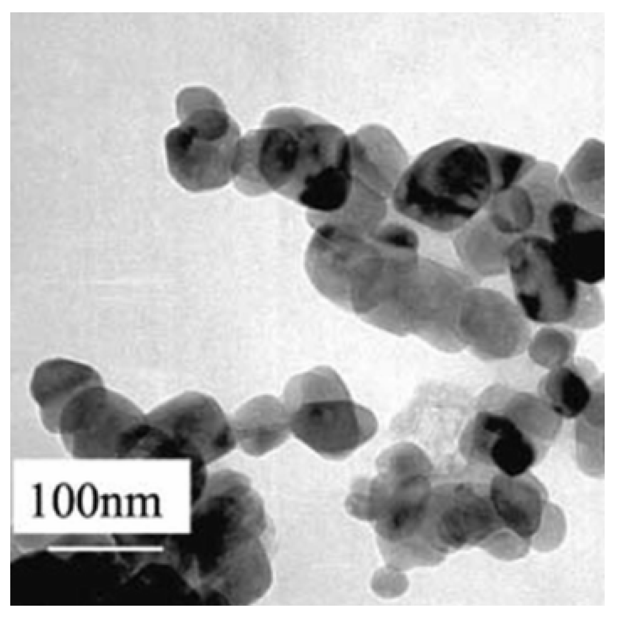
Figure 2.
EMo-NPs antibacterial effects: extracellular/intracellular ROS induction (through metal ion release) with subsequent damage of the cell wall, DNA, proteins, and mitochondria. Sources of the cell organelles: “https://knowgenetics.org/dna-mutations-2/, https://www.cleanpng.com/png-biochemistry-biology-protein-science-pathway-1197074/ (accessed on 15 March 2023)”, “https://www.shutterstock.com/ro/search/mitochondrial (accessed on 15 March 2023)”.
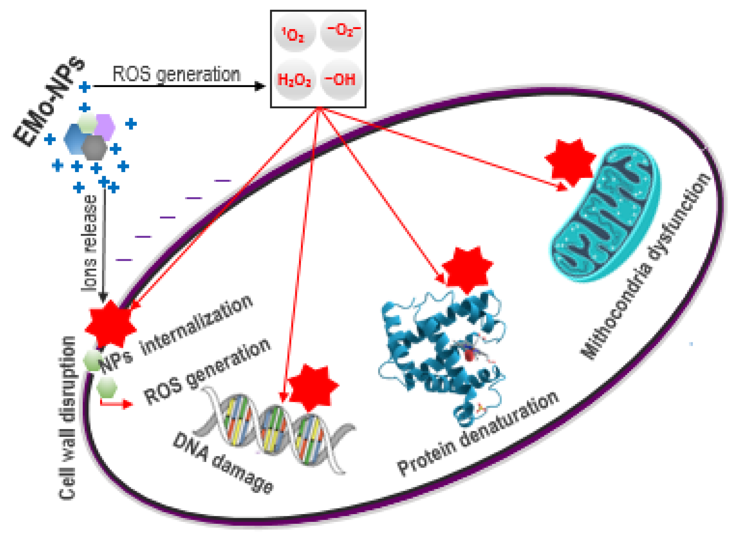
Figure 3.
Possible interactions from a polymer-based bottle/drink/external environment. Migration: blue circles represent molecular-scale migrants (components from polymer matrix, such as monomers); colored hexagons represent EMo-NPs from polymer matrix. Scalping: pink circles represent constituents of drink matrix. Permeation: green circles represent gases (CO2, O2) from both drink matrix and external environment.
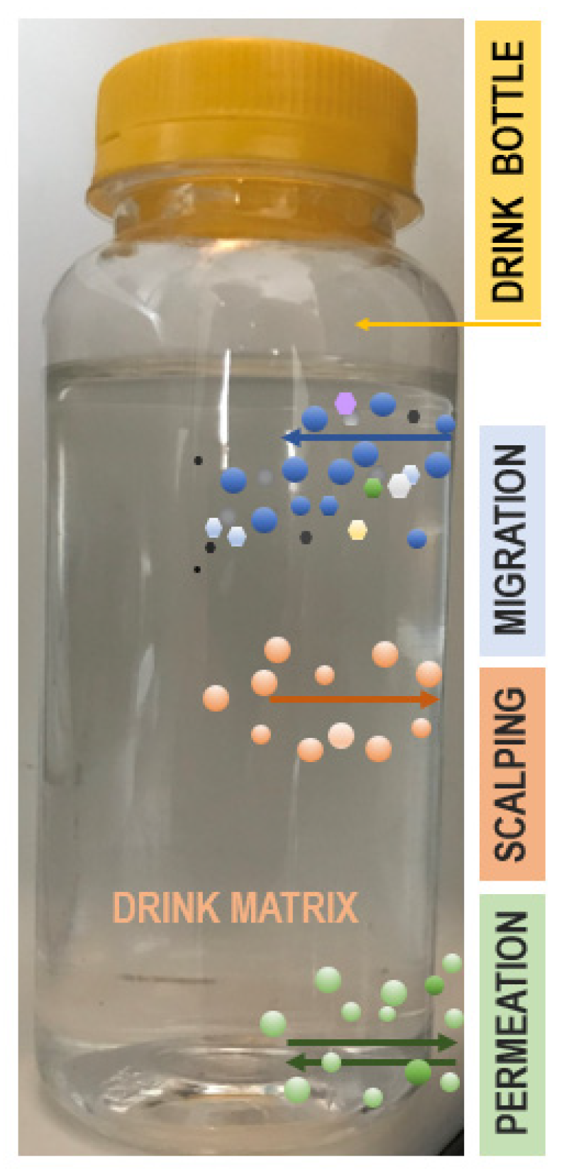
Figure 4.
The oral route of EMo-NPS into the human GI tract. Source of the human digestive system: “https://commons.wikimedia.org/wiki/File:Digestive_system_without_labels.svg. (accessed on 20 March 2023)”.
Figure 4.
The oral route of EMo-NPS into the human GI tract. Source of the human digestive system: “https://commons.wikimedia.org/wiki/File:Digestive_system_without_labels.svg. (accessed on 20 March 2023)”.

Figure 5.
ROS induction inside the eukaryotic cell by EMo-NPs. EMo-NPs are much more toxic than metal ions, serving as Trojan horse-type carriers (green hexagons) which release metal ions inside the cell (gray circles). Source of the cell membrane: “https://smart.servier.com/smart_image/cell (accessed on 22 March 2023)”. Sources of the organelles: “https://www.pngwing.com/en/search?q=mitochondria (accessed on 22 March 2023)”, “https://commons.wikimedia.org/wiki/File:201601_Endoplasmic_reticulum.png, (accessed on 22 March 2023)”, “https://pixabay.com/ro/vectors/dna-biologie-pictogram%C4%83-rna-6177695/ (accessed on 22 March 2023)”.

Table 1.
Types of EMo-NPs and their benefits in the food industry.
| Type | Benefits | References |
|---|---|---|
| CuO-NPs | Excellent antibacterial (against Gram-positive and Gram-negative bacteria), antifungal, and antiviral agent; good reinforcing NPs of polymer/biopolymer matrix | [1,3,4,9,17,30,31,32,33,34,35,36,37,38,39,40,41,42,43,44,45,46,47,48] |
| Fe3O4-NPs | Distinct type of NPs with superior properties compared to α-Fe2O3 (ferric oxide–hematite) and γ-Fe2O3 (ferric oxide–maghemite); possesses strong antibacterial characteristics; source of bioavailable iron; mineral-fortified supplement | |
| MgO-NPs | Strong antimicrobial action similar to nanosilver (depending on their sizes; when the size is smaller than 15 nm, MgO-NPs have a powerful biocidal efficacy); nanoscale MgO is a polymer/biopolymer reinforcement agent; dietary supplement | |
| TiO2-NPs | Top EMo-NPs (major promising NPs player in the food industry, extensively used); photocatalytic antimicrobial activities against bacteria, yeast, and fungi; UV-protective food nanopackaging; enhancer of mechanical and thermal stability of food packaging; oxygen scavenger; biosensor for volatile organic compounds; food coloring agent | |
| ZnO-NPs | Excellent photocatalytical and photocorrosion antimicrobial against bacteria, yeast, and fungi; UV light absorber in food packaging; source of zinc in food supplements |
Table 2.
Main concerns related to EMo-NPS.
| EMo-NPs | Main Concerns | References |
|---|---|---|
| CuO-NPs | Cu, a trace element, is vitally important, playing a significant role in numerous biological activities (hemoglobin production, iron metabolism, hormone synthesis, etc.). However, a high level of Cu ions could be toxic. Cu ions are redox-active, affecting biological systems. Their reactivity is wholly dependent on extrinsic and intrinsic factors (size, shape, surface charge, concentration of NPs). The smaller the size, the more toxic they are. NPs smaller than 40 nm can directly enter cell nuclei from the circulatory system, while NPs greater than 100 nm can cross the cell membrane. Spherical ones are more reactive. CuO-NPs are Trojan horse-type carriers, releasing Cu ions inside the cells. Compared with Fe3O4-NPs, ZnO-NPs, and TiO2-NPs, CuO-NPs are the most potent in terms of cytotoxicity. CuO-NPs’ uptake in many organs (spleen, liver, kidneys, brain, lungs, blood, heart, stomach, bones, marrow), and they could exert: oxidative stress genotoxicity cytotoxicity immunotoxicity inflammation | [104,105,106,107,108,109,110,111,112,113,114] |
| Fe3O4-NPs | Fe is an essential biological trace element not only for human beings but for all other life forms. In the human body, the majority of Fe is in hemoglobin (50–60%); 25% is in an easily mobilizable store, and the remaining 15% is in myoglobin and in numerous enzymes involved in oxidative metabolism and many other cellular activities. In addition, Fe3O4-NPs is used in various biomedical applications (cancer, diabetes, diagnosis of contrast substances, magnetic resonance imaging, inflammatory diseases, targeted drug delivery, gene therapy, biosensors, etc.). Although Fe-based supplements are highly effective for improving iron status and Fe3O4-NPs-based biomedical applications have good potential, there are controversial results regarding the cytotoxic effects and the overall integrity of the cells, once the engineered Fe3O4-NPs are inside the cells. Fe3O4-NPs are less toxic than other metal oxide NPs, but they could effectively enter the cell nucleus. Along similar lines as CuO-NPs, Fe3O4-NPs reactivity is linked to surface modification, concentration, size, shape, dose dependency, obtainment method, etc., and could induce: disruptions of the oxidative balance cytotoxicity immunotoxicity neurobehavioral toxicity inflammation ferroptosis fibrosis/cirrhosis and loss of liver function | [61,115,116,117,118,119,120,121,122,123,124] |
| MgO-NPs | Mg2+ is an important cation for human health. In the human body, the majority of Mg is mainly stored in bones (50–65%) while 34–39% is in muscle, soft tissues, and organs and 1–2% is in blood and extracellular fluids. It plays a significant function in many physiological processes. MgO-NPs are used in a wide range of biomedical applications (cancer therapy, medical imaging, nanocryosurgery, bone regeneration, biosensor, tissue engineering, dental implants, bioactive glasses, etc.). However, MgO-NPs’ toxicity is controversial, depending on the physical and chemical characteristics of the NPs and tested cell type. Concerns about their safety remain (at high concentrations), and refer to: oxidative stress hemolytic activity arteriosclerosis inflammation neronal apoptosis hepatocytotoxicity | [125,126,127,128,129,130,131] |
| TiO2-NPs | TiO2-NPs are one of the most commonly used NPs in consumer products (foods, medicines). The exposure of humans to TiO2-NPs via the oral route is inevitable. The potential toxicity of TiO2-NPs is addressed by multiple studies and is wholly dependent on their size, shape, surface charge, concentration, and solubility. Due to their smaller size, TiO2-NPs are more easily absorbed into cells, where they can become involved in: oxidative stress genotoxicity cytotoxicity immunotoxicity inflammation TiO2-NPs promote photochemical reactions, as they are more active in UV light, but they can induce ROS under dark conditions. If ROS induction can occur under dark conditions, it can likely take place inside the human body. This may lead TiO2-NPs to be toxicologically potent than previously known. | [52,132,133,134,135,136,137,138,139,140,141,142,143,144,145,146,147,148,149] |
| ZnO-NPs | Zn is one of the most essential trace minerals and possesses exceptional properties such as the capacity to modulate immune responses, improve fertility and metabolism, scavenge free radicals, etc. ZnO-NPs are one of the most prevalent metal oxide NPs and possess wide biomedical applications (treatment of various kinds of cancers, drug delivery, etc.). The exposure of humans to ZnO-NPs is very frequent and constitutes an issue of concern to health. The potential toxicity of ZnO-NPs is entirely dependent on their size, shape, surface charge, concentration, and solubility. Studies of their toxicology in in vivo models indicate that ZnO-NPs may cause: oxidative stress genotoxicity cytotoxicity inflammation hepatotoxicity | [150,151,152,153] |
Disclaimer/Publisher’s Note: The statements, opinions and data contained in all publications are solely those of the individual author(s) and contributor(s) and not of MDPI and/or the editor(s). MDPI and/or the editor(s) disclaim responsibility for any injury to people or property resulting from any ideas, methods, instructions or products referred to in the content. |
© 2023 by the authors. Licensee MDPI, Basel, Switzerland. This article is an open access article distributed under the terms and conditions of the Creative Commons Attribution (CC BY) license (https://creativecommons.org/licenses/by/4.0/).
Share and Cite
MDPI and ACS Style
Stuparu-Cretu, M.; Braniste, G.; Necula, G.-A.; Stanciu, S.; Stoica, D.; Stoica, M. Metal Oxide Nanoparticles in Food Packaging and Their Influence on Human Health. Foods 2023, 12, 1882. https://doi.org/10.3390/foods12091882
AMA Style
Stuparu-Cretu M, Braniste G, Necula G-A, Stanciu S, Stoica D, Stoica M. Metal Oxide Nanoparticles in Food Packaging and Their Influence on Human Health. Foods. 2023; 12(9):1882. https://doi.org/10.3390/foods12091882
Chicago/Turabian StyleStuparu-Cretu, Mariana, Gheorghe Braniste, Gina-Aurora Necula, Silvius Stanciu, Dimitrie Stoica, and Maricica Stoica. 2023. "Metal Oxide Nanoparticles in Food Packaging and Their Influence on Human Health" Foods 12, no. 9: 1882. https://doi.org/10.3390/foods12091882
Note that from the first issue of 2016, this journal uses article numbers instead of page numbers. See further details here.