Article

The Bactericidal Efficacy and the Mechanism of Action of Slightly Acidic Electrolyzed Water on Listeria monocytogenes’ Survival

College of Food Science and Biology, Hebei University of Science and Technology, Shijiazhuang 050018, China
*
Author to whom correspondence should be addressed.
Foods 2021, 10(11), 2671; https://doi.org/10.3390/foods10112671
Submission received: 1 October 2021 / Revised: 26 October 2021 / Accepted: 29 October 2021 / Published: 3 November 2021

Abstract

:
In the present work, the bactericidal efficacy and mechanism of slightly acidic electrolyzed water (SAEW) on L. monocytogenes were evaluated. The results showed that the strains of L. monocytogenes were killed completely within 30 s by SAEW whose available chlorine concentration (ACC) was higher than 12 mg/L, and it was confirmed that ACC is the main factor affecting the disinfection efficacy of SAEW. Moreover, our results demonstrated that SAEW could destroy the cell membrane of L. monocytogenes, which was observed by SEM and FT-IR, thus resulting in the leakage of intracellular substances including electrolyte, protein and nucleic acid, and DNA damage. On the other hand, the results found that SAEW could disrupt the intracellular ROS balance of L. monocytogenes by inhibiting the antioxidant enzyme activity, thus promoting the death of L. monocytogenes. In conclusion, the bactericidal mechanism of SAEW on L. monocytogenes was explained from two aspects including the damage of the cell membrane and the breaking of ROS balance.

1. Introduction

Foodborne pathogens have been raising increasing concerns because they are associated with the outbreak of foodborne disease and present an extensive health burden worldwide. Listeria monocytogenes (L. monocytogenes) is one of the main foodborne pathogens threatening human health in refrigerated food and is responsible for Listeriosis in children, the elderly, pregnant women and immunocompromised populations [1]. It has been reported that L. monocytogenes can survive and grow under a variety of extremely adverse conditions including low temperatures (0.4–45 °C), low pH values (4–9.6) and low oxygen levels, which enables it to continue to grow and reproduce in low-temperature preserved food [2,3]. Most seriously, L. monocytogenes enter the human digestive system first, then go across intestinal epithelial cells, enter the liver or other organs through the blood circulation, and can produce infection in the meninges and brain substance[4,5]. Scientists found that the main harmful factors of L. monocytogenes included Listeria hemolysin O, phospholipase and some virulence proteins, and so on [6,7,8,9,10,11].
In order to reduce the microbial contamination caused by foodborne pathogens, especially L. monocytogenes, chlorine sanitizer such as sodium hypochlorite (NaClO) and chlorine dioxide (ClO2) have traditionally been recognized as possessing high disinfection efficacy in the food industry. However, a low chlorine concentration could not reduce the microbial population and a high chlorine concentration may lead to the formation of trihalomethanes and haloacetic acids, which are potentially carcinogenic and teratogenic [12,13,14]. Lan et al. (2019) reported that the application of SAEW could cause a sublethal injury to the recovery of Listeria monocytogenes, and Jeon et al. [15] reported that SAEW can significantly reduce Listeria innocua biofilm cells on food contact surfaces during food processing. As an alternative to the traditional chlorine sanitizer, the strong antibacterial effect of electrolyzed oxidizing water (EOW) has been recognized as and has shown a promising prospect in the food industry.
EOW has two main types including strong acidic electrolyzed water (AEW) and slightly acidic electrolyzed water (SAEW) [16,17]. In the past few years, the disinfection efficacy and mechanism, as well as the application in the food industry, of AEW have been reported and reviewed in many studies [12,18,19]. Furthermore, increasing attentions have been paid to the bactericidal activities of SAEW in recent years because SAEW has a nearly neutral pH of 5.0–6.5 and lower available chlorine concentration (ACC) of 10–30 mg/L compared with AEW, thus demonstrating the advantages of SAEW in comparison with AEW [16,20,21,22]. Generally, the recent studies on SAEW have mainly focused on its application in the disinfection of food materials such as fruits, vegetables, sea foods and so on [23,24,25]. However, few studies about the mechanism of the bactericidal efficacy of SAEW have been systematically reported.
In previous studies, scientists found that the chlorine compounds in EOW are involved in hypochlorous acid (HClO), hypochlorite ion (OCl) and chlorine gas (Cl2), which are regulated by pH, and HClO became the main form with a pH of 5.0–6.5 [16,17,18,19,20,21,22]. Thus, it was recognized that the strong disinfection efficacy of SAEW was attributed to the existence of HClO, which is 80 times more effective as a sanitizer than an equivalent concentration of the hypochlorite ion (OCl) in the inactivation of bacteria [13,26]. On the other hand, researchers investigated the disinfection mechanism of SAEW on Staphylococcus aureus and suggested that the disinfection mechanism on Staphylococcus aureus cells of SAEW was disrupting the permeability of the cell membrane and the cytoplasmic ultrastructures [27]. Further research evaluated the disinfection mechanism of SAEW on Escherichia coli and Staphylococcus aureus, and the results showed that the disinfection behavior was associated with multiple cellular targets including both cell barriers and intracellular components [22]. Our previous study confirmed that SAEW could damage the cell membrane, thus causing the leakage of protein, DNA, RNA and ATP, which suggested that the differences in antibacterial efficacy between SAEW and AEW could be explained by the different impact on RNA of Escherichia coli and Staphylococcus aureus [28]. However, the bactericidal mechanism of SAEW on L. monocytogenes is still unknown.
The aim of the present study was to investigate the bactericidal efficacy and mechanism of SAEW on L. monocytogenes to help to provide results to further research on the target site for the bactericidal mechanism of SAEW. In the present study, the bactericidal effect of SAEW with different ACC on L. monocytogenes was firstly determined by plate count, and then the leakage of intracellular material including K+, intracellular protein and nucleic acid were investigated. Moreover, the DNA damage of SAEW-treated L. monocytogenes was evaluated by flow cytometry and the damage of the cell membrane was observed by scanning electron microscopy (SEM) and Fourier-transform infrared spectroscopy (FTIR). In addition, the levels of antioxidant enzymes including superoxide dismutase (SOD), catalase (CAT) and glutathione peroxidase (GSH-PX) in SAEW-treated L. monocytogenes were assayed, and the reactive oxygen species (ROS) were evaluated by laser scanning confocal microscopy (LSCM).

2. Materials and Methods

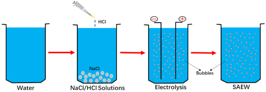
2.1. Preparation of SAEW

SAEW was prepared by using a self-made electrolyzed water device and the preparation process is shown in Figure 1. After generation, SAEW was transferred to polypropylene containers and stored in dark. The pH value was determined by a pH meter (Model 86802, Orion Inc. Boston, MA, USA) and the ACC was measured using the iodometric method. The ACC of the SAEW used in this study are shown in Table 1.

2.2. Preparation of Bacterial Strains and Culture

L. monocytogenes (ATCC19114) was obtained from Solarbio (Beijing Solarbio Life Science Co., Ltd., Beijing, China) and the cultures were kept in a glass tube at −80 °C. Frozen L. monocytogenes was streaked in TSB-YE agar broth, then a single colony of L. monocytogenes was inoculated in 100 mL TSB-YE liquid broth (Beijing Land Bridge Technology, Co., Ltd., Beijing, China), and incubated at 37 °C in an orbital shaker (ZWY-100H, Shanghai Zhicheng Analytical Instruments Manufacturing Co., Ltd., Shanghai, China) at 180 r/min for 24 h.
L. monocytogenes suspension was centrifuged at 8000 r/min for 10 min. The sediment was collected and washed by sterile PBS twice, resuspended by sterile saline to obtain a final cell concentration approximately of 107 CFU/mL. The absorbance of the bacterial suspension was measured at 450 nm using a spectrophotometer (Model Tu-1901, Beijing Purkinje General, Beijing, China) to estimate the bacterial concentration.

2.3. Determination of Antibacterial Effect of SAEW

An amount of 1 mL of inocula of 107 CFU/mL of L. monocytogenes was added to 9 mL of different SAEW for different time lengths. After gradient dilution, the surviving cells were counted by spread-plating 0.1 mL of the sample or dilution onto LB (Luria-Bertani) medium, which is composed of tryptone (10g/L, Oxoid Co., Ltd., Basingstoke, Hampshire, UK), yeast extract (5 g/L, Oxoid Co., Ltd., UK), agar power (2.0% (w/v), Oxoid Co., Ltd., UK) and NaCl (10 g/L, Aoboxing Bioscience Inc., Beijing, China). The plates were incubated at 37 °C for 48 h and then the colonies were counted. In this study, the bacteria counts were expressed in log10CFU/mL.

2.4. Observation of Cell Membrane by SEM and FT-IR

L. monocytogenes suspension was centrifuged at 8000 r/min for 10 min and the sediment was collected and washed by sterile PBS for twice. Based on the above results of the antibacterial efficacy of SAEW on L. monocytogenes strains, the SAEW3 with ACC of 6.03 ± 0.13 and pH of 5.76 ± 0.03 was used in the following measurement. In brief, 1 mL of inocula of approximately 107 CFU/mL of L. monocytogenes was added to 9 mL different SAEW3 solutions for 60 s, 0.1% sodium thiosulfate solution was used to suspend the residual activity of SAEW and then centrifuged at 8000 r/min for 10 min. The same treatment with sterile saline was used as control.
After centrifugation, the sediment was fixed with 2.5% glutaraldehyde overnight, dehydrated in an ascending acetonitrile series (50%, 70%, 80%, 90% and 100% twice for 20 min each) and dried in a lyophilizer (LGJ-10D, Four-Ring Science Instrument Plant Beijing Co., Ltd., Beijing, China). The sample power was sputter-coated with gold and observed with a scanning electron microscope (SEM, S-4800, Hitachi Co., Tokyo, Japan). The sample power was detected using a Fourier-transform infrared spectroscopy (FTIR, Nexus 670, Nicolet co., Madison, WI, America) by attenuated total reflection (ATR) with the following detection conditions: wave number range of 4000–500 cm−1, resolution ratio of 4 cm−1 and scanning numbers of 32 times.

2.5. Measurement of Leakage of Bacterial Intracellular Materials

The preparation of samples and bacterial suspension is described in Section 2.4. After centrifugation, the supernatant was used to take further measurements. The K+ and protein concentration was measured by K+ concentration assay kit and BCA protein assay kit (Jiancheng Bioengineering Institute, Jiangsu, China), respectively. The nucleic acid content was determined by OD260 value with UV spectrophotometer (UV-5200, Shanghai Yuanxi instrument Co. Ltd., Shanghai, China). The sterile saline treatment was used as control.

2.6. Evaluation of DNA Damage by Flow Cytometry (FCM)

The DNA damage of L. monocytogenes was determined using FCM after acridine orange (AO) staining. The preparation of samples and bacterial suspension was performed as described in Section 2.4. Briefly, 170 μL of 0.1 mg/mL AO staining solutions were added to SAEW-treated suspension and the mixtures were incubated at 37 °C for 10 min in dark, then tested by FCM (Accuri C6, BD Biosciences Co., San Jose, CA, USA). The sterile saline treatment was used as control.

2.7. ROS Analysis with Laser Scanning Confocal Microscopic (LSCM)

ROS assay kit (Beijing Solarbio Life Science Co., Ltd., Beijing, China) was used to estimate the intracellular ROS level. The preparation of samples and bacterial suspension is referred to in Section 2.4. A total of 10 μL of DCFH-DA solution was added to samples and control, respectively. The mixtures were incubated at 3 °C for 20 min with every 3-min vortex. After that, the sample was collected by centrifugation of 8000× g r/min for 10 min and washed with 1 mL of sterile saline solution to remove excess DCFH-DA. Finally, samples were observed with CLSM (TCS SP8, Leica Co., Wetzlar, Germany). The excitation wavelength was 488 nm and the emission wavelength was 525 nm.

2.8. Measurement of Intracellular Antioxidant Enzyme Activity

In the present study, three kinds of antioxidant enzymatic activity were measured including SOD, CAT and GSH-PX. The preparation of samples and bacterial suspension is referred to in Section 2.4 and the sterile saline treatment was used as control. The sediment was collected for subsequent treatment. The sediment was resuspended with saline and homogenated by a tissue homogenate machine (Pro 250, pro scientific Co., Oxford, CT, America) for 2 min. The extract was kept in −8 °C for further tests.
The activities of SOD, CAT and GSH-PX were determined by assay kits. All the assay kits were purchased from Jiancheng company (Jiancheng Bioengineering Institute, Jiangsu, China). The results were expressed as U/mg.

2.9. Statistical Analysis

The experiments were performed in triplicate and the data were presented as mean ± standard deviation. Data were analyzed by Duncan’s multiple range tests using SPSS software (SPSS16.0 for Windows). Significant differences between treatments were established at a significance level of p < 0.05.

3. Results and Discussions

3.1. Bactericidal Efficacy of SAEW on Strains of L. monocytogenes

The bactericidal efficacy of SAEW with different ACC on L. monocytogenes was evaluated in the present study and the survival populations of L. monocytogenes are shown in Table 1. The initial counts of L. monocytogenes were about 7.4 log10 CFU/mL. From Table 1, the results showed that SAEW with ACC of more than 12 mg/L (SAEW1 andSAEW2) had significant disinfection efficacy, by which the strains of L. monocytogenes were completely killed within 30 s. However, the disinfection efficacy on L. monocytogenes of SAEW was decreased significantly when the ACC of SAEW was reduced to about 6 mg/L (SAEW3) because the time to completely kill L. monocytogenes was extended to 150 s. Furthermore, as the ACC deceased to less than 5 mg/L (SAEW4, SAEW5 and SAEW6), the survival counts of L. monocytogenes decreased by less than 1 log10 CFU/mL, respectively, in 150 s. Undoubtedly, HClO is the main form of the chlorine compounds in SAEW due to its pH of 5.0–6.5 compared to OCl or Cl2. However, the current method used to determine the chlorine compounds concentration is the iodometric method, which could not accurately determine the HClO contents. Considering that all of the types of SAEW used in the present study have similar pH values but different ACC, we confirm that ACC is the main factor affecting the disinfection efficacy of SAEW on L. monocytogenes, which is in agreement with the previous reports [29]
The strong bactericidal efficacy of SAEW has been recognized by many studies and SAEW has shown that it is a promising prospect in the food industry [20,29,30,31]. Our present study also demonstrated the strong bactericidal efficacy of SAEW on the strains of L. monocytogenes. It was reported that the bactericidal activity of SAEW against Vibrio vulnificus was reduced when the ACC of SAEW was less than 15 mg/L but was maintained in the Vibrio parahaemolyticus when the ACC of SAEW was 0.5 mg/L, which suggested SAEW with an extremely low ACC (even 0.5 mg/L) had still strong bactericidal activity [30]. The results obtained from the present study partly disagree with the previous report. In addition, it has been recognized that SAEW treatment with low ACC could not only cause the sublethal injury of L. monocytogenes and E. coli O157:H7 cells [32,33,34] but also result in the formation of viable but nonculturable (VBNC) on E. coli O157:H7, Salmonella Enteritidis and Yersinia enterocolitica [27,35,36], which set the new challenge to find the antibacterial activities of SAEW. Generally, the bactericidal efficacy of SAEW on L. monocytogenes needs further research.

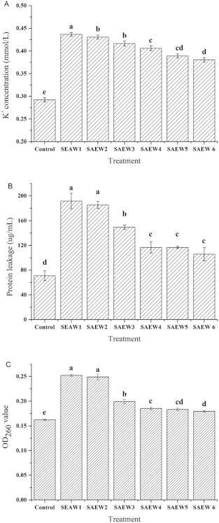
3.2. Effect of SAEW on Intracellular Material Leakages of L. monocytogenes

The cell membrane is an important structure of bacteria, which has the function of controlling material exchange. The intracellular material would leak when the cell membrane was damaged, which would lead to bacterial death [37,38]. As shown in Figure 2, SAEW treatment resulted in the remarkable increase in the extracellular levels of K+ concentration (Figure 2a), proteins (Figure 2b) and nucleic acids (Figure 2c), which indicated that the cell membrane of L. monocytogenes had been injured due to the leakage of intracellular K+, protein and nucleic acids.
It can be seen from Figure 2 that the concentration of each intracellular material in the control was the lowest. After SAEW treatment, the leakage concentration of each intracellular material was increased, and the leakage was positively correlated with the concentration of ACC in the SAEW. Moreover, as the results of the bactericidal effect showed, it was found that the leakage of intracellular material corresponded to the death of L. monocytogenes. It indicated that the cell membrane of dead L. monocytogenes was damaged and SAEW destroyed the cell membrane of L. monocytogenes, which is similar to the previous studies [27,28].

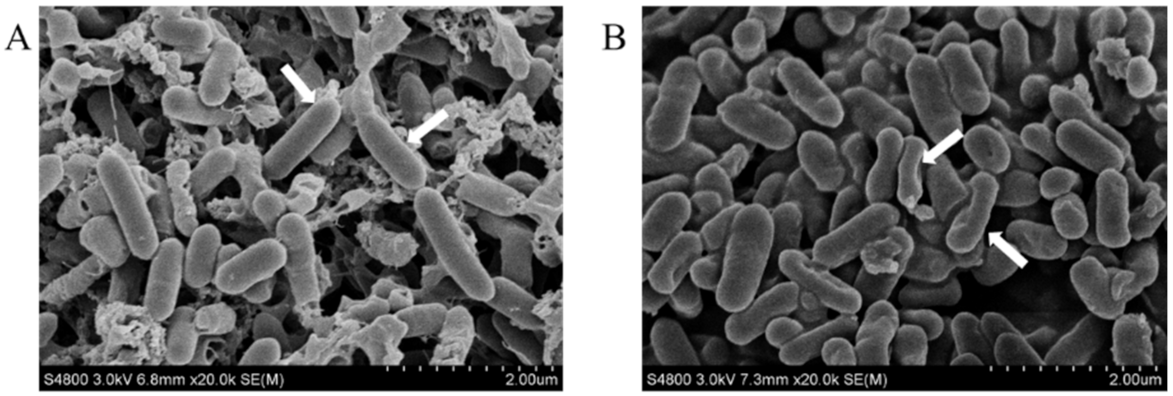
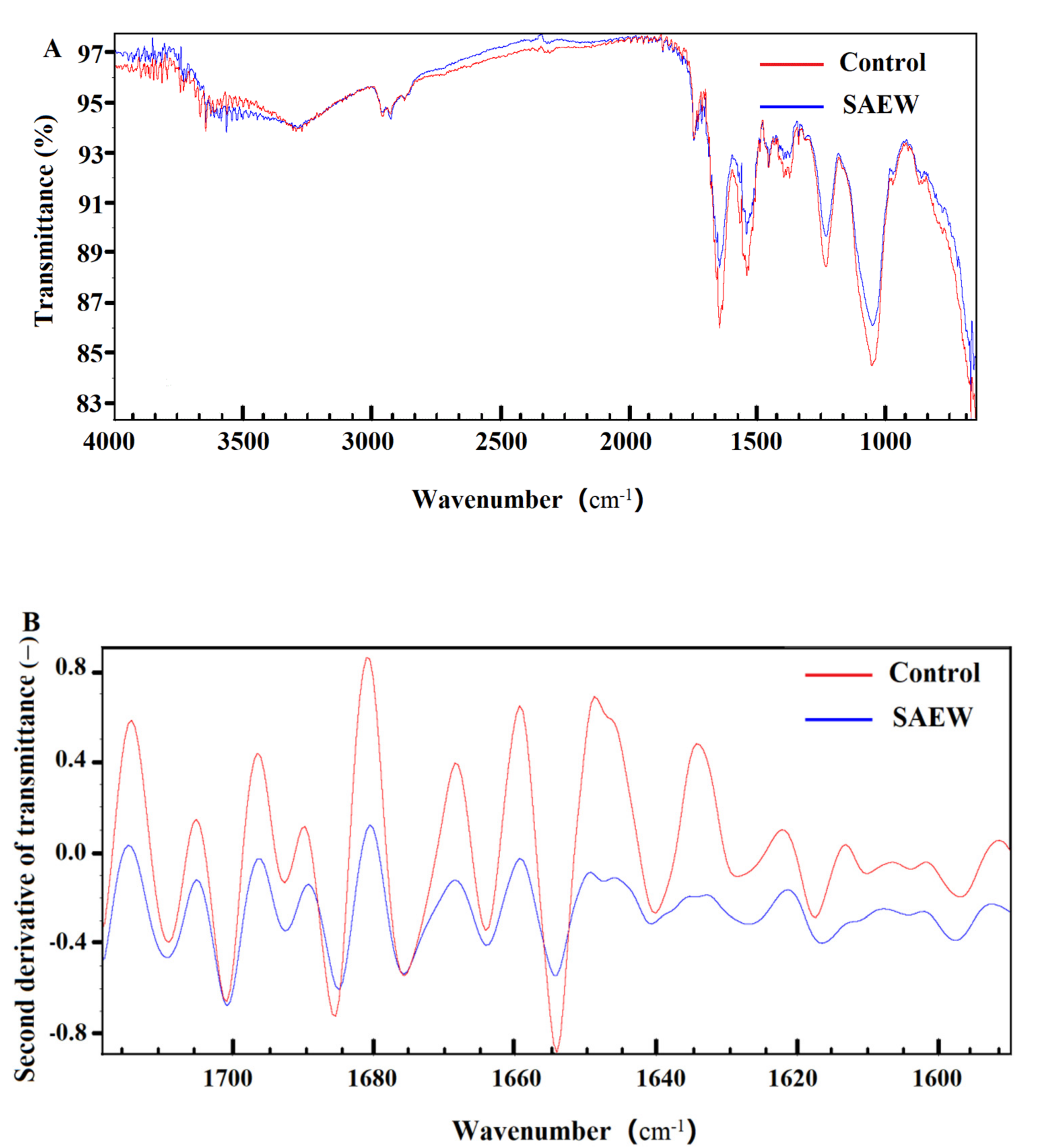
3.3. Effect of SAEW on Cell Membrane of L. monocytogenes

Morphological changes of SAEW-treated and non-treated L. monocytogenes were observed using SEM. As shown in Figure 3, the body cells of L. monocytogenes had a complete morphology, smooth surface and regular texture in the control. However, the images of L. monocytogenes treated with SAEW revealed that the cells were markedly shrunken and partially collapsed. This phenomenon may be caused by the release of intracellular material, which was brought out by the cell membrane damage and the osmotic pressure out of balance [27]. Our previous study also found that SAEW could damage the structure and inner substances of Escherichia coli and Staphylococcus aureus [28].
Moreover, the FTIR analysis was performed to verify the damage on the cell membrane of L. monocytogenes by SAEW, and the results are shown in Figure 4, including the original FTIR graph and the corresponding second derivative infrared spectra graph. In general, the similar peak shapes were observed between the SAEW treatment and the control, but the peak climax showed obvious difference. The results indicated that the cell membrane of L. monocytogenes was affected by SAEW treatment. In the present study, we focused on the range of 1680–1610 cm−1, which is assigned to the bond of C=C [39]. The higher the peak is, the more content of C=C it possesses. As shown in Figure 4, the peak height of L. monocytogenes treated by SAEW including 1638, 1648 and 1668 cm−1 decreased significantly in comparison with that of the control. The results suggested that SAEW could cause the decrease in C=C and the destruction of cell physiological components, thus resulting in the damage to the cell membrane of L. monocytogenes.

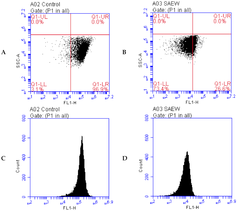
3.4. Effect of SAEW on DNA Damage of L. monocytogenes

Acridine orange (AO) is a fluorochrome, which can enter the cells and combine with DNA to emit green fluorescence [40]. The fluorescence intensity can be measured by FCM to reflect the changes in DNA. The effect of SAEW on the DNA damage of L. monocytogenes was evaluated by FCM, and the results are shown in Figure 5. As presented in Figure 5a,b, the lower right region represents the DNA region, and the lower left region represents the damaged DNA region. It is clear that the ratio of integrated DNA in SAEW-treated L. monocytogenes is about 26.6% instead of about 96.9% in the non-treated strain as a control, which suggests that SAEW treatment could reduce the ratio of the integrated DNA by approximately 70%, thus resulting in the DNA damage of L. monocytogenes.
In Figure 5c,d, the X-axis represents the green fluorescence intensity, and the Y-axis represents the number of cells. After SAEW treatment, the fluorescence intensity of most cells was significantly reduced, and the fluorescence intensity of most cells moved from 105 to 103–104. These results suggest that the DNA of L. monocytogenes was damaged by SAEW treatment. As mentioned above in the present study, SAEW caused oxidative damage in L. monocytogenes.

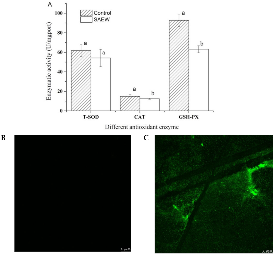
3.5. Effect of SAEW on Antioxidant Enzyme Activity and ROS of L. monocytogenes

SOD and CAT are the two key antioxidant enzymes that remove ROS. SOD catalyzed O2− into H2O2 by the reaction of 4O2− + 2H+ = H2O2 + O2 and H2O2 is further decomposed into H2O by CAT. GSH-PX is an important peroxidase widely found in the organism, which could eliminate hydroperoxides and reduce the toxicity of peroxides to non-toxic hydroxyl compounds. Generally, SOD, CAT and GSH-PX contribute to the enzyme antioxidant defense system together, which were able to effectively remove reactive oxygen radicals [41,42]. Therefore, the activity of SOD, CAT and GSH-PX in the cells of L. monocytogenes was measured in the present work and the results are shown in Figure 6a. There were no significant changes in the SOD activity of the cells of L. monocytogenes between the SAEW treatment and control (p > 0.05). However, the activities of CAT and GSH-PX in the cells of L. monocytogenes treated by SAEW were decreased significantly compared with those of the control (p < 0.05). These results suggest that the first step of the antioxidant chain may have no obvious change and the ROS will be converted to H2O2 by intracellular SOD in the cells of L. monocytogenes treated by SAEW, but the CAT and GSH-PX activity under the influence of SAEW dropped significantly and, thus, inhibited the further decomposition from H2O2 into H2O and O2.
It was reported that SAEW did not result in the accumulation of reactive oxygen species (ROS) inside the microbial cell, indicating that SAEW conducted a ROS-independent behavior on the microbial inactivation, and the chemical oxidants (e.g., hypochlorous acid) played a major role in the microbial intracellular oxidation processing [27]. However, generally, our results show that SAEW treatment could destroy the balance of ROS, which is inconsistent with the previous study. We deduced that the disinfection mechanism of SAEW may be different due to the differences among the bacterial species and this needs further research.
The bacteria could be stained with DCHF-DA, which could combine with ROS to produce green fluorescence. The accumulations of ROS in the cells of L. monocytogenes were evaluated by the laser scanning confocal microscopic (LSCM). As shown in Figure 6, there was almost no green fluorescence in the control (Figure 6b), which indicated that intracellular ROS in L. monocytogenes was at certain levels. Nevertheless, there was a large amount of green fluorescence observed in the SAEW-treated cells (Figure 6c), indicating that SAEW treatment caused a mass of intracellular ROS explosion in L. monocytogenes. Our present study suggests that SAEW could destroy the balance of ROS in L. monocytogenes, which is in agreement with the above study.
ROS were produced by the metabolism of bacterial cells. Excessive ROS would damage the function of bacteria, including affecting the stability of the DNA and inducing oxidative denaturation of lipid. Bacterial cells could manage the ROS level by a free radical scavenging system. To sum up, SAEW can break the intracellular ROS balance of L. monocytogenes by inhibiting the antioxidant enzyme activity. In addition, L. monocytogenes was able to provide a large amount of hydroxyl free radicals [27]. Thus, we deduced that SAEW could break the balance of ROS to promote the death of L. monocytogenes [43,44]. However, the antimicrobial signaling pathways of SAEW on L. monocytogenes need to be further explored.

4. Conclusions

In the present work, the bactericidal efficacy and mechanism of SAEW to L. monocytogenes has been evaluated. The results showed that SAEW had a good bactericidal effect on strains of L. monocytogenes, and ACC played an important role in the bactericidal efficacy. Moreover, our results demonstrated that SAEW could destroy the cell membrane of L. monocytogenes, thus resulting in the leakage of intracellular substances including electrolyte, protein and nucleic acid and DNA damage. On the other hand, the results found that SAEW could disrupt the intracellular ROS balance of L. monocytogenes by inhibiting the antioxidant enzyme activity, thus promoting the death of L. monocytogenes. In conclusion, the bactericidal mechanism of SAEW on L. monocytogenes was explained from two aspects including the damage of the cell membrane and the breaking of ROS balance. The antimicrobial signaling pathways of SAEW on L. monocytogenes need to be further explored.

Author Contributions

Conceptualization, J.H. (Jianxiong Hao); methodology, H.L. and D.L.; validation, J.H. (Jin Huang), C.C. and H.L.; investigation, H.L., D.L., J.H. (Jin Huang), C.C. and H.R.; data curation, J.H. (Jin Huang), C.C. and H.R.; writing—original draft, H.L., D.L. and D.Z.; Project administration, J.H. (Jianxiong Hao). All authors have read and agreed to the published version of the manuscript.

Funding

This research received no external funding.

Acknowledgments

The study was supported by Hebei Natural Science Foundation of China (No. C2018208085) and National Natural Science Foundation of China (No. 31972170).

Conflicts of Interest

The authors declare no conflict of interest.

Abbreviations

SAEW, slightly acidic electrolyzed water; ACC, available chlorine concentration; AEW, acidic electrolyzed; NaClO, sodium hypochlorite; ClO2, chlorine dioxide; EOW, electrolyzed oxidizing water; HClO, hypochlorous acid; OCl-, hypochlorite ion; Cl2, chlorine gas; SOD, antioxidant enzymes including superoxide dismutase; CAT, catalase; GSH-PX, glutathione peroxidase; ROS, reactive oxygen species; SEM, scanning electron microscopy; FTIR, Fourier-transform infrared spectroscopy; LSCM, scanning confocal microscopy.

References

  1. Jadhav, S.; Bhave, M.; Palombo, E. Methods used for the detection and subtyping of Listeria monocytogenes. J. Microbiol. Methods 2012, 88, 327–341. [Google Scholar] [CrossRef] [PubMed]
  2. Sadekuzzaman, M.; Yang, S.; Mizan, F.R.; Kim, H.-S.; Ha, S.-D. Effectiveness of a phage cocktail as a biocontrol agent against L. monocytogenes biofilms. Food Control 2017, 78, 256–263. [Google Scholar] [CrossRef]
  3. Shen, Q.; Pandare, P.; Soni, K.A.; Nannapaneni, R.; Mahmoud, B.S.; Sharma, C.S. Influence of temperature on alkali stress adaptation in Listeria monocytogenes. Food Control 2016, 62, 74–80. [Google Scholar] [CrossRef]
  4. Cossart, P.; Toledo-Arana, A. Listeria monocytogenes, a unique model in infection biology: An overview. Microbes Infect. 2008, 10, 1041–1050. [Google Scholar] [CrossRef]
  5. Drolia, R.; Bhunia, A.K. Crossing the intestinal barrier via Listeria adhesion protein and internalin A. Trends Microbiol. 2019, 27, 408–425. [Google Scholar] [CrossRef]
  6. Dinner, S.; Kaltschmidt, J.; Stump-Guthier, C.; Hetjens, S.; Ishikawa, H.; Tenenbaum, T.; Schroten, H.; Schwerk, C. Mitogen-activated protein kinases are required for effective infection of human choroid plexus epithelial cells by Listeria monocytogenes. Microbes Infect. 2017, 19, 18–33. [Google Scholar] [CrossRef] [PubMed]
  7. Drolia, R.; Tenguria, S.; Durkes, A.C.; Turner, J.R.; Bhunia, A.K. Listeria adhesion protein induces intestinal epithelial barrier dysfunction for bacterial translocation. Cell Host Microbe 2018, 23, 470–484. [Google Scholar] [CrossRef] [PubMed] [Green Version]
  8. Reddy, S.; Akgul, A.; Karsi, A.; Abdelhamed, H.; Wills, R.W.; Lawrence, M.L. The role of Listeria monocytogenes cell wall surface anchor protein LapB in virulence, adherence, and intracellular replication. Microb. Pathog. 2016, 92, 19–25. [Google Scholar] [CrossRef] [PubMed] [Green Version]
  9. Schnupf, P.; Portnoy, D.A. Listeriolysin O: A phagosome-specific lysin. Microbes Infect. 2007, 9, 1176–1187. [Google Scholar] [CrossRef]
  10. Thakur, M.; Asrani, R.K.; Patial, V. Chapter 6—Listeria monocytogenes: A food-borne pathogen. In Foodborne Diseases; Holban, A.M., Grumezescu, A.M., Eds.; Academic Press: Cambridge, MA, USA, 2018; pp. 157–192. [Google Scholar]
  11. Van Troys, M.; Lambrechts, A.; David, V.; Demol, H.; Puype, M.; Pizarro-Cerda, J.; Gevaert, K.; Cossart, P.; Vandekerckhove, J. The actin propulsive machinery: The proteome of Listeria monocytogenes tails. Biochem. Biophys. Res. Commun. 2008, 375, 194–199. [Google Scholar] [CrossRef] [PubMed]
  12. Huang, Y.-R.; Hung, Y.-C.; Hsu, S.-Y.; Huang, Y.-W.; Hwang, D.-F. Application of electrolyzed water in the food industry. Food Control 2008, 19, 329–345. [Google Scholar] [CrossRef]
  13. Koide, S.; Takeda, J.-I.; Shi, J.; Shono, H.; Atungulu, G.G. Disinfection efficacy of slightly acidic electrolyzed water on fresh cut cabbage. Food Control 2009, 20, 294–297. [Google Scholar] [CrossRef]
  14. Waters, B.W.; Hung, Y.-C. Evaluation of different methods for determination of properties of chlorine-based sanitizers. Food Control 2013, 30, 41–47. [Google Scholar] [CrossRef]
  15. Jeon, H.R.; Kwon, M.J.; Yoon, K.S. Control of Listeria innocua Biofilms on Food Contact Surfaces with Slightly Acidic Electrolyzed Water and the Risk of Biofilm Cells Transfer to Duck Meat. J. Food Prot. 2018, 81, 582–592. [Google Scholar] [CrossRef] [PubMed]
  16. Hao, J.; Qiu, S.; Li, H.; Chen, T.; Liu, H.; Li, L. Roles of hydroxyl radicals in electrolyzed oxidizing water (EOW) for the inactivation of Escherichia coli. Int. J. Food Microbiol. 2012, 155, 99–104. [Google Scholar] [CrossRef] [PubMed]
  17. Xiong, K.; Liu, H.-J.; Liu, R.; Li, L.-T. Differences in fungicidal efficiency against Aspergillus flavus for neutralized and acidic electrolyzed oxidizing waters. Int. J. Food Microbiol. 2010, 137, 67–75. [Google Scholar] [CrossRef] [PubMed]
  18. Guentzel, J.L.; Lam, K.L.; Callan, M.A.; Emmons, S.A.; Dunham, V.L. Reduction of bacteria on spinach, lettuce, and surfaces in food service areas using neutral electrolyzed oxidizing water. Food Microbiol. 2008, 25, 36–41. [Google Scholar] [CrossRef] [PubMed]
  19. Liao, L.B.; Chen, W.M.; Xiao, X.M. The generation and inactivation mechanism of oxidation–reduction potential of electrolyzed oxidizing water. J. Food Eng. 2007, 78, 1326–1332. [Google Scholar] [CrossRef]
  20. Ding, T.; Xuan, X.-T.; Li, J.; Chen, S.-G.; Liu, D.-H.; Ye, X.-Q.; Shi, J.; Xue, S.J. Disinfection efficacy and mechanism of slightly acidic electrolyzed water on Staphylococcus aureus in pure culture. Food Control 2016, 60, 505–510. [Google Scholar] [CrossRef]
  21. Issa-Zacharia, A.; Kamitani, Y.; Morita, K.; Iwasaki, K. Sanitization potency of slightly acidic electrolyzed water against pure cultures of Escherichia coli and Staphylococcus aureus, in comparison with that of other food sanitizers. Food Control 2010, 21, 740–745. [Google Scholar] [CrossRef]
  22. Liao, X.; Xuan, X.; Li, J.; Suo, Y.; Liu, D.; Ye, X.; Chen, S.; Ding, T. Bactericidal action of slightly acidic electrolyzed water against Escherichia coli and Staphylococcus aureus via multiple cell targets. Food Control 2017, 79, 380–385. [Google Scholar] [CrossRef]
  23. Klintham, P.; Tongchitpakdee, S.; Chinsirikul, W.; Mahakarnchanakul, W. Combination of microbubbles with oxidizing sanitizers to eliminate Escherichia coli and Salmonella Typhimurium on thai leafy vegetables. Food Control 2017, 77, 260–269. [Google Scholar] [CrossRef]
  24. Liang, D.; Wang, Q.; Zhao, D.; Han, X.; Hao, J. Systematic application of slightly acidic electrolyzed water (SAEW) for natural microbial reduction of buckwheat sprouts. LWT 2019, 108, 14–20. [Google Scholar] [CrossRef]
  25. Mikš-Krajnik, M.; James Feng, L.X.; Bang, W.S.; Yuk, H.-G. Inactivation of Listeria monocytogenes and natural microbiota on raw salmon fillets using acidic electrolyzed water, ultraviolet light or/and ultrasounds. Food Control 2017, 74, 54–60. [Google Scholar] [CrossRef]
  26. Olaimat, A.N.; Holley, R.A. Factors influencing the microbial safety of fresh produce: A review. Food Microbiol. 2012, 32, 1–19. [Google Scholar] [CrossRef] [PubMed]
  27. Li, J.; Ding, T.; Liao, X.; Chen, S.; Ye, X.; Liu, D. Synergetic effects of ultrasound and slightly acidic electrolyzed water against Staphylococcus aureus evaluated by flow cytometry and electron microscopy. Ultrason. Sonochem. 2017, 38, 711–719. [Google Scholar] [CrossRef] [PubMed]
  28. Hao, J.; Wu, T.; Li, H.; Liu, H. Differences of Bactericidal Efficacy on Escherichia coli, Staphylococcus aureus, and Bacillus subtilis of Slightly and Strongly Acidic Electrolyzed Water. Food Bioprocess Technol. 2017, 10, 155–164. [Google Scholar] [CrossRef]
  29. Koide, S.; Shitanda, D.; Note, M.; Cao, W. Effects of mildly heated, slightly acidic electrolyzed water on the disinfection and physicochemical properties of sliced carrot. Food Control 2011, 22, 452–456. [Google Scholar] [CrossRef] [Green Version]
  30. Quan, Y.; Choi, K.-D.; Chung, D.; Shin, I.-S. Evaluation of bactericidal activity of weakly acidic electrolyzed water (WAEW) against Vibrio vulnificus and Vibrio parahaemolyticus. Int. J. Food Microbiol. 2010, 136, 255–260. [Google Scholar] [CrossRef] [PubMed]
  31. Tango, C.N.; Khan, I.; Kounkeu, P.-F.N.; Momna, R.; Hussain, M.; Oh, D.-H. Slightly acidic electrolyzed water combined with chemical and physical treatments to decontaminate bacteria on fresh fruits. Food Microbiol. 2017, 67, 97–105. [Google Scholar] [CrossRef]
  32. Izumi, H.; Inoue, A. Viability of sublethally injured coliform bacteria on fresh-cut cabbage stored in high CO2 at-mospheres following rinsing with electrolyzed water. Int. J. Food Microbiol. 2018, 266, 207–212. [Google Scholar] [CrossRef]
  33. Lan, L.; Zhang, R.; Zhang, X.; Shi, H. Sublethal injury and recovery of Listeria monocytogenes and Escherichia coli O157:H7 after exposure to slightly acidic electrolyzed water. Food Control 2019, 106, 106746. [Google Scholar] [CrossRef]
  34. Xuan, X.-T.; Ding, T.; Li, J.; Ahn, J.-H.; Zhao, Y.; Chen, S.-G.; Ye, X.-Q.; Liu, D.-H. Estimation of growth parameters of Listeria monocytogenes after sublethal heat and slightly acidic electrolyzed water (SAEW) treatment. Food Control 2017, 71, 17–25. [Google Scholar] [CrossRef]
  35. Afari, G.K.; Liu, H.; Hung, Y.-C. The effect of produce washing using electrolyzed water on the induction of the viable but non-culturable (VBNC) state in Listeria monocytogenes and Escherichia coli O157:H7. LWT 2019, 110, 275–282. [Google Scholar] [CrossRef]
  36. Han, D.; Hung, Y.-C.; Wang, L. Evaluation of the antimicrobial efficacy of neutral electrolyzed water on pork products and the formation of viable but nonculturable (VBNC) pathogens. Food Microbiol. 2018, 73, 227–236. [Google Scholar] [CrossRef] [PubMed]
  37. Churklam, W.; Chaturongakul, S.; Ngamwongsatit, B.; Aunpad, R. The mechanisms of action of carvacrol and its synergism with nisin against Listeria monocytogenes on sliced bologna sausage. Food Control 2020, 108, 106864. [Google Scholar] [CrossRef]
  38. Xiang, Q.; Wang, W.; Zhao, D.; Niu, L.; Li, K.; Bai, Y. Synergistic inactivation of Escherichia coli O157:H7 by plasma-activated water and mild heat. Food Control 2019, 106, 106741. [Google Scholar] [CrossRef]
  39. Yonggang, W.; Guangrui, Y.; Xueqing, M.; Minggang, W.; Feifan, L.; Fan, W.; Jianzhong, M.; Jixiang, C. Antibacterial mechanisms of endophytic fungi Alternaria alternata of ethyl acetate extract on Escherichia coli. Chin. Tradit. Herb. Drugs 2018, 49, 374–381. [Google Scholar]
  40. Campana, R.; Baffone, W. Carvacrol efficacy in reducing microbial biofilms on stainless steel and in limiting re-growth of injured cells. Food Control 2018, 90, 10–17. [Google Scholar] [CrossRef]
  41. Hu, M.; Du, J.; Du, L.; Luo, Q.; Xiong, J. Anti-fatigue activity of purified anthocyanins prepared from purple passion fruit (P. edulis Sim) epicarp in mice. J. Funct. Foods 2020, 65, 103725. [Google Scholar] [CrossRef]
  42. Sun, H.; Luo, M.; Zhou, X.; Zhou, Q.; Sun, Y.; Ge, W.; Wei, B.; Cheng, S.; Ji, S. Exogenous glycine betaine treatment alleviates low temperature-induced pericarp browning of ‘Nanguo’ pears by regulating antioxidant enzymes and proline metabolism. Food Chem. 2020, 306, 125626. [Google Scholar] [CrossRef] [PubMed]
  43. Gansukh, E.; Mya, K.K.; Jung, M.; Keum, Y.-S.; Kim, D.H.; Saini, R.K. Lutein derived from marigold (Tagetes erecta) petals triggers ROS generation and activates Bax and caspase-3 mediated apoptosis of human cervical carcinoma (HeLa) cells. Food Chem. Toxicol. 2019, 127, 11–18. [Google Scholar] [CrossRef] [PubMed]
  44. Selvi, S.K.; Vinoth, A.; Varadharajan, T.; Weng, C.F.; Padma, V.V. Neferine augments therapeutic efficacy of cisplatin through ROS- mediated non-canonical autophagy in human lung adenocarcinoma (A549 cells). Food Chem. Toxicol. 2017, 103, 28–40. [Google Scholar] [CrossRef] [PubMed]
Figure 1. Preparation of slightly acidic electrolyzed water (SAEW) by self-made generator. SAEW was generated by passing sodium chloride and hydrochloric acid solutions through the electrolytic chamber of non-membrane.
Foods 10 02671 g001
Figure 2. Effect of SAEW(slightly acidic electrolyzed water) on the leakage of intracellular material in L. monocytogenes including (A) K+ concentration, (B) intracellular protein and (C) intracellular nucleic acid. SAEW with different ACC(available chlorine concentration.) were used for 60 s in the experiment and non-treated L. monocytogenes as control. Each experiment was performed in triplicate and data were presented as mean ± SD (standard deviation). Different letters on the top of the bar mean significant difference between different samples (p < 0.05).
Foods 10 02671 g002
Figure 3. Morphological changes of L. monocytogenes treated by SAEW(slightly acidic electrolyzed water): (A) Control SEM image and (B) SAEW treatment SEM image. SAEW with ACC(available chlorine concentration) of 6.03 ± 0.13 and pH of 5.76 ± 0.03 was used for 60 s in the experiment.
Foods 10 02671 g003
Figure 4. Fourier-transform infrared spectroscopy (FTIR) evaluation on the L. monocytogenes treated by SAEW(slightly acidic electrolyzed water) including (A) original FTIR graph and (B) the corresponding second derivative infrared spectra graph. SAEW with ACC(available chlorine concentration) of 6.03 ± 0.13 and pH of 5.76 ± 0.03 was used for 60 s in the experiment.
Foods 10 02671 g004
Figure 5. Effect of SAEW(slightly acidic electrolyzed water) on DNA damage in L. monocytogenes observed by flow cytometry (FCM) including: (A,C) fluorescence dot plots and counts of L. monocytogenes as control, (B,D) fluorescence dot plots and counts of L. monocytogenes treated by SAEW. SAEW with ACC(available chlorine concentration) of 6.03 ± 0.13 and pH of 5.76 ± 0.03 was used for 60 s in the experiment.
Foods 10 02671 g005
Figure 6. Effect of SAEW on antioxidant enzyme activity and ROS of L. monocytogenes. (A) Changes in the activity of three antioxidant enzymes including SOD, CAT and GSH-PX. Different letters on the top of the bar mean significant difference between different samples (p < 0.05), (B) LSCM image of control, (C) LSCM image of SAEW treatment. SAEW with ACC of 6.03 ± 0.13 and pH of 5.76 ± 0.03 was used for 60 s in the experiment.( SAEW, slightly acidic electrolyzed water; ACC, available chlorine concentration; CAT, catalase; GSH-PX, glutathione peroxidase; ROS, reactive oxygen species; LSCM, scanning confocal microscopy).
Foods 10 02671 g006
Table 1. The survival populations of L. monocytogenes (log10 CFU/mL) treated by different SAEWs for different time.
SAEW
Solutions
pH ValueACC (mg/L)Treatment Time (s)
0306090120150
SAEW 15.73 ± 0.05 A24.81 ± 0.18 A7.34 ± 0.04 Aa0 Ab0 Ab0 Ab0 Ab0 Ab
SAEW 25.75 ± 0.04 A12.35 ± 0.22 B7.28 ± 0.07 Aa0 Ab0 Ab0 Ab0 Ab0 Ab
SAEW 35.76 ± 0.03 A6.03 ± 0.13 C7.45 ± 0.13 Aa5.34 ± 0.04 Bbc5.50 ± 0.20 Bb5.12 ± 0.05 Bc4.09 ± 0.09 Bd0 Ae
SAEW 45.73 ± 0.05 A4.25 ± 0.38 D7.34 ± 0.04 Aa6.88 ± 0.03 Cb6.95 ± 0.07 Cb6.78 ± 0.10 Cbc6.73 ± 0.06 Cbc6.60 ± 0.15 Cc
SAEW 55.80 ± 0.06 A3.54 ± 0.24 E7.37 ± 0.02 Aa7.02 ± 0.05 Db6.94 ± 0.03 Cbc6.85 ± 0.04 Cbc6.81 ± 0.10 Cc6.57 ± 0.11 Cd
SAEW 65.81 ± 0.05 A2.39 ± 0.16 F7.39 ± 0.02 Aa7.11 ± 0.03 Eb6.96 ± 0.06 Cbc6.80 ± 0.10 Cc6.82 ± 0.05 Cc6.62 ± 0.10 Cd
SAEW is the abbreviation of slightly acidic electrolyzed water and ACC is the abbreviation of available chlorine concentration. SAEW 1–6 means different SAEW treatments. The pH and ACC values came from treatment solutions in triplicate and the result was expressed as mean ± SD (standard deviation). Different capital letters mean significant differences in each column (p < 0.05). Different lowercase letters mean significant differences in each row (p < 0.05).
Publisher’s Note: MDPI stays neutral with regard to jurisdictional claims in published maps and institutional affiliations.

