Article

Circulating Tumor Cell Models Mimicking Metastasizing Cells In Vitro: Discrimination of Colorectal Cancer Cells and White Blood Cells Using Digital Holographic Cytometry

1
Department of Pathophysiology, Faculty of Medicine, Masaryk University, 625 00 Brno, Czech Republic
2
Department of Biomedical Sciences, Faculty of Health and Society, Malmö University, 205 06 Malmö, Sweden
3
Biofilms Research Center for Biointerfaces, Malmö University, 205 06 Malmö, Sweden
4
College of Chemistry and Chemical Engineering, Yan’an University, Yan’an 215123, China
5
Department of Molecular Biology, Umeå University, 901 87 Umeå, Sweden
*
Author to whom correspondence should be addressed.
Current Address: Department of Tumor Biology, Institute for Cancer Research, Oslo University Hospital—The Norwegian Radium Hospital, 0424 Oslo, Norway.
Photonics 2022, 9(12), 955; https://doi.org/10.3390/photonics9120955
Submission received: 3 November 2022 / Revised: 5 December 2022 / Accepted: 7 December 2022 / Published: 9 December 2022
(This article belongs to the Special Issue Advances and Application of Imaging on Digital Holography)

Abstract

:
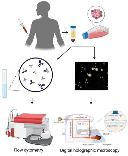
Colorectal cancer (CRC) is the second most metastatic disease with the majority of cases detected in Western countries. Metastases are formed by circulating altered phenotype tumor cells causing 20% of CRC related deaths. Metastatic cells may show higher expression of surface molecules such as CD44, and changes in morphological properties are associated with increased invasiveness and poor prognosis. In this study, we intended to mimic the environment for metastasizing cells. Here, we used digital holographic cytometry (DHC) analysis to determine cellular morphological properties of three metastatic and two non-metastatic colorectal cancer cell lines to show differences in morphology between the CRC cells and peripheral blood mononuclear cells (PBMCs). By establishing differences in cell area, cell thickness, cell volume, and cell irregularity even when the CRC cells were in minority (5% out of PBMCs), DHC does discriminate between CRC cells and the PBMCs in vitro. We also analyzed the epithelial marker EpCAM and migration marker CD44 using flow cytometry and demonstrate that the CRC cell lines and PBMC cells differ in EpCAM and CD44 expression. Here, we present DHC as a new powerful tool in discriminating cells of different sizes in suspension together with a combination of biomarkers.

Graphical Abstract

1. Introduction

Colorectal cancer (CRC), a highly spread tumor disease, is the third and second most common cancer in men and women, respectively [1]. Owing to changes in diet preferences and increasing living standards in Western countries, CRC has become a major cancer disease with more than 1.800.000 of the new cases worldwide [2,3,4]. Despite a monitoring program consisting of stool-based tests and colonoscopy, and intensive chemo- or immunotherapeutic interventions, 20% of patients diagnosed with CRC initially present with metastatic disease and subsequently 35% of patients with stage III eventually relapse and later die from CRC [1,3,5,6].
Circulating tumor cells (CTCs) are detached from the original tumor and spread through the bloodstream to other organs, forming distant lesions to establish metastases [7]. In this process, the epithelial-to-mesenchymal transition (EMT) plays a crucial role since it is characterized by losing intercellular adhesion and epithelial polarization [8,9]. The acquisition of the mesenchymal phenotype is accompanied by increased motility and invasiveness. Besides different cellular morphology, cells may during EMT alter the expression of signaling and surface molecules potentially affecting the tumor microenvironment or contribute to the avoidance of immune surveillance [8,10,11].
Digital holographic cytometry (DHC) is a novel imaging technique for observations of cell samples in their native monolayer settings [12]. DHC functions on the principle of quantitative phase imaging (QPI) providing high spatial resolution and quantification of cellular morphological parameters such as volume, area, and optical thickness from recorded holograms [12,13]. Since it does not require any fluorescent labels or staining, DHC allows for long-term time-lapse experiments with precise observations and with no phototoxic effects on the cells compared to differential interface contrast microscopy [14,15]. Recently, DHC has been used for various biological applications such as evaluation of cell proliferation [13], detection and classification of cell death [16,17,18,19], nanoparticle uptake [20,21], and motility and migration studies [22,23,24]. Moreover, we recently used DHC to discriminate between breast cancer cells and leukemic cells according to their morphological parameters. Leukemic cells were significantly smaller in the investigated parameters optical thickness, volume, and area compared to epithelial breast cancer cells [25]. This finding paved the way for using DHC as a tool for the in vitro circulating tumor cell-model, now further explored in this study by combining morphology measurements with epithelial and mesenchymal surface markers.
Although CTCs have different characteristics, the detection of CTCs is highly complicated since the occurrence in the bloodstream is very sparse compared to human blood cells [26]. Up to date, flow cytometry, Raman spectroscopy [27], microfluidic devices [28,29], immunoassays [30] or methods based on the polymerase chain reaction (PCR) [31] were utilized to discriminate the CTCs from other cells in blood or in liquid biopsies. Indeed, optical tomography provide a more complete mapping of the biological sample in combination with QPI [32]. The 3D refractive index will allow studies of subcellular and micrometer structures of the cells [33,34]. By combining all time-lapse quantitative phase maps, each orientation of the cell can be obtained [35]. For high-throughput 3D cell measurements, microfluidic devices have been integrated to tomographic phase microscopy including work on red blood cells and CTCs [32]. Nissim et al. recently showed in their system that blood cells mixed with colorectal cancer cells could be automatically discriminated on the level of individual cell types, by using label-free holographic flow cytometry [36]. By their custom-built optical system operated under flow, they received single-cell holograms in real time and applied image processing and machine learning. The database created found features that could differ between cancer cells and various blood cells. Moreover, cell sorting was included in a tomographic interferometry approach by rotating cells in a flow with dielectrophoretic forces [37,38]. Interestingly, here the authors used three types of CRCs, HT29, SW-480 and SW-620 together with blood cells [38]. The CRC cell type mixed with blood cells, i.e., the liquid biopsy, were first enriched by filtration and then the CRCs cells were classified during flow using machine learning, and then isolated by using activating DEP. Most elegantly, the authors also built in a fluorescence imaging system for an external validation where only the cancer cells emitted fluorescence light. Moreover, a recent study with an approach aiming for clinical use, also showed classification of cancer cells based on the cell spatial and temporal fluctuations [39]. The classification method can both be used to detect CTCs from blood and analyse cancer cells from tissue or solid tumors.
Despite the recent progress, only the CellSearch protocol is the currently approved method by the US Food and Drug Administration (FDA) for breast, prostate, and CRC [40]. It works on the principle of positive cell enrichment and detection of cell adhesion molecules, but the CellSearch protocol has shown low specificity for the reoccurrence of the CTCs after adjuvant cancer therapy. It has been reported that several drugs, e.g., bevacizumab, alter expression of epithelial cell adhesion molecule (EpCAM) [40,41]. Therefore, there is a demand to identify CTCs using novel parameters allowing their precise detection independently on the cancer treatment or stage.
In the present study, we have performed DHC-based morphological analyses on a set of colon cancer cell lines and peripheral blood mononuclear cells (PBMCs). Besides, we used DHC to distinguish colon cancer cells from white blood cells (WBC) in suspension in vitro, to mimic an environment of the CTCs where they are surrounded by blood cells. In conclusion, we show that DHC serves as a useful technique for discriminating CRC cells from WBC in vitro. Subsequently, we analyzed the expression of the epithelial marker EpCAM and the mesenchymal marker CD44 to better characterize the cell populations.

2. Materials and Methods

2.1. Cell Culture

Five different colon cancer cell lines COLO 205, Caco-2, NCI-H508, LS411N, and SNU-C1 were used. The breast cancer cell line MCF7 was used as a control. All the cell lines were obtained from American Type Culture Collection (ATCC/LGC Standards, Teddington, UK). The cell lines COLO 205, NCI-H508, LS411N, and SNU-C1 were cultured in Roswell Park Memorial Institute (RPMI-1640, Thermo Fisher Scientific, Waltham, MA, USA) media with 10% fetal bovine serum (FBS, Thermo Fisher Scientific). The Caco-2 cell line was cultured in Dulbecco’s Modified Eagle Medium (DMEM, Thermo Fisher Scientific) with 20% FBS, 1% penicillin/streptavidin, 2% L-Glutamine, and 1% non-essential amino acids (Thermo Fisher Scientific). The MCF7 cell line was cultured in RPMI-1640 medium supplemented with 10% FBS and gentamycin 50 µg/mL (Thermo Fisher Scientific). The cell lines were cultured at 37 °C with 5% CO2 in 95% humidity.

2.2. Isolation of Peripheral Blood Mononuclear Cells (PBMCs)

For all experiments, peripheral venous blood was donated from healthy donors and processed within 2 h after collection. All blood samples were drawn with the informed consent of patients according to institutional policies and with approval by the institutional review board. Peripheral blood mononuclear cells PBMCs were separated from whole blood by using Ficoll density gradient centrifugation according to a modified protocol [42]. Blood was transferred to a sterile 50 mL polypropylene tubes (Gibco) and diluted with the same volume of PBS without Ca/Mg. 15 mL of sterile Ficoll Paque (GE Healthcare, Fisher Scientific, Hampton, New Hampshire, USA) was added to 2 new 50 mL tubes and then 20 mL of blood/PBS was transferred to each tube and centrifuged in a swing-out rotor (400× g, 30 min). Thereafter, the first layer of thrombocytes and plasma was removed and the layer with lymphocytes and monocytes was transferred to two new 50 mL tubes. The cells were washed two times (200× g, 10 min) with 20 mL and then 10 mL of RPMI media containing Glutamax, Hepes, gentamicin and FBS was added. The required amount of the cells was used for fluorescence-activated cell sorting and DHC analysis. In this study, PBMCs of three healthy donors was used.

2.3. Fluorescence-Activated Cell Sorting

EpCAM staining: Cells were washed with 5 mL of PBS, harvested by trypsinization and 0.5 × 10⁶ cells per sample was stained with anti-EpCAM-PE (Cat No 130-113-264; Milentyi Biotec GmBH, Bergisch Gladbach, Germany) diluted in PBS to a final concentration of 10 ng/mL and incubated at 4 °C for 30 min in the dark. Thereafter the cells were washed two times with 2 mL PBS and analyzed with fluorescence-activated cell sorting (FACs, BD Bioscience, Accuri C6 Flow Cytometry, NJ, USA).
CD44 staining: Cells were washed with 5 mL of PBS and harvested by trypsinization and 1 × 10⁶ cells per sample were stained with anti-CD44 (Cat No MAB6127; R&D, Minneapolis, MN, USA) diluted in PBS to a final concentration 2.5 µg/mL or left unstained as control with PBS. The samples were incubated at 4 °C for 30 min. Thereafter, the cells were washed two times with 2 mL PBS, incubated with the secondary antibody anti-rat-FITC (Cat No F0104B; R&D). 100 µL of 1/10 diluted anti-rat-FITC in PBS was added to the cells and the negative control. The samples were incubated at 4 °C for 30 min in the dark, washed twice with 2 mL PBS, and analyzed with FACs (BD Bioscience).

2.4. DHC and Image Analysis

Cells were washed with 5 mL of PBS and harvested by trypsinization, washed twice in 2 mL of PBS and 1 × 10⁶ cells were resuspended in 100 µL of PBS. 10 µL of the suspension was added to CountessTM cell chamber slide (ThermoFisher Scientific). Morphological parameters of cells in suspension were then obtained using HoloMonitor M4 (Phase Holographic Imaging AB, PHIAB, Lund, Sweden) and HoloMonitor proprietary software AppSuite (PHIAB). Four separate experiments with 30 images in each were collected during ten minutes, and ten representative images from each experiment were chosen and analyzed. Image capture was performed with a low-intensity 635 nm diode laser, which prevents phototoxicity. The optical magnification used was 10× and the resolution was 0.5 μm. The cells in the images are segmented to extract information on each imaged cell separately, and thereby cellular parameters such as area, optical thickness and optical volume were obtained [43]. For cell mixes, 1 × 10⁶ cells were resuspended and mixed in 100 µL of PBS with ratios of 95 + 5% or 90 + 10% of PBMCs to COLO 205 cells. 10 µL of the mixed suspension was added to the CountessTM cell chamber slide and analyzed using HoloMonitor and AppSuite. Two separate experiments with 30 images in each were collected and ten representative images from one experiment were chosen and analyzed.

2.5. Statistics

Mean and relative standard deviations expressed as a coefficient of variation (CV) were used for the statistical analysis. For the cell morphology studies, at least 10 DHC images from 4 separate experiments were used for the image analysis. The morphology parameters were measured as the mean and CV of the cell mean volume, mean area, mean thickness, mean irregularity, mean eccentricity. Data for DHC are presented as a mean ± standard deviation (STDEV). Statistical significance was determined by a two-tailed unpaired Student’s t-test, with p values ≤ 0.05 considered significant. In Table 1, the percentage for each gated cell subpopulation is calculated from the total cell population of the gated cells per experiment.

3. Results

We have previously analyzed a collection of breast cancer cell lines using an ”in vitro circulating tumor cell model” for analysis of sialic acid as a tumor marker [44] and further determined size parameters using DHC, showing discrimination between large epithelial breast cancer cells and smaller WBC [25].
Here, we analyzed five CRC cell lines, MCF7 breast cancer cells and PBMCs, using DHC for evaluation of size parameters. Thereafter, the CRC cell lines and MCF7 cells were analyzed with PBMCs to distinguish any possible morphological parameters between WBCs and CRCs (Figure 1A–E). The CRC cell lines showed high variability in cell area and volume. Caco-2 and SNU-C1 cells showed larger cell area compared to COLO 205 and LS411N cells. Indeed, PBMCs were the smallest of all the cell lines. Despite significant differences in cell area, the CRC cells had similar optical thickness, as can be seen in Figure 1B. NCI-H508 cells were characterized by slightly increased optical thickness compared to the rest of the cell lines. On the other hand, cell volume shows a similar pattern as cell area. Hence, Caco-2, NCI-H508 and SNU-C1 had higher cell volumes than the three other cell lines and the PBMCs. Moreover, the irregularity and eccentricity of the cell lines were measured. The parameters refer to how much a cellular circumference deviates from a perfect circle, and how much a cell deviates from being spherical (i.e., cell elongation), respectively. No significant eccentricity differences could be detected between CRC cells, except for NCI-H508 and SNU-C1 cells. On the other hand, COLO 205 cells were significantly less irregular compared to other CRC cell lines. Furthermore, it was noted that the PBMCs and COLO 205 cells had had rather comparable irregularity distinguishing them from the rest of the CRC cells.
Based on the results from the morphology studies, the COLO 205 cell line and PBMCs seemed to have the most similar morphological pattern, and we therefore decided to use COLO 205 for our consequent experiments.
The mixed study was performed to find out whether the DHC technique can be used to find out even a minor fraction of COLO 205 cells from PBMC cells dominating in the suspension. The analysis was based on the measurement of the cell area and optical thickness of the two different subpopulations. The individual analysis of COLO 205 and PBMC subpopulations, respectively, revealed that the cells were correctly detected after manual adjustment in the proprietary software. We subsequently analyzed a mixture of the two cell types. Different ratios of the COLO 205 cells and PBMCs were analyzed, starting with 50% of each cell type (data not shown). Thereafter, 90% of PBMC and 10% of COLO 205 cells as well as 95% of PBMC and 5% of COLO 205 cells, respectively, were prepared, mixed and analyzed by using DHC. The COLO 205 cancer cells were clearly identified even in lower concentrations as shown by Figure 2A, top diagrams. 2D and 3D holographic phase images of the cells are shown in Figure 2B. The total number of cells in each gate is displayed in Table 1.
We then aimed to analyze COLO205 cells and PBMCs for the markers EpCAM and CD44 to characterize their EMT status (Figure 3). COLO 205 cells showed high EpCAM expression and histograms were shifted towards higher mean fluorescence intensity (red lines), while the PBMCs were shown to be EpCAM negative. The CD44 analysis (blue lines) revealed that the COLO205 cell line with high EpCAM expression showed minor CD44 expression. As expected, PBMCs were CD44 positive [45].
Our data suggest that DHC in combination with specific cancer cell markers offer high sensitivity and specificity in detecting low abundant metastatic CRC cells in circulating blood.

4. Discussion

We have previously analyzed a collection of breast cancer cell lines in an ”in vitro circulating tumor cell model” for analysis of sialic acid as a tumor marker and size parameters using DHC, thus distinguishing larger epithelial breast cancer cells from small blood cells. The breast cancer cell lines were significantly larger compared to white blood cell lines, Jurkat and THP-1 cells [25,44].
In this study, we have focused on a novel in vitro approaches to evaluate morphological properties and possible markers for metastasizing CRC. Current methodologies of CTC detection are based on the presence of epithelial surface markers or cluster of differentiation (CD) antigens. However, some cancer cells downregulate the epithelial marker EpCAM, so there is a need to find novel ways to specifically detect CTCs [46].
We used DHC to study cellular parameters such as cellular area, thickness, volume, irregularity and eccentricity. As shown in Figure 1, the morphological variability of the CRC cell lines was substantial, especially regarding area, volume and irregularity. We could show that among our set of CRC cell lines, COLO 205 stands out and shares several morphological properties with PBMCs. Hence, COLO 205 cells were therefore chosen to be analyzed together with PBMCs to demonstrate the capability of the DHC technique to perform size discrimination of CRC cells and blood cells. Moreover, the COLO 205 cell line is characterized by mixed growth properties making it even more suitable to mimic the presence of CTCs in the liquid biopsy in vitro model as used in this study. The use of CTC detection for monitoring the effectiveness of cancer therapy with high prognostic potential has been described by others, even though CRC patients have low occurrence of CTCs in the bloodstream [47]. Interestingly, by employing the in vitro model, we were able to discriminate between the COLO 205 cells, liquid biopsy CTC cells, and PBMC cells according to their morphological properties in vitro. In a mixed suspension with PBMCs, COLO 205 cells were detected even at 5% of the total cell amount. Indeed, significant differences in cellular area and optical thickness were determined by DHC when analysing mixed COLO 205 cells and PBMCs. We could also successfully confirm the number of cells in the mixture by using AppSuite’s calculations of the cell types in the images. Since this was performed as a proof-of-concept study with small sample number, further examinations are necessary to validate the results.
Ranc et al. have used Raman spectroscopy to discriminate between a breast CTC line, a CRC cell line and a mononuclear cell line. The approach was successful even the cells were immobilized by the means of chemical fixation successful [27]. We recently used DHC to discriminate between breast cancer and leukemic cells according to their morphological parameters. When compared to breast cancer cells, the WBCs, cell lines Jurkat and THP-1 differed significantly regarding optical thickness, cell volume, and cell area, confirming the results in this study [25]. Singh et al. used the QPI platform coupled with a microfluidics device and advanced mathematical classifiers to identify the breast cancer cell lines MCF7 and MDA-MB-231 among blood cells. Indeed, accurately determined single cell profiles are promising results showing discrimination of tumor cell lines from the two blood cell types [48]. It is noteworthy that suspension cell line COLO 205 used in our study better mimics the conditions of a liquid biopsy, since suspension cells have more similarities to CTCs than adherent cancer cells [49]. According to our morphological analysis by DHC, the COLO 205 cell line showed the most similar properties to PBMCs. It was also important to avoid cellular surface interactions, since surface adherence may alter cell features such as motility or morphology leading to incorrect analysis. It has been reported that different substrates may alter cellular behavior and ultimately change the motility and matrix adhesion. Detachment of cells leads to more rounded morphology, smaller area, or phase increase [50], moreover, it makes the identification of cell subpopulations with similar morphology more complicated.
Despite the possible complications mentioned above, using DHC, we were able to detect the PBMCs and COLO 205 cell subpopulations without any use of advanced machine learning algorithms. The expression profile of COLO 205 and PBMCs shows an inverse correlation of EpCAM and CD44 expression. While COLO 205 cells have increased expression of CD44 proving their metastatic phenotype, PBMCs were negative. Indeed, numerous studies have described the importance of CD44 in tumor progression [51].
Our model to detect CTCs using DHC relied on co-suspensions of PBMCs and COLO 205 cells. We were able to precisely detect both subpopulations even in a 95% PBMC-5% COLO 205 mixed suspension. The significant differences in morphological parameters suggest that DHC can be considered to be a diagnostic tool for distinguishing CTCs from blood cells. Recent techniques for enumeration of CTCs are based on detection of EpCAM and CDs using fluorescent labelling or quantitative PCR [52]. Therefore, there is a need to develop label-free diagnostic tools. DHC working on the principle of quantitative phase imaging coupled with a microfluidic device, presented by Bocanegra et al. [53] would potentially overcome some recent drawbacks such as low sample volume. On the other hand, microfluidic setups suffer from the presence of shear stress which may cause temporary cell deformation [54]. Based on recent reports, we expect that DHC can be included in the diagnostic procedure as a pre-stage component of a diagnostic device to offer qualitative control and discrimination of the cell subpopulations. Although it is unlikely that antigen cell screening could be fully replaced with morphological analysis only, given the current progress in machine learning, neural networks, and classifiers [48,55], morphological evaluation of cells will gradually increase in importance.
In this study, we used DHC to characterize the morphology of CRC cell lines and discriminate between COLO 205 cells and PBMC in suspension at low concentrations. Additionally, we evaluated EpCAM and CD44 expression to establish the EMT status of the cells.

5. Conclusions

In this study, we classified the cellular morphology of a set of CRC cell lines. Next, we compared them regarding the expression of the EMT markers EpCAM and CD44. We demonstrated based on their morphological properties, DHC is a promising tool for discrimination between CRC cells and PBMCs. By this, we believe that the evaluation of cellular morphology and surface markers would add another piece to the CTC detection puzzle.

Author Contributions

A.G.W., Z.E.-S. and M.F. conceived and designed the study; M.F., Z.E.-S. and Y.Z. carried out the cell based studies and performed experiments; M.F. and Z.E.-S. interpreted the digital holographic cytometry data. data; M.F., Z.E.-S., Y.Z. and A.G.W. analyzed the data; J.B. and J.L.P. provided advice and technical assistance; M.F. wrote the original draft of the manuscript; M.F., Z.E.-S., Y.Z. and A.G.W. wrote the final version of the manuscript according to all the other author’s comments. All authors have read and agreed to the published version of the manuscript.

Funding

This research was funded by the Swedish Knowledge Foundation grant number 20160165, The European Union’s Horizon 2020 research and innovation program under the grant agreement: 848098, and under the Marie Sklodowska-Curie grant agreement grant number 721297, the Royal Physiographic Society of Lund, Biofilms Research Center for Biointerfaces and Malmö University.

Institutional Review Board Statement

The study was conducted in accordance with the Declaration of Helsinki. The PBMCs were from unidentified leucocyte concentrates purchased from Skåne University Hospital, Malmö, Sweden.

Informed Consent Statement

Blood samples were drawn with the informed consent of patients according to institutional policies and with approval by the institutional review board.

Data Availability Statement

Not applicable.

Acknowledgments

The authors thank Kersti Alm and Birgit Janicke, Phase Holographic Imaging AB, Lund, Sweden, for critical reading of the manuscript, and for their help and advice with microscopy data curation. Graphical abstract was created with BioRender.com.

Conflicts of Interest

The authors declare no conflict of interest.

References

  1. Kuipers, E.J.; Grady, W.M.; Lieberman, D.; Seufferlein, T.; Sung, J.J.; Boelens, P.G.; van de Velde, C.J.H.; Watanabe, T. Colorectal cancer. Nat. Rev. Dis. Primers 2015, 1, 15065. [Google Scholar] [CrossRef] [Green Version]
  2. Siegel, R.L.; Miller, K.D.; Jemal, A. Cancer statistics, 2019. CA A Cancer J. Clin. 2019, 69, 7–34. [Google Scholar] [CrossRef] [Green Version]
  3. Goodwin, R.A.; Asmis, T.R. Overview of systemic therapy for colorectal cancer. Clin. Colon Rectal Surg. 2009, 22, 251–256. [Google Scholar] [CrossRef] [Green Version]
  4. Sung, H.; Ferlay, J.; Siegel, R.L.; Laversanne, M.; Soerjomataram, I.; Jemal, A.; Bray, F. Global cancer statistics 2020: GLOBOCAN estimates of incidence and mortality worldwide for 36 cancers in 185 countries. CA Cancer J. Clin. 2021, 71, 209–249. [Google Scholar] [CrossRef]
  5. Kuipers, E.J.; Rösch, T.; Bretthauer, M. Colorectal cancer screening—Optimizing current strategies and new directions. Nat. Rev. Clin. Oncol. 2013, 10, 130–142. [Google Scholar] [CrossRef]
  6. Geiger, T.M.; Ricciardi, R. Screening options and recommendations for colorectal cancer. Clin. Colon Rectal Surg. 2009, 22, 209–217. [Google Scholar] [CrossRef] [PubMed] [Green Version]
  7. Donato, C.; Kunz, L.; Castro-Giner, F.; Paasinen-Sohns, A.; Strittmatter, K.; Szczerba, B.M.; Scherrer, R.; Di Maggio, N.; Heusermann, W.; Biehlmaier, O.; et al. Hypoxia Triggers the Intravasation of Clustered Circulating Tumor Cells. Cell Rep. 2020, 32, 108105. [Google Scholar] [CrossRef]
  8. Massagué, J.; Obenauf, A. Metastatic colonization by circulating tumor cells. Nature 2016, 529, 298–306. [Google Scholar] [CrossRef] [Green Version]
  9. Ye, X.; Weinberg, R.A. Epithelial-Mesenchymal Plasticity: A Central Regulator of Cancer Progression. Trends Cell Biol. 2015, 25, 675–686. [Google Scholar] [CrossRef] [Green Version]
  10. Quail, D.F.; Joyce, J.A. Microenvironmental regulation of tumor progression and metastasis. Nat. Med. 2013, 19, 1423–1437. [Google Scholar] [CrossRef]
  11. Clark, A.G.; Vignjevic, D.M. Modes of cancer cell invasion and the role of the microenvironment. Curr. Opin. Cell Biol. 2015, 36, 13–22. [Google Scholar] [CrossRef] [Green Version]
  12. Gjörloff-Wingren, A. Quantitative phase-contrast imaging—A potential tool for future cancer diagnostics. Cytom. Part A 2017, 91, 752–753. [Google Scholar] [CrossRef] [PubMed]
  13. Janicke, B.; Kårsnäs, A.; Egelberg, P.; Alm, K. Label-free high temporal resolution assessment of cell proliferation using digital holographic microscopy. Cytom. Part A 2017, 91, 460–469. [Google Scholar] [CrossRef]
  14. Pastorek, L.; Venit, T.; Hozak, P. Holography microscopy as an artifact-free alternative to phase-contrast. Histochem. Cell Biol. 2018, 149, 179–186. [Google Scholar] [CrossRef]
  15. Kasprowicz, R.; Suman, R.; O’Toole, P. Characterising live cell behaviour: Traditional label-free and quantitative phase imaging approaches. Int. J. Biochem. Cell Biol. 2017, 84, 89–95. [Google Scholar] [CrossRef] [PubMed]
  16. El-Schich, Z.; Molder, A.; Tassidis, H.; Harkonen, P.; Miniotis, M.F.; Wingren, A.G. Induction of morphological changes in death-induced cancer cells monitored by holographic microscopy. J. Struct. Biol. 2015, 189, 207–212. [Google Scholar] [CrossRef] [PubMed]
  17. Balvan, J.; Krizova, A.; Gumulec, J.; Raudenska, M.; Sladek, Z.; Sedlackova, M. Multimodal Holographic Microscopy: Distinction between Apoptosis and Oncosis (vol 10, e0121674, 2015). PLoS ONE 2015, 10, e0121674. [Google Scholar] [CrossRef] [Green Version]
  18. Vicar, T.; Raudenska, M.; Gumulec, J.; Masarik, M.; Balvan, J. Detection and characterization of apoptotic and necrotic cell death by time-lapse quantitative phase image analysis. bioRxiv 2019.
  19. Barker, K.L.; Boucher, K.M.; Judson-Torres, R.L. Label-free classification of apoptosis, ferroptosis and necroptosis using digital holographic cytometry. Appl. Sci. 2020, 10, 4439. [Google Scholar] [CrossRef]
  20. Fojtu, M.; Balvan, J.; Raudenska, M.; Vicar, T.; Bousa, D.; Sofer, Z.; Masarik, M.; Pumera, M. Black Phosphorus Cytotoxicity Assessments Pitfalls: Advantages and Disadvantages of Metabolic and Morphological Assays. Chem. Eur. J. 2019, 25, 349–360. [Google Scholar] [CrossRef]
  21. Sternbæk, L.; Kimani, M.; Gawlitza, K.; Janicke, B.; Alm, K.; Wingren, A. Digital Holographic Cytometry: Macrophage Uptake of Nanoprobes. Imaging Microsc. 2019, 1, 21–23. [Google Scholar]
  22. Wingren, A.G. Moving into a new dimension: Tracking migrating cells with digital holographic cytometry in 3D. Cytom. Part A 2018, 95, 144–146. [Google Scholar] [CrossRef] [PubMed]
  23. Patel, M.; Feith, M.; Janicke, B.; Alm, K.; El-Schich, Z. Evaluation of the impact of imprinted polymer particles on morphology and motility of breast cancer cells by using digital holographic cytometry. Appl. Sci. 2020, 10, 750. [Google Scholar] [CrossRef] [Green Version]
  24. Tolde, O.; Gandalovičová, A.; Křížová, A.; Veselý, P.; Chmelík, R.; Rosel, D.; Brábek, J. Quantitative phase imaging unravels new insight into dynamics of mesenchymal and amoeboid cancer cell invasion. Sci. Rep. 2018, 8, 12020. [Google Scholar] [CrossRef] [Green Version]
  25. El-Schich, Z.; Janicke, B.; Alm, K.; Dizeyi, N.; Persson, J.; Wingren, A. Discrimination between Breast Cancer Cells and White Blood Cells by Non-Invasive Measurements: Implications for a Novel In Vitro-Based Circulating Tumor Cell Model Using Digital Holographic Cytometry. Appl. Sci. 2020, 10, 4854. [Google Scholar] [CrossRef]
  26. Yamada, T.; Matsuda, A.; Koizumi, M.; Shinji, S.; Takahashi, G.; Iwai, T.; Takeda, K.; Ueda, K.; Yokoyama, Y.; Hara, K.; et al. Liquid Biopsy for the Management of Patients with Colorectal Cancer. Digestion 2019, 99, 39–45. [Google Scholar] [CrossRef]
  27. Ranc, V.; Srovnal, J.; Kvitek, L.; Hajduch, M. Discrimination of circulating tumor cells of breast cancer and colorectal cancer from normal human mononuclear cells using Raman spectroscopy. Analyst 2013, 138, 5983–5988. [Google Scholar] [CrossRef]
  28. Huang, Y.Y.; Chen, P.; Wu, C.H.; Hoshino, K.; Sokolov, K.; Lane, N.; Liu, H.Y.; Huebschman, M.; Frenkel, E.; Zhang, J.X.J. Screening and Molecular Analysis of Single Circulating Tumor Cells Using Micromagnet Array. Sci. Rep. 2015, 5, 1–11. [Google Scholar] [CrossRef]
  29. Zhang, H.; Fu, X.; Hu, J.Y.; Zhu, Z.J. Microfluidic bead-based multienzyme-nanoparticle amplification for detection of circulating tumor cells in the blood using quantum dots labels. Anal. Chim. Acta 2013, 779, 64–71. [Google Scholar] [CrossRef]
  30. Gazouli, M.; Lyberopoulou, A.; Pericleous, P.; Rizos, S.; Aravantinos, G.; Nikiteas, N.; Anagnou, N.P.; Efstathopoulos, E.P. Development of a quantum-dot-labelled magnetic immunoassay method for circulating colorectal cancer cell detection. World J. Gastroenterol. 2012, 18, 4419–4426. [Google Scholar] [CrossRef]
  31. Vaiopoulos, A.G.; Kostakis, I.D.; Gkioka, E.; Athanasoula, K.C.; Pikoulis, E.; Papalambros, A.; Christopoulos, P.; Gogas, H.; Kouraklis, G.; Koutsilieris, M. Detection of Circulating Tumor Cells in Colorectal and Gastric Cancer Using a Multiplex PCR Assay. Anticancer Res. 2014, 34, 3083–3092. [Google Scholar] [PubMed]
  32. Merola, F.; Memmolo, P.; Miccio, L.; Mugnano, M.; Ferraro, P. Phase contrast tomography at lab on chip scale by digital holography. Methods 2018, 136, 108–115. [Google Scholar] [CrossRef] [PubMed]
  33. Balasubramani, V.; Kujawińska, M.; Allier, C.; Anand, V.; Cheng, C.-J.; Depeursinge, C.; Hai, N.; Juodkazis, S.; Kalkman, J.; Kuś, A. Roadmap on digital holography-based quantitative phase imaging. J. Imaging 2021, 7, 252. [Google Scholar] [CrossRef] [PubMed]
  34. Balasubramani, V.; Kuś, A.; Tu, H.-Y.; Cheng, C.-J.; Baczewska, M.; Krauze, W.; Kujawińska, M. Holographic tomography: Techniques and biomedical applications. Appl. Opt. 2021, 60, B65–B80. [Google Scholar] [CrossRef]
  35. Merola, F.; Memmolo, P.; Miccio, L.; Savoia, R.; Mugnano, M.; Fontana, A.; D’ippolito, G.; Sardo, A.; Iolascon, A.; Gambale, A. Tomographic flow cytometry by digital holography. Light Sci. Appl. 2017, 6, e16241. [Google Scholar] [CrossRef] [Green Version]
  36. Nissim, N.; Dudaie, M.; Barnea, I.; Shaked, N.T. Real-Time Stain-Free Classification of Cancer Cells and Blood Cells Using Interferometric Phase Microscopy and Machine Learning. Cytom. Part A 2021, 99, 511–523. [Google Scholar] [CrossRef]
  37. Habaza, M.; Kirschbaum, M.; Guernth-Marschner, C.; Dardikman, G.; Barnea, I.; Korenstein, R.; Duschl, C.; Shaked, N.T. Rapid 3D refractive-index imaging of live cells in suspension without labeling using dielectrophoretic cell rotation. Adv. Sci. 2017, 4, 1600205. [Google Scholar] [CrossRef]
  38. Dudaie, M.; Nissim, N.; Barnea, I.; Gerling, T.; Duschl, C.; Kirschbaum, M.; Shaked, N.T. Label-free discrimination and selection of cancer cells from blood during flow using holography-induced dielectrophoresis. J. Biophotonics 2020, 13, e202000151. [Google Scholar] [CrossRef]
  39. Ben Baruch, S.; Rotman-Nativ, N.; Baram, A.; Greenspan, H.; Shaked, N.T. Cancer-Cell Deep-Learning Classification by Integrating Quantitative-Phase Spatial and Temporal Fluctuations. Cells 2021, 10, 3353. [Google Scholar] [CrossRef]
  40. Allen, J.E.; El-Deiry, W.S. Circulating Tumor Cells and Colorectal Cancer. Curr. Color. Cancer Rep. 2010, 6, 212–220. [Google Scholar] [CrossRef] [Green Version]
  41. Nicolazzo, C.; Massimi, I.; Lotti, L.V.; Vespa, S.; Raimondi, C.; Pulcinelli, F.M.; Gradilone, A.; Gazzaniga, P. Impact of chronic exposure to bevacizumab on EpCAM-based detection of circulating tumor cells. Chin. J. Cancer Res. 2015, 27, 491–496. [Google Scholar] [CrossRef] [PubMed]
  42. Panda, S.K.; Ravindran, B. Isolation of Human PBMCs. Bio-Protoc. 2013, 3, e323. [Google Scholar] [CrossRef]
  43. El-Schich, Z.; Leida Mölder, A.; Gjörloff Wingren, A. Quantitative phase imaging for label-free analysis of cancer cells—Focus on digital holographic microscopy. Appl. Sci. 2018, 8, 1027. [Google Scholar] [CrossRef] [Green Version]
  44. El-Schich, Z.; Zhang, Y.; Göransson, T.; Dizeyi, N.; Persson, J.; Johansson, E.; Caraballo, R.; Elofsson, M.; Shinde, S.; Sellergren, B.; et al. Sialic Acid as a Biomarker Studied in Breast Cancer Cell Lines In Vitro Using Fluorescent Molecularly Imprinted Polymers. Appl. Sci. 2021, 11, 3256. [Google Scholar] [CrossRef]
  45. Levesque, M.C.; Haynes, B.F. In vitro culture of human peripheral blood monocytes induces hyaluronan binding and up-regulates monocyte variant CD44 isoform expression. J. Immunol. 1996, 156, 1557. [Google Scholar]
  46. Keller, L.; Werner, S.; Pantel, K. Biology and clinical relevance of EpCAM. Cell Stress 2019, 3, 165. [Google Scholar] [CrossRef] [Green Version]
  47. Cohen, S.J.; Punt, C.J.A.; Iannotti, N.; Saidman, B.H.; Sabbath, K.D.; Gabrail, N.Y.; Picus, J.; Morse, M.A.; Mitchell, E.; Miller, M.C.; et al. Prognostic significance of circulating tumor cells in patients with metastatic colorectal cancer. Ann. Oncol. 2009, 20, 1223–1229. [Google Scholar] [CrossRef]
  48. Singh, D.K.; Ahrens, C.C.; Li, W.; Vanapalli, S.A. Label-free, high-throughput holographic screening and enumeration of tumor cells in blood. Lab Chip 2017, 17, 2920–2932. [Google Scholar] [CrossRef]
  49. Park, J.Y.; Jeong, A.L.; Joo, H.J.; Han, S.; Kim, S.-H.; Kim, H.-Y.; Lim, J.-S.; Lee, M.-S.; Choi, H.-K.; Yang, Y. Development of suspension cell culture model to mimic circulating tumor cells. Oncotarget 2017, 9, 622–640. [Google Scholar] [CrossRef] [Green Version]
  50. Lam, V.K.; Nguyen, T.C.; Chung, B.M.; Nehmetallah, G.; Raub, C.B. Quantitative assessment of cancer cell morphology and motility using telecentric digital holographic microscopy and machine learning. Cytom. Part A 2018, 93, 334–345. [Google Scholar] [CrossRef] [Green Version]
  51. Chen, C.; Zhao, S.; Karnad, A.; Freeman, J. The biology and role of CD44 in cancer progression: Therapeutic implications. J. Hematol. Oncol. 2018, 11, 1–23. [Google Scholar] [CrossRef] [PubMed] [Green Version]
  52. Politaki, E.; Agelaki, S.; Apostolaki, S.; Hatzidaki, D.; Strati, A.; Koinis, F.; Perraki, M.; Saloustrou, G.; Stoupis, G.; Kallergi, G.; et al. A Comparison of Three Methods for the Detection of Circulating Tumor Cells in Patients with Early and Metastatic Breast Cancer. Cell. Physiol. Biochem. 2017, 44, 594–606. [Google Scholar] [CrossRef] [PubMed] [Green Version]
  53. Bocanegra Evans, H.; Gorumlu, S.; Aksak, B.; Castillo, L.; Sheng, J. Holographic microscopy and microfluidics platform for measuring wall stress and 3D flow over surfaces textured by micro-pillars. Sci. Rep. 2016, 6, 28753. [Google Scholar] [CrossRef]
  54. Armistead, F.J.; Gala De Pablo, J.; Gadêlha, H.; Peyman, S.A.; Evans, S.D. Cells Under Stress: An Inertial-Shear Microfluidic Determination of Cell Behavior. Biophys. J. 2019, 116, 1127–1135. [Google Scholar] [CrossRef] [PubMed] [Green Version]
  55. Chen, C.; Mahjoubfar, A.; Tai, L.-C.; Blaby, I.; Huang, A.; Niazi, K.; Jalali, B. Deep Learning in Label-free Cell Classification. Sci. Rep. 2016, 6, 21471. [Google Scholar] [CrossRef]
Figure 1. Morphological parameters for the five CRC cell lines COLO 205, Caco-2, NCI-H508, LS411N, SNU-C1, the cell line MCF7, and PBMCs, obtained using DHC. The different morphological parameters were area (A), optical thickness (B), volume (C), irregularity (D), and eccentricity (E).
Photonics 09 00955 g001
Figure 2. Detection of PBMCs and COLO 205 cells individually and in mixed suspension. (A). Area in µm2 and optical thickness avg. in µm. (B). Presenting representative segmented DHC holographic phase image in 2D with a zoomed in detail image in 3D. The color scale bars represent the thickness of the cells in µm, and the scale bars represent 50 µm.
Photonics 09 00955 g002
Figure 3. EpCAM and CD44 expression of COLO 205 cells and PBMCs. The flow cytometry histograms show the mean fluorescence intensity (MFI) and % positive cells of unstained (black lines) and stained cells (EpCAM-red, CD44-blue), respectively. One representative experiment out of two performed is shown.
Photonics 09 00955 g003
Table 1. DHC analysis of the PBMCs and COLO 205 cells detection in suspension. Percentages in parentheses show the mixing ratio of the cells detected by DHC. Area and optical thickness are given as average values.
Pure Suspension
PBMC
Pure Suspension
COLO 205
Mixed Suspension
PBMC (90%) and COLO 205 (10%)
Mixed Suspension
PBMC (95%) and COLO 205 (5%)
Gated cells2488243615121851736104
% gated cells1001008911946
Area (µm2)36.5162.733.6163.538.2169.3
Optical thickness (µm)4.18.84.68.34.18.0
Publisher’s Note: MDPI stays neutral with regard to jurisdictional claims in published maps and institutional affiliations.