Article

Role of Biosynthesized Silver Nanoparticles with Trigonella foenum-graecum Seeds in Wastewater Treatment

1
King Abdullah Institute for Nanotechnology, King Saud University, Riyadh 11451, Saudi Arabia
2
Department of Zoology, King Saud University, Riyadh 11451, Saudi Arabia
3
Department of Physics, College of Science, Princess Nourah bint Abdulrahman University, Riyadh 11671, Saudi Arabia
4
Department of Chemical Engineering, King Saud University, Riyadh 11421, Saudi Arabia
5
Department of Chemistry, College of Science, Princess Nourah bint Abdulrahman University, P.O. Box 84428, Riyadh 11671, Saudi Arabia
*
Authors to whom correspondence should be addressed.
Processes 2023, 11(8), 2394; https://doi.org/10.3390/pr11082394
Submission received: 10 June 2023 / Revised: 10 July 2023 / Accepted: 3 August 2023 / Published: 9 August 2023

Abstract

:
As the human population continues to escalate, its requirement for clean water is also increasing. This has resulted in an increased dependency on wastewater effluent to maintain the base flow of urban streams, especially in water-stressed regions. The present study reports the synthesis of AgNPs with green credentials using an aqueous extract of Trigonella foenum-graecum seeds. The observance of surface plasmon resonance (SPR) with UV–Vis spectrophotometry confirmed the presence of spherical/oblong particles with a mean diameter of 43.8 nm and low polydispersity index (PDI) of 0.391 measured by transmission electron microscopy (TEM) and DLS (dynamic light scattering) technique, respectively. The elemental map of AgNPs was demonstrated with energy-dispersive spectroscopy (EDS) and the constituent functional groups were identified by the FTIR spectra, which were similar to the bulk seed extract with a slight shift in the pattern. The emission spectrum of nanoparticles was recorded for the excitation wavelength of 349 using fluorescence microscopy and the crystalline structure was assessed using X-ray diffraction. The potential wastewater remedial efficacy of the synthesized AgNPs was evaluated based on the water quality parameters (pH, EC, BOD, COD) of the sewage effluent collected from a local Sewage Treatment Plant (STP). Furthermore, the photo degradative efficacy was investigated using the degradation percentage of Crystal Violet (CV) dye, which was recorded as 94.5% after 20 min. In addition, the antimicrobial activity of the NPs versus bulk seed extract was assessed against two bacterial strains, Escheria coli and Staphylococcus aureus, using the disc diffusion method. The AgNPs showed a profound modulatory effect on the water quality parameters, coupled with marked antimicrobial and photodegradative activity. Thus, the biogenically synthesized AgNPs offer a prospective potential for use in wastewater remediation strategies.

1. Introduction

An increase in the human population and subsequent global warming has led to a rapid depletion of water resources on the planet. The demand for urban water has escalated due to rapid development, population increase, and climate change, particularly in the arid and semiarid regions of the world. The water requirements in water-stressed regions are met by urban streams or reservoirs that are increasingly dependent on wastewater from sewage treatment plants (STPs) to maintain the base flows into aquatic habitats [1]. Since freshwater resources are limited, modern methods of conserving them include the selective use of water resources and the reuse of treated wastewater for a variety of applications [2,3].
Nanotechnology is a promising scientific discipline that has made significant advances in several areas, including wastewater treatment. The primary goal of nanotechnology is to create unique structures with improved electrical, optical, magnetic, conductive, and mechanical capabilities by manipulating matter at the molecular and atomic scales [4]. Heavy metals, organic and inorganic solvents, biological toxins, and pathogens are common pollutants in wastewater that have been shown to be effectively cleaned up by nanomaterials [5,6]. The most extensively investigated nanomaterials in current times for water and wastewater treatment processes include carbon nanotubes (CNTs), metal oxide nanoparticles, zero-valent metal nanoparticles, and nanocomposites [7,8]. In the field of wastewater treatment, silver nanoparticles (AgNPs) and their hybrids are currently gaining impetus [9]. Silver nanoparticles (Ag NPs) exhibit remarkable toxicity to microbes and thus possess potent microbicidal activity against a broad spectrum of microbes, including viruses, bacteria, and fungi [10,11]. Furthermore, their improved adsorption capacities distinguish them as viable alternatives in eliminating contaminants such as the dyes rhodamine, Congo red, and methyl orange commonly found in wastewater bodies [12]. Furthermore, the conventional chemical and physical approaches in synthesis of AgNPs consume time, energy, and capital in addition to the toxic side effects generated with repeated use. Currently, green modes of synthesis, also termed biosynthesis, utilize microbes, fungi and medicinal plants/herbs which are easily available, easy to handle, and provide a wide array of metabolites for the synthesis of AgNPs, as well as being nontoxic and ecofriendly [13,14,15,16].
The major focus in biosynthesis has been on the utilization of plant extracts due to their abundant availability and phytochemical profile [17,18]. In order to reduce the silver in silver nitrate, which is employed as a precursor in the biosynthesis of AgNPs, phytocompounds are typically used as alternative reducing or capping agents. Several plant extracts have been used in the past to synthesize AgNPs, including Papaver somniferum [19], Bauhinia variegata L. [20], Hevea brasiliensis [21], Aloe vera [22], and Moringa oliefera [14], as well as stem extract from the cotton plant Gossypium hirsutum and Portulaca oleracea leaves [23,24]. The plant Trigonella foenum-graecum, also referred to as fenugreek, is a member of the Leguminoseae (Fabaceae) family. The phyto-constituent profile of the seeds and leaves of fenugreek reveal that they are nutrient-dense and packed with vital phytochemicals that provide the herb its therapeutic effects. These include proteins, lipids, alkaloids, flavonoids, fibers, saponins, steroidal saponins, vitamins, and minerals. Additionally, it exhibits antioxidant, antibacterial, anti-diabetic, and anticancer effects [25,26]. For various biomedical and environmental remediation applications, fenugreek seed extract and leaves have been used in several prior studies to biogenically synthesize silver nanoparticles (AgNPs) [27,28,29,30].
From this premise, the present study reports a novel biogenic synthesis of AgNPs with Trigonella foenum-graecum seeds with further effective nanonization which has received a patent. Furthermore, this study includes an assessment of their potential in wastewater remediation based on photodegradative efficacy, modulatory effect on water quality parameters, and antimicrobial potency.

2. Materials and Methods

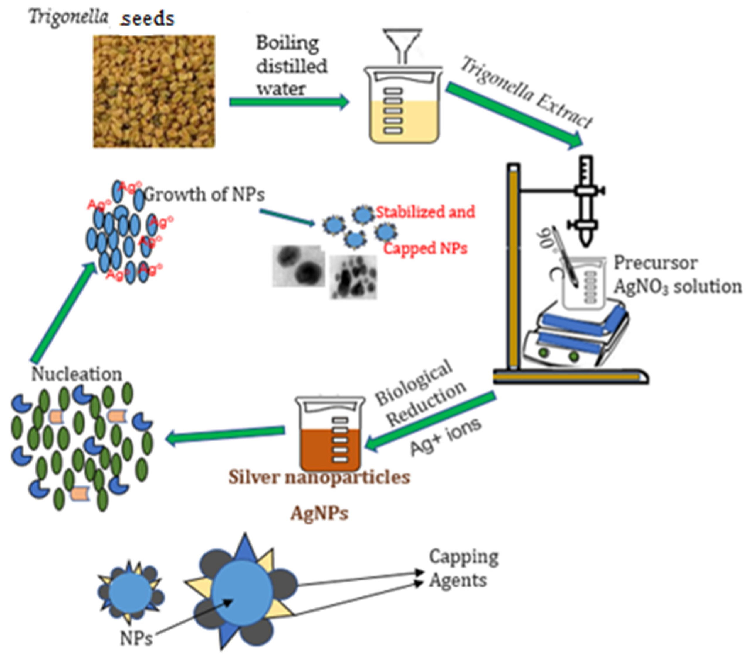
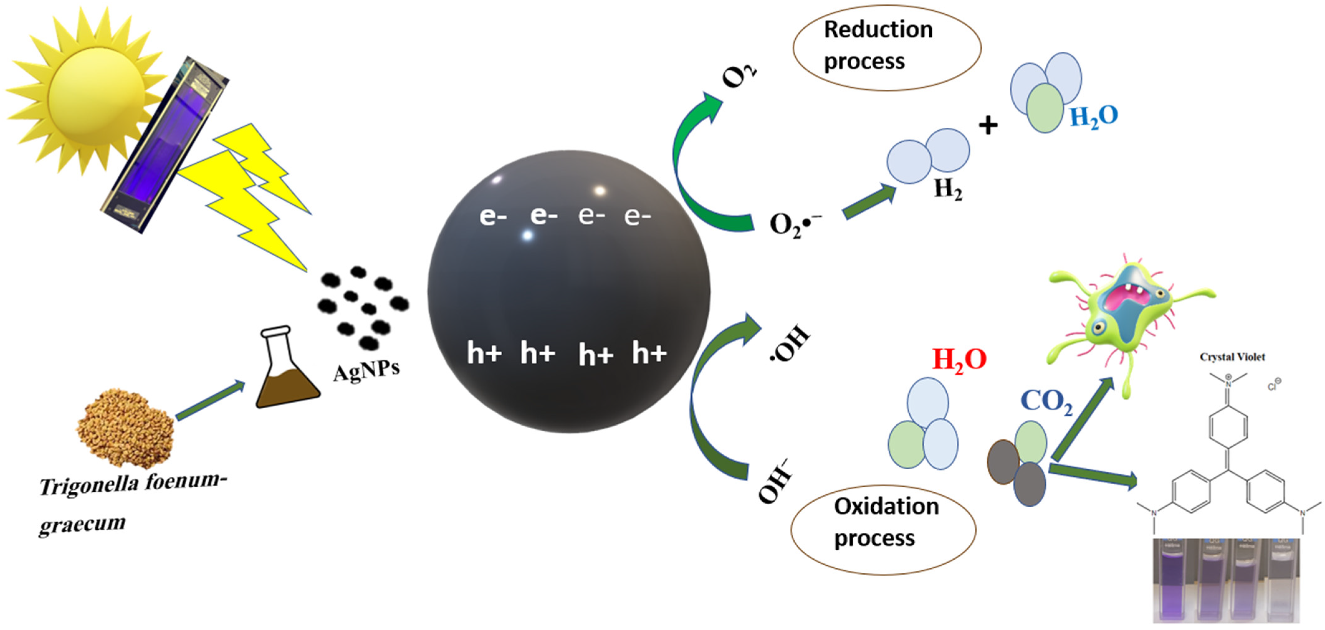
2.1. Green Synthesis of Ag Nanoparticles

Trigonella foenum-graecum seeds were purchased from the neighborhood market in Riyadh, Saudi Arabia. Cleansed, dried, and powdered seeds were used. We boiled 10 g of powdered seeds in 100 mL of distilled water (pH 6.10) for 2 min to prepare the aqueous extract. The extract was then filtered through Whatman 40 filter paper, and the filtrate was stored at 4 °C for further use. The silver nanoparticles (AgNPs) (pH 5.32) were then prepared using the aqueous seed extract according to Awad et al. [27] with certain modifications (Figure 1).

2.2. Characterization of Synthesized AgNPs

The UV–Vis absorbance using ultraviolet–visible spectroscopy (Perkin Elmer UV–Vis spectrometer, Buckinghamshire, UK) confirmed the reduction of silver ions to silver nanoparticles in the presence of Trigonella seed extract. The average particle size of AgNPs was analyzed by Zetasizer, Nano series, HT Laser, ZEN3600 (Molvern Instruments, Malvern, UK). Transmission and Scanning electron microscopy (TEM and SEM) was used to assess the size, shape, and morphologies (JEOL-FE-SEM TEM, Tokyo, Japan), and energy dispersive spectrometer (EDS) analysis was carried out for the detection of elemental silver. Fourier transform infrared (FTIR) spectra in the range of 400–4000 cm−1 were recorded using a Perkin-Elmer 100 spectrophotometer [27,31,32].

2.3. Treatment of Sewage Effluent by Synthesized AgNPs

Grab samples of sewage effluent were collected from the Sewage Treatment Plant (STP) at Al-Hahsa City, Saudi Arabia, in plastic barrels. Samples of tap water were collected in polyethylene bottles directly from the laboratory. Three bottles of 3 L capacity were taken in the laboratory; in two bottles, the sewage effluent was added, and in one, the normal tap water was added. About 50 mgL−1 of synthesized AgNPs were added to one of the bottles which contained 3 L the sewage effluent. The bottles were then kept aside for 72 h, after which the treated effluent was filtered. Water quality parameters were assessed for the treated effluent, untreated effluent and tap water.

2.4. Antibacterial Activity of Silver Nanoparticles

To assess the microbicidal efficacy of the silver nanoparticles, pure cultures of Gram-positive and Gram-negative bacteria, Escherichia coli and Staphylococcus aureus were utilized. The disc diffusion method was used to evaluate the antibacterial activity [33]. Plates containing nutrient agar were prepared, sterilized, and solidified. Thereafter, the test bacterial cultures were inoculated onto the plates. The sterile discs were coated with nanoparticle solutions at two doses of 1 and 0.5 gmL−1 and then placed in nutritional agar medium, where they were cultured for 24 h at 370 °C. A zone of clearing surrounding the wells, which was measured as the diameter of the inhibition zone, was used to demonstrate the inhibitory activity of the bulk aqueous extract of Trigonella seeds and the nanoparticles (AgNPs).

2.5. Statistical Analysis

The mean and standard deviation (Mean ± SD) for all parameters relating to water quality are used to express the data. To analyze group differences, a one-way analysis of variance was conducted, followed by a Tukey’s test (SPSS 22.0 statistical program, Chicago, IL, USA). The threshold for significance was fixed at p ≤ 0.05.

3. Results and Discussion

3.1. Synthesis and Characterization of the Nanoparticles

Synthesis of AgNPs through the reduction of silver nitrate by phyto-constituents of aqueous Trigonella seed extract was confirmed by an observable change in the color of the solution to brown [32,34].

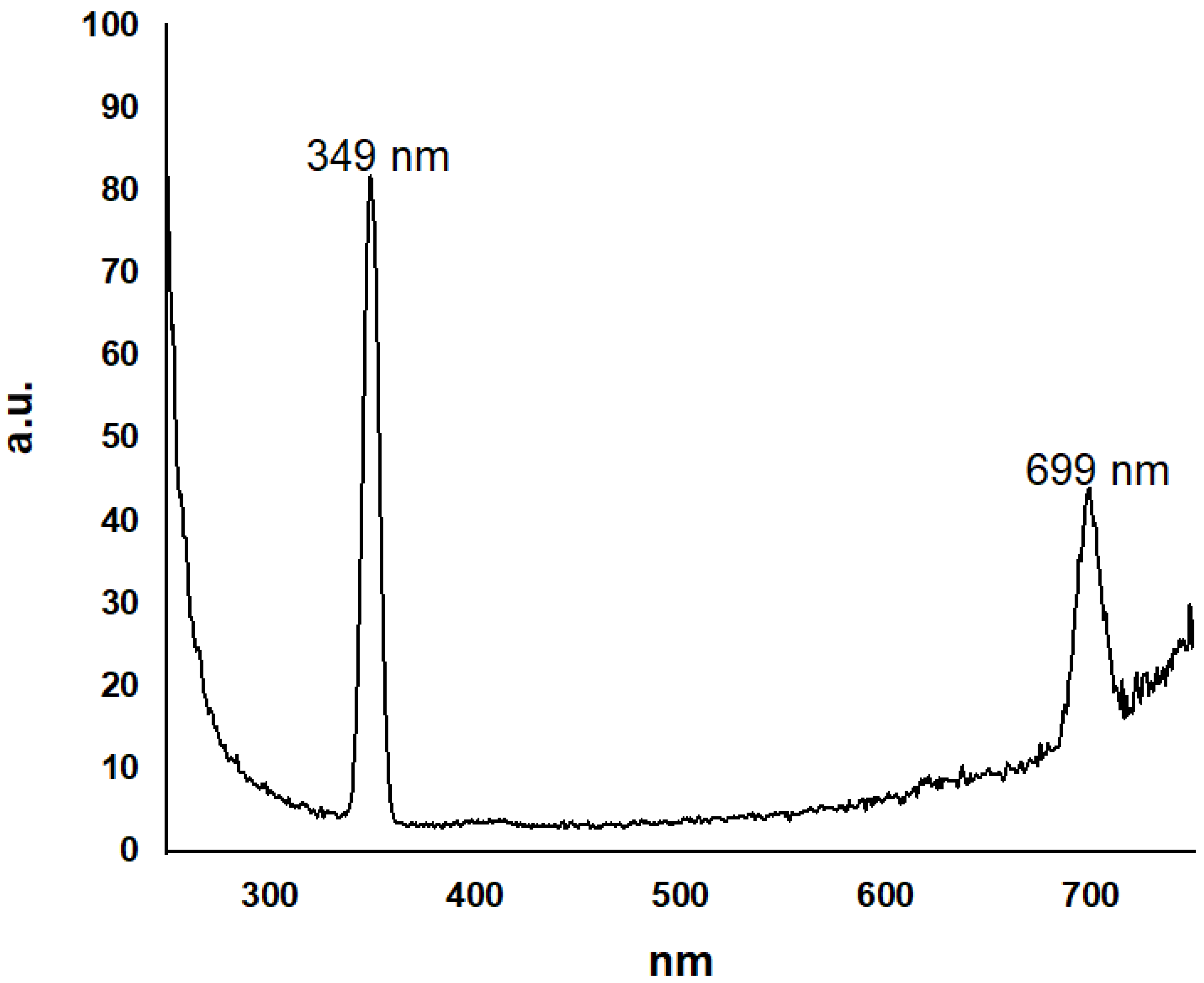
3.1.1. UV–Vis Spectrophotometry

The observed surface plasmon resonance (SPR) by the UV–Vis spectrophotometry confirmed the formation of AgNPs. A strong and broad surface plasmon resonance (SPR) has been seen in metal nanoparticles between 2 and 100 nm in size. Spherical nanoparticles demonstrate a single SPR band, in agreement with Mie’s theory [35,36]. Figure 2 shows the absorption peak of the AgNPs solution corresponds to the SPR obtained in a visible range at 439.29 nm. In line with these results, it has been reported that AgNPs exhibit a distinctive absorption peak at about 400 nm, attributed to SPR. The free electrons of the metal NPs are responsible for the SPR absorption peak attributed to the combined vibration of electrons with the light wave [26]. The refractive index of the surrounding medium, particle size, and adsorbed species on their surfaces affects the position of the SPR absorption peak (kmax) of spherical AgNPs [37,38].

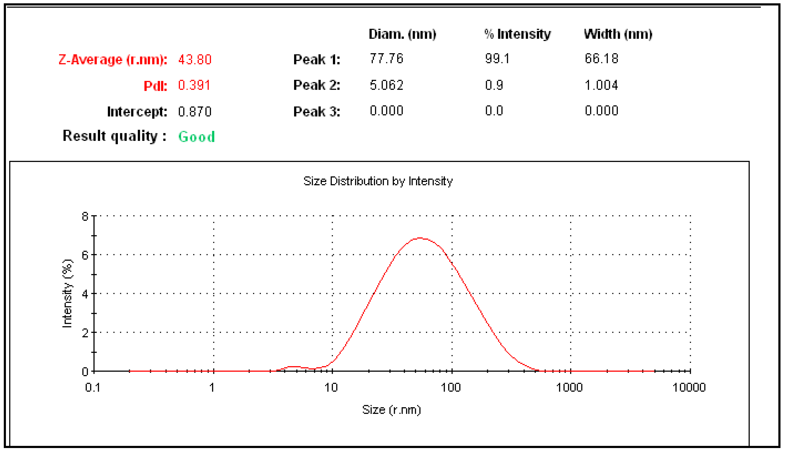
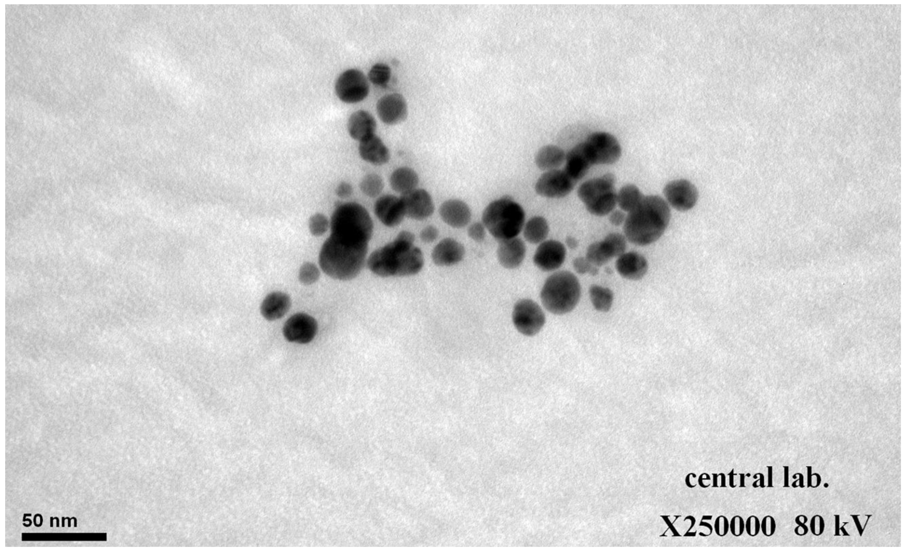
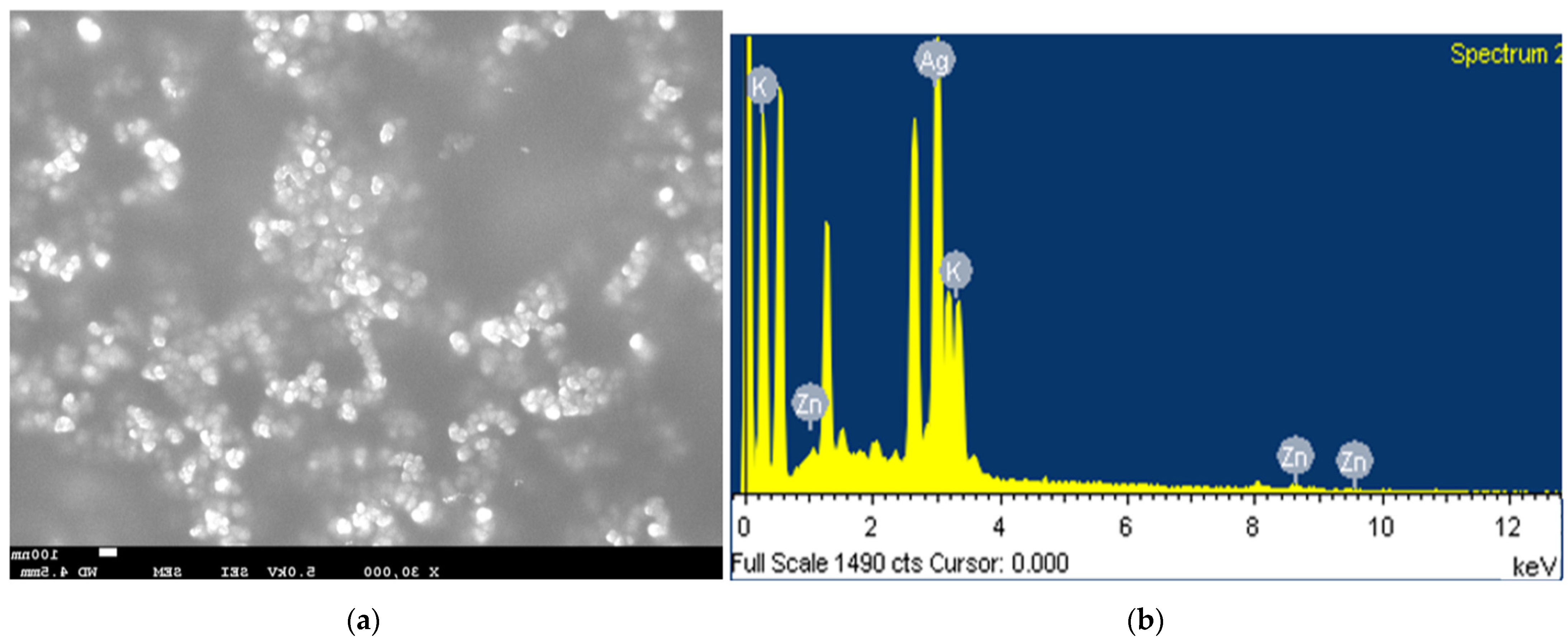
3.1.2. Size and Morphology of Nanoparticles

The particle size distribution by intensity determined using the DLS technique is illustrated in Figure 3. The average hydrodynamic diameter of the prepared AgNPs using Trigonella seeds was 43.8 d.nm with an intercept of 0.870 and low polydispersity index (PDI) of 0.391. Similar hydrodynamic radii of AgNPs obtained with DLS in the range of 9–42 nm were reported by Ahani and Khatibzadeh [39]. In comparison to the TEM measurement of the particle size, the particles were moderately larger in DLS, as it measures the hydrodynamic radius of the particles [40,41].
The TEM micrographs illustrated that the synthesized Ag NPs were dispersed without agglomeration. In addition, their morphotype was spherical and oblong, with an average particle size that ranged between 9 and 21 nm (Figure 4). The particle sizes observed in the TEM images are in consensus with a previous study that reported green synthesized Ag NPs with an average size of 20–50 nm [41], and are smaller than those reported by Goyal et al. [32].
The energy-dispersive spectroscopy (EDS) revealed the elemental map of AgNPs. The silver ions in AgNPs are represented by a prominent peak at 3 keV. AgNPs often show a typical absorption peak at 3 keV because of the surface plasmon resonance. In addition to K and Zn, 87.45% of the Ag was found to be present in AgNPs at 3 keV (Figure 5a,b). The presence of K and Zn atoms in the aqueous extract of Trigonella seeds could possibly be due to the presence of other bioactive components [42,43].

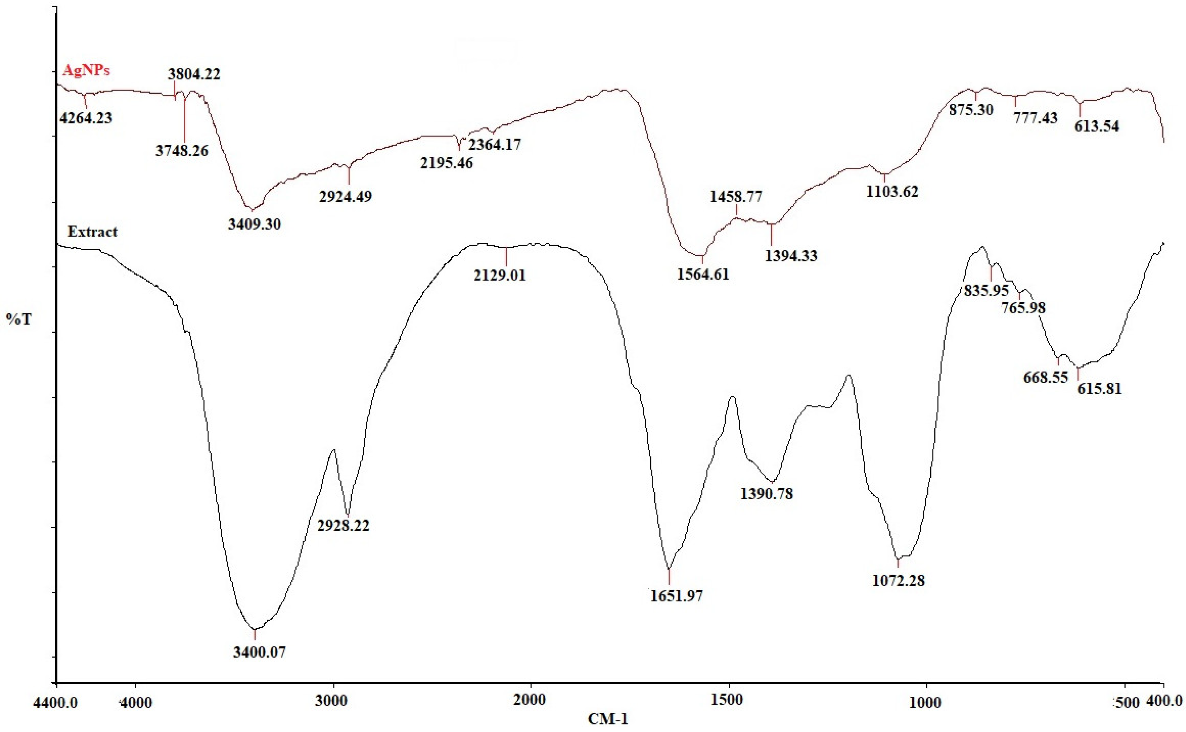
3.1.3. FTIR Analysis

FTIR is vital structural tool used to assess the chemical interactions between the metal particles and biomolecules in plant extracts. This gives an insight into the surface chemical composition of the silver nanoparticles along with an identification of the biomolecules that are involved in the capping and stabilization of the metal nanoparticles [44].
The absorption band intensities in the spectra for bulk Trigonella seed extract and AgNPs are illustrated in Figure 6. The FTIR spectra of both the bulk Trigonella seed extract and AgNPs show different major peaks positioned at 3409.30, 3400.07, 2924.49, 2928.22, 1564.61, 1651.97, 1394.33, 1390.78, 1103.62, 1072.28, 875.30, 835.95, 777.43, 765.98, 613.54, and 615.81 cm−1. The presence of the bio-components from the seed extract in the sample acting as a capping agent for the AgNPs is clearly demonstrated by certain similarities between the spectra, with small marginal shifts in peak positions. Aliphatic primary amines are responsible for the medium N-H stretching that causes the peak at 3400–3409 cm−1. The band at 2924–2928 cm−1 is recognized as medium C-H stretching of the alkane group. The peaks at 2129–2195 cm−1 correspond to strong C=N=C stretching of the carbodiimide group. The broad peaks at 1564–1651 cm−1 correspond to medium N-H bending of the amine group. Peaks between 1072–1103 cm−1 are ascribed to strong C-O stretching of secondary alcohol. Peaks below 800 cm−1 correspond to strong C=C bending of the alkene group. The alkyl halides band, particularly the C-Cl bond, has a peak in the 628 cm−1 range [45]. Therefore, it may be concluded that these biomolecules aid in capping and effectively stabilize the synthesized Ag-NPs [32,44].

3.1.4. Fluorescence Spectroscopy

One technique for evaluating the optical characteristics of nanoparticles as photonic materials is the fluorescence (FL) spectrum. The most important criterion for surface plasmon resonance is frequently the collective oscillation of the silver-conducting electrons. AgNPs have a greater visible light resolution than any other known organic or inorganic chromophore. The nature, size, and shape of the surrounding particles in suspension strongly influence surface plasmon resonance [46].
Furthermore, the optical properties and electronic characteristics of the AgNPs were assessed (Figure 7). The synthesis of silver nanoparticles further increases the intensity of emission. Synthesized colloidal AgNPs are dispersed in water, and the fluorescence emission spectrum is recorded for the excitation wavelength at 349 nm. The three stages of the fluorescence phenomenon are photoexcitation of an electron, relaxation of the excited electron, and lastly, fluorescence emission. A shift in the emission fluorescence spectra at 699 nm was detected, and the effect of photoexcitation at a wavelength of 349 nm from states above the Fermi level was assessed. The process of electron phonon and hole phonon scattering was related to the fluorescence shift [45]. The transition of the lowest unoccupied molecular orbital (LUMO) to the highest occupied molecular orbital (HOMO), and the transition from HOMO to LUMO+3 and LUMO+4, were all reported to correspond to the band in UV–Vis spectroscopy. Since LUMO+3/+4’s orbital compositions are comparable to those of LUMO, the p-sp transition (ligand-to-metal charge transfer, or LMCT) is likewise the primary cause of the transition. These two bands are hence the result of the sp-sp interband transition. AgNPs exhibit intense red emission in their crystalline state, with a maximum emission wavelength in the fluorescence spectrum [47].

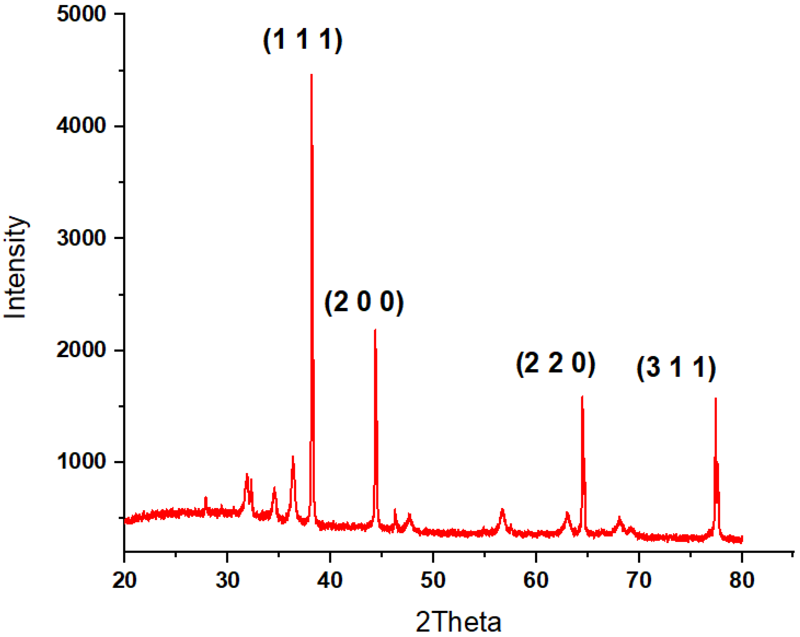
3.1.5. X-ray Diffraction Analysis

The Bragg’s law, which is based on a wide-angle elastic scattering of X-rays, is the fundamental idea behind the XRD technique. In other words, when exposed to X-rays, the crystal creates a variety of diffraction patterns that show the physicochemical characteristics of the crystal structure. The crystalline nature of AgNPs was investigated and validated in the current work through XRD analysis, as shown in Figure 8. As can be observed, the X-ray diffraction spectrum demonstrated the existence of prominent peaks in the diffraction values that were at 2 theta 38.185 ° (1 1 1), 44.381 ° (2 0 0), 64.573 ° (2 2 0), 77.557 ° (3 1 1), and 81.715 ° (2 2 2), along with the corresponding patterns, respectively. This documents and evidences the crystal planes of the face-centered cubic silver structure and crystalline nature (JCPDS COD 9011607) with high purity. Similar patterns for the AgNPs have been also reported in previous studies [48,49].

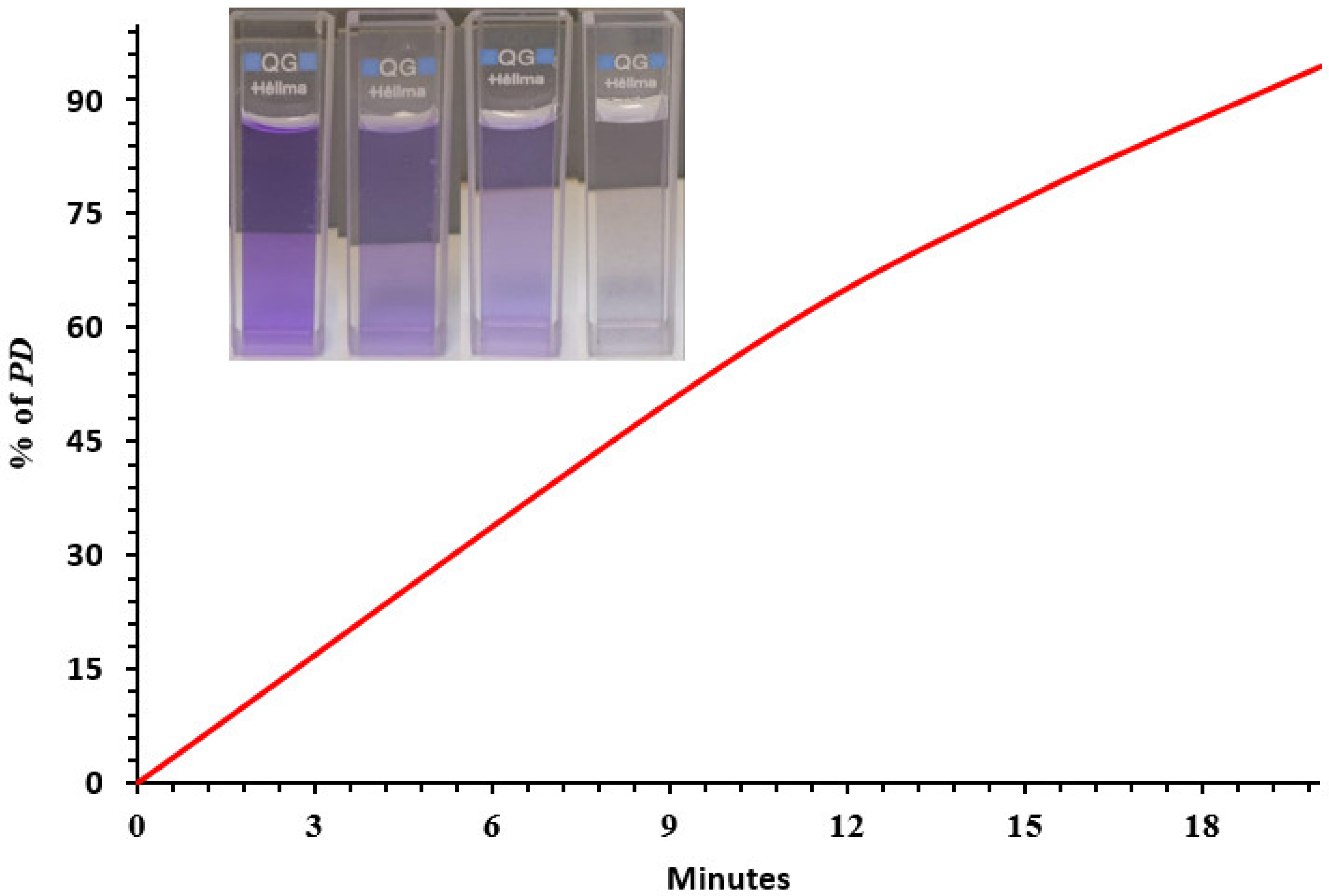
3.2. Photocatalytic Degradation of Crystal Violet (CV) Dye by AgNPs

The noble metal, silver has the highest potent localized surface plasmon resonance with low optical loss in comparison to other metals. Hot electrons from the silver nanoparticles (Ag NPs) move to the oxygen absorbed on the Ag NPs during localized surface plasmon resonance (LSPR), which causes the oxygen-absorbed Ag nanostructure to become negatively charged. In addition, the heated electrons accelerate the plasmonic Ag NPs’ catalytic oxidation reaction [50,51]. As shown in Figure 9, the AgNPs exhibited appreciable degradation of crystal violet dye under UV irradiation. The computed percentage of degradation efficiency was 94.5%.
Figure 10 presents an illustration of the photocatalytic mechanism of green synthesized AgNPs, and could be summarized as
AgNPs + Photon → Ag+ + e electron
e + O2 oxygen →O2 oxygen radicals
Ag+ silver ions → Ag + hole (h+)
h+ + H2O water / OH hydroxide ions → OH radicals
CV dye + OH → Decolorization of CV dye
Table 1 provides a comparison of photodegradative efficacy of various plant-based AgNPs to validate the potential of the AgNPs used in the present study.

3.3. Water Quality Parameters of the Sewage Effluent

Table 2 represents the effect of the synthesized AgNPs on the basic water quality parameters of the sewage effluent. There was a marked alteration in the parameters assessed in terms of pH, EC, inorganic salts, and COD and BOD. It was clearly observed that treatment with AgNPs significantly improved the water quality parameters of the effluent, which is best attributed to the enhanced adsorption activity of the AgNPs coupled with its strong antimicrobial potency [12]. The enhanced adsorption ability of the Ag NPs was evident by the reduction in the EC and inorganic ions. While the decrease in the BOD of the effluent demonstrated a decrease in the microbial count brought about by the AgNPs [10,12,30,57]. Additionally, a significant decrease in the COD values was observed in the effluent on treatment with AgNPs. In consensus with this, recent studies have reported elimination of coliform bacteria and heterotrophic bacteria, with the reduction in the COD of wastewater treatment plants using silver-loaded magnetic nanoparticles [24,58]. The nanoscale size of the nanoparticles provides an increased surface area for an effective adsorption of inorganic/organic matrix in the effluent sample.

3.4. Antibacterial Activity of Trigonella/Ag-NPs

Rampant use of antibiotics in recent times, especially during the pandemic, has made the WWTPs act as reservoirs for antibiotic-resistant bacteria, and their potential horizontal gene transfer could have grave implications on human health. Silver nanoparticles have been known for their remarkable antimicrobial activity for decades, being able to combat bacterial infections both in vitro and in vivo. Both Gram-negative and Gram-positive bacteria, as well as strains that are multidrug resistant, are susceptible to AgNPs’ antibacterial properties. Above a certain dose, they showed negligible toxicity towards mammalian cells while exhibiting strong toxicity towards bacteria like E. coli [10]. Accordingly, the antimicrobial activity of the AgNPs with Trigonella seeds in the current study was clearly defined, as the nanoparticles showed a larger diameter of the inhibition zone at two different concentrations than was seen for the bulk Trigonella seed extract against two bacterial strains, E. coli and S. aureus (Table 3). The antibacterial effect on the Gram -positive strain, S.aureus, was more pronounced. This is consistent with other studies that found AgNPs made from either Trigonella seed or leaf extract to have strong microbicidal activity [27,28,32]. Additionally, AgNPs synthesized by several modes have been extensively documented to have comparable antibacterial properties [23,59]. The electrostatic attraction between the Ag+ ion and the negative charge on the bacterial cell membrane clarifies the antibacterial ability of the Ag-NPs. Thus, the Ag+ ion interferes with bacterial DNA replication and respiration by interacting with enzymes that have thiol groups to form disulfide bridges, permeating the cell membrane, and killing the bacteria [60]. Furthermore, Sabry et al. [61], reported that contact between AgNPs with the bacterial surface leads to an interaction with the bacterial protein in the cell wall which could interfere with the DNA replication and also trigger the generation of reactive oxygen species which eventually leads to oxidative damage of the bacterial cells. The variability observed in the susceptibility of microbes to the ag-NPs could be attributed to the nature of their cell wall structure, which affects the permeability. The major constituents of the outer membrane of Gram-negative bacteria are lipopolysaccharides that limit the entry of macro/hydrophilic molecules. Conversely, the Gram-positive bacteria have negligible or no lipopolysaccharide content. Furthermore, these bacteria possess multiple peptidoglycan layers and chains of negatively charged glycerin in their cell wall that interacts with the Ag+ ions [32].
Currently, water pollution from the sewage of the textile industry in wastewater is one of the major global environmental challenges. Textile wastewater contains a large number of toxic dyes, heavy metals such as mercury, chromium, cadmium, lead, and arsenic, color pigments, and aromatic compounds [29,62]. These toxic chemicals are transported over long distances along with wastewater, and are persistent pollutants in soil and water that have adverse health effects on aquatic fauna, degrade soil fertility, and impair primary production in the aquatic environment with subsequent anoxic conditions [63]. Textile dyes deteriorate the aesthetics of the water bodies, resulting in an increase in BOD and COD, interfering with the food chains, bioaccumulating, and progressing carcinogenicity and mutagenicity [64,65]. In the present study, the Trigonella/AgNPs effectively photodegraded the dye, crystal violet, thereby adding to its potential wastewater remediation capacity along with modulation of the water quality parameters of the sewage effluent. Furthermore, its potent anti-microbial activity supported the previous reports on the ability of AgNPs in effectively eliminating over 700 microorganisms found in WWTPs [59]. Thus, synthesis of AgNPs integrating green technology using plant extracts/herbs offers several advantages such as eco-friendliness, biocompatibility, and cost-effectiveness. These distinct properties of AgNPs could prospectively play a vital role in several nanomaterial-based wastewater treatment strategies in modern times.

4. Conclusions

In the current study, a low-cost biogenic synthesis of AgNPs with Trigonella seed extract was successfully attempted. The nanoparticles were analytically characterized and their remedial efficacy on wastewater quality and degradative potential of an organic dye showed substantial outcomes that were notable in terms of environmental sustainability. The nanoparticles demonstrated the ability to degrade crystal violet with effective decrease in BOD and COD of the wastewater under optimized conditions. Taken together, the results of the present study show a prospective role of AgNPs using Trigonella seeds as an environmentally benign treatment approach for domestic/textile industry wastewater. However, further investigations are imperative to determine the environmental effects of these nanostructures, as well as their viability, via techno-economic feasibility studies.

Author Contributions

Conceptualization, M.A.A. and P.V.; Data curation, A.A.H. and K.M.O.; Formal analysis, K.M.O.; Investigation, M.A.A., A.A.H., N.A. and T.S.A.; Methodology, M.A.A.; Resources, N.A. and T.S.A.; Software, K.M.O.; Validation, A.A.H.; Writing—original draft, M.A.A. and P.V.; Writing—review and editing, P.V. All authors have read and agreed to the published version of the manuscript.

Funding

The authors extend their appreciation to the Deputyship for Research & Innovation, Ministry of Education in Saudi Arabia for funding this research work through the project number RI-44-0191.

Institutional Review Board Statement

Not relevant.

Informed Consent Statement

Not applicable.

Data Availability Statement

Data will be provided on request.

Conflicts of Interest

The authors declare no conflict of interest.

References

  1. Luthy, R.G.; Sedlak, D.L.; Plumlee, M.H.; Austin, D.; Resh, V.H. Wastewater-effluent-dominated streams as ecosystem-management tools in a drier climate. Front. Ecol. Environ. 2015, 13, 477–485. [Google Scholar] [CrossRef] [Green Version]
  2. Kalkan, C.; Yapsakl, K.; Mertoglu, B.; Tufan, D.; Saatci, A. Evaluation of Biological ActivatedCarbon (BAC) process in wastewater treatment secondary effluent for reclamation purposes. Desalination 2011, 265, 8. [Google Scholar] [CrossRef]
  3. Shittu, K.O.; Ihebunna, O. Purification of simulated waste water using green synthesized silver nanoparticles of Piliostigma thonningii aqueous leave extract. Adv. Nat. Sci. Nanosci. Nanotechnol. 2017, 8, 045003. [Google Scholar] [CrossRef] [Green Version]
  4. Jain, K.; Patel, A.S.; Pardhi, V.P.; Flora, S.J.S. Nanotechnology in Wastewater Management: A New Paradigm TowardsWastewater Treatment. Molecules 2021, 26, 1797. [Google Scholar] [CrossRef]
  5. Kumar, S.; Ahlawat, W.; Bhanjana, G.; Heydarifard, S.; Nazhad, M.M.; Dilbaghi, N. Nanotechnology-Based Water Treatment Strategies. J. Nanosci. Nanotechnol. 2014, 14, 1838–1858. [Google Scholar] [CrossRef]
  6. Ahmed, S.F.; Mofijur, M.; Ahmed, B.; Mehnaz, T.; Mehejabin, F.; Maliat, D.; Hoang, A.T.; Shafiullah, G.M. Nanomaterials as a sustainable choice for treating wastewater. Environ. Res. 2022, 214, 113807. [Google Scholar] [CrossRef]
  7. Lu, H.; Wang, J.; Stoller, M.; Wang, T.; Bao, Y.; Hao, H. An Overview of Nanomaterials for Water and Wastewater Treatment. Adv. Mater. Sci. Eng. 2016, 2016, 4964828. [Google Scholar] [CrossRef] [Green Version]
  8. Anjum, M.; Miandad, R.; Waqas, M.; Gehany, F.; Barakat, M.A. Remediation of wastewater using various nano-materials. Arab. J. Chem. 2019, 12, 4897–4919. [Google Scholar] [CrossRef] [Green Version]
  9. Malik, S.B.; Saggu, J.I.; Gul, A.; Abbasi, B.A.; Iqbal, J.; Waris, S.; Jardan, Y.A.B.; Chalgham, W. Synthesis and Characterization of Silver and Graphene Nanocomposites and Their Antimicrobial and Photocatalytic Potentials. Molecules 2022, 27, 5184. [Google Scholar] [CrossRef]
  10. Bruna, T.; Maldonado-Bravo, F.; Jara, P.; Caro, N. Silver Nanoparticles and Their Antibacterial Applications. Int. J. Mol. Sci. 2021, 22, 7202. [Google Scholar] [CrossRef]
  11. Haris, M.; Fatima, N.; Iqbal, J.; Chalgham, W.; Mumtaz, A.S.; El-Sheikh, M.A.; Tavafoghi, M. Oscillatoria limnetica Mediated Green Synthesis of Iron Oxide (Fe2O3) Nanoparticles and Their Diverse In Vitro Bioactivities. Molecules 2023, 28, 2091. [Google Scholar] [CrossRef]
  12. Ganguly, K.; Dutta, S.D.; Patel, D.K.; Lim, K. Chapter 18—Silver nanoparticles for wastewater treatment. In Micro and Nano Technologies, Aquananotechnology; Abd-Elsalam, K.A., Muhammad Zahid, M., Eds.; Elsevier: Amsterdam, The Netherlands, 2021; pp. 385–401. ISBN 9780128211410. [Google Scholar] [CrossRef]
  13. Galatage, S.T.; Hebalkar, A.S.; Dhobale, S.V.; Mali, O.R.; Kumbhar, P.S.; Nikade, S.V.; Killedar, S.G. Silver Nanoparticles: Properties, Synthesis, Characterization, Applications and Future Trends. In Silver Micro-Nanoparticles—Properties, Synthesis, Characterization, and Applications; IntechOpen: Rijeka, Croatia, 2021. [Google Scholar] [CrossRef]
  14. Mohammed, A.B.A.; Mohamed, A.; El-Naggar, N.E.; Mahrous, H.; Nasr, G.M.; Abdella, A.; Ahmed, R.H.; Irmak, S.; Elsayed, M.S.A.; Selim, S.; et al. Antioxidant and Antibacterial Activities of Silver Nanoparticles Biosynthesized by Moringa oleifera through Response Surface Methodology. J. Nanomater. 2022, 2022, 9984308. [Google Scholar] [CrossRef]
  15. Krishnaraj, C.; Jagan, E.G.; Rajasekar, S.; Selvakumar, P.; Kalaichelvan, P.T.; Mohan, N.J.C.S.B.B. Synthesis of silver nanoparticles using Acalypha indica leaf extracts and its antibacterial activity against water borne pathogens. Colloids Surf. B 2010, 76, 50–56. [Google Scholar]
  16. Ullah, Z.; Gul, F.; Iqbal, J.; Abbasi, B.A.; Kanwal, S.; Chalgham, W.; El-Sheikh, M.A.; Diltemiz, S.E.; Mahmood, T. Biogenic Synthesis of Multifunctional Silver Oxide Nanoparticles (Ag2ONPs) Using Parieteria alsinaefolia Delile Aqueous Extract and Assessment of Their Diverse Biological Applications. Microorganisms 2023, 11, 1069. [Google Scholar] [CrossRef]
  17. Vanlalveni, C.; Lallianrawna, S.; Biswas, A.; Selvaraj, M.; Changmai, B.; Rokhum, S.L. Green synthesis of silver nanoparticles using plant extracts and their antimicrobial activities: A review of recent literature. RSC Adv. 2021, 11, 2804–2837. [Google Scholar]
  18. Abbasi, B.A.; Iqbal, J.; Yaseen, T.; Zahra, S.A.; Ali, S.; Uddin, S.; Mahmood, T.; Kanwal, S.; El-Serehy, H.A.; Chalgham, W. Exploring Physical Characterization and Different Bio-Applications of Elaeagnus angustifolia Orchestrated Nickel Oxide Nanoparticles. Molecules 2023, 28, 654. [Google Scholar] [CrossRef]
  19. Vijayaraghavan, K.; Kamala Nalini, S.P.; Kannaian, U.P.N.; Dhakshinamoorthy, M. One step green synthesis of silver nano/microparticles using extracts of Trachyspermum ammi and Papaver somniferum. Colloids Surf. B 2012, 94, 114–117. [Google Scholar]
  20. Kumar, V.; Yadav, S.K. Synthesis of different-sized silver nanoparticles by simply varying reaction conditions with leaf extracts of Bauhinia variegata L. IET Nanobiotechnology 2010, 6, 1–8. [Google Scholar] [CrossRef]
  21. Guidelli, E.J.; Ramos, A.P.; Zaniquelli, M.E.D.; Baffa, O. Green synthesis of colloidal silver nanoparticles using natural rubber latex extracted from Hevea brasiliensis. Spectrochim. Acta Par A Mol. Biomol. Spectrosc. 2011, 82, 140–145. [Google Scholar]
  22. Tippayawat, P.; Phromviyo, N.; Boueroy, P.; Chompoosor, A. Green synthesis of silver nanoparticles in Aloe vera plant extract prepared by a hydrothermal method and their synergistic antibacterial activity. PeerJ 2016, 4, e2589. [Google Scholar]
  23. Vanti, G.L.; Nargund, V.B.; Basavesha, K.N.; Vanarchi, R.; Kurjogi, M.; Mulla, S.I.; Tubaki, S.; Patil, R.R. Synthesis of Gossypium hirsutum-derived silver nanoparticles and their antibacterial efficacy against plant pathogens. Appl. Organomet. Chem. 2019, 33, e4630. [Google Scholar] [CrossRef] [Green Version]
  24. Saratale, G.D.; Saratale, R.J.; Cho, S.; Ghodake, G.; Bharagava, R.N.; Park, Y.; Mulla, S.I.; Kim, D.; Kadam, A.; Nair, S.; et al. Investigation of photocatalytic degradation of reactive textile dyes by Portulaca oleracea-functionalized silver nanocomposites and exploration of their antibacterial and antidiabetic potential. J. Alloys Compd. 2020, 833, 155083. [Google Scholar] [CrossRef]
  25. Nagulapalli Venkata, K.C.; Swaroop, A.; Bagchi, D.; Bishayee, A. A small plant with big benefits: Fenugreek (Trigonella foenum-graecum Linn.) for disease prevention and health promotion. Mol. Nutr. Food Res. 2017, 61, 1600950. [Google Scholar] [CrossRef]
  26. Basu, T.K.; Srichamroen, A. Chapter—Health Benefits of Fenugreek (Trigonella foenum-graecum leguminosse). In Preedy, Bioactive Foods in Promoting Health; Watson, R.R., Victor, R., Eds.; Academic Press: Cambridge, MA, USA, 2010; pp. 425–435. ISBN 9780123746283. [Google Scholar] [CrossRef]
  27. Awad, M.A.; Hendi, A.A.; Ortashi, K.M.; Alzahrani, B.; Soliman, D.; Alanazi, A.; Alenazi, W.; Taha, R.M.; Ramadan, R.; El-Tohamy, M.; et al. Biogenic synthesis of silver nanoparticles using Trigonella foenum-graecum seed extract: Characterization, photocatalytic and antibacterial activities. Sens. Actuators A Phys. 2021, 323, 112670. [Google Scholar] [CrossRef]
  28. Rizwana, H.; Alwhibi, M.S.; Aldarsone, H.A.; Awad, M.A.; Soliman, D.A.; Bhat, R.S. Green synthesis, characterization, and antimicrobial activity of silver nanoparticles prepared using Trigonella foenum-graecum L. leaves grown in Saudi Arabia. Green Process. Synth. 2021, 10, 421–429. [Google Scholar] [CrossRef]
  29. Moond, M.; Singh, S.; Sangwan, S.; Devi, P.; Beniwal, A.; Rani, J.; Kumari, A.; Rani, S. Biosynthesis of Silver Nanoparticles Utilizing Leaf Extract of Trigonella foenum-graecum L. for Catalytic Dyes Degradation and Colorimetric Sensing of Fe3+/Hg2+. Molecules 2023, 28, 951. [Google Scholar] [CrossRef]
  30. Varghese, R.; Almalki, M.A.; Ilavenil, S.; Rebecca, J.; Choi, K.C. Silver nanopaticles synthesized using the seed extract of Trigonella foenum-graecum L. and their antimicrobial mechanism and anticancer properties. Saudi J. Biol. Sci. 2019, 26, 148–154. [Google Scholar] [CrossRef]
  31. Bhat, R.S.; Alghamdi, J.M.; Aldbass, A.M.; Aljebrin, N.A.; Alangery, A.B.; Soliman, D.A.; Al-Daihan, S. Biochemical and FT-IR profiling of Tritium aestivum L. seedling in response to sodium fluoride treatment. Fluoride 2022, 55, 81–89. [Google Scholar]
  32. Goyal, S.; Gupta, N.; Kumar, A.; Chatterjee, S.; Nimesh, S. Antibacterial, anticancer and antioxidant potential of silver nanoparticles engineered using Trigonella foenum-graecum seed extract. IET Nanobiotechnol. 2018, 12, 526–533. [Google Scholar] [CrossRef] [PubMed]
  33. Balouiri, M.; Sadiki, M.; Ibnsouda, S.K. Methods for in vitro evaluating antimicrobial activity: A review. J. Pharm. Anal. 2016, 6, 71–79. [Google Scholar] [CrossRef] [Green Version]
  34. Bratovcic, A. Biosynthesis of Green Silver Nanoparticles and Its UV-Vis Characterization. Int. J. Innov. Sci. Eng. Technol. 2020, 7, 170–176. [Google Scholar]
  35. Xia, Y.; Halas, N.J. Shape-controlled synthesis and surface plasmonic properties of metallic nanostructures. MRS Bull. 2005, 30, 338–348. [Google Scholar] [CrossRef] [Green Version]
  36. Duan, X.; Liu, N. Magnesium for dynamic nanoplasmonics. Acc. Chem. Res. 2019, 52, 1979–1989. [Google Scholar] [CrossRef] [PubMed] [Green Version]
  37. Bastús, N.G.; Piella, J.; Puntes, V. Quantifying the sensitivity of multipolar (dipolar, quadrupolar, and octapolar) surface plasmon resonances in silver nanoparticles: The effect of size, composition, and surface coating. Langmuir 2015, 32, 290–300. [Google Scholar] [PubMed]
  38. El-Desouky, N.; Shoueir, K.; El-Mehasseb, I.; El-Kemary, M. Synthesis of silver nanoparticles using bio valorization coffee waste extract: Photocatalytic flow-rate performance, antibacterial activity, and electrochemical investigation. Biomass Conv. Bioref. 2022, 1–15. [Google Scholar] [CrossRef]
  39. Ahani, M.; Khatibzadeh, M. Green synthesis of silver nanoparticles using gallic acid as reducing and capping agent: Effect of pH and gallic acid concentration on average particle size and stability. Inorg. Nano-Met. Chem. 2022, 52, 234–240. [Google Scholar]
  40. Ravichandran, V.; Vasanthi, S.; Shalini, S.; Shah, S.A.A.; Tripathyd, M.; Paliwala, N. Green synthesis, characterization, antibacterial, antioxidant and photocatalytic activity of Parkia speciosa leaves extract mediated silver nanoparticles. Results Phys. 2019, 15, 102565. [Google Scholar] [CrossRef]
  41. Meena, R.K.; Chouhan, N. Biosynthesis of silver nanoparticles from plant (fenugreek seeds) reducing method and their optical properties. Res. J. Recent. Sci. 2015, 2277, 2502. [Google Scholar]
  42. Bilal, M.; Khan, S.; Ali, J.; Ismail, M.; Khan, M.I.; Asiri, A.M.; Khan, S.B. Biosynthesized silver supported catalysts for disinfection of Escherichia coli and organic pollutant from drinking water. J. Mol. Liq. 2019, 281, 295–306. [Google Scholar] [CrossRef]
  43. Vijayakumar, M.; Priya, K.; Ilavenil, S.; Janani, B.; Vedarethinam, V.; Ramesh, T.; Arasu, M.V.; Al-Dhabi, N.A.; Kim, Y.O.; Kim, H.J. Shrimp shells extracted chitin in silver nanoparticle synthesis: Expanding its prophecy towards anticancer activity in human hepatocellular carcinoma HepG2 cells. Int. J. Biol. Macromol. 2020, 165, 1402–1409. [Google Scholar]
  44. Ikram, S.A.S. Silver Nanoparticles: One Pot Green Synthesis Using Terminalia arjuna Extract for Biological Application. J. Nanomed. Nanotechnol. 2015, 6, 1000309. [Google Scholar] [CrossRef] [Green Version]
  45. El-Kemary, M.; Zahran, M.; Khalifa, S.A.; El-Seedi, H.R. Spectral characterisation of the silver nanoparticles biosynthesized using Ambrosia maritima plant. Micro Nano Lett. 2016, 11, 311–314. [Google Scholar] [CrossRef]
  46. Kumar, P.P.N.V.; Pammi, S.V.N.; Pratap Kollu, S.K.V.V.; Shameem, U. Green synthesis and characterization of silver nanoparticles using Boerhaavia diffusa plant extract and their antibacterial activity. Ind. Crops Prod. 2014, 52, 562–566. [Google Scholar]
  47. Hay, P.J.; Wadt, W.R. Ab initio effective core potentials for molecular calculations. Potentials for the transition metal atoms Sc to Hg. J. Chem. Phys. 1985, 82, 270. [Google Scholar]
  48. Cao, G.; Wang, Y. Characterization and Properties of Nanomaterials. Nanostructures and Nanomaterials: Synthesis, Properties, and Applications; Volume 2 of World Scientific series in nanoscience and nanotechnology; World Scientific: Singapore, 2011; pp. 508, 581. ISBN 9814322504/9789814322. [Google Scholar]
  49. Swetha, V.; Lavanya, S.; Sabeena, G.; Pushpalaksmi, E.; Jenson, S.J.; Annadurai, G. Synthesis and characterization of silver nanoparticles from Ashyranthus aspera extract for antimicrobial activity studies. J. Appl. Sci. Environ. Manag. 2020, 24, 1161–1167. [Google Scholar]
  50. Ma, Y.; Tao, L.; Bai, S.; Hu, A. Green Synthesis of Ag Nanoparticles for Plasmon-Assisted Photocatalytic Degradation of Methylene Blue. Catalysts 2021, 11, 1499. [Google Scholar]
  51. Lee, S.H.; Jo, J.S.; Park, J.H.; Lee, S.W.; Jang, J.W. A hot-electron-triggered catalytic oxidation reaction of plasmonic silver nanoparticles evidenced by surface potential mapping. J. Mater. Chem. A 2018, 6, 20939–20946. [Google Scholar] [CrossRef]
  52. Rani, P.; Kumar, V.; Singh, P.P.; Matharu, A.S.; Zhang, W.; Kim, K.H.; Rawat, M. Highly stable AgNPs prepared via a novel green approach for catalytic and photocatalytic removal of biological and non-biological pollutants. Environ. Int. 2020, 143, 105924. [Google Scholar]
  53. Singh, J.; Kumar, V.; Jolly, S.S.; Kim, K.H.; Rawat, M.; Kukkar, D.; Tsang, Y.F. Biogenic synthesis of silver nanoparticles and its photocatalytic applications for removal of organic pollutants in water. J. Ind. Eng. Chem. 2019, 80, 247–257. [Google Scholar] [CrossRef]
  54. Jose, V.; Raphel, L.; Aiswariya, K.S.; Mathew, P. Green synthesis of silver nanoparticles using Annona squamosa L. seed extract: Characterization, photocatalytic and biological activity assay. Bioprocess. Biosyst. Eng. 2021, 44, 1819–1829. [Google Scholar]
  55. Kathiravan, V. Green synthesis of silver nanoparticles using different volumes of Trichodesma indicum leaf extract and their antibacterial and photocatalytic activities. Res. Chem. Intermed. 2018, 44, 4999–5012. [Google Scholar] [CrossRef]
  56. Anjana, V.N.; Joseph, M.; Francis, S.; Joseph, A.; Koshy, E.P.; Mathew, B. Microwave assisted green synthesis of silver nanoparticles for optical, catalytic, biological and electrochemical applications. Artif. Cells Nanomed. Biotechnol. 2021, 49, 438–449. [Google Scholar] [CrossRef] [PubMed]
  57. Perez, M. The Effects of Silver Nanoparticles on Wastewater Treatment and Escherichia Coli Growth. Bachelor’s Thesis, Florida State University, Tallahassee, FL, USA, 2012. [Google Scholar]
  58. Najafpoor, A.; Norouzian-Ostad, R.; Alidadi, H.; Rohani-Bastami, T.; Davoudi, M.; Barjasteh-Askari, F.; Zanganeh, J. Effect of magnetic nanoparticles and silver-loaded magnetic nanoparticles on advanced wastewater treatment and disinfection. J. Mol. Liq. 2020, 303, 112640. [Google Scholar] [CrossRef]
  59. Epelle, E.I.; Okoye, P.U.; Roddy, S.; Gunes, B.; Okolie, J.A. Advances in the Applications of Nanomaterials for Wastewater Treatment. Environments 2022, 9, 141. [Google Scholar] [CrossRef]
  60. Li, H.; Gao, Y.; Li, C.; Ma, G.; Shang, Y.; Sun, Y. A comparative study of the antibacterial mechanisms of silver ion and silver nanoparticles by Fourier transform infrared spectroscopy. Vib. Spectrosc. 2016, 85, 112–121. [Google Scholar] [CrossRef]
  61. Sabry, N.; Tolba, S.; Kh, F.; Abdel-Gawad, F.; Bassem, S.; Hossam, F.; El-Taweel, G.E.; Okasha, A.; Ibrahim, M. Interaction between nano silver and bacteria: Modeling approach. Biointerface Res. Appl. Chem. 2018, 8, 3570–3574. [Google Scholar]
  62. Singha, K.; Pandit, P.; Maity, S.; Sharma, S.R. Harmful environmental effects for textile chemical dyeing practice. In Green Chemistry for Sustainable Textiles; Woodhead Publishing: Cambridge, UK, 2021; pp. 153–164. [Google Scholar]
  63. Dutta, S.; Bhattacharjee, J. A comparative study between physicochemical and biological methods for effective removal of textile dye from wastewater. In Development in Wastewater Treatment Research and Processes; Elsevier: Amsterdam, The Netherlands, 2022; pp. 1–2. [Google Scholar]
  64. Patil, R.; Zahid, M.; Govindwar, S.; Khandare, R.; Vyavahare, G.; Gurav, R.; Desai, N.; Pandit, S.; Jadhav, J. Constructed wetland: A promising technology for the treatment of hazardous textile dyes and effluent. In Development in Wastewater Treatment Research and Processes; Elsevier: Amsterdam, The Netherlands, 2022; pp. 173–198. [Google Scholar]
  65. Al-Tohamy, R.; Ali, S.S.; Li, F.; Okasha, K.M.; Mahmoud, Y.A.G.; Elsamahy, T.; Jiao, H.; Fu, Y.; Sun, J. A critical review on the treatment of dye-containing wastewater: Ecotoxicological and health concerns of textile dyes and possible remediation approaches for environmental safety. Ecotoxicol. Environ. Saf. 2022, 231, 13160. [Google Scholar] [CrossRef]
Figure 1. Illustration of the mode of synthesis of the AgNPs using Trigonella seed extract.
Processes 11 02394 g001
Figure 2. UV–Vis spectrum of the synthesized Trigonella AgNPs.
Processes 11 02394 g002
Figure 3. Representative DLS spectrum of synthesized Trigonella AgNPs showing the average size of the nanoparticles.
Processes 11 02394 g003
Figure 4. TEM micrograph showing the morphotype of the Trigonella AgNPs.
Processes 11 02394 g004
Figure 5. (a) SEM micrograph of Trigonella AgNPs with the (b) corresponding EDS spectrum.
Processes 11 02394 g005
Figure 6. FTIR spectra of Trigonella AgNPs and the bulk seed extract.
Processes 11 02394 g006
Figure 7. Fluorescence spectrum exhibiting the optical properties and electronic characteristics of the Trigonella AgNPs.
Processes 11 02394 g007
Figure 8. X-ray diffractogram of Trigonella AgNPs.
Processes 11 02394 g008
Figure 9. Time-dependent photodegradative percentage of CV dye under UV irradiation by the AgNPs.
Processes 11 02394 g009
Figure 10. Illustration of the mechanism involved in the photocatalytic degradation of CV dye by AgNPs.
Processes 11 02394 g010
Table 1. Comparative assessment of photodegradative ability of several plant-derived AgNPs reported in the literature with the present study.
PlantSize (nm)Illumination SourceDye/ConcentrationDegradation Time/PercentageReference
Phaseolus vulgaris (kidney beans)10–20 nm Sunlight RR-141 (50 mL, 20 ppm) 150 min
97%
(Rani et al., 2020) [52]
Trigonella foenum-graecum leaf5–20 nm Sunlight Blue 19 (RB19) and reactive yellow 186 (RY186); respectively180 min/88% and 86%; respectively(Singh et al., 2019) [53]
Annona squamosa L.22 nm Sunlight Coomassie brilliant blue(CBB)/5 mL of 1% CBBGradually degradation/(10, 20,
30 min)
(Jose et al., 2021) [54]
Trichodesma indicum leaf35–33 nmSolar lightMethylene blue (MB) dye/200 mL aqueous solution of MB82% of MB within 210 min(Kathiravan et al., 2018) [55]
Cyanthillium cinereum leaf 5–40 nm  Without light irradiationMethylene blue and fuchsine in the presence of aq.NaBH4/(0.08 × 10−3 M) of the dyes Gradual degradation(Anjana et al., 2021) [56]
Trigonella foenum-graecum seeds82.53 nmUV lightRhodamine B dye/10 μgL−1(93%) with decoloration after 216 h(Awad et al., 2021) [27]
Trigonella foenum-graecum seeds43.80 nmUV lightCrystal violet/10−6 M of dye94.5% with in 20 minPresent study
Table 2. Effect of Trigonella AgNPs on the water quality parameters of sewage effluent.
ParameterTap WaterUntreated Sewage EffluentSewage Effluent Treated with Trigonella Ag-NPs
pH7.96 ± 0.04160 a8.77 ± 01966 b7.15 ± 0.1101 c
EC (ppm)692.23 ± 4.6801 a1269.36 ± 2.6350 b726.06 ± 3.5232 c
SO4 (ppm)48.43 ± 0.5132 a166.900 ± 1.3892 b46.67 ± 0.7571 a
Cl (ppm)75.70 ± 0.6083 a552.93 ± 1.6773 b182.93 ± 2.2679 c
HCO3 (ppm)64.63 ± 0.6806 a57.53 ± 0.6658 b66.03 ± 0.3055 a
Mg+2 (ppm)29.73 ± 0.2309 a81.55 ± 0.4500 b47.53 ± 0.5033 c
Ca+2 (ppm)82.87 ± 1.0263 a136.03 ± 1.0016 b104.10 ± 0.3605 c
COD (mg/L)0.77 ± 0.0577 a1135.67 ± 8.6217 b805.20 ± 4.9518 c
BOD (mg/L)0.00 ± 0.0000 a49.17 ± 0.9609 b21.57 ± 0.5131 c
EC: electrical conductivity; COD: chemical oxygen demand; BOD: biological oxygen demand. Values are expressed as mean ± SD. Different superscripts in a row show significant difference (p ≤ 0.05).
Table 3. Diameter of inhibition zone at different concentrations of AgNPs and bulk Trigonella seed extract against Gram-positive and -negative bacterial strains.
Bacterial StrainAgNpsBulk Trigonella Seed Extract
1 µg/mL0.5 µg/mL
Escherichia coli (Gram-negative)24 mm20 mm15 mm
Staphylococcus aureus (Gram-positive)30 mm27 mm14 mm
Disclaimer/Publisher’s Note: The statements, opinions and data contained in all publications are solely those of the individual author(s) and contributor(s) and not of MDPI and/or the editor(s). MDPI and/or the editor(s) disclaim responsibility for any injury to people or property resulting from any ideas, methods, instructions or products referred to in the content.

