Review

Hybrid Materials for Tissue Repair and Replacement: Another Frontier in Biomaterial Exploitation Focusing on Cardiovascular and Urological Fields

by
Martina Casarin
1,†,
Martina Todesco
2,†,
Chiara Giulia Fontanella
3,
Alessandro Morlacco
1,
Fabrizio Dal Moro
1 and
Andrea Bagno
3,*
1
Department of Surgery, Oncology and Gastroenterology, University of Padua, Via Giustiniani 2, 35128 Padua, Italy
2
Department of Civil, Environmental and Architectural Engineering, University of Padua, Via Marzolo 9, 35131 Padua, Italy
3
Department of Industrial Engineering, University of Padua, Via Marzolo 9, 35131 Padua, Italy
*
Author to whom correspondence should be addressed.
These authors contributed equally to this work.
Processes 2023, 11(7), 2013; https://doi.org/10.3390/pr11072013
Submission received: 19 May 2023 / Revised: 26 June 2023 / Accepted: 3 July 2023 / Published: 5 July 2023
(This article belongs to the Special Issue Synthesis and Applications of Novel Functional Materials)

Abstract

:
The main purpose of tissue engineering is to fabricate and exploit engineered constructs suitable for the effective replacement of damaged tissues and organs to perfectly integrate with the host’s organism without eliciting any adverse reaction. Ideally, autologous materials represent the best option, but they are often limited due to the low availability of compatible healthy tissues. So far, one therapeutic approach relies on the exploitation of synthetic materials as they exhibit good features in terms of impermeability, deformability, and flexibility, but present chronic risks of infections and inflammations. Alternatively, biological materials, including naturally derived ones and acellular tissue matrices of human or animal origin, can be used to induce cells growth and differentiation, which are needed for tissue regeneration; however, this kind of material lacks satisfactory mechanical resistance and reproducibility, affecting their clinical application. In order to overcome the above-mentioned limitations, hybrid materials, which can be obtained by coupling synthetic polymers and biological materials, have been investigated with the aim to improve biological compatibility and mechanical features. Currently, the interest in these materials is growing, but the ideal ones have not been found yet. The present review aims at exploring some applications of hybrid materials, with particular mention to urological and cardiovascular fields. In the first case, the efforts to find a construct that can guarantee impermeability, mechanical resistance, and patency is herein illustrated; in the second case, the search for impermeability, hemocompatibility and adequate compliance is disclosed.

1. Introduction: The Birth of Tissue Engineering and the Challenging Choice of Adequate Biomaterials

Tissue engineering is becoming the answer to several clinical needs related to the restoration and replacement of injured tissues and organs. As formally established at a National Science Foundation workshop in 1998, tissue engineering is “the application of principles and methods of engineering and life sciences toward the fundamental understanding of structure-function relationships in normal and pathological mammalian tissues and the development of biological substitutes to restore, maintain or improve tissue function” [1]. Consequently, the choice of the right biomaterial to use as a scaffold is of crucial importance. However, the term “biomaterial” did not receive a univocal definition. It changed over time starting from the indication of a material used for implantable devices, with the exception of drugs and soft biological tissues [2], to “a systematically, pharmacologically inert substance designed for implantation within or incorporation with a living system” [3]. In the last decades, biomaterial has been defined as “a substance (other than a drug) or combination of substances, synthetic or natural in origin, which can be used for any period of time, as a whole or as a part of a system which treats, augments, or replaces any tissue, organ, or function of the body” [4].
For tissue engineering purposes, the materials are fabricated with the aim of preserving the remaining normal tissue and replacing the diseased ones [5,6]. Therefore, the biocompatibility issue is of foremost importance in order to avoid (or at least minimize) adverse reactions after implantation. In this context, it is worthy to consider not only the “biological” compatibility, but also the overall functionality of the implantable system [7].
For these reasons, several kinds of biomaterials have been tested both unseeded and seeded with living cells in order to restore, maintain, or enhance damaged or missing anatomical structures. From a general point of view, biomaterials for tissue engineering can be divided into two classes: synthetic and biological. In turn, biological materials can be grouped into naturally derived ones and acellular tissue matrices.
Among a wide variety of synthetic materials, polymers such as polyglycolic acid (PGA), polylactic acid (PLA), poly-(lactic-co-glycolic) acid (PLGA), polycaprolactone (PCL), polyethylene glycol (PEG), polyvinyl alcohol (PVA), and polyurethane (PU) [8,9,10] have been tested. Major advantages of these materials are due to easy fabrication with tailored structural conformation and geometry beyond the biodegradability rate that can be adjusted by controlling the chemical composition. Unfortunately, synthetic polymers often lack regeneration properties since they do not provide cells the appropriate signals to adhere, migrate, and differentiate.
Biological materials such as alginate, collagen, proteoglycans, chitosan, fibroin, agarose, and gelatin have been used to overcome these limitations [11,12,13]. In fact, natural polymers have the advantage of being biologically active and can promote cell adhesion and growth. However, they possess weak mechanical properties, which are limited by fair reproducibility due to their inherent biological variability. Acellular matrices have been proposed as alternative types of biological materials. These matrices are produced by removing all cellular and nuclear components of the donor (which can be both human and animal), at the same time keeping the extra-cellular matrix (ECM) structure intact. The procedure for removing cellular and nuclear components is called “decellularization”; it aims at avoiding host immune response once the acellular matrices are implanted in vivo, and enhancing tissue regeneration by means of the natural growth factors that are normally present within the ECM. Moreover, appropriately decellularized matrices maintain the structural conformation and mechanical features of the original tissues, which are important for damaged tissue repair [14,15,16,17]. After implantation, acellular matrices provide an appropriate environment for cell adhesion and growth, and degrade over time being progressively replaced and remodeled by cells [5]. However, the main drawback of decellularized tissues is due to the high level of batch-to-batch variability, which greatly limits the repeatability of clinical outcomes.
To overcome the limitations of both synthetic and biological materials, and take advantage of their favorable features, a new approach was recently suggested, which combines biological tissues with synthetic polymers [18,19,20,21,22,23,24,25,26]. These new materials can be termed “hybrid materials” and can be fabricated by joining biologically derived materials with synthetic ones in order to merge the superior biocompatibility of biological substances and the mechanical resistance of synthetic ones.
In the present review, the most important hybrid materials, and current applications thereof, are presented with a specific focus on the urological and cardiovascular fields.

2. Moving toward a New Concept of Biocompatible Materials: Hybrid Materials

Hybrid materials can be conveniently used in tissue engineering in order to repair and/or replace damaged or diseased tissues/organs using a combination of cells, growth factors, and scaffolds.
While it is unknown where the definition of hybrid materials derived from at first, organic and inorganic materials have been combined for thousands of years; for example, bright and colorful paints have been created by mixing colors with inorganic pigments. Moreover, hybrid membranes have been exploited in biotechnology for a long time, but only recently their distinctive physicochemical features have been investigated thanks to the introduction of sophisticated analytical instruments [27].
In the scientific literature, the concept of hybrid materials emerged in the 2000s, and the interest in their advantageous exploitation has increased over time. Figure 1 shows the increasing trend in the number of articles published from 2000 to 2023 concerning hybrid materials in regenerative medicine. The following words were used as keywords for searching PubMed databases: hybrid membrane, hybrid materials in tissue engineering, and regenerative medicine. The keywords were searched in the article title and abstract. Research has been performed between September 2022 and June 2023, with a total result of 28,924 articles. Articles not matching the provided definition of hybrid materials and not related to urological and cardiovascular fields were excluded.
Hybrid materials can be considered a subgroup of composite materials. By definition, these latter are obtained by joining two or more constituent materials with different chemical and/or physical properties, which are somehow combined on a macroscopic scale to obtain a new material with better features, specifically oriented for a given application. One of the most widely acknowledged definitions of hybrid materials refers to the synergistic combination of organic and inorganic components on a microscopic (molecular) scale to create novel material properties [28,29]. The intimate mixture of inorganic components, organic components, or both types of components [30], allows generating novel compounds with superior properties compared with the characteristics of the original components.
As for the purpose of the present review, we suggest an extension of the above-mentioned definition of hybrid materials: they are composite materials obtained by coupling synthetic polymers with biological tissues. Therefore, they merge structural, chemical, and physical characteristics of both kinds of components, which are separated on a macroscopic scale [31,32].
Combining the beneficial functionalities of each material to produce a construct with improved properties such as mechanical functionality, biocompatibility, and (bio)degradability [33], the proposed hybrid materials have the novel ability to exhibit special properties not found in any individual component. Table 1 summarizes the studies published on biological-synthetic hybrid materials and their clinical applications. Indeed, Table 1 includes not only the materials fitting the given definition, but also those that have been defined “hybrid” by the authors.
Some of these novel materials include combinations of organic–inorganic and organic–organic individual materials: Figure 2 illustrates possible associations of the components used for the production of hybrid materials. For example, with regard to hybrid materials based on coupling organic and inorganic components, the hydroxyapatite/chitosan material was created by Zhang et al. in 2017 to regenerate bone tissue [34]. The aim of Zhang’s work was to design a biomimetic and bioactive scaffold in order to take advantage of the mechanical properties and osteoinductivity of hydroxyapatite combined with chitosan chemoattractant properties due to its structural similarity with bone glycosaminoglycans. In vitro, it appreciably stimulated both cell growth and mineral deposition. Hydroxyapatite was also used in combination with fibroin for its excellent and intrinsic properties that are suitable in biotechnological and biomedical fields [56].
Ghosal et al. [37] used polycaprolactone/TiO2 electrospun fibers coated with collagen type I for skin tissue engineering. Nanofibers were obtained by electrospinning to obtain the scaffold, which was successively immersed in a collagen type I solution. The material was then dried in air and characterized from the physicochemical point of view. According to the study, biomaterials fabricated of natural components, including collagen, demonstrated the capability to effectively coat synthetic biomaterials with nanofibers. Indeed, collagen-coated nanofibers showed higher hydrophilicity than those without collagen; moreover, collagen promoted cell attachment and proliferation.
PCL has been extensively used for tissue engineering applications. In 2014, Cardoso et al. [36] proposed a new hybrid membrane based on chitosan and PCL. Vero cells were used to assess the cytocompatibility of a PCL mesh covered with chitosan. The results showed that hybrid membranes obtained by coupling PCL with chitosan provided better outcomes than PCL alone. Thus, chitosan not only guaranteed proper biodegradation rates, but also exhibited the inherent ability to act as an antibacterial agent. Moreover, it promoted cell growth, allowing the creation of a 3D hybrid structure.
In 2009, Ananta et al. [22] produced a biodegradable hybrid scaffold for tissue repair, which was fabricated of poly(lactic acid-co-caprolactone) (PLACL) in the internal layer, and two plastically compressed hyperhydrated collagen gels on the external sides. Neonatal (foreskin) fibroblasts (NNFs) were seeded inside and on the top of the collagen gels to mimic one interstitial, one epithelial, and one composite interstitial-epithelial tissue. After 7 days, the cells seeded within the scaffold proliferated, suggesting that the construct acted as a porous and interconnected network through which oxygen and nutrients can be efficiently supplied.
In the same year, Lawrence et al. [35] proposed a multilayered scaffold to mimic small intestinal submucosa (SIS) mechanical features. This scaffold achieved huge interest for several tissue engineering applications due to its promising properties, but its clinical exploitation is limited because of its heterogeneity and permeability to urea, which can cause inflammation in the surrounding tissues. To overcome these limitations, the authors created a hybrid material by sandwiching a PLGA film (to provide mechanical resistance) between two external porous chitosan matrices (to obtain biological activity). Fibroblasts and canine bladder SMCs growth was then evaluated in vitro for 7 days, showing promising results in terms of shape, viability, and functionality. Unfortunately, in vivo tests were not performed to confirm these appealing results.
Hong et al. [57] offered an example of animal-derived tissues used in combination with synthetic materials. They created a solution using decellularized ECM from pig skin that was electrospun onto a surface, which they refer to as a “biohybrid membrane”. The same technique was used to deposit a poly(ester-urethane)urea (PEUU) solution concurrently. The two components were deposited separately using a two-nozzle system. A bonding with the biodegradable PEUU, which has good mechanical properties but limited cellular infiltration and tissue integration capacity, was used in combination with the decellularized ECM from porcine skin, which presents interesting biocompatibility and bioactivity, also ensuring rapid degradation rates. The electrospinning process has been thoroughly investigated as an effective technique to create fibrous scaffolds on micrometric and submicrometric scales that are structurally similar to the ECM. This study revealed that the obtained structure can demonstrate rapid cellular infiltration without any sign of inflammation.
Two examples of hybrid membranes developed for cardiovascular [26] and urological applications [46] by coupling a polycarbonate urethane with decellularized porcine tissues are reported in Figure 3.
The rationale for using hybrid membranes that combine polymeric materials and biological tissues is explained in Figure 4. Biocompatible polymeric materials are able to guarantee mechanical resistance and impermeability to the hybrid construct; after being properly decellularized, biological tissues can be repopulated by the patient’s own circulating cells.
The above-mentioned examples demonstrate the potential of hybrid materials, which are emerging in the field of regenerative medicine for a variety of different settings, including orthopedics (e.g., bone and cartilage regeneration), skin lesion repair, and urological and cardiovascular surgeries. These two areas are covered in more detail in the following paragraphs.

3. Urological Applications of Hybrid Materials: From Urological Conduits to the Regeneration of the Urinary Bladder

Next to the general characteristics needed to obtain the ideal scaffold for implantation (i.e., biocompatibility, biodegradability, non-immunogenicity, adequate blood supply and vascularization, cell growth promotion, and mechanical features similar to those of native tissue), other important characteristics must be achieved in the urological field. First, the impermeability of the hybrid construct must be ensured since it must function as a barrier against urine, which is toxic for the surrounding tissues. Secondly, depending on the tissue site (i.e., a conduit such as ureter or urethra, or a whole organ such as urinary bladder), adequate mechanical resistance must be assured [58,59]. In the case of urinary conduits, patency is crucial to prevent stenosis that can hamper urine transport with subsequent renal damage [60]. On the other hand, urinary bladder substitution needs a functional support for an adequate dynamic mechanical and chemical resistance during both filling and emptying phases. In fact, engineered bladders have to support urine storage at low pressures, keeping contractile properties to allow physiologic voiding; consequently, an appropriate compliant muscular wall reconstruction with a highly specialized urothelium has to be obtained [61]. The epithelium allows protecting the scaffold from urine toxicity, while the muscular wall must provide the peristaltic activity for the physiologic urine transport. For these reasons, a complete and functional repopulation of all bladder components is required during bladder regeneration [62]. A rapid urothelial repopulation and differentiation are necessary to restore the impermeable barrier against urine and to limit its leakage, which can lead to inflammatory responses and graft shrinkage [63]. At the same time, blood vessel regeneration is required to provide oxygen and nutrients and to remove wastes and damaged cells. Moreover, it is essential to regenerate the smooth muscle layers to perform bladder compliance and contractility [62].
The necessity for both impermeability and adequate scaffold porosity to promote cell ingrowth is really challenging [58]. However, the formation of a urothelial lining is not only important for the re-establishment of a barrier for urine [64], but also for the regeneration of all bladder wall components [65].
The best scaffold for urinary applications has been sought after using a variety of strategies, including both synthetic materials (e.g., poly(glycolic acid) and poly(lactic-glycolic acid [66,67,68,69,70])) and biological ones (e.g., naturally derived polymers such as silk [71,72], alginate [73] and collagen [74,75,76,77] or acellular tissue matrices such as small intestinal submucosa (SIS) [78,79,80,81,82,83,84,85], bladder acellular matrix (BAM) [86,87], amniotic membrane (AM) [88], and dermis [89,90]).
On one hand, acellular matrices offer excellent trophic factors since they are naturally provided with a wide variety of growth factors, thereby stimulating tissue regeneration and growth [91,92]. Moreover, they prevent permeation from luminal to abdominal cavities and undergo biodegradation after implantation, being remodeled by usual activity of cells [93]. However, these matrices cannot shelter a high density of SMCs [94]. On the other hand, synthetic polymers can be fabricated reproducibly with customized mechanical features, degradation properties, and porosity [95,96]. However, synthetic materials lack biological competence due to the absence of trophic factors and natural barrier function of luminal endothelium.
Unfortunately, the individual limitations of both types of materials impaired their applications in clinics. Consequently, researchers’ attention has been focused on hybrid materials. Indeed, synthetic materials can provide reproducible and tailored mechanical properties while biological materials provide growth factors and cytokines to promote cell ingrowth and differentiation.
One of the first studies evaluating a hybrid graft for urological purposes was published in 2007, when Kanatani et at. [40] created two types of urethral substitutes with optimal biodegradability and biocompatibility by combining a copoly(L-lactide/ε-caprolactone) [P(LA/CL)] tube with a collagen type I sponge following two different approaches: the first substitute was waved in a vascular stent style (type 1), while the second was tailored for the urethral tube (type 2). In detail, the tubes of P(LA/CL) were dipped in a collagen solution, frozen at −80 °C and then lyophilized to create a P(LA/CL)-collagen scaffold tube of 8 mm diameter, which was then cross-linked with glutaraldehyde. Afterwards, the grafts were implanted to replace 1.5 cm urethral defects in 28 male rabbits (14 for each group). Type 2 grafts demonstrated more encouraging results compared with type 1: the fibers were more tightly knitted and eventual prolapsed fibers into the lumen would have degraded without dragging the remaining fibers. Moreover, whereas rabbits with implanted type 1 scaffolds developed fistulae, stenoses, or urethral stones, animals implanted with type 2 ones never developed such complications. Thus, authors found that type 1 scaffolds partially degraded, causing the collapse of the remaining fibers, leading to stone formation or stenosis, while type 2 were more tightly knitted. This study pinpointed not only the importance of biomaterial composition for urethral tissue regeneration (which needs the formation of an adequate inner epithelial layer to prevent the formation of urinary stones and fistulas and of an adequate smooth muscle regeneration to restore the reservoir of conduit functionality), but also the significance of the fabrication technique. In fact, P(LA/CL) alone has high hydrophobicity, impairing its biocompatibility for cell adhesion, thus requiring surface modifications to improve initial cell attachment. For this reason, authors decided to combine collagen with this material to overcome the limitations of both materials, exploiting their advantages, resulting in good biocompatibility of the hybrid material.
Soon after, Eberli et al. [18] fabricated a hybrid scaffold by bonding bladder acellular matrix (BAM) obtained from porcine bladder conveniently processed with a multi-step detergent protocol, to a thick layer of PGA with threaded collagen fiber stitches. In detail, authors avoided heat bonding followed by lyophilization to join BAM and PGA in order to prevent collagen denaturation. Thus, the bonding technique chosen by the authors provided for the perforation of the matrix using a needle along the thickness of the hybrid material. Moreover, they intended to accommodate a large number of cells on one side, while the other served as a barrier against urine. Thus, urothelial cells (UCs) were seeded onto the BAM side, while bladder smooth muscle cells (SMCs) were seeded on the PGA side. Re-cellularized scaffolds were then implanted in mice, comparing hybrid scaffolds to BAM and PGA alone (controls groups). The authors concluded that only the hybrid scaffolds maintained the specific organization of a normal bladder tissue. Three distinct layers were shown: urothelial layer, dense collagen layer, and thick muscle compartment, where SMCs had begun to align and form compact muscle bundles. On the contrary, seeded BAM alone allowed the attachment of both cell types, but a thick muscular compartment was absent; seeded PGA alone allowed both cell types attachment with the development of a smooth muscle layer, but with a less distinct interface between UCs and SMCs, with UCs deeply penetrating into the muscle layer. The authors concluded that hybrid scaffolds fabricated of BAM and PGA possess the ideal features for hollow organ replacement, recognizing the superiority of hybrid material in comparison with each individual component.
In 2012 Horst et al. [19] developed a bilayered hybrid scaffold composed by BAM and PLGA, with the aim to support various cell type growth and to provide an effective barrier for urine. BAM was chosen to improve scaffold stability, to provide barrier function, and to promote the attachment of UCs; PLGA was chosen for improving structural support to cellular infiltration. In particular, PLGA microfibers were electrospun directly onto the abluminal surface of porcine BAM (previously treated with a multi-step detergent washing procedure), which was pre-fixed on a cylindrical collector with the luminal side in direct contact with the mandrel. In detail, three spinning procedures were tested to determine the best way to create a stable hybrid graft: continuous spinning of PLGA microfibers on dry BAM, continuous spinning of PLGA microfibers on wet BAM, and layer-by-layer spinning of PLGA fibers on continuously rehydrated BAM. Despite SEM, analysis showed no differences among the three conditions, the stability of the hybrid materials was significantly dependent by the spinning procedure, since the cross-sectional analysis demonstrated stable attachment of the various layers of the grafts obtained by layer-by-layer spinning on rehydrated BAM, whereas the microfibrous layer detached from BAM in the grafts obtained by continuous electrospinning on dry and wet BAM. Regarding cells proliferation on the grafts, the resulting hybrid scaffold provided good support for primary bladder SMCs growth, attachment, and proliferation, which was less evident in the case of cells seeded on BAM alone (control group), reaching conclusions similar to those presented by Eberli et al. In the work by Horst et al., 4 and 8 weeks after implantation in rats for bladder reconstruction after partial cystectomy, the regeneration of bladder tissue structures consisting of urothelium, smooth muscle, and collagen-rich layers infiltrated with host cells and micro vessels, was evident. Moreover, hybrid scaffolds were able to maintain normal bladder capacity, whereas BAM recipients showed a significant distension of the bladder, demonstrating how this hybrid scaffold can support bladder regeneration. Afterwards, the same authors [24] performed more specific studies on the hybrid material by investigating the role of scaffold porosity on tissue ingrowth using hybrid scaffolds produced through the direct electrospinning of polymer microfibers on the external side of BAM, as described in a previous study [19]. They compared two types of scaffolds obtained by the electrospinning of PLGA on wet BAM, which was kept hydrated during the entire procedure to guarantee the stability of the graft: single-spun (SS) PLGA, and more porous co-spun (CS) PLGA. Scaffolds were then seeded with SMCs and implanted in rats undergoing augmentation cystoplasty. They demonstrated that SMCs penetrated into deeper regions of the CS scaffolds compared with the SS ones thanks to their increased porosity. Moreover, cell distribution throughout the CS sections was more homogeneous. This evidence suggested how scaffold porosity can support superior cell seeding and migration. Four weeks after implantation, tissue regeneration was observed with a multilayered composition, typical of the bladder wall, in both SS and CS scaffolds. SS scaffolds exhibited significant shrinkage, whereas CS ones maintained their size after 4 weeks. Furthermore, the same research group [44] proposed a hybrid microporous scaffold obtained by co-spinning non-water-soluble polyester urethane or PLGA and water-soluble PEG to improve pore size directly onto the external side of hydrated porcine BAM (subjected to the same multi-step detergent washing procedure used elsewhere [19,24]). Differently from previous studies, scaffolds were treated with glutaraldehyde to achieve an increased crosslinking. The scaffolds were then seeded before testing them in a rat cystoplasty model. Most notably, the authors applied an innovative technique to seed SMCs and let them infiltrate into the scaffolds by using a series of centrifuges, finding no significant differences in vitro between the two types of scaffolds. Instead, in vivo they found better results in terms of healing and smooth muscle and urothelial regeneration on polyester urethane scaffolds after 8 weeks from surgery. Whereas the regeneration in the PLGA group decreased during time, in the case of polyester urethane the regeneration significantly increased between week 4 and 8, demonstrating the superiority of polyester urethane for bladder reconstruction.
A different type of material was proposed in 2012: Geutjes et al. [42] tested a collagen-polymer conduit as urinary diversion in the porcine model. They created a conduit (12 cm in length and 15 mm in diameter) using bovine collagen type I coupled with Vypro II synthetic polymer mesh in a cylindrical mold. The construct was then subjected to freezing, freeze-drying, and crosslinked with carbodiimide, freeze-dried again, and finally sterilized before seeding with UCs in the lumen side and cultured for 6 days. In this study, Vypro II mesh was used to reinforce the fragile and easily collapsible collagenic conduit. Unfortunately, the authors noticed that Vypro II mesh was not incorporated in the tissue because of its limited biocompatibility. Moreover, there was an evident hydroureter and a hydronephrotic kidney on the urostomy side in all animals and histologically, no differences were noticed between unseeded and seeded groups. The authors also suggested the importance of seeding other cell types such as SMCs to allow peristaltic movement and prevent hydronephrosis.
The same year, Basu et al. [43] reported the successful application of a sutured PGA-shaped tube coated with PLGA, seeded with SMCs from porcine adipose, bladder, and peripheral blood for 6 days within a bioreactor, and then tested in a porcine cystectomy model. The authors ascertained the superiority of seeded constructs compared with unseeded ones in terms of muscle regeneration. Non-seeded scaffolds remained patent and developed a urothelial layer mainly composed of fibrous connective tissue, but only limited smooth muscle growth was observed, while all seeded groups developed a luminal urothelial cell lining surrounded by multiple smooth muscle layers. No differences were detected among the different SMCs-seeded source.
The following year, Engelhardt et al. [41] realized a collagen-poly(lactic acid-co-ε-caprolactone) (PLAC) hybrid scaffold for bladder tissue regeneration. A sterilized PLAC mesh was placed on the top of a collagen type I layer and then covered with a second layer of collagen type I. It was placed on a porous filter paper and exposed to plastic compression to remove water excess by loading it with a 120 g weight for 5 min. In this way, the collagen layers did not always penetrate between the polymer fibers, but the three distinct layers remained well-attached to each other, without the need to involve the use of crosslinking solutions (i.e., glutaraldehyde or freeze-drying procedure). Human bladder SMCs and UCs were cultured on and inside the collagen-PLAC hybrid scaffold in vitro for 14 days. Both cell types were able to proliferate in and on the construct, forming dense cell layers on the top after two weeks. Afterwards, seeded scaffolds were implanted subcutaneously in the backs of nude mice. In vivo, hybrid constructs showed a lower inflammatory reaction compared with PLAC meshes alone. Moreover, the first signs of degradation were visible after six months. The authors concluded that these hybrid scaffolds have the potential to regenerate the urinary bladder, as they showed efficient cell proliferation and appropriate mechanical properties, being able to withstand internal bladder pressures.
Differently from other studies, in 2013 Franck et al. [23] compared several groups of silk scaffolds produced by the gel spinning process. They consisted of smooth, compact multi-laminates (group 1) or rough, porous lamellar-like sheets (group 2). Aqueous silk fibroin solution was spun onto a rotating (200 rpm) mandrel with a diameter of 6 mm. Group 1 was then treated with methanol, while group 2 was then subjected to lyophilization and subsequent methanol treatment. After sterilization with 70% ethanol, both groups were assessed alone or coated with collagen type I or type IV or fibronectin in order to evaluate attachment, proliferation, and differentiation of SMCs, UCs, murine embryonic stem cells (ESCs), and induced pluripotent stem (iPS) cells. The best results were achieved in the case of fibronectin-coated group 2 scaffolds, which promoted the highest levels of SMCs and UCs attachment and growth, and facilitated ESCs and iPS cells differentiation toward both urothelial and smooth muscle bladder-associated lineages, which can be useful in case of urinary bladder reconstruction when the exploitation of primary cells is not applicable. For this reason, the authors concluded that fibroin-coated group 2 scaffolds represent a promising scaffold for cell-seeded bladder tissue engineering.
In the same year, Ajalloueian et al. [38] proposed an innovative approach to reduce the preparation time of the constructs by introducing minced mucosal bladder tissue as part of a hybrid material consisting of PCL-knitted mesh integrated with 2 layers of collagen that were plastically compressed. In detail, dried PCL was compressed into cylindrical mandrel, followed by melt-spinning at 180 °C. PCL-knitted mesh was then alkaline hydrolyzed and sterilized in 70% ethanol for 30 min. For the preparation of the hybrid scaffold, collagen type I solution was mixed with cells medium and cast in a rectangular mold (20 × 30 × 10 mm3) and incubated for 10 min at 37 °C to create a semi-rigid hydrogel onto which the sterilized PCL-knitted mesh was placed. Afterwards, the remaining collagen solution was cast into the mold to generate a second layer of collagen. Gel formation was finished at 37 °C for 20 min. The obtained gel with PCL mesh in the middle was then covered with another nylon mesh and a loading plate for 5 min to obtain the plastic-compressed hybrid graft. Combining PCL mesh with collagen allowed improving the mechanical properties of collagen alone, while the use of minced tissue allowed reducing the time for cells preparation and expansion by directly placing minced tissue inside the plastically compressed collagen (plastic compression was performed after placing the minced particles on the gel to overcome the problem of construct contraction after 6 weeks of in vitro culture). This innovative seeding method resulted in optimal proliferation of UCs and epithelial cells and compression assisted in a slight penetration of minced particles into the collagen. However, the use of autologous minced mucosal bladder tissue remains limited to non-oncologic cases, impairing its application to a wider range of patients. The following year, the same research group [25] presented a hybrid electrospun PLGA-plastically compressed (PC) collagen scaffold for bladder mucosa expansion. In particular, the authors optimized the electrospinning process in order to increase pore size and scaffold porosity with the aim of supporting neovascularization and tissue ingrowth, by electrospinning the PLGA solution onto a PCL winded tube on the collecting mandrel (differently from the non-optimized one in which PLGA was electrospun directly onto the collecting mandrel). The PLGA was placed between two collagen gels and the minced bladder mucosa was distributed on the top, or both on the top and inside, the construct prior to plastic compression, as already performed in the previous study [38]. The scaffolds were then cultured for 4 weeks. Improved mechanical properties in comparison to PC collagen alone were assessed. The strength of the hybrid PLGA-PC collagen construct was comparable to human bladder tissue. Moreover, they were able to demonstrate that cells from minced tissue migrated, expanded, and re-organized to a confluent cell layer on the top of the construct after 2 weeks and formed a multilayered urothelium after 4 weeks.
Another tissue source was evaluated in 2016 by Adamowicz et al. [20], who proposed a novel material obtained by coupling frozen human amniotic membrane with two-layered membranes prepared from electrospun poly-(L-lactide-co-E-caprolactone) (PLCL) on both external and internal sides. In detail, two consecutive perpendicular to each other layers of PLCL were used as substrate for amniotic membrane and then an additional two perpendicular layers of PLCL nanofiber were applied in order to create a sandwiched structure. Subsequently, bone marrow MSCs were seeded onto the scaffolds and the seeding procedure was repeated after 4 and 6 days, and then cultured for 7–14 days during which MSCs tended to migrate towards the amniotic membrane. The constructs were then implanted in rats, which underwent hemi-cystectomy and bladder augmentation. The authors demonstrated the effective regeneration of urothelium and smooth muscle thanks to the presence of the amniotic membrane, which provided the appropriate surface to regenerate the urinary bladder wall, achieving the requirements for a normal bladder contraction and compliance. In fact, PLCL nanofibers formed an elastic three-dimensional frame, which assured the necessary strength, shape, and protection of amniotic membrane, which alone lacks adequate mechanical resistance, but it guarantees a regeneration-enhancing effect.
In 2019, a new material based on the use of P(3-hydroxybutyrate-co-4hydroxybutyrate) copolymer (P) with gelatin (G) was investigated by comparing two versions of the hybrid material [45]: the first group (PG) with gelatin electrospun on the electrospun copolymer, and the second one (PGP) with gelatin sandwiched between two external layers of copolymer. PG exhibited increased hydrophilic properties in comparison to PGP and copolymer alone; however, it presented lower solution stability than PGP, which has good water-resistant features. PG, PGP, and P(3-hydroxybutyrate-co-4hydroxybutyrate) copolymer alone (control group) were then seeded with murine fibroblasts and cultured for 3 days, resulting in increased cell growth in case of PG and PGP compared with the control group, demonstrating to provide a better environment for supporting cell growth and proliferation. Moreover, the presence of gelatin, which includes the adhesive amino acid sequence Arg-Gly-Asp (RGD), provided excellent biocompatibility and favored cell attachment and proliferation.
The following year, another innovative graft obtained by coupling graphene with amniotic membrane was proposed to replace the neuronal network of tissue-engineered urinary bladder [21]. The bio-composite material was created with a sandwiched structure covering the stromal side (chosen for its irregular ultrastructure that provides a suitable substrate for graphene attachment) of the frozen human amniotic membrane with two layers of graphene. Scaffolds were seeded with UCs and SMCs, showing good organization on the graphene surface, significantly increasing the electrical conductivity of the material. Moreover, the electrical stimulation applied in vitro allowed improving SMCs growth and linear arrangement. However, as in a previous study [20], amniotic membrane was not subjected to a decellularization procedure in order to avoid an immunological rejection once implanted in vivo. This issue was recently faced in the case of SIS, which was proposed for the first time in combination with two different polycarbonate urethanes [46]. In this study, porcine SIS was decellularized to remove cells and DNA fragments as previously reported [85], but since its application in the urological field is hampered by its permeability, it was proposed to combine decellularized porcine SIS with two commercially available polycarbonate urethanes (i.e., Chronoflex AR and Chronoflex AR-LT) [46]. Both the proposed SIS-based hybrid membranes demonstrated promising features, suitable for the creation of a tissue-engineered urinary diversion. The presence of polymers in combination with the decellularized SIS enhanced the mechanical resistance, but also significantly increased bone marrow MSCs growth in vitro compared with SIS and polymers alone until 14 days.
To our knowledge, there are no cases of clinical translation of hybrid materials in urology. This demonstrates the extreme innovativeness of this kind of materials but, at the same time, it reveals the strong need for further assessments in order to investigate their potential application into the clinical practice.

4. Hybrid Materials in the Cardiovascular Field: A Challenge for Material–Blood Interaction

Many materials are used in the cardiovascular field for heart valves and blood vessels replacements. Either mechanical or biological valves are currently used in clinics, but both present several drawbacks. In particular, mechanical valves significantly impact patient quality of life since they represent long-term risk factors for thrombosis and thromboembolism [97], thus requiring lifelong anticoagulation therapy. Moreover, both durability and functionality of current bioprosthetic heart valves are limited by their calcification potential [98,99]. The rate of calcification is inversely related to the age of the patient at the time of implantation [100,101,102]. Additionally, commercially available bioprosthetic valves are chemically fixed with glutaraldehyde to stabilize the biological tissue and mask xenogeneic epitopes to prevent immune rejection, causing possible cytotoxic effects.
Autologous blood vessels (e.g., radial artery or saphenous vein) are the preferred conduits for grafting in vascular surgery, but their availability is limited due to their poor quality, and their withdrawal results in donor site morbidity [103,104]. Currently, synthetic grafts are used as a feasible alternative, but they are limited by low patency rates [105]. Thrombosis is the most frequent cause of vascular graft failure, which can be also associated with intimal hyperplasia; it develops around the anastomosis and can be due to a variety of factors, such as a mismatch in vessel compliance or in the diameter between the native and grafted vessels. After one year from surgery, graft failure appears to be significantly influenced by atherosclerosis, which is mostly due to the reaction of the immune system’s cells that cause the formation of atherosclerotic plaques [106,107,108].
The main issue in the use of any material of synthetic or chemically fixed biological origins, is that they cannot perfectly integrate with the patient, and they are not able to adapt to the patient’s somatic growth. They lack remodeling and regenerative properties, and this prevents their application in the pediatric population [54]. Hence, the major challenge of tissue engineering in cardiovascular tissue repair is the development of a material that can overcome the drawbacks of currently available devices.
As previously mentioned, there are two approaches for the realization of cardiovascular constructs: the use of decellularized matrices [109,110,111,112], which has the advantage of preserving anatomical architecture [113] and growth factors, and the use of bioresorbable biopolymers [114,115,116,117]. Decellularized tissues frequently lack patency, are not impermeable, and do not possess appropriate mechanical strength, whereas resorbable biopolymers often exhibit a degradation rate that is too high in comparison with the time needed for tissue regeneration.
Hybrid materials have been proposed to produce constructs with the required technical features and the correct physicochemical behavior [26,118,119]. In light of the necessity to obtain a scaffold as close to the native tissue in terms of both mechanical and biological functionalities, researchers have been able to adapt conventional regenerative biomaterials to maintain biological stability or functional activity by combining tissue components with polymers [5,37].
As mentioned, synthetic materials (e.g., polytetrafluoroethylene (PTFE)) are frequently utilized in cardiovascular tissue reconstruction; however, they possess a high risk of graft failure due to their potential for early development of thrombosis and intimal hyperplasia [120,121,122].
Several approaches have been investigated in order to facilitate the deposition of cells to increase the thromboresistance of implanted grafts. To this purpose, the first attempt was the direct seeding of endothelial cells to promote the growth of an endothelial layer, which is the only perfectly hemocompatible surface. However, the cells must be isolated from the patient’s vessels several weeks before surgery, increasing the time needed for the graft preparation and preventing the use of a ready-to-use device [123].
In order to improve hemocompatibility [124], heparin is employed in several clinical treatments and sometimes it can be covalently bound to synthetic grafts, such as polyethylene terephthalate (PET); in this way, the functionalized polymers are coated with an ultra-thin layer of heparin and in vitro tests indicated an improved hemocompatibility with a significant decrease in the number of adhered platelets if compared with non-functionalized material. Unfortunately, other studies have demonstrated that a high quantity of heparin can result in the risk of heparin-induced thrombocytopenia [125,126].
Alt et al. [47] and Herrmann et al. [48] added with anticoagulants polymers such as PLA and PDLLA (poly(DL-lactide)), which are known for their biocompatibility and their suitability as scaffolding materials able to promote cell growth and proliferation. These two polymers have been used to improve the interaction of synthetic grafts with surrounding tissues and blood, like polytetrafluoroethylene (PTFE) can. In order to take advantage of their function, PTFE vascular grafts were coated using the dip coating technique with PLA and PDLLA, respectively, which antithrombotic drugs were added to in known concentrations. The combination of polymer degradation and anticoagulant reactivation effectively reduced the formation of thrombi on the material surface. Coated vascular grafts were tested in vivo in sheep and pig animal models, the results of the studies demonstrate beneficial effects of a polymeric stent coating.
For this reason, Heise et al. [49] decided to continue the work and, by means of the dip-coating technique, a PLA containing polyethylene glycol (PEG)-hirudin/iloprost combination was applied to an ePTFE vascular graft [48]. As previously reported, anticoagulants were used to prevent thrombus formation while PLA was chosen for its biocompatibility and biodegradability [47,48]. The grafts were implanted in a pig animal model and no blockage was found after 90 days; the patency was 90% and it was observed a pseudo-neointima development within the explanted grafts.
While the aforementioned groups prepared what they called hybrid material, which does not perfectly fit our definition, in 2009 Heidenhain et al. [50] coated a cross-linked decellularized pig aorta with PDLLA, which also contained lepirudin as anticoagulant drug. Biological tissue was decellularized to reduce immunogenicity and the ECM of the final product was chemically cross-linked to stabilize it and limit the ECM absorption protecting it against macrophages’ fast deterioration [127].
Instead of glutaraldehyde, the 10,000-times less toxic fixative genepin was used, which has a proliferative capability 5000 times higher than glutaraldehyde [128]. PDLLA-lepirudin was used to coat the decellularized and fixed tissue using a dipping method. In this way, a layer of polymer and anticoagulant drug covered the chemically fixed and decellularized tissue. PDLLA was used for its biocompatibility, whereas lepirudin was chosen because it has been proven to successfully reduce the thrombogenicity of vascular prostheses in vitro [48]. The major weakness of the proposed approach is due to the rapid rate of polymer degradation, especially due to PDLLA breaking via hydrolytic digestion and non-enzymatic activities [129]. Following in vivo implantation in pigs, the luminal side of each graft developed a pseudo intima producing stenosis, which may have been caused by the rapid PDLLA degradation.
Reid et al. [51] adopted a different approach: they combined a polymer with an ECM generated from decellularized tissues (i.e., bovine heart and aorta) to create a scaffold suitable for vascular tissue engineering. Minced decellularized tissue was dissolved in a PCL solution to produce an electrospun ECM/PCL scaffold. Nevertheless, the authors referred to this material as a hybrid material, even though it does not fit our definition, which calls for the existence of two distinct constituents. The polymer has been employed for its favorable chemical and physical qualities, whereas decellularized tissue has been used for its advantageous physical and chemical ability to sustain cells [130,131]. According to the experimental evidence, the ECM/PCL scaffold exhibited cell adhesion superior to the control (polymeric scaffold), and cell survival assays supported this observation. Human Umbilical Vein Endotherial Cells (HUVECs) were seeded on the scaffold that were able to increase cells adhesion and proliferation after 10 days. Regarding the material’s mechanical characteristics, uniaxial tensile tests were performed. The polymer made the scaffold stiffer and more hydrophobic. This last characteristic was confirmed by contact angle measurement.
Heydarkhan-Hagvall et al. [39] used a strategy similar to that presented by Reid et al. to create a material combining synthetic and natural materials by hybridization or bio-hybridization. This group used the electrospinning technique with natural proteins to create fibrous scaffolds for various applications. This method is promising since it combines natural proteins with PCL. During the manufacturing process, PCL was added to a mixture of collagen type I, elastin, and gelatin type B to create the electrospun scaffold. Indeed, electrospinning has a great potential for the effective and affordable creation of 3D fibrous matrices, with a high surface area to volume ratio. When the electrospun scaffold was exposed to glutaraldehyde, it intermolecularly cross-linked, enabling cell culture; however, the cross-linking process significantly decreased the porosity, and this can influence material-cell interactions.
Stamm et al. [52] made further efforts to find the ideal material in terms of increased integration with the surrounding tissues but with a decreased potential for thrombogenesis. They were aware that decellularized tissues cannot be directly implanted in vivo since collagen fibers, which are exposed in the decellularized tissue, are highly thrombogenic and induce platelet adhesion and activation [132]. In order to improve the mechanical properties of this construct, the research team decided to enzymatically decellularize pig aortic valves and saturate them with biodegradable poly(hydroxybutyrate) via a stepwise solvent exchange method. A dip-coating process was used for the fabrication of hybrid tissue: decellularized and lyophilized tissue was repeatedly immersed in a polymer solution followed by solvent evaporation. According to in vitro biocompatibility tests, human blood vessel cells were found to survive and thrive on matrix/polymer hybrid tissue. Matrix/polymer patches were implanted in rabbit to assess proinflammatory activity in vivo; the sheep model was used for the functional in vivo test. Tests on rabbits provided positive results and confirmed that the material does not lead to the formation of thrombi. However, less encouraging results were obtained in the larger animal model since discrete fibrinous deposits were found on the inflamed leaflets.
Grabow et al. [53] applied the same strategy by coating a decellularized plus lyophilized aortic valve with two biopolymers: poly(4-hydroxybutyrate) (P[4HB]) and poly(3-hydroxybutyrate-co4-hydroxybutyrate) (P[3HB-co4HB]). The selected polymers are bioresorbable, and the degradation product is a naturally occurring human metabolite found in heart, brain, and several other organs [133]. Lyophilized aortic valves were immersed in polymers solution at different concentrations in order to study the influence on mechanical behavior. These materials possess excellent pliability and elasticity, that make them very good candidates for use in soft tissue engineering. Actually, regarding other mechanical properties, the structure had a lower resistance compared with decellularized valves, and this was likely due to the lyophilization process, which involved microscopic shrinking effects that have been thought to impair the three-dimensional tissue architecture, changing the structural characteristics of the decellularized matrix. This effect was shown during the functional tests of heart valves conducted under physiological hemodynamic load conditions in a pulse duplicator system. A large transvalvular pressure gradient was observed with an important regurgitation due to restricted leaflet motion and inadequate valve function caused by lyophilization-induced leaflet shrinking.
Jahnavi et al. [54] created a heart valve fabricated of Bio-Hybrid scaffold. It was obtained by combining polymers with decellularized bovine pericardium. In detail, the scaffold was fabricated by electrospinning polycaprolactone-chitosan (PCL-CH) on the surface of decellularized pericardium. The combination exploits the biocompatibility of decellularized tissue, which has weak mechanical features and quickly degrades, and the characteristics of polymeric nanofibers. The creation of hydrogen bonds between chitosan hydroxyl groups and the ester groups of PCL can explain the reason why PCL-CH adheres to ECM of decellularized bovine pericardium. Delamination was thus prevented. Dip-coating of biological tissues with biodegradable polymers revealed higher mechanical capabilities; however, the organic solvents used to dissolve the polymer disrupted the structural integrity of the ECM, thus the authors opted for the electrospinning process [52,53,134]. Cytocompatibility direct contact tests in vitro revealed an increased cell adhesion and proliferation on Bio-Hybrid materials without any evidence of lysis or alteration in cell morphology. Meanwhile, physicochemical analyses showed that the Bio-Hybrid scaffold possessed biomechanical properties similar to those of native valve leaflets, including contact angle, fiber diameter, and mechanical resistance (tensile strength, Young’s modulus, and burst strength). In addition, the scaffold demonstrated an increased capacity to absorb water compared with the decellularized pericardium. Minimum hemolysis was experienced, thus proving a sufficient hemocompatibility level.
Pericardium has been extensively used not only for heart valve reconstruction, but also for the creation of vessel grafts, and the internal chamber of circulatory support devices, as in the case of the CARMAT Total Artificial Heart (TAH), developed by Carpentier et al. [55]. In terms of both materials and automation, this TAH holds the distinction of being a real innovative device. Commercial bioprosthetic heart valves are used to control the blood flow and a hybrid membrane separates the blood compartments of the ventricular chambers from the actuation fluid. The membrane was obtained by combining a synthetic polymer with animal pericardium chemically treated with glutaraldehyde [55]. After fixation, pericardium was immersed in polyethylene glycol (PEG) to prevent solvent reaction with water. Finally, the pericardium was coupled with the polymer (Chronoflex AR, a polyurethane carbonate supplied by AdvanceSource Biomaterials, Wilmington, MA, USA) using the solution casting technique. This invention aimed at producing a fully hemocompatible material with good mechanical and sealing properties. In the CARMAT TAH, blood-contacting surfaces showed good hemocompatibility without clot formation after being exposed to human blood in vitro. However, pericardium is fixed with glutaraldehyde, which has all the previously mentioned drawbacks.
Recently, Todesco et al. [135] combined decellularized pericardium with polycarbonate urethanes (Chronoflex AR and Chronoflex ARLT) to avoid the use of glutaraldehyde [26]. These two polymers are widely used in the biomedical field, particularly in cardiovascular applications, and their biocompatibility has already undergone extensive in vitro assessment [136]. The ability to stimulate thrombin production and activate platelets has been preliminarily tested in vitro and the results demonstrated that the proposed hybrid membrane possesses good blood compatibility.
A similar approach was used by Mudigonda et al. [118], who functionalized a pericardial matrix with a layer of polymeric nanofibers to obtain the mechanical strength needed for implantation in the circulatory system, also improving cell homing capacity. A PCL solution was electrospun onto a decellularized pericardial core mounted on a rotating mandrel. Subsequent analyses and characterization confirmed an appropriate mechanical strength, associated with biocompatibility and hemocompatibility of the material.
As stated by definition for the purposes of the present review, hybrid materials are those materials obtained by merging biological and synthetic components that can be coupled together by different techniques. The dip-coating technique allows one material to be coated with the other, which can sometimes degrade over time and result in the initial reaction of the cells. However, this technique does not allow for the precise control over the final thickness of the coating layer. The electrospinning technique makes it possible to deposit a layer of polymer over a fibrous sheet in order to re-create a scaffold similar to the desired ECM thus harnessing the biochemical cues from the ECM and the mechanical integrity of the polymer to mimic the structural properties of the biological tissue. Finally, the solution casting technique allows a given amount of material to be deposited on top of the other by controlling its final thickness consequently.
Recent improvements in hybrid materials open new possibilities in the cardiovascular field. To the authors’ knowledge, these materials have not yet been adopted in the clinical practice despite extensive in vitro and in vivo investigations to better understand their chemical and physical characteristics, their interactions with cells, and their interactions with an organism.

5. Conclusions

An increasing number of studies have already acknowledged the limitations of biological and synthetic materials, taken individually, for many biomedical applications. Thus, recent efforts have been focused on the optimization of grafts’ properties, by combining synthetic and biological materials in order to exploit the strengths and to overcome the disadvantages of both.
Differently from composite materials, this review defined hybrid materials as those in which two distinct components (one synthetic and one biological) can be distinguished, providing unique properties compared with each individual material alone. Moreover, the main advantage of hybrid constructs with respect to composites consists of the possibility to provide distinct biological and mechanical properties on the different sides of the graft. This is of particular importance in urological and cardiovascular applications, where tubular conduits must be characterized by an outer layer providing adequate mechanical features and impermeability, and an inner layer able to assure compatibility with urine and blood being prone to be repopulated with circulating cells; therefore, the inner layer is expected to develop into a urothelial tissue and an endothelial tissue. This is the main therapeutic potential associated with hybrid materials.
Hybrid materials can be specifically tailored addressing the desired properties, which can be optimized for each particular application by choosing the right combination of individual materials. Therefore, mechanical properties and biocompatibility can be improved in comparison with the single components, making hybrid constructs suitable for a wider number of medical applications by enhancing tissue regeneration and promoting cell growth and tissue formation. However, the properties of hybrid materials can be more complex than those of each individual component; as a consequence, it is necessary to perform further research and development to fully understand their potential applications. Moreover, some limitations of this kind of material cannot be neglected. First, it must be pinpointed the complexity of hybrid material realization, which is more complicated than that of individual components. Indeed, more advanced processing techniques and materials science expertise are required. Secondly, the hybrid material must be stable over time without degrading, reducing its effectiveness, and increasing the risk of adverse reactions. A further issue can be associated with long realization times, implying that the product may not be immediately available for the clinical use. Therefore, it is necessary to find an appropriate storage to provide hybrid scaffolds off-the-shelf.
The present review illustrated recent advancements in the realization and optimization of hybrid materials for tissue engineering applications, with particular regard to the urological and cardiovascular fields, also discussing their strengths and weaknesses. Hybrid materials for both urological and cardiovascular fields are found to be innovative while complying with the constraints dictated by the specific field of application. In the urological field, the materials, in addition to impermeability and patency, must be able to resist the contact with urine; therefore, the internal epithelium formation serves to protect the scaffold from urine toxicity and to restore the impermeable barrier against urine limiting its leakage. With regard to the cardiovascular field, the materials have not only to be able to resist physiological pressure, but also to be non-thrombogenic. For this reason, in the final application, the biological tissue of the hybrid construct, which is in permanent contact with blood, must be repopulated by circulating cells, promoting the formation of a newly grown endothelial layer.
At present, the proposed hybrid materials are far from the clinical translation: as specified in the paragraphs above, a long sequence of in vitro and in vivo assessments must be performed to ascertain their biocompatibility and functionality for both urological and cardiovascular applications. Moreover, further efforts must be made to optimize material selection and fabrication techniques for the production of biomedical devices. However, promising results, even though preliminary, suggest the potentiality of hybrid materials for future clinical applications and authorize continuous studies and research in this direction.

Author Contributions

Conceptualization, M.C., M.T. and A.B.; literature search and data collection, M.C. and M.T.; manuscript writing, M.C. and M.T.; review and editing, all authors; supervision, C.G.F., A.M., F.D.M. and A.B. All authors have read and agreed to the published version of the manuscript.

Funding

This research received no external funding.

Data Availability Statement

No new data were created.

Conflicts of Interest

The authors declare no conflict of interest.

References

  1. Skalak, R. Tissue Engineering: Proceedings of a Workshop Held at Granlibakken, Lake Tahoe, California, February 26–29, 1988; UCLA Symposia on Molecular and Cellular, Biology; Skalak, R., Fox, C.F., Eds.; Liss: New York, NY, USA, 1988; ISBN 978-0-8451-4706-1. [Google Scholar]
  2. Cohen, J. Biomaterials in Orthopedic Surgery. Am. J. Surg. 1967, 114, 31–41. [Google Scholar] [CrossRef] [PubMed]
  3. Park, J.B. Biomaterials Science and Engineering; Springer: Boston, MA, USA, 1984; ISBN 978-1-4612-9710-9. [Google Scholar]
  4. Bergmann, C.P.; Stumpf, A. Dental Ceramics: Microstructure, Properties and Degradation; Topics in Mining, Metallurgy and Materials Engineering; Springer: Berlin/Heidelberg, Germany, 2013; ISBN 978-3-642-38223-9. [Google Scholar]
  5. Murphy, S.V.; Atala, A. Organ Engineering—Combining Stem Cells, Biomaterials, and Bioreactors to Produce Bioengineered Organs for Transplantation. BioEssays News Rev. Mol. Cell. Dev. Biol. 2013, 35, 163–172. [Google Scholar] [CrossRef] [PubMed]
  6. Suh, H. Tissue Restoration, Tissue Engineering and Regenerative Medicine. Yonsei Med. J. 2000, 41, 681. [Google Scholar] [CrossRef] [PubMed] [Green Version]
  7. Todros, S.; Todesco, M.; Bagno, A. Biomaterials and Their Biomedical Applications: From Replacement to Regeneration. Processes 2021, 9, 1949. [Google Scholar] [CrossRef]
  8. Place, E.S.; George, J.H.; Williams, C.K.; Stevens, M.M. Synthetic Polymer Scaffolds for Tissue Engineering. Chem. Soc. Rev. 2009, 38, 1139–1151. [Google Scholar] [CrossRef]
  9. Liu, S.; Qin, S.; He, M.; Zhou, D.; Qin, Q.; Wang, H. Current Applications of Poly(Lactic Acid) Composites in Tissue Engineering and Drug Delivery. Compos. Part B Eng. 2020, 199, 108238. [Google Scholar] [CrossRef]
  10. Reddy, M.S.B.; Ponnamma, D.; Choudhary, R.; Sadasivuni, K.K. A Comparative Review of Natural and Synthetic Biopolymer Composite Scaffolds. Polymers 2021, 13, 1105. [Google Scholar] [CrossRef]
  11. O’Brien, F.J. Biomaterials & Scaffolds for Tissue Engineering. Mater. Today 2011, 14, 88–95. [Google Scholar] [CrossRef]
  12. Nikolova, M.P.; Chavali, M.S. Recent Advances in Biomaterials for 3D Scaffolds: A Review. Bioact. Mater. 2019, 4, 271–292. [Google Scholar] [CrossRef]
  13. Gomez-Florit, M.; Pardo, A.; Domingues, R.M.A.; Graça, A.L.; Babo, P.S.; Reis, R.L.; Gomes, M.E. Natural-Based Hydrogels for Tissue Engineering Applications. Molecules 2020, 25, 5858. [Google Scholar] [CrossRef]
  14. Crapo, P.M.; Gilbert, T.W.; Badylak, S.F. An Overview of Tissue and Whole Organ Decellularization Processes. Biomaterials 2011, 32, 3233–3243. [Google Scholar] [CrossRef] [Green Version]
  15. Keane, T.J.; Swinehart, I.T.; Badylak, S.F. Methods of Tissue Decellularization Used for Preparation of Biologic Scaffolds and in Vivo Relevance. Methods San Diego Calif. 2015, 84, 25–34. [Google Scholar] [CrossRef] [Green Version]
  16. Mendibil, U.; Ruiz-Hernandez, R.; Retegi-Carrion, S.; Garcia-Urquia, N.; Olalde-Graells, B.; Abarrategi, A. Tissue-Specific Decellularization Methods: Rationale and Strategies to Achieve Regenerative Compounds. Int. J. Mol. Sci. 2020, 21, 5447. [Google Scholar] [CrossRef]
  17. Rabbani, M.; Zakian, N.; Alimoradi, N. Contribution of Physical Methods in Decellularization of Animal Tissues. J. Med. Signals Sens. 2021, 11, 1–11. [Google Scholar] [CrossRef]
  18. Eberli, D.; Freitas Filho, L.; Atala, A.; Yoo, J.J. Composite Scaffolds for the Engineering of Hollow Organs and Tissues. Methods San Diego Calif. 2009, 47, 109–115. [Google Scholar] [CrossRef] [Green Version]
  19. Horst, M.; Madduri, S.; Milleret, V.; Sulser, T.; Gobet, R.; Eberli, D. A Bilayered Hybrid Microfibrous PLGA--Acellular Matrix Scaffold for Hollow Organ Tissue Engineering. Biomaterials 2013, 34, 1537–1545. [Google Scholar] [CrossRef] [Green Version]
  20. Adamowicz, J.; Pokrywczyńska, M.; Tworkiewicz, J.; Kowalczyk, T.; van Breda, S.V.; Tyloch, D.; Kloskowski, T.; Bodnar, M.; Skopinska-Wisniewska, J.; Marszałek, A.; et al. New Amniotic Membrane Based Biocomposite for Future Application in Reconstructive Urology. PLoS ONE 2016, 11, e0146012. [Google Scholar] [CrossRef] [Green Version]
  21. Adamowicz, J.; Pasternak, I.; Kloskowski, T.; Gniadek, M.; Van Breda, S.V.; Buhl, M.; Balcerczyk, D.; Gagat, M.; Grzanka, D.; Strupinski, W.; et al. Development of a Conductive Biocomposite Combining Graphene and Amniotic Membrane for Replacement of the Neuronal Network of Tissue-Engineered Urinary Bladder. Sci. Rep. 2020, 10, 5824. [Google Scholar] [CrossRef] [Green Version]
  22. Ananta, M.; Aulin, C.E.; Hilborn, J.; Aibibu, D.; Houis, S.; Brown, R.A.; Mudera, V. A Poly(Lactic Acid-Co-Caprolactone)–Collagen Hybrid for Tissue Engineering Applications. Tissue Eng. Part A 2009, 15, 1667–1675. [Google Scholar] [CrossRef]
  23. Franck, D.; Gil, E.S.; Adam, R.M.; Kaplan, D.L.; Chung, Y.G.; Estrada, C.R.; Mauney, J.R. Evaluation of Silk Biomaterials in Combination with Extracellular Matrix Coatings for Bladder Tissue Engineering with Primary and Pluripotent Cells. PLoS ONE 2013, 8, e56237. [Google Scholar] [CrossRef]
  24. Horst, M.; Milleret, V.; Nötzli, S.; Madduri, S.; Sulser, T.; Gobet, R.; Eberli, D. Increased Porosity of Electrospun Hybrid Scaffolds Improved Bladder Tissue Regeneration: The Role of Scaffold Porosity on Tissue Ingrowth. J. Biomed. Mater. Res. A 2014, 102, 2116–2124. [Google Scholar] [CrossRef] [PubMed]
  25. Ajalloueian, F.; Zeiai, S.; Fossum, M.; Hilborn, J.G. Constructs of Electrospun PLGA, Compressed Collagen and Minced Urothelium for Minimally Manipulated Autologous Bladder Tissue Expansion. Biomaterials 2014, 35, 5741–5748. [Google Scholar] [CrossRef] [PubMed]
  26. Todesco, M.; Zardin, C.; Iop, L.; Palmosi, T.; Capaldo, P.; Romanato, F.; Gerosa, G.; Bagno, A. Hybrid Membranes for the Production of Blood Contacting Surfaces: Physicochemical, Structural and Biomechanical Characterization. Biomater. Res. 2021, 25, 26. [Google Scholar] [CrossRef] [PubMed]
  27. Kickelbick, G. Hybrid Materials: Synthesis, Characterization, and Applications; Kickelbick, G., Ed.; Wiley-VCH: Weinheim, Germany, 2007; ISBN 978-3-527-31299-3. [Google Scholar]
  28. Kickelbick, G. Hybrid Materials—Past, Present and Future. Hybrid Mater. 2014, 1, 39–51. [Google Scholar] [CrossRef]
  29. Saleh, T.A. Hybrid Materials: Fundamentals and Classifications. In Polymer Hybrid Materials and Nanocomposites; Elsevier: Amsterdam, The Netherlands, 2021; pp. 147–176. ISBN 978-0-12-813294-4. [Google Scholar]
  30. Alemán, J.V.; Chadwick, A.V.; He, J.; Hess, M.; Horie, K.; Jones, R.G.; Kratochvíl, P.; Meisel, I.; Mita, I.; Moad, G.; et al. Definitions of Terms Relating to the Structure and Processing of Sols, Gels, Networks, and Inorganic-Organic Hybrid Materials (IUPAC Recommendations 2007). Pure Appl. Chem. 2007, 79, 1801–1829. [Google Scholar] [CrossRef]
  31. Chowdhury, S.; Pal, B.; Datta, P. Composite Biomaterials for Bone Grafting and Other Biomedical Applications. In Encyclopedia of Materials: Plastics and Polymers; Elsevier: Amsterdam, The Netherlands, 2022; pp. 697–716. ISBN 978-0-12-823291-0. [Google Scholar]
  32. Knight, M.; Curliss, D. Composite Materials. In Encyclopedia of Physical Science and Technology; Elsevier: Amsterdam, The Netherlands, 2003; pp. 455–468. ISBN 978-0-12-227410-7. [Google Scholar]
  33. Coenen, A.M.J.; Bernaerts, K.V.; Harings, J.A.W.; Jockenhoevel, S.; Ghazanfari, S. Elastic Materials for Tissue Engineering Applications: Natural, Synthetic, and Hybrid Polymers. Acta Biomater. 2018, 79, 60–82. [Google Scholar] [CrossRef]
  34. Zhang, Y.; Venugopal, J.R.; El-Turki, A.; Ramakrishna, S.; Su, B.; Lim, C.T. Electrospun Biomimetic Nanocomposite Nanofibers of Hydroxyapatite/Chitosan for Bone Tissue Engineering. Biomaterials 2008, 29, 4314–4322. [Google Scholar] [CrossRef]
  35. Lawrence, B.J.; Maase, E.L.; Lin, H.; Madihally, S.V. Multilayer Composite Scaffolds with Mechanical Properties Similar to Small Intestinal Submucosa. J. Biomed. Mater. Res. A 2009, 88A, 634–643. [Google Scholar] [CrossRef]
  36. Cardoso, G.B.; Machado-Silva, A.B.; Sabino, M.; Santos, A.R., Jr.; Zavaglia, C.A. Novel Hybrid Membrane of Chitosan/Poly (ε-Caprolactone) for Tissue Engineering. Biomatter 2014, 4, e29508. [Google Scholar] [CrossRef] [Green Version]
  37. Ghosal, K.; Thomas, S.; Kalarikkal, N.; Gnanamani, A. Collagen Coated Electrospun Polycaprolactone (PCL) with Titanium Dioxide (TiO2) from an Environmentally Benign Solvent: Preliminary Physico-Chemical Studies for Skin Substitute. J. Polym. Res. 2014, 21, 410. [Google Scholar] [CrossRef]
  38. Ajalloueian, F.; Zeiai, S.; Rojas, R.; Fossum, M.; Hilborn, J. One-Stage Tissue Engineering of Bladder Wall Patches for an Easy-to-Use Approach at the Surgical Table. Tissue Eng. Part C Methods 2013, 19, 688–696. [Google Scholar] [CrossRef]
  39. Heydarkhan-Hagvall, S.; Schenke-Layland, K.; Dhanasopon, A.P.; Rofail, F.; Smith, H.; Wu, B.M.; Shemin, R.; Beygui, R.E.; MacLellan, W.R. Three-Dimensional Electrospun ECM-Based Hybrid Scaffolds for Cardiovascular Tissue Engineering. Biomaterials 2008, 29, 2907–2914. [Google Scholar] [CrossRef] [Green Version]
  40. Kanatani, I.; Kanematsu, A.; Inatsugu, Y.; Imamura, M.; Negoro, H.; Ito, N.; Yamamoto, S.; Tabata, Y.; Ikada, Y.; Ogawa, O. Fabrication of an Optimal Urethral Graft Using Collagen-Sponge Tubes Reinforced with Copoly(L-Lactide/ε-Caprolactone) Fabric. Tissue Eng. 2007, 13, 2933–2940. [Google Scholar] [CrossRef]
  41. Engelhardt, E.-M.; Micol, L.A.; Houis, S.; Wurm, F.M.; Hilborn, J.; Hubbell, J.A.; Frey, P. A Collagen-Poly(Lactic Acid-Co-ɛ-Caprolactone) Hybrid Scaffold for Bladder Tissue Regeneration. Biomaterials 2011, 32, 3969–3976. [Google Scholar] [CrossRef]
  42. Geutjes, P.; Roelofs, L.; Hoogenkamp, H.; Walraven, M.; Kortmann, B.; de Gier, R.; Farag, F.; Tiemessen, D.; Sloff, M.; Oosterwijk, E.; et al. Tissue Engineered Tubular Construct for Urinary Diversion in a Preclinical Porcine Model. J. Urol. 2012, 188, 653–660. [Google Scholar] [CrossRef]
  43. Basu, J.; Jayo, M.J.; Ilagan, R.M.; Guthrie, K.I.; Sangha, N.; Genheimer, C.W.; Quinlan, S.F.; Payne, R.; Knight, T.; Rivera, E.; et al. Regeneration of Native-Like Neo-Urinary Tissue from Nonbladder Cell Sources. Tissue Eng. Part A 2012, 18, 1025–1034. [Google Scholar] [CrossRef]
  44. Horst, M.; Milleret, V.; Noetzli, S.; Gobet, R.; Sulser, T.; Eberli, D. Polyesterurethane and Acellular Matrix Based Hybrid Biomaterial for Bladder Engineering: Hybrid Biomaterial for Bladder Engineering. J. Biomed. Mater. Res. B Appl. Biomater. 2017, 105, 658–667. [Google Scholar] [CrossRef]
  45. Azuraini, M.J.; Huong, K.-H.; Khalil, H.P.S.A.; Amirul, A.A. Fabrication and Characterization of P(3HB-Co-4HB)/Gelatine Biomimetic Nanofibrous Scaffold for Tissue Engineering Application. J. Polym. Res. 2019, 26, 257. [Google Scholar] [CrossRef]
  46. Casarin, M.; Todesco, M.; Sandrin, D.; Romanato, F.; Bagno, A.; Morlacco, A.; Dal Moro, F. A Novel Hybrid Membrane for Urinary Conduit Substitutes Based on Small Intestinal Submucosa Coupled with Two Synthetic Polymers. J. Funct. Biomater. 2022, 13, 222. [Google Scholar] [CrossRef]
  47. Alt, E.; Seliger, C. Antithrombotic Stent Coatings: Hirudin/Iloprost Combination. Semin. Interv. Cardiol. SIIC 1998, 3, 177–183. [Google Scholar]
  48. Herrmann, R.; Schmidmaier, G.; Märkl, B.; Resch, A.; Hähnel, I.; Stemberger, A.; Alt, E. Antithrombogenic Coating of Stents Using a Biodegradable Drug Delivery Technology. Thromb. Haemost. 1999, 82, 51–57. [Google Scholar] [CrossRef] [PubMed]
  49. Heise, M.; Schmidmaier, G.; Husmann, I.; Heidenhain, C.; Schmidt, J.; Neuhaus, P.; Settmacher, U. PEG-Hirudin/Iloprost Coating of Small Diameter EPTFE Grafts Effectively Prevents Pseudointima and Intimal Hyperplasia Development. Eur. J. Vasc. Endovasc. Surg. 2006, 32, 418–424. [Google Scholar] [CrossRef] [PubMed] [Green Version]
  50. Heidenhain, C.; Weichert, W.; Schmidmaier, G.; Wildemann, B.; Hein, M.; Neuhaus, P.; Heise, M. Polymer Coating of Porcine Decellularized and Cross-Linked Aortic Grafts. J. Biomed. Mater. Res. B Appl. Biomater. 2010, 94B, 256–263. [Google Scholar] [CrossRef] [PubMed]
  51. Reid, J.A.; Callanan, A. Hybrid Cardiovascular Sourced Extracellular Matrix Scaffolds as Possible Platforms for Vascular Tissue Engineering. J. Biomed. Mater. Res. B Appl. Biomater. 2020, 108, 910–924. [Google Scholar] [CrossRef] [Green Version]
  52. Stamm, C.; Khosravi, A.; Grabow, N.; Schmohl, K.; Treckmann, N.; Drechsel, A.; Nan, M.; Schmitz, K.-P.; Haubold, A.; Steinhoff, G. Biomatrix/Polymer Composite Material for Heart Valve Tissue Engineering. Ann. Thorac. Surg. 2004, 78, 2084–2092, discussion 2092–2093. [Google Scholar] [CrossRef]
  53. Grabow, N.; Schmohl, K.; Khosravi, A.; Philipp, M.; Scharfschwerdt, M.; Graf, B.; Stamm, C.; Haubold, A.; Schmitz, K.-P.; Steinhoff, G. Mechanical and Structural Properties of a Novel Hybrid Heart Valve Scaffold for Tissue Engineering. Artif. Organs 2004, 28, 971–979. [Google Scholar] [CrossRef]
  54. Jahnavi, S.; Kumary, T.V.; Bhuvaneshwar, G.S.; Natarajan, T.S.; Verma, R.S. Engineering of a Polymer Layered Bio-Hybrid Heart Valve Scaffold. Mater. Sci. Eng. C 2015, 51, 263–273. [Google Scholar] [CrossRef]
  55. Jansen, P.; van Oeveren, W.; Capel, A.; Carpentier, A. In Vitro Haemocompatibility of a Novel Bioprosthetic Total Artificial Heart. Eur. J. Cardiothorac. Surg. 2012, 41, e166–e172. [Google Scholar] [CrossRef] [Green Version]
  56. Li, L.; Wei, K.-M.; Lin, F.; Kong, X.-D.; Yao, J.-M. Effect of Silicon on the Formation of Silk Fibroin/Calcium Phosphate Composite. J. Mater. Sci. Mater. Med. 2008, 19, 577–582. [Google Scholar] [CrossRef]
  57. Hong, Y.; Huber, A.; Takanari, K.; Amoroso, N.J.; Hashizume, R.; Badylak, S.F.; Wagner, W.R. Mechanical Properties and in Vivo Behavior of a Biodegradable Synthetic Polymer Microfiber–Extracellular Matrix Hydrogel Biohybrid Scaffold. Biomaterials 2011, 32, 3387–3394. [Google Scholar] [CrossRef] [Green Version]
  58. Casarin, M.; Morlacco, A.; Dal Moro, F. Bladder Substitution: The Role of Tissue Engineering and Biomaterials. Processes 2021, 9, 1643. [Google Scholar] [CrossRef]
  59. Casarin, M.; Morlacco, A.; Moro, F.D. Tissue Engineering and Regenerative Medicine in Pediatric Urology: Urethral and Urinary Bladder Reconstruction. Int. J. Mol. Sci. 2022, 26, 6360. [Google Scholar] [CrossRef]
  60. Kloskowski, T.; Kowalczyk, T.; Nowacki, M.; Drewa, T. Tissue Engineering and Ureter Regeneration: Is It Possible? Int. J. Artif. Organs 2013, 36, 392–405. [Google Scholar] [CrossRef]
  61. Serrano-Aroca, Á.; Vera-Donoso, C.D.; Moreno-Manzano, V. Bioengineering Approaches for Bladder Regeneration. Int. J. Mol. Sci. 2018, 19, 1796. [Google Scholar] [CrossRef] [Green Version]
  62. Lin, H.-K.; Madihally, S.V.; Palmer, B.; Frimberger, D.; Fung, K.-M.; Kropp, B.P. Biomatrices for Bladder Reconstruction. Adv. Drug Deliv. Rev. 2015, 82–83, 47–63. [Google Scholar] [CrossRef]
  63. Sievert, K.D.; Tanagho, E.A. Organ-Specific Acellular Matrix for Reconstruction of the Urinary Tract. World J. Urol. 2000, 18, 19–25. [Google Scholar] [CrossRef]
  64. Lavelle, J.; Meyers, S.; Ramage, R.; Bastacky, S.; Doty, D.; Apodaca, G.; Zeidel, M.L. Bladder Permeability Barrier: Recovery from Selective Injury of Surface Epithelial Cells. Am. J. Physiol. Ren. Physiol. 2002, 283, F242–F253. [Google Scholar] [CrossRef] [Green Version]
  65. Baskin, L.S.; Hayward, S.W.; Young, P.; Cunha, G.R. Role of Mesenchymal-Epithelial Interactions in Normal Bladder Development. J. Urol. 1996, 156, 1820–1827. [Google Scholar] [CrossRef]
  66. Oberpenning, F.; Meng, J.; Yoo, J.J.; Atala, A. De Novo Reconstitution of a Functional Mammalian Urinary Bladder by Tissue Engineering. Nat. Biotechnol. 1999, 17, 149–155. [Google Scholar] [CrossRef]
  67. Lai, J.-Y.; Yoon, C.Y.; Yoo, J.J.; Wulf, T.; Atala, A. Phenotypic and Functional Characterization of in Vivo Tissue Engineered Smooth Muscle from Normal and Pathological Bladders. J. Urol. 2002, 168, 1853–1857, discussion 1858. [Google Scholar] [CrossRef]
  68. Nakanishi, Y.; Chen, G.; Komuro, H.; Ushida, T.; Kaneko, S.; Tateishi, T.; Kaneko, M. Tissue-Engineered Urinary Bladder Wall Using PLGA Mesh-Collagen Hybrid Scaffolds: A Comparison Study of Collagen Sponge and Gel as a Scaffold. J. Pediatr. Surg. 2003, 38, 1781–1784. [Google Scholar] [CrossRef] [PubMed]
  69. Atala, A.; Freeman, M.R.; Vacanti, J.P.; Shepard, J.; Retik, A.B. Implantation in Vivo and Retrieval of Artificial Structures Consisting of Rabbit and Human Urothelium and Human Bladder Muscle. J. Urol. 1993, 150, 608–612. [Google Scholar] [CrossRef] [PubMed]
  70. Atala, A.; Bauer, S.B.; Soker, S.; Yoo, J.J.; Retik, A.B. Tissue-Engineered Autologous Bladders for Patients Needing Cystoplasty. Lancet Lond. Engl. 2006, 367, 1241–1246. [Google Scholar] [CrossRef] [PubMed]
  71. Xie, M.; Song, L.; Wang, J.; Fan, S.; Zhang, Y.; Xu, Y. Evaluation of Stretched Electrospun Silk Fibroin Matrices Seeded with Urothelial Cells for Urethra Reconstruction. J. Surg. Res. 2013, 184, 774–781. [Google Scholar] [CrossRef] [PubMed]
  72. Algarrahi, K.; Franck, D.; Ghezzi, C.E.; Cristofaro, V.; Yang, X.; Sullivan, M.P.; Chung, Y.G.; Affas, S.; Jennings, R.; Kaplan, D.L.; et al. Acellular Bi-Layer Silk Fibroin Scaffolds Support Functional Tissue Regeneration in a Rat Model of Onlay Esophagoplasty. Biomaterials 2015, 53, 149–159. [Google Scholar] [CrossRef] [PubMed] [Green Version]
  73. Yang, B.; Zhang, Y.; Zhou, L.; Sun, Z.; Zheng, J.; Chen, Y.; Dai, Y. Development of a Porcine Bladder Acellular Matrix with Well-Preserved Extracellular Bioactive Factors for Tissue Engineering. Tissue Eng. Part C Methods 2010, 16, 1201–1211. [Google Scholar] [CrossRef]
  74. Micol, L.A.; Arenas da Silva, L.F.; Geutjes, P.J.; Oosterwijk, E.; Hubbell, J.A.; Feitz, W.F.J.; Frey, P. In-Vivo Performance of High-Density Collagen Gel Tubes for Urethral Regeneration in a Rabbit Model. Biomaterials 2012, 33, 7447–7455. [Google Scholar] [CrossRef]
  75. Sayeg, K.; Freitas-Filho, L.G.; Waitzberg, Â.F.L.; Arias, V.E.A.; Laks, M.; Egydio, F.M.; Oliveira, A.S. Integration of Collagen Matrices into the Urethra When Implanted as Onlay Graft. Int. Braz. J. Urol. Off. J. Braz. Soc. Urol. 2013, 39, 414–423. [Google Scholar] [CrossRef] [Green Version]
  76. Pinnagoda, K.; Larsson, H.M.; Vythilingam, G.; Vardar, E.; Engelhardt, E.-M.; Thambidorai, R.C.; Hubbell, J.A.; Frey, P. Engineered Acellular Collagen Scaffold for Endogenous Cell Guidance, a Novel Approach in Urethral Regeneration. Acta Biomater. 2016, 43, 208–217. [Google Scholar] [CrossRef]
  77. Aufderklamm, S.; Vaegler, M.; Kelp, A.; Maurer, S.; Gustafsson, L.; Mundhenk, J.; Busch, S.; Daum, L.; Stenzl, A.; Amend, B.; et al. Collagen Cell Carriers Seeded with Human Urothelial Cells for Urethral Reconstructive Surgery: First Results in a Xenograft Minipig Model. World J. Urol. 2017, 35, 1125–1132. [Google Scholar] [CrossRef]
  78. Kropp, B.P.; Eppley, B.L.; Prevel, C.D.; Rippy, M.K.; Harruff, R.C.; Badylak, S.F.; Adams, M.C.; Rink, R.C.; Keating, M.A. Experimental Assessment of Small Intestinal Submucosa as a Bladder Wall Substitute. Urology 1995, 46, 396–400. [Google Scholar] [CrossRef]
  79. Kropp, B.P.; Rippy, M.K.; Badylak, S.F.; Adams, M.C.; Keating, M.A.; Rink, R.C.; Thor, K.B. Regenerative Urinary Bladder Augmentation Using Small Intestinal Submucosa: Urodynamic and Histopathologic Assessment in Long-Term Canine Bladder Augmentations. J. Urol. 1996, 155, 2098–2104. [Google Scholar] [CrossRef]
  80. Campodonico, F.; Benelli, R.; Michelazzi, A.; Ognio, E.; Toncini, C.; Maffezzini, M. Bladder Cell Culture on Small Intestinal Submucosa as Bioscaffold: Experimental Study on Engineered Urothelial Grafts. Eur. Urol. 2004, 46, 531–537. [Google Scholar] [CrossRef]
  81. Drewa, T. The Artificial Conduit for Urinary Diversion in Rats: A Preliminary Study. Transplant. Proc. 2007, 39, 1647–1651. [Google Scholar] [CrossRef]
  82. Ayyildiz, A.; Akgül, K.T.; Huri, E.; Nuhoğlu, B.; Kiliçoğlu, B.; Ustün, H.; Gürdal, M.; Germiyanoğlu, C. Use of Porcine Small Intestinal Submucosa in Bladder Augmentation in Rabbit: Long-Term Histological Outcome. ANZ J. Surg. 2008, 78, 82–86. [Google Scholar] [CrossRef]
  83. Wu, S.; Liu, Y.; Bharadwaj, S.; Atala, A.; Zhang, Y. Human Urine-Derived Stem Cells Seeded in a Modified 3D Porous Small Intestinal Submucosa Scaffold for Urethral Tissue Engineering. Biomaterials 2011, 32, 1317–1326. [Google Scholar] [CrossRef]
  84. Wang, Y.; Liao, L. Histologic and Functional Outcomes of Small Intestine Submucosa-Regenerated Bladder Tissue. BMC Urol. 2014, 14, 69. [Google Scholar] [CrossRef] [Green Version]
  85. Casarin, M.; Fortunato, T.M.; Imran, S.; Todesco, M.; Sandrin, D.; Borile, G.; Toniolo, I.; Marchesan, M.; Gerosa, G.; Bagno, A.; et al. Porcine Small Intestinal Submucosa (SIS) as a Suitable Scaffold for the Creation of a Tissue-Engineered Urinary Conduit: Decellularization, Biomechanical and Biocompatibility Characterization Using New Approaches. Int. J. Mol. Sci. 2022, 23, 2826. [Google Scholar] [CrossRef]
  86. Liu, Y.; Bharadwaj, S.; Lee, S.J.; Atala, A.; Zhang, Y. Optimization of a Natural Collagen Scaffold to Aid Cell-Matrix Penetration for Urologic Tissue Engineering. Biomaterials 2009, 30, 3865–3873. [Google Scholar] [CrossRef]
  87. Liao, W.-B.; Song, C.; Li, Y.-W.; Yang, S.-X.; Meng, L.-C.; Li, X.-H. Tissue-Engineered Conduit Using Bladder Acellular Matrix and Bladder Epithelial Cells for Urinary Diversion in Rabbits. Chin. Med. J. Engl. 2013, 126, 335–339. [Google Scholar]
  88. Chen, C.; Zheng, S.; Zhang, X.; Dai, P.; Gao, Y.; Nan, L.; Zhang, Y. Transplantation of Amniotic Scaffold-Seeded Mesenchymal Stem Cells and/or Endothelial Progenitor Cells From Bone Marrow to Efficiently Repair 3-Cm Circumferential Urethral Defect in Model Dogs. Tissue Eng. Part A 2018, 24, 47–56. [Google Scholar] [CrossRef] [PubMed]
  89. Bhargava, S.; Chapple, C.R.; Bullock, A.J.; Layton, C.; MacNeil, S. Tissue-Engineered Buccal Mucosa for Substitution Urethroplasty. BJU Int. 2004, 93, 807–811. [Google Scholar] [CrossRef] [PubMed]
  90. Kimuli, M.; Eardley, I.; Southgate, J. In Vitro Assessment of Decellularized Porcine Dermis as a Matrix for Urinary Tract Reconstruction. BJU Int. 2004, 94, 859–866. [Google Scholar] [CrossRef] [PubMed]
  91. Ziats, N.P.; Miller, K.M.; Anderson, J.M. In Vitro and in Vivo Interactions of Cells with Biomaterials. Biomaterials 1988, 9, 5–13. [Google Scholar] [CrossRef]
  92. Chun, S.Y.; Lim, G.J.; Kwon, T.G.; Kwak, E.K.; Kim, B.W.; Atala, A.; Yoo, J.J. Identification and Characterization of Bioactive Factors in Bladder Submucosa Matrix. Biomaterials 2007, 28, 4251–4256. [Google Scholar] [CrossRef]
  93. Eberli, D.; Susaeta, R.; Yoo, J.J.; Atala, A. Tunica Repair with Acellular Bladder Matrix Maintains Corporal Tissue Function. Int. J. Impot. Res. 2007, 19, 602–609. [Google Scholar] [CrossRef]
  94. Atala, A. Tissue Engineering of Human Bladder. Br. Med. Bull. 2011, 97, 81–104. [Google Scholar] [CrossRef]
  95. Dong, Y.; Yong, T.; Liao, S.; Chan, C.K.; Stevens, M.M.; Ramakrishna, S. Distinctive Degradation Behaviors of Electrospun Polyglycolide, Poly(DL-Lactide-Co-Glycolide), and Poly(L-Lactide-Co-Epsilon-Caprolactone) Nanofibers Cultured with/without Porcine Smooth Muscle Cells. Tissue Eng. Part A 2010, 16, 283–298. [Google Scholar] [CrossRef]
  96. Dvir, T.; Timko, B.P.; Kohane, D.S.; Langer, R. Nanotechnological Strategies for Engineering Complex Tissues. Nat. Nanotechnol. 2011, 6, 13–22. [Google Scholar] [CrossRef]
  97. Coyan, G.N.; da Mota Silveira-Filho, L.; Matsumura, Y.; Luketich, S.K.; Katz, W.; Badhwar, V.; Wagner, W.R.; D’Amore, A. Acute In Vivo Functional Assessment of a Biodegradable Stentless Elastomeric Tricuspid Valve. J. Cardiovasc. Transl. Res. 2020, 13, 796–805. [Google Scholar] [CrossRef]
  98. Grunkemeier, G.L.; Furnary, A.P.; Wu, Y.; Wang, L.; Starr, A. Durability of Pericardial versus Porcine Bioprosthetic Heart Valves. J. Thorac. Cardiovasc. Surg. 2012, 144, 1381–1386. [Google Scholar] [CrossRef] [Green Version]
  99. Makkar, R.R.; Fontana, G.; Jilaihawi, H.; Chakravarty, T.; Kofoed, K.F.; De Backer, O.; Asch, F.M.; Ruiz, C.E.; Olsen, N.T.; Trento, A.; et al. Possible Subclinical Leaflet Thrombosis in Bioprosthetic Aortic Valves. N. Engl. J. Med. 2015, 373, 2015–2024. [Google Scholar] [CrossRef]
  100. Kheradvar, A.; Zareian, R.; The Regents of the University of California. Methods for Development of Hybrid Tissue Engineered Valve with Polyurethane Core. United States Patent US 2019/0151509 A1, 23 May 2019. [Google Scholar]
  101. Stephens, E.H.; de Jonge, N.; McNeill, M.P.; Durst, C.A.; Grande-Allen, K.J. Age-Related Changes in Material Behavior of Porcine Mitral and Aortic Valves and Correlation to Matrix Composition. Tissue Eng. Part A 2010, 16, 867–878. [Google Scholar] [CrossRef] [Green Version]
  102. Ong, S.H.; Mueller, R.; Iversen, S. Early Calcific Degeneration of a CoreValve Transcatheter Aortic Bioprosthesis. Eur. Heart J. 2012, 33, 586. [Google Scholar] [CrossRef]
  103. Conte, M.S. Critical Appraisal of Surgical Revascularization for Critical Limb Ischemia. J. Vasc. Surg. 2013, 57, 8S–13S. [Google Scholar] [CrossRef] [Green Version]
  104. Klinkert, P.; Post, P.N.; Breslau, P.J.; van Bockel, J.H. Saphenous Vein Versus PTFE for Above-Knee Femoropopliteal Bypass. A Review of the Literature. Eur. J. Vasc. Endovasc. Surg. 2004, 27, 357–362. [Google Scholar] [CrossRef] [Green Version]
  105. Pashneh-Tala, S.; MacNeil, S.; Claeyssens, F. The Tissue-Engineered Vascular Graft—Past, Present, and Future. Tissue Eng. Part B Rev. 2016, 22, 68–100. [Google Scholar] [CrossRef] [Green Version]
  106. Davies, M.G.; Hagen, P.-O. Pathophysiology of Vein Graft Failure: A Review. Eur. J. Vasc. Endovasc. Surg. 1995, 9, 7–18. [Google Scholar] [CrossRef] [Green Version]
  107. FitzGibbon, G.M.; Leach, A.J.; Kafka, H.P.; Keon, W.J. Coronary Bypass Graft Fate: Long-Term Angiographic Study. J. Am. Coll. Cardiol. 1991, 17, 1075–1080. [Google Scholar] [CrossRef] [Green Version]
  108. Vanderwal, A.; Becker, A.; Elbers, J.; Das, P. An Immunocytochemical Analysis of Rapidly Progressive Atherosclerosis in Human Vein Grafts. Eur. J. Cardiothorac. Surg. 1992, 6, 469–474. [Google Scholar] [CrossRef]
  109. Steinhoff, G.; Stock, U.; Karim, N.; Mertsching, H.; Timke, A.; Meliss, R.R.; Pethig, K.; Haverich, A.; Bader, A. Tissue Engineering of Pulmonary Heart Valves on Allogenic Acellular Matrix Conduits: In Vivo Restoration of Valve Tissue. Circulation 2000, 102, Iii-50–Iii-55. [Google Scholar] [CrossRef]
  110. Wilson, G.J.; Courtman, D.W.; Klement, P.; Michael Lee, J.; Yeger, H. Acellular Matrix: A Biomaterials Approach for Coronary Artery Bypass and Heart Valve Replacement. Ann. Thorac. Surg. 1995, 60, S353–S358. [Google Scholar] [CrossRef] [PubMed]
  111. Bader, A.; Schilling, T.; Teebken, O.E.; Brandes, G.; Herden, T.; Steinhoff, G.; Haverich, A. Tissue Engineering of Heart Valves—Human Endothelial Cell Seeding of Detergent Acellularized Porcine Valves1. Eur. J. Cardiothorac. Surg. 1998, 14, 279–284. [Google Scholar] [CrossRef] [PubMed] [Green Version]
  112. Mirsadraee, S.; Wilcox, H.E.; Korossis, S.A.; Kearney, J.N.; Watterson, K.G.; Fisher, J.; Ingham, E. Development and Characterization of an Acellular Human Pericardial Matrix for Tissue Engineering. Tissue Eng. 2006, 12, 763–773. [Google Scholar] [CrossRef] [PubMed]
  113. Bechtel, J.F.M.; Müller-Steinhardt, M.; Schmidtke, C.; Brunswik, A.; Stierle, U.; Sievers, H.-H. Evaluation of the Decellularized Pulmonary Valve Homograft (SynerGraft). J. Heart Valve Dis. 2003, 12, 734–739, discussion 739–740. [Google Scholar] [PubMed]
  114. Hoerstrup, S.P.; Sodian, R.; Daebritz, S.; Wang, J.; Bacha, E.A.; Martin, D.P.; Moran, A.M.; Guleserian, K.J.; Sperling, J.S.; Kaushal, S.; et al. Functional Living Trileaflet Heart Valves Grown In Vitro. Circulation 2000, 102, III-44–III-49. [Google Scholar] [CrossRef]
  115. Shinoka, T.; Breuer, C.K.; Tanel, R.E.; Zund, G.; Miura, T.; Ma, P.X.; Langer, R.; Vacanti, J.P.; Mayer, J.E. Tissue Engineering Heart Valves: Valve Leaflet Replacement Study in a Lamb Model. Ann. Thorac. Surg. 1995, 60, S513–S516. [Google Scholar] [CrossRef]
  116. Stock, U.A.; Nagashima, M.; Khalil, P.N.; Nollert, G.D.; Herdena, T.; Sperling, J.S.; Moran, A.; Lien, J.; Martin, D.P.; Schoen, F.J.; et al. Tissue-Engineered Valved Conduits in the Pulmonary Circulation. J. Thorac. Cardiovasc. Surg. 2000, 119, 732–740. [Google Scholar] [CrossRef] [Green Version]
  117. Sodian, R.; Loebe, M.; Hein, A.; Martin, D.P.; Hoerstrup, S.P.; Potapov, E.V.; Hausmann, H.; Lueth, T.; Hetzer, R. Application of Stereolithography for Scaffold Fabrication for Tissue Engineered Heart Valves. ASAIO J. 2002, 48, 12–16. [Google Scholar] [CrossRef]
  118. Mudigonda, J.; Xu, D.; Amedi, A.; Lane, B.A.; Corporan, D.; Wang, V.; Padala, M. A Biohybrid Material With Extracellular Matrix Core and Polymeric Coating as a Cell Honing Cardiovascular Tissue Substitute. Front. Cardiovasc. Med. 2022, 9, 807255. [Google Scholar] [CrossRef]
  119. Zheng, Z.; Eglin, D.; Alini, M.; Richards, G.R.; Qin, L.; Lai, Y. Visible Light-Induced 3D Bioprinting Technologies and Corresponding Bioink Materials for Tissue Engineering: A Review. Engineering 2021, 7, 966–978. [Google Scholar] [CrossRef]
  120. Goldman, M.D.; Simpson, D.; Hawker, R.J.; Norcott, H.C.; Mccollum, C.N. Aspirin and Dipyridamole Reduce Platelet Deposition on Prosthetic Femoro-Popliteal Grafts in Man. Ann. Surg. 1983, 198, 713–716. [Google Scholar] [CrossRef]
  121. Pumphrey, C.W.; Chesebro, J.H.; Dewanjee, M.K.; Wahner, H.W.; Hollier, L.H.; Pairolero, P.C.; Fuster, V. In Vivo Quantitation of Platelet Deposition on Human Peripheral Arterial Bypass Grafts Using Indium-111-Labeled Platelets. Am. J. Cardiol. 1983, 51, 796–801. [Google Scholar] [CrossRef]
  122. Sayers, R.D.; Raptis, S.; Berce, M.; Miller, J.H. Long-Term Results of Femorotibial Bypass with Vein or Polytetrafluoroethylene. Br. J. Surg. 2003, 85, 934–938. [Google Scholar] [CrossRef]
  123. Salacinski, H.J.; Tiwari, A.; Hamilton, G.; Seifalian, A.M. Cellular Engineering of Vascular Bypass Grafts: Role of Chemical Coatings for Enhancing Endothelial Cell Attachment. Med. Biol. Eng. Comput. 2001, 39, 609–618. [Google Scholar] [CrossRef]
  124. Kolar, M.; Mozetič, M.; Stana-Kleinschek, K.; Fröhlich, M.; Turk, B.; Vesel, A. Covalent Binding of Heparin to Functionalized PET Materials for Improved Haemocompatibility. Materials 2015, 8, 1526–1544. [Google Scholar] [CrossRef] [Green Version]
  125. Calaitges, J.G.; Liem, T.K.; Spadone, D.; Nichols, W.K.; Silver, D. The Role of Heparin-Associated Antiplatelet Antibodies in the Outcome of Arterial Reconstruction. J. Vasc. Surg. 1999, 29, 779–785; discussion 785–786. [Google Scholar] [CrossRef] [Green Version]
  126. Curi, M.A.; Skelly, C.L.; Baldwin, Z.K.; Woo, D.H.; Baron, J.M.; Desai, T.R.; Katz, D.; McKinsey, J.F.; Bassiouny, H.S.; Gewertz, B.L.; et al. Long-Term Outcome of Infrainguinal Bypass Grafting in Patients with Serologically Proven Hypercoagulability. J. Vasc. Surg. 2003, 37, 301–306. [Google Scholar] [CrossRef] [Green Version]
  127. Liang, H.-C.; Chang, Y.; Hsu, C.-K.; Lee, M.-H.; Sung, H.-W. Effects of Crosslinking Degree of an Acellular Biological Tissue on Its Tissue Regeneration Pattern. Biomaterials 2004, 25, 3541–3552. [Google Scholar] [CrossRef]
  128. Schmidmaier, G.; Lucke, M.; Schwabe, P.; Raschke, M.; Haas, N.P.; Wildemann, B. Collective Review: Bioactive Implants Coated with Poly(D,L-Lactide) and Growth Factors IGF-I, TGF-Β1, or BMP-2 for Stimulation of Fracture Healing. J. Long. Term Eff. Med. Implants 2006, 16, 61–69. [Google Scholar] [CrossRef]
  129. Tamada, J.A.; Langer, R. Erosion Kinetics of Hydrolytically Degradable Polymers. Proc. Natl. Acad. Sci. USA 1993, 90, 552–556. [Google Scholar] [CrossRef] [PubMed] [Green Version]
  130. Tapias, L.F.; Ott, H.C. Decellularized Scaffolds as a Platform for Bioengineered Organs. Curr. Opin. Organ Transplant. 2014, 19, 145–152. [Google Scholar] [CrossRef] [PubMed] [Green Version]
  131. Lu, T.-Y.; Lin, B.; Kim, J.; Sullivan, M.; Tobita, K.; Salama, G.; Yang, L. Repopulation of Decellularized Mouse Heart with Human Induced Pluripotent Stem Cell-Derived Cardiovascular Progenitor Cells. Nat. Commun. 2013, 4, 2307. [Google Scholar] [CrossRef] [PubMed] [Green Version]
  132. van der Meijden, P.E.J.; Munnix, I.C.A.; Auger, J.M.; Govers-Riemslag, J.W.P.; Cosemans, J.M.E.M.; Kuijpers, M.J.E.; Spronk, H.M.; Watson, S.P.; Renné, T.; Heemskerk, J.W.M. Dual Role of Collagen in Factor XII–Dependent Thrombus Formation. Blood 2009, 114, 881–890. [Google Scholar] [CrossRef] [Green Version]
  133. Nelson, T.; Kaufman, E.; Kline, J.; Sokoloff, L. The Extraneural Distribution of?-Hydroxybutyrate. J. Neurochem. 1981, 37, 1345–1348. [Google Scholar] [CrossRef]
  134. Wu, S.; Liu, Y.-L.; Cui, B.; Qu, X.-H.; Chen, G.-Q. Study on Decellularized Porcine Aortic Valve/Poly (3-Hydroxybutyrate-Co-3-Hydroxyhexanoate) Hybrid Heart Valve in Sheep Model. Artif. Organs 2007, 31, 689–697. [Google Scholar] [CrossRef]
  135. Todesco, M.; Pontara, E.; Cheng, C.; Gerosa, G.; Pengo, V.; Bagno, A. Preliminary Hemocompatibility Assessment of an Innovative Material for Blood Contacting Surfaces. J. Mater. Sci. Mater. Med. 2021, 32, 86. [Google Scholar] [CrossRef]
  136. Reed, A.M.; Potter, J.; Szycher, M. A Solution Grade Biostable Polyurethane Elastomer: ChronoFlex® AR. J. Biomater. Appl. 1994, 8, 210–236. [Google Scholar] [CrossRef]
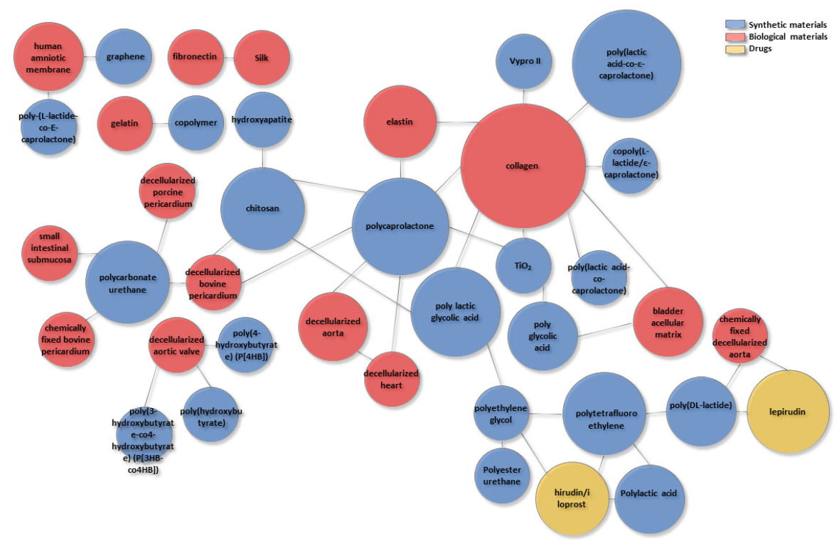
Figure 1. Numbers of articles on hybrid materials published between 2000 and June 2023 (*).
Processes 11 02013 g001
Figure 2. Possible associations of components (synthetic materials in blue, biological materials in red, and drugs in yellow) for the production of hybrid materials. Circles dimensions are proportional to the materials occurrence found in literature and reported in Table 1.
Processes 11 02013 g002
Figure 3. Examples of the hybrid materials obtained by coupling: (A) porcine decellularized pericardium with polycarbonate urethane (Chronoflex AR) for cardiovascular tissue reconstruction; (B) porcine decellularized SIS with polycarbonate urethane (Chronoflex AR-LT) for urological applications.
Processes 11 02013 g003
Figure 4. Representation of the fate of a hybrid material after in vivo implantation: on the left, the schematic composition of the hybrid material fabricated by coupling a decellularized biological tissue with a synthetic polymer: circulating cells are chemo-attracted by the acellular ECM, whose mechanical resistance is sustained by the polymer. On the right, the hybrid material is effectively repopulated with the host’s circulating cells.
Processes 11 02013 g004
Table 1. Summary of the main studies on hybrid materials for different clinical applications.
Biological MaterialsSynthetic MaterialsApplicationsType of StudyReferences
Hydroxyapatite + Chitosan-Orthopedicin vitro[34]
ChitosanPoly lactic glycolic acid-in vitro[35]
Chitosan Polycaprolactone-in vitro[36]
CollagenPolycaprolactone + TiO2Dermatologicalin vitro[37]
CollagenPolycaprolactoneUrologicalin vitro[38]
Collagen and elastinPolycaprolactoneCardiovascularin vitro[39]
Collagen Copoly(L-lactide/ε-caprolactone)Urological in vitro and
in vivo
[40]
CollagenPoly(lactic acid-co-caprolactone)Dermatologicalin vitro[22]
CollagenPoly(lactic acid-co-ε-caprolactone)Urologicalin vitro and
in vivo
[41]
Collagen Vypro IIUrologicalin vitro and
in vivo
[42]
Silk + collagen or fibronectin-Urologicalin vitro[23]
CollagenPoly lactic glycolic acidUrologicalin vitro[25]
-poly glycolic acid + poly lactic glycolic acidUrologicalin vivo[43]
Bladder acellular matrix + collagenPoly glycolic acidUrologicalin vivo[18]
Bladder acellular matrix Poly lactic glycolic acidUrologicalin vitro and
in vivo
[19,24]
-Polyester urethane or poly lactic glycolic acid + Polyethylene glycolUrologicalin vivo[44]
Human amniotic membrane Poly-(L-lactide-co-E-caprolactone)Urologicalin vivo[20]
Human amniotic membraneGrapheneUrologicalin vitro [21]
GelatinCopolymerUrologicalin vitro [45]
Small intestinal submucosaPolycarbonate urethaneUrologicalin vitro [46]
-Polytetrafluoroethylene + Polylactic acidCardiovascularin vitro[47]
-Polytetrafluoroethylene + Poly(DL-lactide)Cardiovascularin vitro and in vivo[48]
-Polytetrafluoroethylene + polylactic acid with Polyethylene glycol-hirudin/iloprostCardiovascularin vitro and
in vivo
[49]
Chemically fixed decellularized porcine aorta Poly(D,L-lactide) with lepirudinCardiovascularin vitro and
in vivo
[50]
Decellularized bovine heart and aortaPolycaprolactoneCardiovascularin vitro[51]
Decellularized porcine aortic valve Poly(hydroxybutyrate)Cardiovascularin vitro and
in vivo
[52]
Decellularized aortic valvePoly(4-hydroxybutyrate) (P[4HB]) or poly(3-hydroxybutyrate-co4-hydroxybutyrate) (P[3HB-co4HB])Cardiovascularin vitro [53]
Decellularized bovine pericardium + ChitosanPolycaprolactone Cardiovascularin vitro[54]
Fixed bovine pericardiumPolycarbonate urethaneCardiovascularin vitro [55]
Decellularized bovine or porcine pericardium Polycarbonate urethaneCardiovascularin vitro [26]
Disclaimer/Publisher’s Note: The statements, opinions and data contained in all publications are solely those of the individual author(s) and contributor(s) and not of MDPI and/or the editor(s). MDPI and/or the editor(s) disclaim responsibility for any injury to people or property resulting from any ideas, methods, instructions or products referred to in the content.

