
Neuroprotective Activities of New Monoterpenoid Indole Alkaloid from Nauclea officinalis
by
 , , , , and
Sook Yee Liew
1,2,*,
Wen Qi Mak
3,
Hin Yee Thew
3,
Kooi Yeong Khaw
3,*,
Hazrina Hazni
2,
Marc Litaudon
4 and
and
Khalijah Awang
2,5 1
Chemistry Division, Centre for Foundation Studies in Science, Universiti Malaya, Kuala Lumpur 50603, Malaysia
2
Centre for Natural Products Research and Drug Discovery (CENAR), Universiti Malaya, Kuala Lumpur 50603, Malaysia
3
School of Pharmacy, Monash University Malaysia, Bandar Sunway 47500, Malaysia
4
Institut de Chimie des Substances Naturelles, CNRS-ICSN UPR2301, University Paris-Saclay, CEDEX, 91198 Gif-sur-Yvette, France
5
Department of Chemistry, Faculty of Science, Universiti Malaya, Kuala Lumpur 50603, Malaysia
*
Authors to whom correspondence should be addressed.
Processes 2023, 11(3), 646; https://doi.org/10.3390/pr11030646
Submission received: 29 December 2022
/
Revised: 15 February 2023
/
Accepted: 16 February 2023
/
Published: 21 February 2023
Abstract
:Phytochemical investigation on the bark of Nauclea officinalis led to the isolation of a new monoterpenoid indole alkaloid, nauclediol. The structure of the compound was identified through extensive spectroscopic analysis. Nauclediol displayed cholinesterase-inhibitory activities towards AChE and BChE with IC50 values of 15.429 and 8.756 µM, respectively. Statistical analysis revealed that the mode of inhibition of nauclediol was non-competitive inhibitor for both AChE and BChE. Molecular docking revealed that nauclediol interacts with the choline-binding site and the catalytic triad of TcAChE and hBChE. This study also demonstrated the neuroprotective potential of nauclediol against amyloid beta-induced cytotoxicity and LPS-induced neuroinflammation activity in a dose-dependent manner.
1. Introduction
Alzheimer’s disease (AD) is the most common type of dementia, and it is estimated to affect 131 million people globally by the year 2050. Over the years, several hypotheses have been proposed to slow down the progression of AD, including by improving type 2 diabetes mellitus [1] and the discovery of next-generation therapeutics for the management of AD. It includes cholinesterase, β-amyloid, tau, and neuroinflammation hypotheses. However, the current drugs available to slow down the progression of AD are cholinesterase inhibitors (Donepezil, Rivastigmine, and Galantamine) and Memantine (NMDA antagonist); they are associated with limitations such as adverse effects, low bioavailability, and inconsistent efficacy [2]. These drugs are based on a one drug-one target hypothesis, whilst the etiology of AD is multifactorial [3]. Therefore, this necessitates the need for novel agents with multi-targeted potential. Natural products are of particular interest due to their ability to reduce oxidative stress and anti-inflammatory activity, and could potentially slow down the progression of AD [4,5]. This further reinforces the possibility of discovering new agents derived from natural sources.
Natural products are also known as secondary phytometabolites. These organic chemicals do not directly affect cell growth and proliferation, but they enhance survival mechanisms in living organisms [6]. Investigations into molecular mechanisms and docking analysis have shed light on the molecular activities of these secondary phytometabolites [7]. Most secondary phytometabolites are known to interfere with inflammatory mediators such as cytokines and peptides and to inhibit macrophage activity [8]; indole alkaloids, topsentins, and dragmacidins [9] are some good examples. Nauclea officinalis (Pierre ex Pitard) Merr. and Chun, a traditional Chinese medicine from the Rubiaceae family, is widely used to treat exogenous fever, pink eye, pneumonia and acute jaundice in China. It has anti-inflammatory and anti-malarial properties [10,11,12]. In addition, Nauclea officinalis has been known to contain many indole alkaloids with biological activities. For example, 17-oxo-19-(Z)-naucline and naucleoffieine H displayed anti-inflammatory effects by decreasing the LPS-stimulated production of nitric oxide in RAW264.7 cells [13,14]. Thus, in the continuous search for bioactive indole alkaloids from the Rubiaceae family [15,16,17], a new monoterpenoid indole alkaloid, nauclediol, was isolated from Nauclea officinalis. In the present study, the isolation and structural elucidation of nauclediol is reported together with the neuroprotective potential of the compound.
2. Materials and Methods
2.1. General Procedures
The general procedures were the same as previously described [18].
2.2. Plant Materials
The barks of Nauclea officinalis from Hutan Simpan Mersing, Johor were collected by a phytochemical group of the Department of Chemistry, Faculty of Science, Universiti Malaya. Botanical identification was conducted by the botanist, Mr. Teo Leong Eng. The voucher specimen (KL 4745) of the plant was deposited at the Herbarium of the Department of Chemistry, Faculty of Science, Universiti Malaya, Kuala Lumpur, Malaysia.
2.3. Extraction, Isolation and Purification of Compound
The extraction procedure for 1.5 kg of dried and grounded bark of Nauclea officinalis was the same as previously described [18,19]. An amount of 6.2 g of CH2Cl2 extract was obtained. The CH2Cl2 extract was then subjected to column chromatography over silica gel using CH2Cl2 and MeOH solvent (100:0, 99:1, 98:2, 97:3, 96:4, 95:5, 94:6, 90:10, 83:17, and 75:25 v/v), and 20 fractions were finally obtained. Further purification from fraction 6 by PTLC yielded nauclediol (8.2 mg, CH2Cl2:MeOH; 97:3 v/v saturated with NH4OH). The structure of nauclediol was elucidated using various spectroscopic methods, such as NMR, UV, IR, and LCMS-IT-TOF.
2.4. Cholinesterase Enzyme Inhibitory Activity
Nauclediol was evaluated for its enzyme inhibition potential against acetylcholinesterase from Electrophorus electricus (EeAChE) and butyrylcholinesterase from Equine serum (eBChE). Cholinesterase inhibition was conducted using Ellman’s calorimetry method as described previously [20]. In a 96-well plate, buffer solution, DTNB (Ellman’s reagent), ACh/BCh, and nauclediol were added in ascending concentrations across the row. Then, 1% DMSO was added as the control for this experiment. Lastly, the cholinesterase enzyme was added to the assigned enzymatic columns. The absorbance of the plate was then measured using a spectrophotometer at a wavelength of 412 nm for 30 min after the addition of enzymes. Each test was conducted in triplicate to ensure consistency of the results.
The enzyme kinetic mechanism of nauclediol was assessed with compound concentrations from 1.56 to 25 uM. The enzyme and DTNB concentrations were in accordance with the method described above. The statistical analysis was performed using GraphPad Prism v8.0e (Dotmatics, Boston, United Kingdom) and Excel [21].
2.5. Molecular Docking
Molecular docking was carried out following the method described by Abdul Wahab et al. [22]. Briefly, molecular docking of nauclediol was performed using Autodock 3.0.5 (The Scripps Research Institute, La Jolla, CA, USA) along with AutoDockTools (ADT) [23]. Hyperchem 8 was used to build the 3D-crystal structure of the nauclediol, and the structure was subjected to energy minimization with a convergence criterion of 0.05 kcal/(molA). The three-dimensional crystal structures of AChE from Torpedo californica (TcAChE) (PDB ID: 1W6R) [24] and BChE from Homo sapiens (hBChE) (PDB ID: 2WIJ) [25] were retrieved from the Protein Data Bank. The proteins were edited using ADT to add hydrogen atoms and remove all water molecules. Non-polar hydrogens and lone pairs were then merged, and each atom was assigned Gasteiger partial charges. A grid box was generated at the center of the active site gorge with 60 × 60 × 60 points and spacing of 0.375 Ǻ. In total, 100 independent dockings were carried out for each docking experiment, with a population size of 150 and 2,500,000 energy evaluations. The best conformation with the lowest docked energy in the most populated cluster was selected. Analysis and visualization of the conformations from the docking experiments were conducted using Acceryls Discovery Studio 2.5 (Accelrys Inc., San Diego, CA, USA).
2.6. Neuroprotective Potential of Nauclediol on Aβ-(1-42)-Induced Toxicity on SH-SY5Y Cells
2.6.1. Cell Culture and Cell Viability Assay
Human neuroblastoma cells (SH-SY5Y) were cultured in a complete culture medium containing DMEM nutrient mixture F-12 (1:1) supplemented with 10% fetal calf serum, glutamine, and 1% antibiotics (penicillin–streptomycin), and kept at 37 °C under 5% CO2.
A cell viability assay was performed to determine the non-cytotoxic concentration of nauclediol in SH-SY5Y cells. Cells were seeded with a cell density of 25,000 cells/well in 96-well plates for 24 h. The cells were then treated with nauclediol at concentrations of 1, 2, 5, and 10 μM for 24 h. An MTT assay was used to assess cell viability after drug treatment. In brief, 20 μL of the MTT solution (5 mg/mL) was added to each well and incubated at 37 °C for 4 h. Then, 100 μL of DMSO was added to lyse the cells after the removal of the supernatant from each well. The amount of formazan generated was quantified using a microplate reader (SpectraMax ID3, Pennsylvania, USA) at an absorbance of 595 nm. All experiments were carried out 3 times.
2.6.2. Neuroprotective Assay against Beta Amyloid-Induced Toxicity
Aβ-(1-42) oligomers were prepared according to the protocol of Huang et al. [26], with some modifications. Briefly, lyophilized Aβ-(1-42) powder was initially dissolved to 1.0 mM in hexafluoro-2-isopropanol (HFIP) and incubated at room temperature for 30 min. The solution was ensured to be clear and colorless. The HFIP was allowed to evaporate from the open tubes overnight into the fume hood. A thin, clear film was formed at the bottom of the tube. The film was initially dissolved in DMSO and subsequently adjusted to 13 μM in DMEM/Hams F-12 media containing 1% Pen/strep and 1% FBS. The solution was incubated at 37 °C under 5% CO2.
The MTT assay was used to determine the protection of nauclediol toward SH-SY5Y cells against Aβ-(1-42) aggregate toxicity. In a 96-well plate, 25,000 cells/well were seeded and incubated overnight. Cells were treated with nauclediol at concentrations of 0.1, 1, 5, and 10 μM for 24 h. Aβ-(1-42) aggregates were added to the wells and incubated for another 24 h; then, 20 μL of the MTT solution (5 mg/mL) was added to each well and incubated at 37 °C for 4 h. Finally, 100 μL of DMSO was added to lyse the cells after the removal of the supernatant in each well. The amount of formazan generated was quantified using a microplate reader (SpectraMax ID3, Pennsylvania, USA) at an absorbance of 595 nm. All experiments were carried out in triplicate.
2.7. Anti-Neuroinflammatory Effect of Nauclediol on LPS-Induced Neuro-Inflammation in BV2 Cells
2.7.1. Cell Culture and Cell Viability Assay
C57/BL6 microglial cells (BV2) were cultured in a complete culture medium containing DMEM supplemented with 10% fetal calf serum, glutamine, and 1% antibiotics (penicillin–streptomycin), and kept at 37 °C under 5% CO2.
A cell viability assay was performed to determine the non-cytotoxic concentration of nauclediol in BV2 cells. Cells were seeded with a cell density of 20,000 cells/well in 96-well plates for 24 h. The cells were then treated with nauclediol at concentrations of 0.625, 1.25, 2.5, 5, and 10 μM for 24 h. An MTT assay was used to assess cell viability after drug treatment. In brief, 20 μL of the MTT solution (5 mg/mL) was added to each well and incubated at 37 °C for 4 h; then, 100 μL of DMSO was added to lyse the cells after the removal of the supernatant from each well. The amount of formazan generated was quantified using a microplate reader (SpectraMax ID3, Pennsylvania, USA) at an absorbance of 595 nm. All experiments were carried out in triplicate.
2.7.2. Neuroprotective Assay against LPS-Induced Inflammation
In a 96-well plate, 20,000 cells/well were seeded and incubated overnight. Cells were treated with nauclediol at concentrations of 0.0625, 0.125, 0.25, and 0.5 μM for 24 h. LPS (1 ug/mL) were added to the wells and incubated for another 24 h. A second 96-well plate was used for the measurement of nitride oxide. Sulfanilamide and NED solutions were equilibrated to room temperature for 30 min before use. A 50 μL sample of the solution was transferred from the first plate to the second plate. Then, 50 μL of sulfanilamide solution was added to all wells in the second plate and incubated for 5 min. Subsequently, 50 μL of NED solution was added and incubated for another 5 min. The plate was then read at an absorbance of 540 nm using a microplate reader (SpectraMax ID3, Pennsylvania, USA). All experiments were carried out in triplicate. The absorbance was converted into a percentage of nitrite to determine the anti-inflammatory effects of nauclediol.
2.7.3. Statistical Analysis
For this cell-based study, the results were analyzed using R programming. Data are expressed as mean ± standard deviation. Differences between groups are analyzed using one-way ANOVA followed by Student’s t-test for MTT assay and nitrite production. A p-value less than 0.05 was considered statistically significant.
3. Results and Discussion
3.1. Structural Elucidation of Nauclediol
Nauclediol (Figure 1) was isolated as a brownish amorphous solid. The LCMS-IT-TOF spectrum showed a pseudomolecular ion peak [M+Na]+ at m/z 359.1275 (calc. 359.1372) corresponding to the molecular formula of C20H20N2O3. The UV spectrum of nauclediol demonstrated strong absorptions at 374, 308, 277, 256, and 220 nm. In the IR spectrum, an absorption band at 1643 cm−1 was observed, indicative of a corynanthe type of indole alkaloid with an N4-C=O amide group [27,28]. In addition, a broad hydroxyl absorption band appeared at 3401 cm−1, suggesting the presence of a hydrogen-bonded OH group [28].
In the 1H-NMR spectrum (Table 1), the presence of four aromatic proton signals at the downfield region and a -CH2-CH2-N- group were observed, indicating the existence of a tetrahydro-β-carboline skeleton [10,28]. In ring A, 2 of the 4 aromatic protons appeared as doublets at δH 7.53 and 7.37, corresponding to H-9 (1H, d, J = 7.8 Hz) and H-12 (1H, d, J = 7.8 Hz), respectively. The other two protons, H-10 (δH 7.04, 1H, t, J = 7.8 Hz) and H-11 (δH 7.19, 1H, t, J = 7.8 Hz), appeared as triplets. Furthermore, the COSY spectrum also showed the correlations between the four aromatic protons; H-9 with H-10, H-10 with H-11, and H-11 with H-12 (Figure 2). In addition, 2 sets of triplet signals, which appeared at δH 4.33 (2H, t, J = 6.9 Hz) and δH 3.06 (2H, t, J = 6.9 Hz), were attributable to methylene protons H2-5 and H2-6, respectively. H-14, in ring D, appeared as a singlet at δH 7.00, indicating the presence of neighboring quaternary carbons C-3 (δC 135.6) and C-15 (δC 141.2). Furthermore, the presence of a downfield quartet at δH 6.46 (1H, q, J = 6.9 Hz, H-17), coupled with an upfield doublet at δH 1.86 (3H, d, J = 6.9 Hz, H3-18), implied the presence of a trisubstituted olefin group. Two downfield singlets were observed at δH 4.45 (2H, s, H2-19) and 4.55 (2H, s, H2-21) which correlated with the carbon signals at δC 64.1 (C-19) and δC 64.3 (C-21), respectively, in the HSQC spectrum, suggesting the existence of two hydroxyl groups (a diol).
As indicated in the 13C-NMR and DEPT-135 spectra, nauclediol has a total of twenty carbon signals comprising one methyl, one carbonyl, four methylenes, six methines, and eight quaternary carbons. The signal of a carbonyl carbon was observed at δC 160.9 (C-22). The HMBC spectrum showed a correlation of H2-19 and H2-21 with C-20 (δC 129.2), indicating the connectivity of the two methylenes with quaternary carbon C-20, as shown in Figure 2. In addition, HMBC correlations between H3-18 and C-16 (δC 120.0), H-19 and C-15, and H-21 and C-15 were observed, thus supporting the connectivity of the trisubstituted olefin group and propane-1,3-diol group to ring D through C-16 and C-15, respectively. The COSY spectrum revealed correlation peaks between H2-5 and H2-6 and H3-18 and H-17, indicating that the respective protons were coupled with each other (Figure 2).
Based on the aforementioned and extensive analysis of all spectral data, the structure of nauclediol was established identified as a new corynanthe type of monoterpenoid indole alkaloid. The biogenetic precursor of nauclediol is strictosidine. Strictosidine may undergo cleavage of glycoside to strictosidine aglycone, followed by cleavage of ring D, to form dialdehyde intermediate. Subsequently, a series of rearrangement could lead to the formation of 21-oxogeissoschizine, followed by dehydration, to form nauclediol, as shown in Figure 3.
3.2. Cholinesterase Inhibition of Nauclediol
Cholinesterase inhibitors (ChEIs) are the first and most commonly recognized treatment strategy for AD. They work by reducing the metabolism of cholinergic neurotransmitters and acetylcholine released from the cholinergic neurons, thus increasing the synaptic levels of ACh and BCh [29]. In this study, nauclediol was tested for its inhibitory potential against AChE and BChE, respectively. Nauclediol showed inhibition on cholinesterase enzymes with IC50 values of 15.429 and 8.756 µM, which was two-fold more selective toward BChE (Figure 4). Furthermore, nauclediol was three times more potent as an inhibitor of BChE compared to galanthamine (standard or drug), with a reported IC50 value of 28.29 µM [19].
The mode of inhibition was determined by means of Michaelis–Menten kinetic. An analysis using Graphpad revealed that the mechanism of action of naucleodiol against AChE and BChE was non-competitive based on the greatest goodness of fit (Figure 5). This result was further supported and strengthened by statistical analysis using Excel, which showed that naucleodiol was best suited to be a noncompetitive inhibitor based on the lowest sum of squared residuals (RSS), relative standard error (RSE) for regression, and the Bayesian information criterion (BIC) (Supplementary Materials). The Ki values of nauclediol were 6.9 and 35.41 µM against AChE and BChE, respectively (Supplementary Materials), indicating a stronger binding affinity of naucleodiol towards AChE. This is in opposition to the IC50 values, as nauclediol possessed better IC50 values for BChE than for AChE.
By comparing with the cholinesterase-inhibitory activities of other indole alkaloids from N. officinalis [19], a brief structure–activity relationship study can be conducted. Angustidine, angustine, angustoline, and naucline are also corynanthe types of indole alkaloids [19], with the same precursors as nauclediol. According to the proposed biogenesis pathway, the difference between nauclediol and the four known indole alkaloids begins to appear after the formation of strictosidine. Rotation at C-14 of strictosidine is needed to form strictosidine lactam, and is followed by the formation of the four known indole alkaloids. On the other hand, formation of nauclediol does not require the rotation at C-14 of strictosidine, but involves a ring opening in strictosidine aglycone. Thus, the four known indole alkaloids possess ring E (pyridine or pyran ring), but nauclediol does not. Based on the previously reported data, angustidine, angustine, angustoline, and naucline exhibited moderate to weak AChE inhibition, with angustidine having the lowest IC50 value (21.72 µM) [19]. In comparison with the four known indole alkaloids, nauclediol possesses a better inhibitory effect towards AChE. It was suggested that the potency of nauclediol in AChE inhibition may be due to the presence of a ring-opened system in ring E and the two hydroxyl groups. For BChE, angustidine and angustine (IC50 = 1.03 and 4.98, respectively) [19] showed better inhibitory effects compared to nauclediol. However, nauclediol was shown to be 2.5 and 4 times more potent than angustoline and naucline, respectively, as angustoline has only one hydroxyl group in its structure, while naucline has a pyran ring instead of a pyridine ring (ring E). Based on structure–relationship studies, its potency in inhibiting BChE may be increased by the presence of a pyridine ring and two hydroxyl groups.
3.3. Molecular Docking of Nauclediol
Since nauclediol is potent in inhibiting both enzymes, molecular docking analysis was carried out in order to understand its binding interaction with the enzymes. The results revealed that nauclediol docked well in the catalytic triad and choline binding site of TcAChE (Table 2). The hydroxyl group of C-19 formed a hydrogen bond with His 440 at a distance of 2.60 Å in the catalytic triad (Table 2). The catalytic triad serves as the active site of the enzyme, where acetylcholine, the substrate, is hydrolyzed into choline and acetate [22]. In addition, hydrogen bonds and hydrophobic interactions were observed between the compound and the amino acid residues in the choline binding site, which anchored this compound to the TcAChE active site gorge (Figure 6). An oxygen atom of the carbonyl group C-22 interacted with Tyr 130 through a hydrogen bond at a distance of 2.26 Å, while rings B and D of the compound formed a π–π stacking interaction with Trp 84. For hBChE, nauclediol also bound with amino acid residues of the catalytic triad and choline binding site with a hydrophobic interaction (Table 2). A π–π stacking interaction was observed between the indole ring of nauclediol and His 438 from the active site of hBChE, as shown in Figure 7. In addition, a π–σ interaction was observed between Trp 82 and C-17 of nauclediol.
3.4. Nauclediol Reduces Aβ-(1-42)-Induced Toxicity on SH-SY5Y Cells
As illustrated in Figure 8A, nauclediol had no cytotoxic effect on SH-SY5Y cells from 1 to 10 μM. Further, the cells were pretreated with 1 to 10 μM of nauclediol followed by Aβ-(1-42) aggregates. Figure 8B shows that 13 μM Aβ-(1-42) aggregates significantly reduced cell viability to 54.5 ± 6.52%, compared to the untreated cells. The cell viability of neuroblastoma SH-SY5Y cells was increased by 33.28 to 64.87% at the tested concentrations (0.1 to 10 μM) compared to cells treated with Aβ-(1-42) aggregates alone. Overall, our study showed that nauclediol protected neuroblastoma SH-SY5Y cells against Aβ-(1-42)-induced cytotoxicity.
3.5. Nauclediol Reduces LPS-Induced Neuroinflammation in BV2 Cells
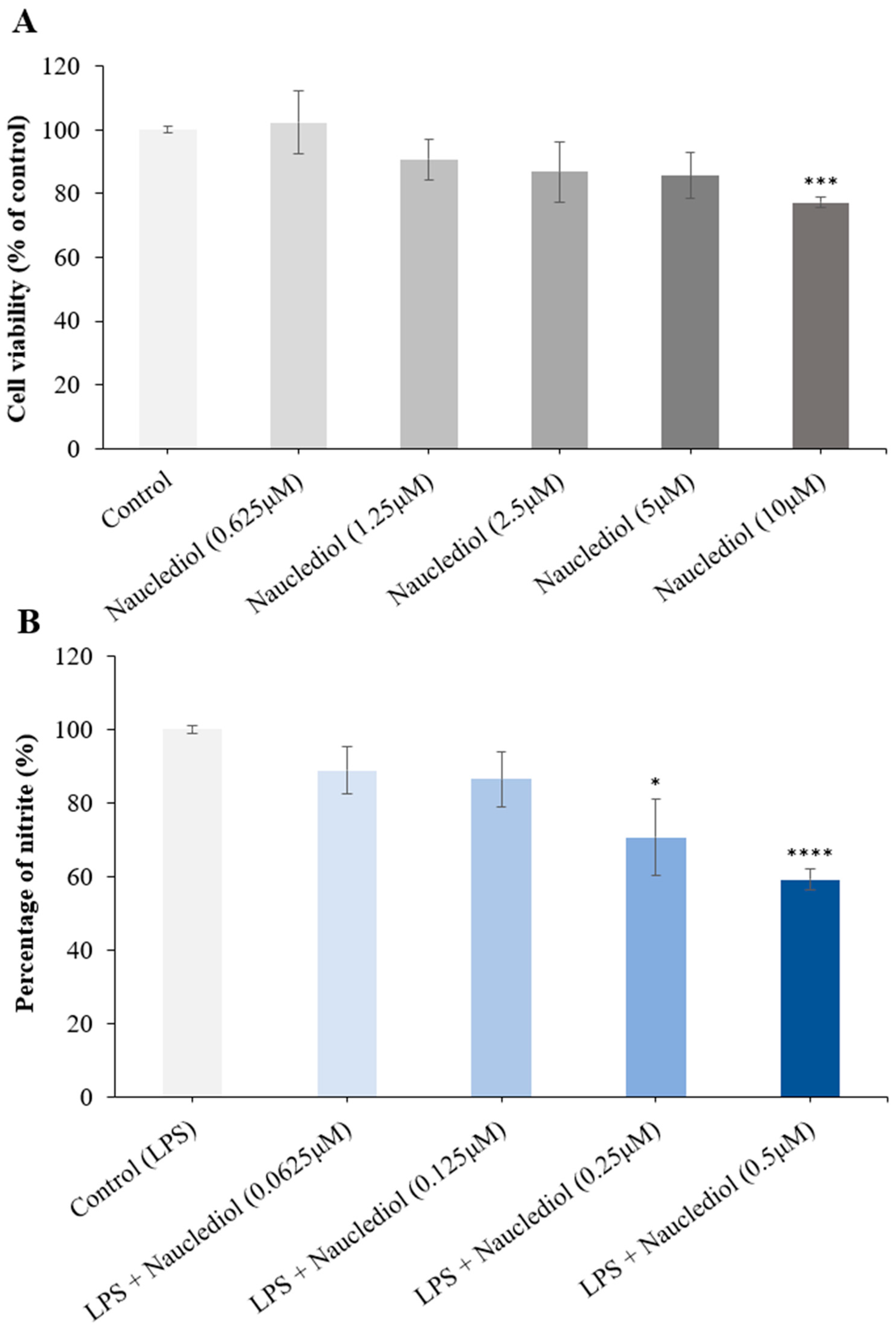
Nauclediol was tested for its cytotoxic effect on BV2 cells at 1.25 to 10 μM. Figure 9 shows the BV2 cell viabilities upon exposure to nauclediol. The treated doses, at 1.25 to 10 μM, were cytotoxic towards BV2 cells from 9.28 to 22.7% when compared to untreated cells. Non-toxic doses, at 0.0625 to 0.5 μM, were used for further experiments. Nauclediol demonstrated a statistically significant reduction in nitrite at doses of 0.25 to 0.5 μM.
In this test, we sought to evaluate whether nauclediol could reduce the elevated levels of nitric oxide production by LPS-induced inflammation in BV2 cells. Several studies have shown the relationship between nitric oxide and the pathogenesis of AD, with effects such as neuronal death and S-nitrosylation of cellular components [30]. In addition, in post mortem brain tissues from AD patients, Aβ plaques are strongly associated with reactive microglia and nitric oxide production from activated microglia [31].
4. Conclusions
In conclusion, the present study has yielded a new monoterpenoid indole alkaloid, nauclediol, from Nauclea officinalis. It is a dual cholinesterase inhibitor and is two-fold more selective against the butyrylcholinesterase enzyme. Upon a molar-based comparison with galantamine, a drug or medication used in the treatment of Alzheimer disease, nauclediol was three times more potent in inhibiting BChE. In addition, Michaelis–Menten kinetics showed that nauclediol is a non-competitive inhibitor with a higher affinity towards AChE. In addition, nauclediol significantly protected cells against beta amyloid-induced toxicity and neuroinflammation at submicromolar levels. Overall, the findings in this study are worth further investigation for nauclediol’s potential in assays related to other neurodegenerative diseases.
Supplementary Materials
The following supporting information can be downloaded at: https://www.mdpi.com/article/10.3390/pr11030646/s1, Table S1: statistical analysis of kinetic mechanism of nauclediol against AChE, Figure S1: (a) Correlation plot and (b) line-weaver burk plot of nauclediol against AChE, Table S2: statistical analysis of kinetic mechanism of nauclediol against BChE, Figure S2: (a) Correlation plot and (b) line-weaver burk plot of nauclediol against AChE, Figure S3: Statistical analysis of kinetic mechanism of nauclediol against AChE by GraphPad prism, Figure S4: Statistical analysis of kinetic mechanism of nauclediol against BChE by GraphPad prism.
Author Contributions
Conceptualization, S.Y.L., K.Y.K. and K.A.; methodology, S.Y.L., K.Y.K., H.H., M.L. and K.A.; investigation, S.Y.L. and K.Y.K.; data analysis, S.Y.L., W.Q.M., H.Y.T., K.Y.K., H.H., M.L. and K.A.; writing—original draft preparation, S.Y.L., W.Q.M., H.Y.T. and K.Y.K.; writing—review and editing, S.Y.L., W.Q.M., H.Y.T., K.Y.K., H.H., M.L. and K.A.; project administration, S.Y.L., K.Y.K., M.L. and K.A. All authors have read and agreed to the published version of the manuscript.
Funding
This work was supported by Universiti Malaya Research Grant (RK011-2020).
Institutional Review Board Statement
Not applicable.
Informed Consent Statement
Not applicable.
Data Availability Statement
Not applicable.
Acknowledgments
The authors sincerely thank D. M. Nor and Rafly bin Syamsir (Universiti Malaya) for the collection of plant material and Teo Leong Eng for the botanical identification. The authors would also like to thank the International French Malaysia Natural Product Laboratory (IFM-NatProLab) between CNRS-ICSN, the French Embassy in Malaysia, and Universiti Malaya for the collaborative work.
Conflicts of Interest
The authors declare no conflict of interest.
References
- Wee, S.A.; Nhu, D.T.; Khaw, Y.K.; Tang, S.K.; Yeong, Y.K. Linking Diabetes to Alzheimer’s Disease: Potential Roles of Glucose Metabolism and Alpha-Glucosidase. Curr. Neuropharmacol. 2023. ahead of print. [Google Scholar] [CrossRef]
- Mukherjee, P.K.; Kumar, V.; Mal, M.; Houghton, P.J. Acetylcholinesterase inhibitors from plants. Phytomedicine 2007, 14, 289–300. [Google Scholar] [CrossRef] [PubMed]
- Iqubal, A.; Rahman, S.O.; Ahmed, M.; Bansal, P.; Haider, M.R.; Iqubal, M.K.; Najmi, A.K.; Pottoo, F.H.; Haque, S.E. Current Quest in Natural Bioactive Compounds for Alzheimer’s Disease: Multi-Targeted-Designed-Ligand Based Approach with Preclinical and Clinical Based Evidence. Curr. Drug Targets 2021, 22, 685–720. [Google Scholar] [CrossRef] [PubMed]
- Kong, Y.R.; Tay, K.C.; Su, Y.X.; Wong, C.K.; Tan, W.N.; Khaw, K.Y. Potential of Naturally Derived Alkaloids as Multi-Targeted Therapeutic Agents for Neurodegenerative Diseases. Molecules 2021, 26, 728. [Google Scholar] [CrossRef]
- Kong, Y.R.; Jong, Y.X.; Balakrishnan, M.; Bok, Z.K.; Weng, J.K.K.; Tay, K.C.; Goh, B.H.; Ong, Y.S.; Chan, K.G.; Lee, L.H.; et al. Beneficial Role of Carica papaya Extracts and Phytochemicals on Oxidative Stress and Related Diseases: A Mini Review. Biology 2021, 10, 287. [Google Scholar] [CrossRef]
- Williams, D.H.; Stone, M.J.; Hauck, P.R.; Rahman, S.K. Why Are Secondary Metabolites (Natural Products) Biosynthesized? J. Nat. Prod. 1989, 52, 1189–1208. [Google Scholar] [CrossRef]
- Croteau, R.; Kutchan, T.M.; Lewis, N.G. Natural Products (Secondary Metabolites); American Society of Plant Physiologists: Rockville, MD, USA, 2000; Volume 24, pp. 1250–1318. [Google Scholar]
- Calixto, J.B.; Otuki, M.F.; Santos, A.R.S. Anti-Inflammatory Compounds of Plant Origin. Part I. Action on Arachidonic Acid Pathway, Nitric Oxide and Nuclear Factor κ B (NF-κB). Planta Med. 2003, 69, 973–983. [Google Scholar] [CrossRef]
- Mcconnell, O.J.; Saucy, G.; Jacobs, R. Use for Topsentin Compounds and Pharmaceutical Compositions Containing Same. U.S. Patent US5290777A, March 1994. [Google Scholar]
- Sun, J.Y.; Lou, H.X.; Dai, S.J.; Xu, H.; Zhao, F.; Liu, K. Indole alkoloids from Nauclea officinalis with weak antimalarial activity. Phytochemistry 2008, 69, 1405–1410. [Google Scholar] [CrossRef]
- Sun, J.Y.; Lou, H.X.; Xu, H.; Dai, S.J.; Liu, K. Two new indole alkaloids from Nauclea officinalis. Chin. Chem. Lett. 2007, 18, 1084–1086. [Google Scholar] [CrossRef]
- Su, K.; Gong, M.; Zhou, J.; Deng, S. Study of Chemical Composition of Nauclea Officinalis Leaves. Int. J. Chem. 2009, 1, 77–81. [Google Scholar] [CrossRef] [Green Version]
- Song, L.-L.; Mu, Y.-L.; Zhang, H.-C.; Wu, G.-Y.; Sun, J.-Y. A new indole alkaloid with anti-inflammatory from the branches of Nauclea officinalis. Nat. Prod. Res. 2018, 34, 2283–2288. [Google Scholar] [CrossRef] [PubMed]
- Song, S.; Liu, P.; Wang, L.; Li, D.; Fan, H.; Chen, D.; Zhao, F. In vitro anti-inflammatory activities of naucleoffieine H as a natural alkaloid from Nauclea officinalis Pierrc ex Pitard, through inhibition of the iNOS pathway in LPS-activated RAW 264.7 macrophages. Nat. Prod. Res. 2020, 34, 2694–2697. [Google Scholar] [CrossRef] [PubMed]
- Liew, S.Y.; Looi, C.Y.; Paydar, M.; Cheah, F.K.; Leong, K.H.; Wong, W.F.; Mustafa, M.R.; Litaudon, M.; Awang, K. Subditine, a New Monoterpenoid Indole Alkaloid from Bark of Nauclea subdita (Korth.) Steud. Induces Apoptosis in Human Prostate Cancer Cells. PLoS ONE 2014, 9, e87286. [Google Scholar] [CrossRef]
- Qureshi, A.K.; Mukhtar, M.R.; Hirasawa, Y.; Hosoya, T.; Nugroho, A.E.; Morita, H.; Shirota, O.; Mohamad, K.; Hadi, A.H.A.; Litaudon, M.; et al. Neolamarckines A and B, New Indole Alkaloids from Neolamarckia Cadamba. Chem. Pharm. Bull. 2011, 59, 291–293. [Google Scholar] [CrossRef] [Green Version]
- Mukhtar, M.R.; Osman, N.; Awang, K.; Hazni, H.; Qureshi, A.K.; Hadi, A.H.A.; Zaima, K.; Morita, H.; Litaudon, M. Neonaucline, a New Indole Alkaloid from the Leaves of Ochreinauclea maingayii (Hook. f.) Ridsd. (Rubiaceae). Molecules 2011, 17, 267–274. [Google Scholar] [CrossRef] [PubMed]
- Liew, S.Y.; Mukhtar, M.R.; Hadi, A.H.A.; Awang, K.; Mustafa, M.R.; Zaima, K.; Morita, H.; Litaudon, M. Naucline, a New Indole Alkaloid from the Bark of Nauclea officinalis. Molecules 2012, 17, 4028–4036. [Google Scholar] [CrossRef] [Green Version]
- Liew, S.Y.; Khaw, K.Y.; Murugaiyah, V.; Looi, C.Y.; Wong, Y.L.; Mustafa, M.R.; Litaudon, M.; Awang, K. Natural indole butyrylcholinesterase inhibitors from Nauclea officinalis. Phytomedicine 2015, 22, 45–48. [Google Scholar] [CrossRef]
- Khaw, K.Y.; Chong, C.W.; Murugaiyah, V. LC-QTOF-MS analysis of xanthone content in different parts of Garcinia mangostana and its influence on cholinesterase inhibition. J. Enzyme Inhib. Med. Chem. 2020, 35, 1433–1441. [Google Scholar] [CrossRef]
- Walsh, R. Comparing enzyme activity modifier equations through the development of global data fitting templates in Excel. PeerJ 2018, 6, e6082. [Google Scholar] [CrossRef]
- Abdul Wahab, S.M.; Sivasothy, Y.; Liew, S.Y.; Litaudon, M.; Mohamad, J.; Awang, K. Natural cholinesterase inhibitors from Myristica cinnamomea King. Bioorganic Med. Chem. Lett. 2016, 26, 3785–3792. [Google Scholar] [CrossRef]
- Morris, G.M.; Goodsell, D.S.; Halliday, R.S.; Huey, R.; Hart, W.E.; Belew, R.K.; Olson, A.J. Automated docking using a Lamarckian genetic algorithm and an empirical binding free energy function. J. Comput. Chem. 1998, 19, 1639–1662. [Google Scholar] [CrossRef]
- Greenblatt, H.M.; Guillou, C.; Guénard, D.; Argaman, A.; Botti, S.; Badet, B.; Thal, C.; Silman, I.; Sussman, J.L. The Complex of a Bivalent Derivative of Galanthamine with Torpedo Acetylcholinesterase Displays Drastic Deformation of the Active-Site Gorge: Implications for Structure-Based Drug Design. J. Am. Chem. Soc. 2004, 126, 15405–15411. [Google Scholar] [CrossRef] [PubMed]
- Carletti, E.; Aurbek, N.; Gillon, E.; Loiodice, M.; Nicolet, Y.; Fontecilla-Camps, J.-C.; Masson, P.; Thiermann, H.; Nachon, F.; Worek, F. Structure–activity analysis of aging and reactivation of human butyrylcholinesterase inhibited by analogues of tabun. Biochem. J. 2009, 421, 97–106. [Google Scholar] [CrossRef] [Green Version]
- Chunhui, H.; Dilin, X.; Ke, Z.; Jieyi, S.; Sicheng, Y.; Dapeng, W.; Qinwen, W.; Wei, C. A11-positive β-amyloid Oligomer Preparation and Assessment Using Dot Blotting Analysis. J. Vis. Exp. 2018, 135, e57592. [Google Scholar] [CrossRef] [Green Version]
- Au, T.Y.; Cheung, H.T.; Sternhell, S. New corynanthé alkaloids from Strychnos angustiflora. J. Chem. Soc. Perkin Trans. 1 1973, 0, 13–16. [Google Scholar] [CrossRef]
- Shigemori, H.; Kagata, T.; Ishiyama, H.; Morah, F.; Ohsaki, A.; Kobayashi, J.i. Naucleamides A-E, New Monoterpene Indole Alkaloids from Nauclea latifolia. Chem. Pharm. Bull. 2003, 51, 58–61. [Google Scholar] [CrossRef] [Green Version]
- Cavalli, A.; Bolognesi, M.L.; Minarini, A.; Rosini, M.; Tumiatti, V.; Recanatini, M.; Melchiorre, C. Multi-target-directed ligands to combat neurodegenerative diseases. J. Med. Chem. 2008, 51, 347–372. [Google Scholar] [CrossRef]
- Doherty, G.H. Nitric oxide in neurodegeneration: Potential benefits of non-steroidal anti-inflammatories. Neurosci. Bull. 2011, 27, 366–382. [Google Scholar] [CrossRef]
- Sparrow, J.R. Inducible nitric oxide synthase in the central nervous system. J. Mol. Neurosci. 1994, 5, 219–229. [Google Scholar] [CrossRef]
Figure 1.
Structure of nauclediol.
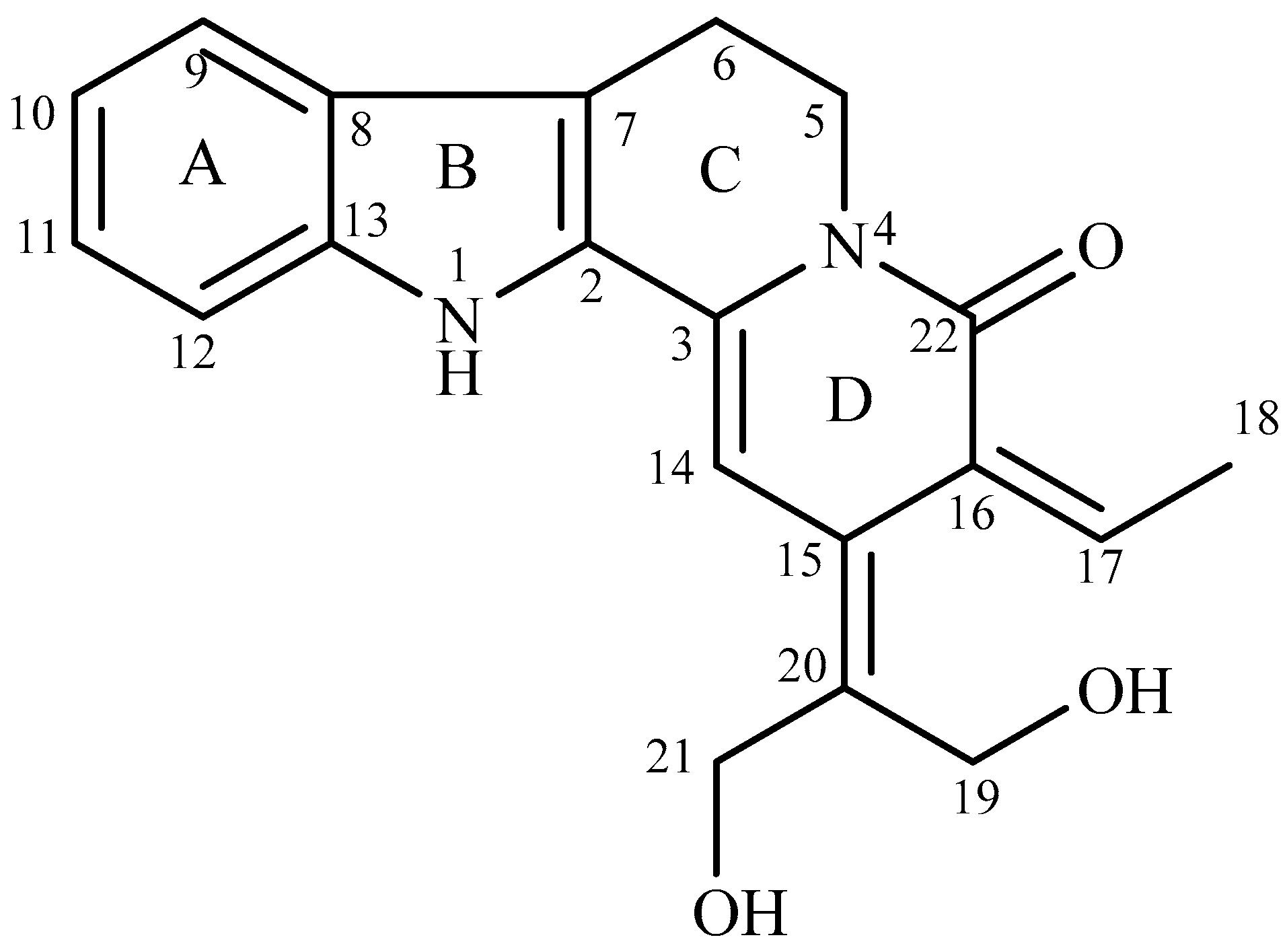
Figure 2.
1H-1H COSY and selected 1H-13C HMBC correlations of nauclediol.
Figure 3.
A proposed biogenesis process of nauclediol.
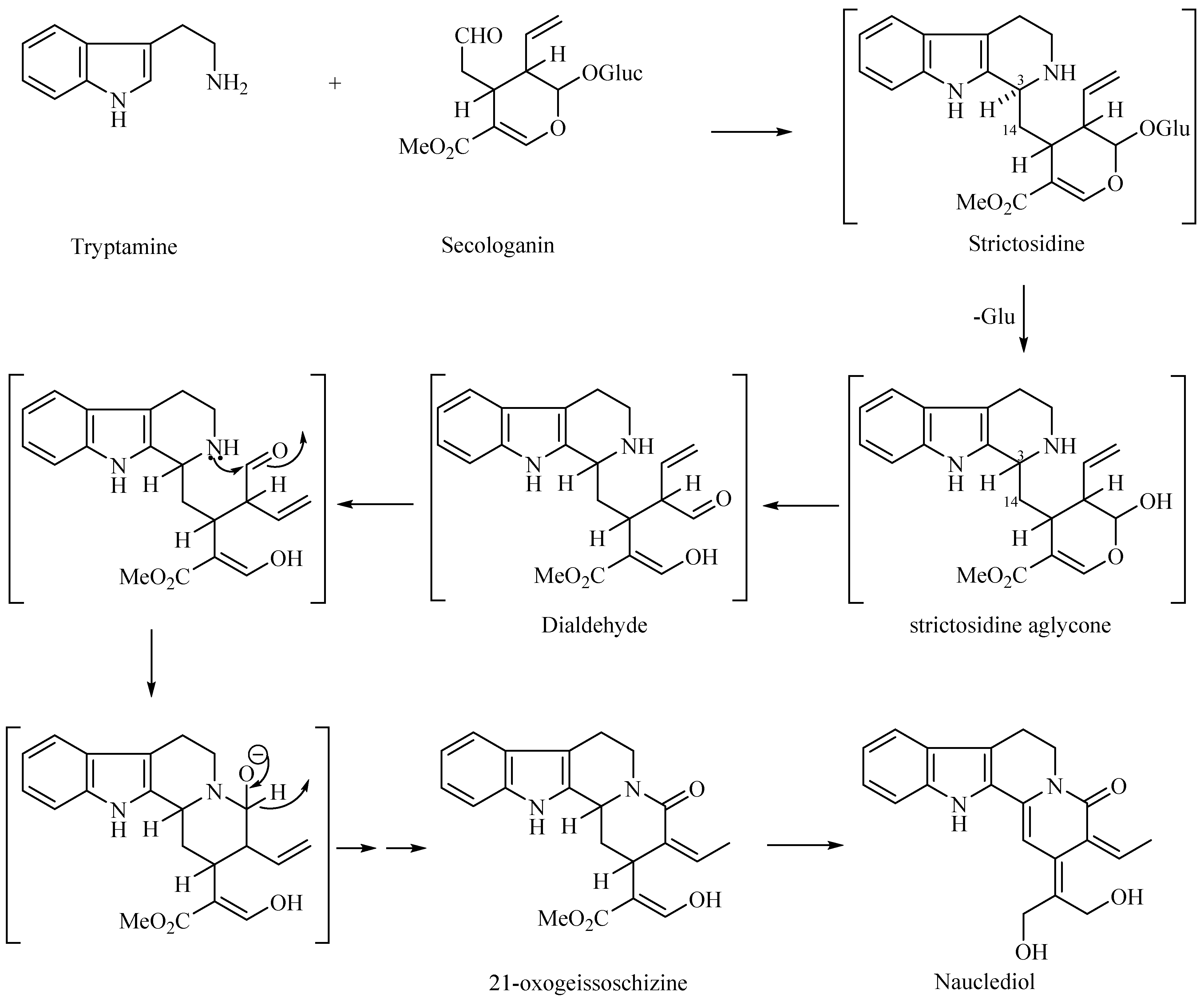
Figure 4.
Cholinesterase inhibition of nauclediol against (a) acetylcholinesterase and (b) butrylcholinesterase enzymes at concentrations from 3.12 to 100 µM. The results were repeated in triplicates (n = 3).
Figure 4.
Cholinesterase inhibition of nauclediol against (a) acetylcholinesterase and (b) butrylcholinesterase enzymes at concentrations from 3.12 to 100 µM. The results were repeated in triplicates (n = 3).
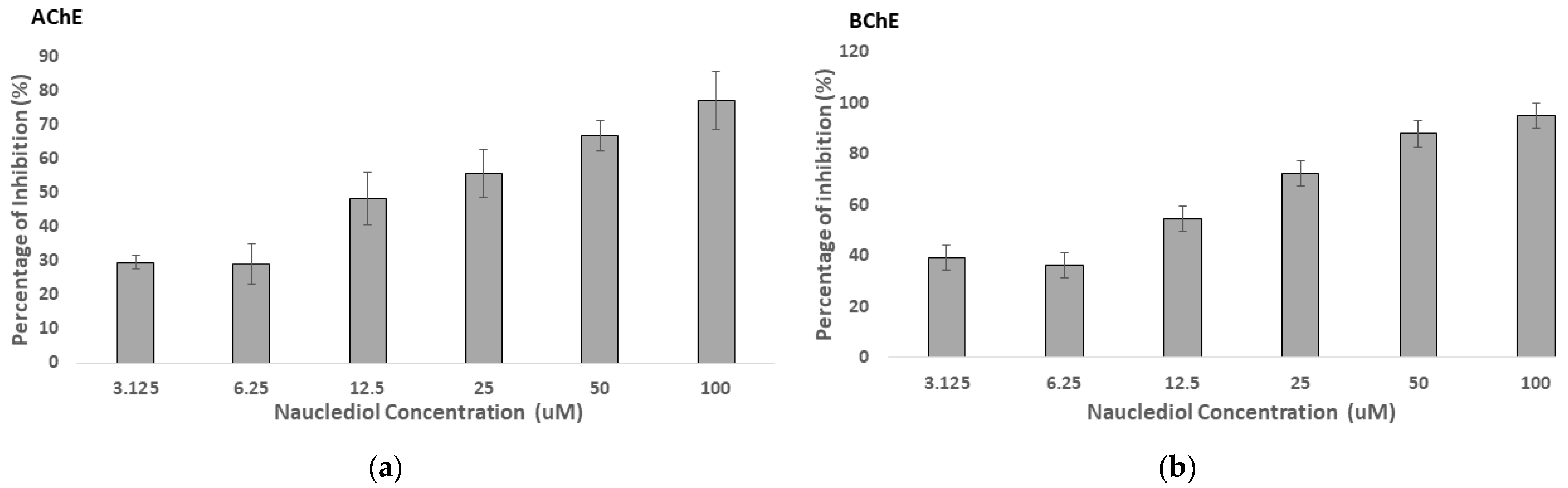
Figure 5.
Enzyme kinetic study of nauclediol against (a) acetylcholinesterase and (b) butrylcholinesterase enzymes. The plots show the fit of the primary data to the non-competitive inhibition model of nauclediol. The experimental data points were fit using the non-linear curve-fitting algorithm of GraphPad Prism v 8.0e.
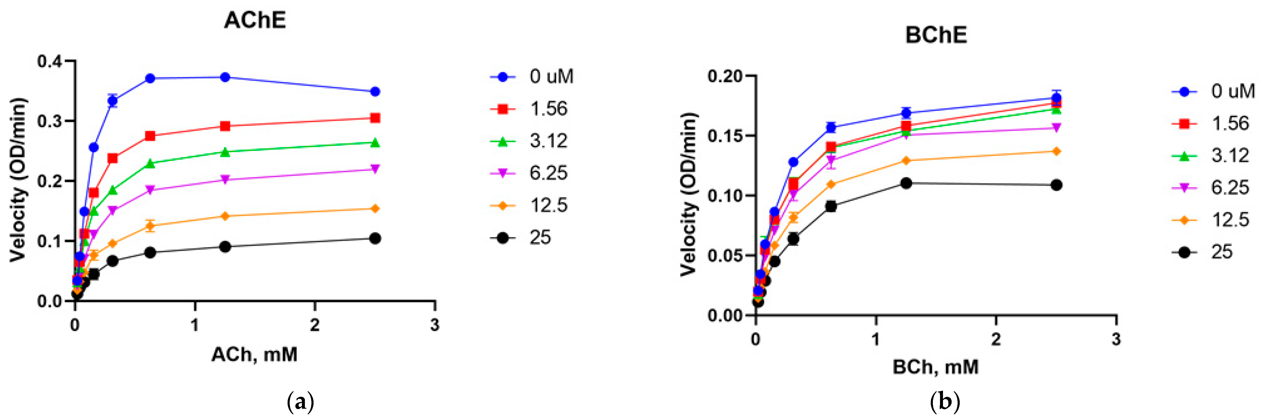
Figure 6.
Binding interaction of nauclediol (in yellow) with amino acid residues of TcAChE. The hydrogen bond between the ligand and amino acid residues are shown by the red line.
Figure 6.
Binding interaction of nauclediol (in yellow) with amino acid residues of TcAChE. The hydrogen bond between the ligand and amino acid residues are shown by the red line.

Figure 7.
Binding interaction of nauclediol (in yellow) with amino acid residues of hBChE.
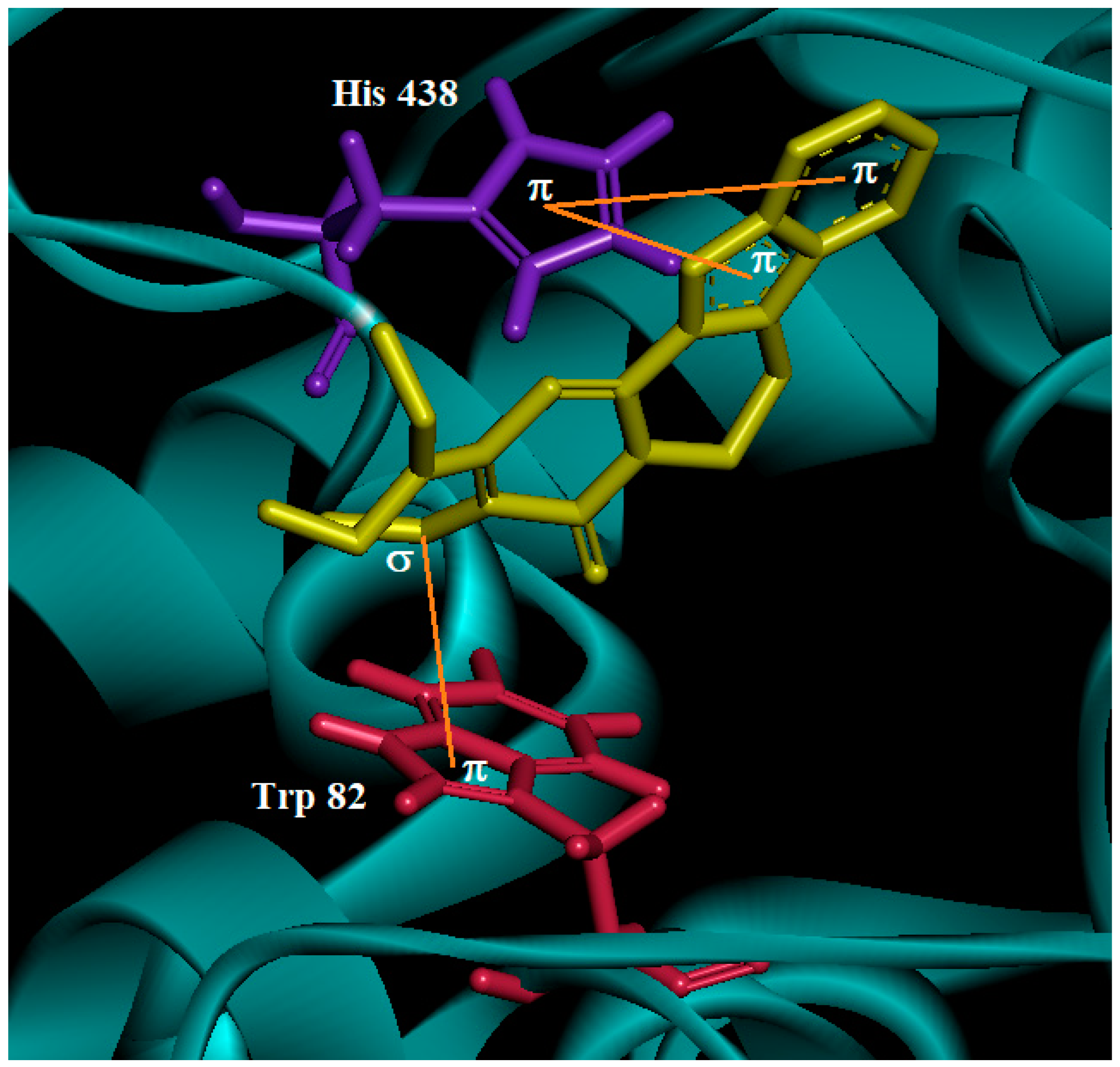
Figure 8.
Neuroprotective activity of nauclediol on SH-SY5Y cells. (A) Cells were treated with various doses of nauclediol for 24 h. (B) Cells were pre-treated with non-detrimental concentrations of nauclediol for 24 h before exposure to 13 μM Aβ-(1-42) aggregates for 24 h. Data are expressed as mean ± standard deviation of three independent experiments; statistical significance was performed by one-way ANOVA followed by Student’s t-test. ** and *** indicate significant statistical differences between treated and untreated control cells, or versus Aβ-(1-42) aggregates (p < 0.05).

Figure 9.
Anti-neuroinflammation activity of nauclediol. (A) BV2 cells were treated with various doses of nauclediol for 24 h. (B) BV2 cells were pre-treated with non-detrimental concentrations of nauclediol for 24 h before exposure to LPS (1 µg/mL) for 24 h. Data are expressed as mean ± standard deviation of three independent experiments; statistical significance was performed by one-way ANOVA followed by Student’s t-test. *, ***, and **** indicate significant statistical differences between treated and untreated control cells or versus LPS (p < 0.05).

Table 1.
1H-NMR (400 MHz) and 13C-NMR (100 MHz) spectral data of nauclediol in MeOD.
| Position | 1H, δH (Multiplicity, J in Hz) | 13C (δC) |
|---|---|---|
| 2 | - | 127.5 |
| 3 | - | 135.6 |
| 5 | 4.33 (t, 6.9) | 40.5 |
| 6 | 3.06 (t, 6.9) | 19.0 |
| 7 | - | 113.0 |
| 8 | - | 125.7 |
| 9 | 7.53 (d, 7.8) | 118.9 |
| 10 | 7.04 (t, 7.8) | 119.6 |
| 11 | 7.19 (t, 7.8) | 124.0 |
| 12 | 7.37 (d, 7.8) | 111.3 |
| 13 | - | 138.6 |
| 14 | 7.00 (s) | 95.5 |
| 15 | - | 141.2 |
| 16 | - | 120.0 |
| 17 | 6.46 (q, 6.9) | 123.8 |
| 18 | 1.86 (d, 6.9) | 12.2 |
| 19 | 4.45 (s) | 64.1 |
| 20 | - | 129.2 |
| 21 | 4.55 (s) | 64.3 |
| 22 | - | 160.9 |
Table 2.
Binding interaction data for nauclediol with amino acid residues of TcAChE and hBChE.
| Ligand/ Compound | Enzyme | Binding Energy (kcal/mol) | Number of Conformations in the Cluster | Interacting Site | Residue | Type of Interaction | Distance (Å) | Ligand Interacting |
|---|---|---|---|---|---|---|---|---|
| Nauclediol | TcAChE | −13.01 | 64 | Catalytic triad | His 440 | Hydrogen | 2.60 | Hydroxyl group at C-19 |
| Choline binding site | Trp 84 Tyr 130 | Hydrophobic Hydrogen | - 2.26 | Ring B and D Oxygen atom of the carbonyl group C-22 | ||||
| hBChE | −12.19 | 60 | Catalytic triad | His 438 | Hydrophobic | - | Ring A and B | |
| Choline binding site | Trp 82 | Hydrophobic | - | C-17 |
Disclaimer/Publisher’s Note: The statements, opinions and data contained in all publications are solely those of the individual author(s) and contributor(s) and not of MDPI and/or the editor(s). MDPI and/or the editor(s) disclaim responsibility for any injury to people or property resulting from any ideas, methods, instructions or products referred to in the content. |
© 2023 by the authors. Licensee MDPI, Basel, Switzerland. This article is an open access article distributed under the terms and conditions of the Creative Commons Attribution (CC BY) license (https://creativecommons.org/licenses/by/4.0/).
Share and Cite
MDPI and ACS Style
Liew, S.Y.; Mak, W.Q.; Thew, H.Y.; Khaw, K.Y.; Hazni, H.; Litaudon, M.; Awang, K. Neuroprotective Activities of New Monoterpenoid Indole Alkaloid from Nauclea officinalis. Processes 2023, 11, 646. https://doi.org/10.3390/pr11030646
AMA Style
Liew SY, Mak WQ, Thew HY, Khaw KY, Hazni H, Litaudon M, Awang K. Neuroprotective Activities of New Monoterpenoid Indole Alkaloid from Nauclea officinalis. Processes. 2023; 11(3):646. https://doi.org/10.3390/pr11030646
Chicago/Turabian StyleLiew, Sook Yee, Wen Qi Mak, Hin Yee Thew, Kooi Yeong Khaw, Hazrina Hazni, Marc Litaudon, and Khalijah Awang. 2023. "Neuroprotective Activities of New Monoterpenoid Indole Alkaloid from Nauclea officinalis" Processes 11, no. 3: 646. https://doi.org/10.3390/pr11030646
Note that from the first issue of 2016, this journal uses article numbers instead of page numbers. See further details here.