Article

Characterization of Streptomyces Species and Validation of Antimicrobial Activity of Their Metabolites through Molecular Docking

1
Central Department of Chemistry, Tribhuvan University, Kirtipur 44618, Kathmandu, Nepal
2
Department of Chemistry, University of Virginia, Charlottesville, VA 22903, USA
3
Department of Medical Microbiology and Immunology, University of Wisconsin-Madison, Madison, WI 53706, USA
4
Department of Chemistry, Florida Agricultural and Mechanical University, Tallahassee, FL 32307, USA
*
Authors to whom correspondence should be addressed.
These authors contributed equally to this work.
Processes 2022, 10(10), 2149; https://doi.org/10.3390/pr10102149
Submission received: 23 August 2022 / Revised: 7 October 2022 / Accepted: 19 October 2022 / Published: 21 October 2022
(This article belongs to the Section Biological Processes and Systems)

Abstract

:
Finding new antibacterial agents from natural products is urgently necessary to address the growing cases of antibiotic-resistant pathogens. Actinomycetes are regarded as an excellent source of therapeutically important secondary metabolites including antibiotics. However, they have not yet been characterized and explored in great detail for their utility in developing countries such as Nepal. In silico molecular docking in addition to antimicrobial assays have been used to examine the efficacy of chemical scaffolds biosynthesized by actinomycetes. This paper depicts the characterization of actinomycetes based on their morphology, biochemical tests, and partial molecular sequencing. Furthermore, antimicrobial assays and mass spectrometry-based metabolic profiling of isolates were studied. Seventeen actinomycete-like colonies were isolated from ten soil samples, of which three isolates showed significant antimicrobial activities. Those isolates were subsequently identified to be Streptomyces species by partial 16S rRNA gene sequencing. The most potent Streptomyces species_SB10 has exhibited an MIC and MBC of 1.22 μg/mL and 2.44 μg/mL, respectively, against each Staphylococcus aureus and Shigella sonnei. The extract of S. species_SB10 showed the presence of important metabolites such as albumycin. Ten annotated bioactive metabolites (essramycin, maculosin, brevianamide F, cyclo (L-Phe-L-Ala), cyclo (L-Val-L-Phe), cyclo (L-Leu-L-Pro), cyclo (D-Ala-L-Pro), N6, N6-dimethyladenosine, albumycin, and cyclo (L-Tyr-L-Leu)) were molecularly docked against seven antimicrobial target proteins. Studies on binding energy, docking viability, and protein-ligand molecular interactions showed that those metabolites are responsible for conferring antimicrobial properties. These findings indicate that continuous research on the isolation of the Streptomyces species from Nepal could lead to the discovery of novel and therapeutically relevant antimicrobial agents in the future.

1. Introduction

Antibiotics have been considered powerful tools to fight against microbial infections and have revolutionized the healthcare system globally. However, the misuse and overuse of antibiotics have accelerated the development of antibiotic resistance. Such resistance is one of the most serious problems confronting healthcare facilities in the twenty-first century [1]. Several bacterial resistance mechanisms, such as the multidrug efflux system [2], the production of β-lactamases [3], and aminoglycoside-modifying enzymes [4] have emerged in the last decades. The microbial world has shown incredible resilience in prevalent antibiotics, and the development of resistance is virtually impossible to avoid. The relentless development of resistance can render valuable antibiotics useless. This scenario demands an urgent need for the development of new antibiotics that have effective therapeutic activities against infectious diseases [5,6].
Actinomycetes have proven particularly beneficial to the pharmaceutical industry due to their seemingly limitless ability to make secondary metabolites with diverse chemical structures and biological functions. Among actinomycetes, Streptomyces is the most prolific genus used in therapeutic applications and pharmaceutical industries [7,8]. Approximately 60% of antibiotics used today are derived from the genus Streptomyces [9]. Streptomyces are filamentous, Gram-positive, and sporulating bacteria with high GC content in a range of 55–75% [10]. Streptomyces species are saprophytic organisms that spend the majority of their life cycles as dormant spores [11]. Though Streptomyces are valuable prokaryotes of clinical and medicinal importance, novel antibiotics have not been discovered in the last few decades. This is because of the repeated rediscovery of similar secondary metabolites from bacteria of similar ecological niches [12]. We believe the continuous screening of actinomycetes from different habitats could lead to the discovery of alternative antibiotics.
The process of new drug discovery is an arduous task; however, the use of computer-aided tools in drug discovery is now gaining more popularity and appreciation [13]. To find medications from natural sources, a computational platform has emerged as a key area of research. The computer-aided drug discovery (CADD) approach entails the discovery of potential drug targets, high-throughput screening, optimization of lead compounds, and examination of potential side effects and toxicity [14]. Molecular docking can be used to model the interaction between a small molecule and a target protein, thereby allowing the characterization of the behavior of small molecules at the binding site of target proteins [15].
Nepal, being a country with a wide range of geographies, altitudes, and vegetation along with many unexplored and untouched ecosystems, has a high probability of isolating actinomycetes producing new and potent secondary metabolites. Soil is the habitat for a large number of Streptomyces species, and a majority of soils have 104 to 107 Streptomyces colony-forming units per gram of soil, accounting for 1 to 20% or even more of the total viable counts [16]. Nevertheless, Nepalese ecosystems have not been extensively explored in the search for actinomycetes.
In the present paper, an attempt was made to isolate, characterize, and validate the antimicrobial properties of Streptomyces species isolated from soil collected from untouched habitats of Nepal covering higher altitudes to lowlands. The focus was on characterizing the Streptomyces species based on morphology, biochemical tests, molecular sequencing, and molecular annotation of secondary metabolites in some potent isolates. Finally, those metabolites were used to dock against some target proteins of several bacteria to understand the mechanism of action of these metabolites for the exploration and validation of antimicrobial properties.

2. Materials and Methods

2.1. Collection of Samples and Isolation of Streptomyces

Ten soil samples (SB1 to SB10) were collected from different geographical locations in Nepal covering different altitudes and untouched habitats, as shown in Table 1.

2.2. Isolation of Streptomyces

One gram of soil was taken and suspended in 10 mL of autoclaved water by vortexing. The vegetative cells in the soil sample were killed by heat treatment at 80 °C for 30 min. The suspension was further diluted up to 1000-fold in sterilized water and 100 µL of suspension was spread into individual ISP4 (International Streptomyces Project 4) plates and incubated at 28 °C for 7 to 14 days. The actinomycetes were isolated on the ISP4 medium with the addition of nalidixic acid (20 mg/mL) and cycloheximide (50 mg/mL). The growth of Gram-negative bacteria was inhibited by nalidixic acid, and the growth of the fungus was controlled by cycloheximide. Based on morphological characterization, the colonies were sub-cultured and the glycerol stocks were prepared for further use. The details of the soil sample collection and isolation of Streptomyces species were carried out as described in the literature [17,18].

2.3. Morphological Characterization

Macroscopic and microscopic studies were implemented for the morphological characterization of the isolated colonies. The isolates were preliminarily identified by macroscopic characterization in which aerial and substrate mycelium, color, shape, pigmentation, and appearance of the colonies were observed [19]. The appearance of the colonies was noted based on dryness, roughness, toughness, and elevation. Microscopic identification was done by Gram staining and observing under 100× magnification [20]. Likewise, several biochemical tests including lipase, gelatin, amylase, sulfur test, indole, nitrate reduction, motility, urease, catalase, and MRVP tests were performed (Tables S3 and S4) [21].

2.4. Molecular Characterization

The isolates were cultured in a tryptic soy broth (TSB) medium in an Erlenmeyer flask at 28 °C and 180 rpm for 4–5 days with glass beads. Then, 15 mL of the culture was taken in a falcon tube and the cells were separated by centrifugation at 8000 rpm for 10 min. Genomic DNA was isolated using the phenol-chloroform method [22]. Universal primers 27F: AGAGTTTGATCCTGGCTCAG and 1492R: GGTTACCTTGTTACGACTT were used for the amplification of 16S rDNA. The PCR amplification was performed in 29 cycles in a 50 µL reaction mixture containing 10 μM oligonucleotides, 5× premix having Taq polymerase, and 2 µL (100 ng) genomic DNA. A cycle was run at 95 °C initial denaturation, 51.4 °C annealing, and 95 °C extensions. The resulting PCR products were then purified using Monarch® PCR and a DNA Cleanup kit (New England Biolabs Inc., Ipswich, MA, USA). The unidirectional sequencing of 16S rDNA was carried out in Macrogen, Seoul, South Korea. The BLAST search tool was used for the annotation of 16S rDNA partial sequences in the database [23].

2.5. Shake Flask Fermentation

Bacterial isolates were cultured in TSB broth (30 g/L) for 3 to 4 days at 140 rpm, 28 °C in a shaking incubator. After the full growth of the bacteria, its 1 mL suspension was transferred to freshly prepared TSB broth (100 mL) for fermentation and kept for 5 to 7 days (until bacterial growth meets stationary phase) at 28 °C with constant shaking at 140 rpm [24]. The suspension was mixed with an equal volume of ethyl acetate for the extraction of secondary metabolites. Finally, the organic phase was further used for antimicrobial assays.

2.6. Antimicrobial Assays

The antimicrobial activity of 17 Streptomyces isolates was initially tested by the primary screening method. The pathogens examined were Staphylococcus aureus ATCC 43300, Escherichia coli ATCC 2591, Kleibsella pneumonia ATCC 700603, Salmonella typhi ATCC 14028, Shigella sonnei ATCC 25931, and Acinetobacter baumanii ATCC 19606. The antimicrobial assays of potent bacterial isolates in perpendicular screenings were performed by using the agar well diffusion method. The assays were carried out in triplicate with 5 µg of Streptomyces extract in each well along with 1 mg/mL neomycin and 50% DMSO as a positive and negative control, respectively. The minimum inhibitory concentration (MIC) and minimum bactericidal concentration (MBC) of the most potent bacterial isolates were determined using broth microdilution [25].

2.7. Mass Spectrometry

The mass spectrometry of the ethyl acetate extracts of S. species_SB1 and S. species_SB3 was performed in the Sophisticated Analytical Instrument Facility (SAIF), Lucknow, India using an Agilent 6520 Q-TOF mass spectrometer. The data were acquired using positive electron spray ionization (ESI) mode. The mobile phase consisted of 5 to 80% gradient acetonitrile and 5 mM acetate buffer [25]. For SB10, low resolution (LR)-LCMS was obtained by using an HPLC system (1100 Series, Agilent Technologies, Waldbronn, Germany) coupled with an MS (ABSCIEx 3200 Q Trap, Darmstadt, Germany). Likewise, the extract was also subjected to HR-LC-ESI-MS/MS (Bruker TOF-MS MaXis impact ESI-HR-MS) profiling [18]. The positive mode ESI spectrum was deposited in the Global Natural Products Social (GNPS) Molecular Networking Platform (https://gnps.ucsd.edu) (accessed on 1 October 2022).

2.8. Molecular Annotation

The molecular profiling of the extracts from isolates S. species_SB1 and S. species_SB3 was executed by the previously described procedure [26]. The raw data of LC-HRMS obtained from extracts of isolates S. species_SB1 and S. species_SB3 were analyzed using MestreNova 12.0 software (Santiago de Compostela, SPAIN) for peak detection, alignment, and annotation. They were then compared to the database library, literature, dictionary of natural products, and METLIN metabolite searching cloud. The molecular formula for m/z and MS/MS fragmentation of isolate S. species_SB10 was read using Bruker Compass Data Analysis 4.4 and the molecule annotation was carried out in SIRIUS 4.9.12 through CSI: FingerID user interface by exporting a .mgf file [18].

2.9. Molecular Docking for the Connection with Antimicrobial Assays

2.9.1. Binding Site Prediction

The binding site residues of the target proteins were chosen in this investigation based on previous findings as well as through the co-crystallized ligand in the retrieved protein [27,28]. After discovering binding sites, the coordinates of the binding pocket for each target protein were generated using the define and edit binding site tool of BIOVIA Discovery Studio.

2.9.2. Ligand Preparation

The annotated compounds were downloaded in .sdf format through PubChem [29,30] and translated to .pdb format via Pymol [31,32]. For docking purposes, polar hydrogens and Gasteiger charges were added, and the compounds were saved in PDBQT format using the AutoDock application.

2.9.3. Receptor Preparation

The protein targets, 1JIJ (crystal structure of S. aureus TyrRS in complex with SB-239629); 6J33 (crystal structure of ligand-free PulA from Klebsiella pneumoniae); 3TTZ (crystal structure of a topoisomerase ATPase inhibitor); 3SRW (S.aureus Dihydrofolate Reductase complexed with novel 7-aryl-2,4-diaminoquinazolines); 4UMW (crystal structure of zinc-transporting PIB-type ATPase in E2.PI state); 3UDI (crystal structure of Acinetobacter baumannii PBP1a in complex with penicillin G); and 7KRK (putative FabG from Acinetobacter baumannii) were attained from the RCSB PDB database. The proteins were prepared in BIOVIA Discovery Studio by detaching water components and other associated ligands before the docking experiments. The structures were saved in PDBQT format after optimizing with Kollmann charges and adding polar hydrogen in AutoDock tools [33]. A three-dimensional affinity grid box was generated around the binding sites of the target proteins with the size of 40 × 40 × 40 Angstrom and centered on the important residues of all target proteins where potent metabolites would bind.

2.9.4. Molecular Docking and Validation

AutoDock tools version 1.5.6 was used to dock the annotated compounds into the binding site of the target proteins. The outcome is a list of poses that are arranged according to ΔG, the predicted binding energy in kcal/mol. Using the superimposition technique, the root-mean-square deviation (RMSD) values of the first and second docked low-energy protein-ligand complex postures were determined. If the RMSD values are under 2 Å, the validation is said to have been successful. The poses of compounds in target proteins were analyzed using BIOVIA Discovery studio, and the best pose was chosen based on hydrogen bonding, hydrophobic, and π–π interactions with the binding residues.

3. Results

3.1. Morphological Characterization

A total of 17 isolates showing the characteristics of Streptomyces were isolated from 10 different soil samples. The detail and morphological features of all the isolates are presented in Supplementary Tables S1 and S2. The aerial and substrate mycelium of S. species_SB10 were grayish and white, respectively, along with no diffusible pigments (Figure 1).

3.2. Molecular Characterization of Isolates

Universal primers were used to amplify the 16S rRNA. The 16S rRNA gene sequencing of SB1, SB3, and SB10 showed that the isolates resembled the genus Streptomyces on the BLAST sequence search generated from NCBI. A DNA sequence analysis showed that SB1, SB3, and SB10 were closely related to Streptomyces bungoensis, Streptomyces aureus, and Streptomyces griseoplanus with percentage similarities of 84.31, 95.79, and 95.92, respectively. Thus, according to morphological and molecular characterization, it was concluded that isolates from the soil samples SB1, SB3, and SB10 were members of the genus Streptomyces.

3.3. Antimicrobial Assays

Out of 17 isolates, S. species_SB1, S. species_SB3, and S. species_SB10 showed potential antimicrobial activities in the primary screening and hence were considered for further studies. The antimicrobial assays performed on the three Streptomyces isolates showed moderate to strong activity against Gram-positive (S. aureus) and Gram-negative (S. typhi, K. pneumoniae, S. sonnei, A. baumannii, and E. coli) bacteria. The zones of inhibition (ZoIs) measured were recorded as shown in Table 2 and Supplementary Figure S1. The antibacterial activity of S. species_SB10 was the highest against S. aureus and S. sonnei with a ZoI of 30 mm for each bacterium. The results showed that the extract of S. species_SB10 was more potent than the extracts of S. species_SB1 and S. species_SB3. Therefore, only the MIC and MBC of the S. species_SB10 were determined. The MIC of S. species_SB10 against S. aureus and S. sonnei was found to be 1.22 μg/mL, while the MBC was 2.44 μg/mL (Figure S2). On the other hand, the MIC of neomycin (positive control) against S. aureus and S. sonnei was 1.56 μg/mL, and the MBC was 12.5 and 6.25 μg/mL, respectively.

3.4. Liquid Chromatography–Mass Spectrometry Analysis

Streptomyces species showing good antimicrobial properties (S. species_SB1, S. species_SB3, and S. species_SB10) were further processed for mass analysis in ethyl acetate extracts. The annotated metabolites are shown in Table 3 and Table 4. The metabolites were found to contain multiple chemical scaffolds. Extracts of S. species_SB1 and S. species_SB3 primarily consisted of diketopiperazines (DKPs). The extract of S. species_SB10 showed the presence of the valuable metabolite albumycin and aminocoumarin antibiotics. At least 20 different secondary metabolites were identified from these three isolates.

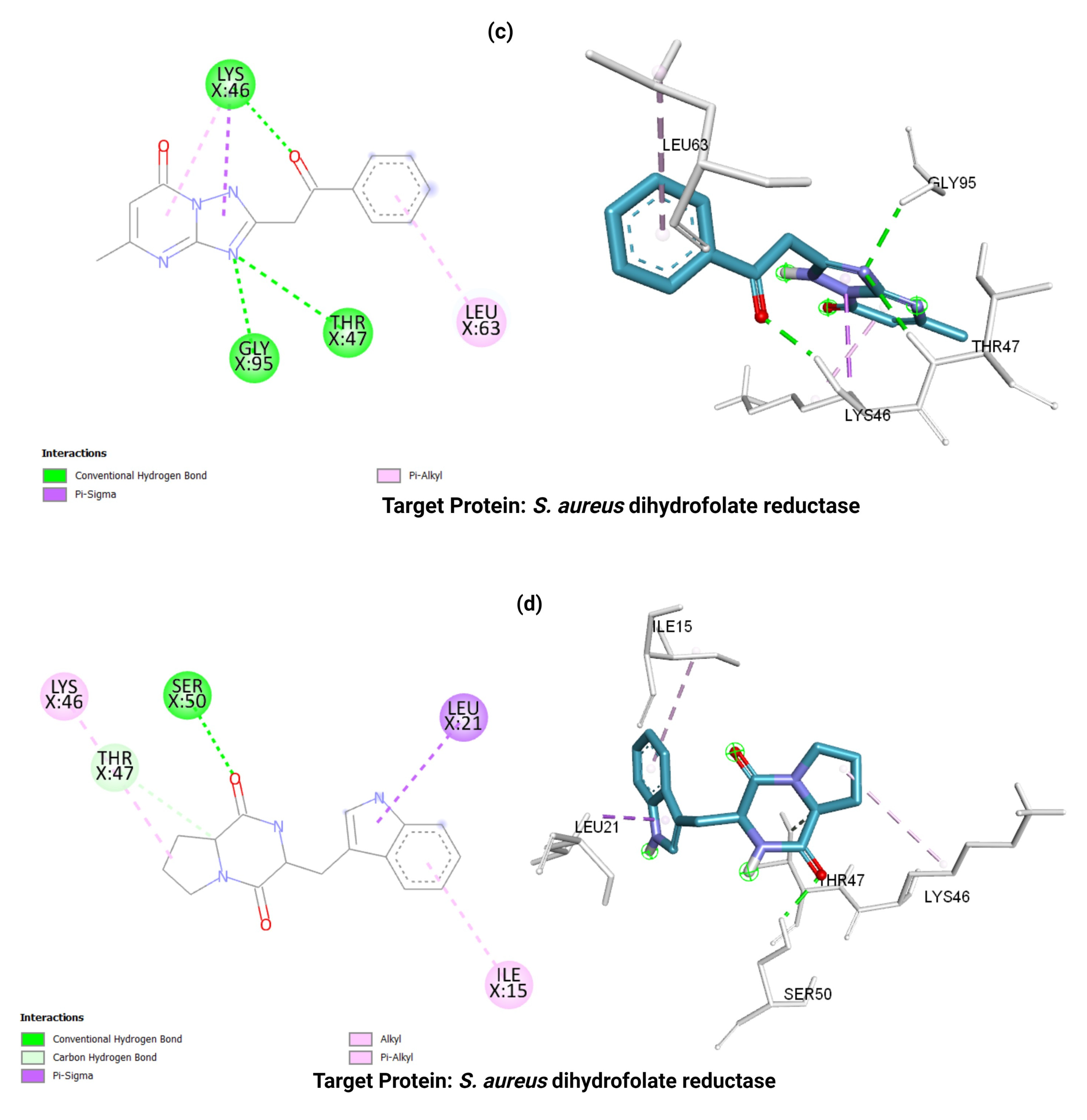
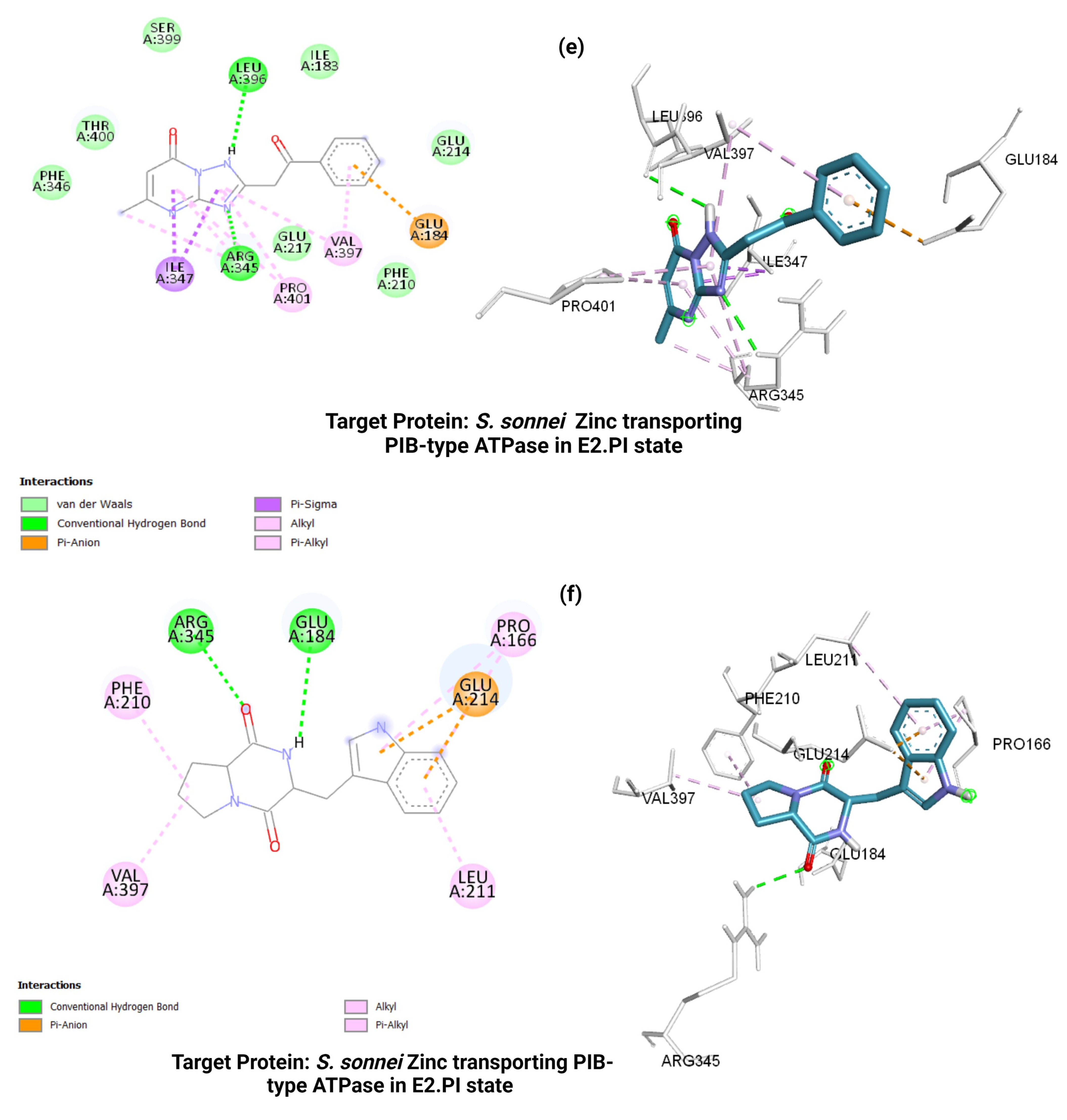
3.5. Molecular Docking Analysis

The major ten annotated compounds in mass spectrometry were docked through AutoDock tools with the target proteins mentioned earlier. The binding energies of ligands (docked compounds) with proteins are displayed in Table S5. The results of the docking analysis suggested that brevianamide F, essramycin, cyclo (L-Phe-L-Ala), and cyclo (L-Val-L-Phe) were the potential candidates that could inhibit the target proteins of various antimicrobials. These compounds exhibited suitable binding affinities with acceptable binding interactions along with one or more hydrogen-bonding interactions with binding residues of proteins. Most of the interacting residues fell under binding sites of co-crystallized ligands on target proteins, which are in bold in Table 5. The binding interactions of essramycin and Brevianamide F complexed with target proteins, S. aureus TyrRS (PDB ID: 1JIJ); S. aureus dihydrofolate reductase (3SRW); and S. sonnei Zinc transporting PIB-type ATPase in E2.PI state (4UMW), respectively, are shown in Figure 2 and the other interactions of potent complexes were displayed in supplementary Figures S3–S6.

4. Discussion

Microbial species are the source of secondary metabolites with undeniable biological activities. Most importantly, actinomycetes are given particular attention for their role in the production of various bioactive secondary metabolites. Streptomyces species are continually used to discover novel compounds with various biological functions [53]. Antibiotics are the primary and most crucial output of Streptomyces species [54]. So, in this work, we have investigated the antibiotic-producing Streptomyces species from untouched habitats in Nepal.
Despite the development of antibiotics and the success of their use, infectious diseases continue to be the second biggest cause of death globally. Bacterial infections account for over 17 million fatalities each year, primarily affecting the elderly and young [54]. In addition to this, multi-drug resistance (MDR) has posed a serious issue in treating infectious diseases. This situation highlights the urgent need to investigate new antimicrobial agents to tackle MDR. The main contender for antibiotic discovery is still soil microorganisms [55]. Hence, this study is focused on the isolation and characterization of Streptomyces species and the validation of their antimicrobial activities using the metabolites present in their extracts through a computational approach.
We have isolated 17 different strains of actinomycetes from varying locations. Among these, three Streptomyces species, namely, S. species_SB1, S. species_SB3, and S. species_SB10 were determined to possess potential antibacterial properties. S. species_SB10 showed an interesting antibacterial effect against S. aureus and S. sonnei with ZoIs even higher than neomycin (1 mg/mL). This isolate showed an MIC and MBC of 1.22 μg/mL and 2.44 μg/mL, respectively, against S. aureus and S. sonnei. The potential of S. species_SB10 as well as S. species_SB1 and S. species_SB3 to inhibit the pathogens establishes them as effective candidates for antibiotic production that can combat MDR.
These three potent Streptomyces strains were found to contain DKPs such as maculosin, cyclo (L-Phe-L-Ala), cyclo (L-Val-L-Phe), cyclo (L-Leu-L-Pro), cyclo (D-Ala-L-Pro), cyclo (L-Tyr-L-Leu), and brevianamide F. DKPs are known for their rich source of new biologically active compounds. They show a wide spectrum of biological activities, suggesting them as potential therapeutic candidates [56]. They are known to function as antibacterial [57], antiviral [58], antifungal [59], anti-hyperglycemic [60], and antitumor agents [61]. These 2,5 DKPs are also involved as enzyme modulators and biochemical mediators [62]. The availability of donor and acceptor atoms for hydrogen bonding, rigid conformation, and resistance towards proteolysis favor interaction with biological targets [62]. Likewise, Brevianamide F is used as a medicament for cardiovascular dysfunction and cognitive enhancement [62]. The presence of these chemical scaffolds might be the reason for the effective antibacterial activity shown by the extracts. Furthermore, the isolation and characterization of these compounds are needed for other bioactivity-related studies. Moreover, to obtain the full potential of isolated strains, different media cultivation might be required in the future.
Albumycin was previously noted to have antibacterial activities [50]. Similarly, aminocoumarins were also described to show potential antibacterial effects against Staphylococci, including a methicillin-resistant Staphylococcus aureus strain [63]. Reports show that 3-aminocoumarin has been used for the synthesis of metal complexes possessing antimicrobial and antioxidant activities [64]. The presence of these chemical scaffolds increases the likelihood of S. species_SB1, SB3, and SB10 being employed as therapeutic drugs, particularly as antimicrobial agents. For further validation of antimicrobial activity, molecular docking was performed.
Molecular docking helps minimize the binding energy by optimizing the conformation of both the receptor and ligand and the orientation between the ligand and receptor. Although activity assays are still required to verify the activities of the ligands, molecular docking is a theoretical and reliable approach to anticipating the interactions between the receptor and ligand [65]. To find possible inhibitors for target proteins, we employed molecular docking analysis to predict binding sites and the potential activity of the top-scoring compounds [66]. The results were evaluated according to the docking poses and the protein-ligand interactions [65]. Furthermore, the lower the binding energy, the higher the stability of the complex [67]. The present study suggests brevianamide F, essramycin, cyclo (L-Phe-L-Ala), and cyclo (L-Val-L-Phe), as potent inhibitors for target proteins with significant binding energy and appropriate interactions.
The compounds brevianamide F and essramycin have comparatively lower binding energies with the target proteins, and most of the interacted residues fall into binding sites on comparison with the interacted residues of co-crystallized ligands of retrieved protein from RCSB and also with the important residues of target proteins, according to the literature [68,69]. The orientation of ligands in the target proteins within the binding site was analyzed through hydrogen-bonding, hydrophobic, and π–π interactions. According to earlier in silico and in vitro studies, natural compounds such as acacetin and chrysin can prevent the growth of S. aureus by inhibiting tyrosyl-tRNA synthetases (TyrRSs) [70]. In addition, through the molecular docking method too, ciprofloxacin analogs were identified as potential candidates against S. aureus DNA gyrase [66]. Furthermore, a molecular docking study revealed acetylated abietane quinone as a suitable inhibitor of S. aureus clumping factor A (ClfA) with good binding interactions and binding energy [71]. Hence, molecular docking methods can be taken into account to corroborate the potent metabolites.
The secondary metabolites from Streptomyces are significant products that can be utilized to combat various MDR pathogens. The production and commercial viability of these secondary metabolites are not yet fully established, but shortly, their application may accelerate, specifically in the manufacturing of antibiotics. It has been estimated that only 10% of discovered antibiotics are from screened bacterial strains and only 1% from all microbes [72]. Actinomycetes found in terrestrial soil comprise around two-thirds of all natural antibiotics. According to recent estimates, to find the next new antibiotics class, 107 actinomycetes strains would need to be screened [73]. Hence, for the utilization of the soil actinomycetes for antibiotics production, the pace of screening and validation should be intensified. Considering this fact, the use of computational techniques may be extended into validating newer antibiotic-potent products showing a broad spectrum of activity.

5. Conclusions

The present study depicts that based on the variations of altitude and soil types and their contents, there is a huge probability of obtaining a varying diversity of antimicrobial-producing Streptomyces. In this study, a total of 17 distinct strains were isolated based on the morphological characteristics of 10 different soil samples collected from different habitats. Three samples were selected for further study based on the antimicrobial screening test. The three Streptomyces species SB1, SB3, and SB10 exhibited significant antibacterial activity against S. aureus, S. sonnei, and A. baumanii. At least 24 different secondary metabolites were identified from these three isolates, and these metabolites included different chemical scaffolds such as DKPs and aminocoumarin antibiotics. These metabolites were further subjected to docking to validate their antibacterial properties. Docking positively signified brevianamide F, essramycin, cyclo(L-Phe-L-Ala), and cyclo(L-Val-L-Phe) as potent inhibitors for target proteins, thereby showing the potential of isolated Streptomyces species for use in the production of antibiotics.

Supplementary Materials

The following supporting information can be downloaded at: https://www.mdpi.com/article/10.3390/pr10102149/s1, Table S1: Soil profile, total isolated actinomycetes and characterization method of 10 samples; Table S2: Culture characteristic and Gram staining of samples SB1–SB10; Table S3: Biochemical test of microbial strains; Table S4: Cultural and physiological characteristics of microbial strains; Table S5: The binding energies of target proteins with potent compounds; Figure S1: Zone of inhibition exhibited by SB1, SB3, and SB10 against S. aureus and S. sonnei; Figure S2: MIC and MBC of the extract SB10 against S. sonnei and S. aureus; Figure S3: (A–D) represents 2D structures of essramycin with target proteins: PulA from Klebsiella pneumonia (6J33), Topoisomerase ATPase inhibitor (3TTZ), A. baumannii PBP1a (3UDI), and Putative FabG from A. baumannii (7KRK), respectively; Figure S4: (A–D) represent 2D structures of brevianamide F with target proteins: PulA from Klebsiella pneumonia (6J33), Topoisomerase ATPase inhibitor (3TTZ), A. baumannii PBP1a (3UDI), and Putative FabG from A. baumannii (7KRK), respectively; Figure S5: (A–G) represent 2D structures of cyclo(L-Val-L-Phe) with target proteins: S. aureus TyrRS (1JIJ), PulA from Klebsiella pneumonia (6J33), Topoisomerase ATPase inhibitor (3TTZ), S.aureus Dihydrofolate Reductase (3SRW), Zinc transporting PIB-type ATPase in E2.PI state (4UMW), A. baumannii PBP1a (3UDI), and Putative FabG from A. baumannii (7KRK), respectively; Figure S6: (A–E) represent 2D structures of cyclo(L-Phe-L-Ala) with target proteins: PulA from Klebsiella pneumonia (6J33), Topoisomerase ATPase inhibitor (3TTZ), S.aureus Dihydrofolate Reductase (3SRW), A. baumannii PBP1a (3UDI), and Putative FabG from A. baumannii (7KRK), respectively.

Author Contributions

Conceptualization, N.P., B.R.B. and S.B.; methodology, B.R.B., S.B. and K.K.; validation, B.R.B., U.L. and A.A. (Ashma Adhikari); formal analysis, B.R.B., B.A., A.S., N.A. and R.T.; investigation, S.B., B.R.B., K.K. and U.L.; resources, N.P. and B.R.B.; data curation, N.A. and B.R.B.; writing—original draft preparation, S.B. and B.R.B.; writing—review and editing, N.P., N.A., B.P.R., A.A. (Achyut Adhikari), R.P.Y. and B.B.T.; visualization, B.R.B. and B.P.R.; supervision, N.P.; project administration, N.P.; funding acquisition, N.P. All authors have read and agreed to the published version of the manuscript.

Funding

This research was funded by the University Grants Commission, Nepal under grant number CRG-75/76-S&T-1 to Niranjan Parajuli.

Data Availability Statement

The data presented in this study are available on request from the corresponding author.

Acknowledgments

Authors would like to acknowledge Ishwor Pathak, Shreesti Shrestha, Ganesh BK, and Sajan Shakya for their help. We are very thankful to Gross lab, University of Tübingen, Germany, and SAIF, India for the acquisition of MS data.

Conflicts of Interest

The authors declare no conflict of interest.

References

  1. Uddin, T.M.; Chakraborty, A.J.; Khusro, A.; Zidan, B.R.M.; Mitra, S.; Emran, T.B.; Dhama, K.; Ripon, M.K.H.; Gajdács, M.; Sahibzada, M.U.K.; et al. Antibiotic Resistance in Microbes: History, Mechanisms, Therapeutic Strategies and Future Prospects. J. Infect. Public Health 2021, 14, 1750–1766. [Google Scholar] [CrossRef]
  2. Baucheron, S.; Monchaux, I.; Le Hello, S.; Weill, F.-X.; Cloeckaert, A. Lack of Efflux Mediated Quinolone Resistance in Salmonella Enterica Serovars Typhi and Paratyphi A. Front. Microbiol. 2014, 5, 12. [Google Scholar] [CrossRef] [Green Version]
  3. Chuma, T.; Miyasako, D.; Hesham, D.; Takayama, T.; Nakamoto, Y.; Shahada, F.; Akiba, M.; Okamoto, K. Chronological Change of Resistance to β-Lactams in Salmonella Enterica Serovar Infantis Isolated from Broilers in Japan. Front. Microbiol. 2013, 4, 113. [Google Scholar] [CrossRef] [PubMed] [Green Version]
  4. Frontiers|Prospects for Circumventing Aminoglycoside Kinase Mediated Antibiotic Resistance. Available online: https://www.frontiersin.org/articles/10.3389/fcimb.2013.00022/full (accessed on 5 August 2022).
  5. Bassetti, M.; Vena, A.; Castaldo, N.; Righi, E.; Peghin, M. New Antibiotics for Ventilator-Associated Pneumonia. Curr. Opin. Infect. Dis. 2018, 31, 177–186. [Google Scholar] [CrossRef] [PubMed]
  6. Terreni, M.; Taccani, M.; Pregnolato, M. New Antibiotics for Multidrug-Resistant Bacterial Strains: Latest Research Developments and Future Perspectives. Molecules 2021, 26, 2671. [Google Scholar] [CrossRef]
  7. Al-shaibani, M.M.; Radin Mohamed, R.M.S.; Sidik, N.M.; Enshasy, H.A.E.; Al-Gheethi, A.; Noman, E.; Al-Mekhlafi, N.A.; Zin, N.M. Biodiversity of Secondary Metabolites Compounds Isolated from Phylum Actinobacteria and Its Therapeutic Applications. Molecules 2021, 26, 4504. [Google Scholar] [CrossRef] [PubMed]
  8. Rajivgandhi, G.N.; Vimala, R.T.V.; Ramachandran, G.; Kanisha, C.C.; Manoharan, N.; Li, W.-J. An Overview on Natural Product from Endophytic Actinomycetes. In Natural Products from Actinomycetes: Diversity, Ecology and Drug Discovery; Rai, R.V., Bai, J.A., Eds.; Springer: Singapore, 2022; pp. 151–165. ISBN 9789811661327. [Google Scholar]
  9. Arefa, N.; Sarker, A.K.; Rahman, M.A. Resistance-guided Isolation and Characterization of Antibiotic-producing Bacteria from River Sediments. BMC Microbiol. 2021, 21, 116. [Google Scholar] [CrossRef]
  10. Connell, N.D. Expression Systems for Use in Actinomycetes and Related Organisms. Curr. Opin. Biotechnol. 2001, 12, 446–449. [Google Scholar] [CrossRef]
  11. Vikineswary, S.; Nadaraj, P.; Wong, W.H.; Balabaskaran, S. Actinomycetes from a Tropical Mangrove Ecosystem- Antifungal Activity of Selected Strains. Asia-Pac. J. Mol. Biol. Biotechnol. 1997, 5, 81–86. [Google Scholar]
  12. Belknap, K.C.; Park, C.J.; Barth, B.M.; Andam, C.P. Genome Mining of Biosynthetic and Chemotherapeutic Gene Clusters in Streptomyces Bacteria. Sci. Rep. 2020, 10, 2003. [Google Scholar] [CrossRef] [Green Version]
  13. Chaudhary, K.K.; Mishra, N. A Review on Molecular Docking: Novel Tool for Drug Discovery. Databases 2016, 3, 1029. [Google Scholar]
  14. Segall, M.D.; Barber, C. Addressing Toxicity Risk When Designing and Selecting Compounds in Early Drug Discovery. Drug Discov. Today 2014, 19, 688–693. [Google Scholar] [CrossRef]
  15. Pagadala, N.S.; Syed, K.; Tuszynski, J. Software for Molecular Docking: A Review. Biophys. Rev. 2017, 9, 91–102. [Google Scholar] [CrossRef]
  16. Kurtböke, İ. Bacteriophages; BoD—Books on Demand; InTech: Rijeka, Croatia, 2012; ISBN 978-953-51-0272-4. [Google Scholar]
  17. Khadayat, K.; Sherpa, D.D.; Malla, K.P.; Shrestha, S.; Rana, N.; Marasini, B.P.; Khanal, S.; Rayamajhee, B.; Bhattarai, B.R.; Parajuli, N. Molecular Identification and Antimicrobial Potential of Streptomyces Species from Nepalese Soil. Int. J. Microbiol. 2020, 2020, 8817467. [Google Scholar] [CrossRef]
  18. Bhattarai, B.R.; Khadayat, K.; Aryal, N.; Aryal, B.; Lamichhane, U.; Bhattarai, K.; Rana, N.; Regmi, B.P.; Adhikari, A.; Thapa, S.; et al. Untargeted Metabolomics of Streptomyces Species Isolated from Soils of Nepal. Processes 2022, 10, 1173. [Google Scholar] [CrossRef]
  19. Bergey’s Manual® of Systematic Bacteriology; Goodfellow, M.; Kämpfer, P.; Busse, H.-J.; Trujillo, M.E.; Suzuki, K.; Ludwig, W.; Whitman, W.B. (Eds.) Springer: New York, NY, USA, 2012; ISBN 978-0-387-95043-3. [Google Scholar]
  20. Sitachitta, N.; Gadepalli, M.; Davidson, B.S. New α-Pyrone-Containing Metabolites from a Marine-Derived Actinomycete. Tetrahedron 1996, 52, 8073–8080. [Google Scholar] [CrossRef]
  21. Maheshwari, D.K. Practical Microbiology; S. Chand Publishing: New Delhi, India, 2002; ISBN 978-81-219-2153-4. [Google Scholar]
  22. Nybo, S.E.; Shepherd, M.D.; Bosserman, M.A.; Rohr, J. Genetic Manipulation of Streptomyces Species. Curr. Protoc. Microbiol. 2010, 19, 10E.3.1–10E.3.26. [Google Scholar] [CrossRef]
  23. Tamura, K.; Peterson, D.; Peterson, N.; Stecher, G.; Nei, M.; Kumar, S. MEGA5: Molecular Evolutionary Genetics Analysis Using Maximum Likelihood, Evolutionary Distance, and Maximum Parsimony Methods. Mol. Biol. Evol. 2011, 28, 2731–2739. [Google Scholar] [CrossRef] [Green Version]
  24. Klausen, C.; Nicolaisen, M.H.; Strobel, B.W.; Warnecke, F.; Nielsen, J.L.; Jørgensen, N.O.G. Abundance of Actinobacteria and Production of Geosmin and 2-Methylisoborneol in Danish Streams and Fish Ponds. FEMS Microbiol. Ecol. 2005, 52, 265–278. [Google Scholar] [CrossRef] [Green Version]
  25. Aryal, B.; Adhikari, B.; Aryal, N.; Bhattarai, B.R.; Khadayat, K.; Parajuli, N. LC-HRMS Profiling and Antidiabetic, Antioxidant, and Antibacterial Activities of Acacia Catechu (L.f.) Willd. BioMed Res. Int. 2021, 2021, e7588711. [Google Scholar] [CrossRef]
  26. Aryal, B.; Niraula, P.; Khadayat, K.; Adhikari, B.; Khatri Chhetri, D.; Sapkota, B.K.; Bhattarai, B.R.; Aryal, N.; Parajuli, N. Antidiabetic, Antimicrobial, and Molecular Profiling of Selected Medicinal Plants. Evid.-Based Complement. Altern. Med. 2021, 2021, e5510099. [Google Scholar] [CrossRef]
  27. Egbert, M.; Porter, K.A.; Ghani, U.; Kotelnikov, S.; Nguyen, T.; Ashizawa, R.; Kozakov, D.; Vajda, S. Conservation of Binding Properties in Protein Models. Comput. Struct. Biotechnol. J. 2021, 19, 2549–2566. [Google Scholar] [CrossRef]
  28. Belkin, S.; Kundrotas, P.J.; Vakser, I.A. Inhibition of Protein Interactions: Co-Crystalized Protein-Protein Interfaces Are Nearly as Good as Holo Proteins in Rigid-Body Ligand Docking. J. Comput. Aided Mol. Des. 2018, 32, 769–779. [Google Scholar] [CrossRef]
  29. PubChem. Available online: https://pubchem.ncbi.nlm.nih.gov/ (accessed on 23 July 2022).
  30. Kim, S. Exploring Chemical Information in PubChem. Curr. Protoc. 2021, 1, e217. [Google Scholar] [CrossRef]
  31. PyMOL|Pymol.Org. Available online: https://pymol.org/2/ (accessed on 14 August 2022).
  32. Yuan, S.; Chan, H.C.S.; Hu, Z. Using PyMOL as a Platform for Computational Drug Design. WIREs Comput. Mol. Sci. 2017, 7, e1298. [Google Scholar] [CrossRef]
  33. Morris, G.M.; Huey, R.; Lindstrom, W.; Sanner, M.F.; Belew, R.K.; Goodsell, D.S.; Olson, A.J. AutoDock4 and AutoDockTools4: Automated Docking with Selective Receptor Flexibility. J. Comput. Chem. 2009, 30, 2785–2791. [Google Scholar] [CrossRef] [Green Version]
  34. Li, B.; Chen, G.; Bai, J.; Jing, Y.-K.; Pei, Y.-H. A Bisamide and Four Diketopiperazines from a Marine-Derived Streptomyces Sp. J. Asian Nat. Prod. Res. 2011, 13, 1146–1150. [Google Scholar] [CrossRef]
  35. Fang, Q.; Maglangit, F.; Wu, L.; Ebel, R.; Kyeremeh, K.; Andersen, J.H.; Annang, F.; Pérez-Moreno, G.; Reyes, F.; Deng, H. Signalling and Bioactive Metabolites from Streptomyces Sp. RK44. Molecules 2020, 25, 460. [Google Scholar] [CrossRef] [Green Version]
  36. Sunaryanto, R.; Marwoto, B.; Irawadi, T.T.; Mas’ud, Z.A.; Hartoto, L. Antibiotic Compound from Marine Actinomycetes (Streptomyces Sp. A11): Isolation and Structure Elucidaton. Indones. J. Chem. 2010, 10, 219–225. [Google Scholar] [CrossRef]
  37. Alshaibani, M.M.; MohamadZin, N.; Jalil, J.; Sidik, N.M.; Ahmad, S.J.; Kamal, N.; Edrada-Ebel, R. Isolation, Purification, and Characterization of Five Active Diketopiperazine Derivatives from Endophytic Streptomyces SUK 25 with Antimicrobial and Cytotoxic Activities. J. Microbiol. Biotechnol. 2017, 27, 1249–1256. [Google Scholar] [CrossRef] [Green Version]
  38. Tan, L.T.-H.; Chan, K.-G.; Pusparajah, P.; Yin, W.-F.; Khan, T.M.; Lee, L.-H.; Goh, B.-H. Mangrove Derived Streptomyces Sp. MUM265 as a Potential Source of Antioxidant and Anticolon-Cancer Agents. BMC Microbiol 2019, 19, 38. [Google Scholar] [CrossRef] [PubMed] [Green Version]
  39. El-Gendy, M.M.A.; Shaaban, M.; Shaaban, K.A.; El-Bondkly, A.M.; Laatsch, H. Essramycin: A First Triazolopyrimidine Antibiotic Isolated from Nature†. J. Antibiot. 2008, 61, 149–157. [Google Scholar] [CrossRef] [PubMed]
  40. Qiu, L.; Li, J.-J.; Li, Z.; Wang, J.-J. Production and Characterization of Biocontrol Fertilizer from Brewer’s Spent Grain via Solid-State Fermentation. Sci. Rep. 2019, 9, 480. [Google Scholar] [CrossRef] [Green Version]
  41. Wang, G.; Dai, S.; Chen, M.; Wu, H.; Xie, L.; Luo, X.; Li, X. Two Diketopiperazine Cyclo(pro-Phe) Isomers from Marine Bacteria Bacillus Subtilis Sp. 13-2. Chem. Nat. Compd. 2010, 46, 583–585. [Google Scholar] [CrossRef]
  42. Pérez-Picaso, L.; Olivo, H.F.; Argotte-Ramos, R.; Rodríguez-Gutiérrez, M.; Rios, M.Y. Linear and Cyclic Dipeptides with Antimalarial Activity. Bioorganic Med. Chem. Lett. 2012, 22, 7048–7051. [Google Scholar] [CrossRef]
  43. Řezanka, T.; Spížek, J.; Přikrylová, V.; Dembitsky, V.M. Four New Derivatives of Trihomononactic Acids from Streptomyces Globisporus. Eur. J. Org. Chem. 2004, 2004, 4239–4244. [Google Scholar] [CrossRef]
  44. Takamatsu, S.; Lin, X.; Nara, A.; Komatsu, M.; Cane, D.E.; Ikeda, H. Characterization of a Silent Sesquiterpenoid Biosynthetic Pathway in Streptomyces Avermitilis Controlling Epi -Isozizaene Albaflavenone Biosynthesis and Isolation of a New Oxidized Epi -Isozizaene Metabolite: Characterization of a Silent Sesquiterpenoid Biosynthetic Pathway. Microb. Biotechnol. 2011, 4, 184–191. [Google Scholar] [CrossRef] [PubMed]
  45. Bae, M.; Park, S.; Kwon, Y.; Lee, S.; Shin, J.; Nam, J.-W.; Oh, D.-C. QM-HiFSA-Aided Structure Determination of Succinilenes A–D, New Triene Polyols from a Marine-Derived Streptomyces Sp. Mar. Drugs 2017, 15, 38. [Google Scholar] [CrossRef] [Green Version]
  46. Dickschat, J.S.; Martens, T.; Brinkhoff, T.; Simon, M.; Schulz, S. Volatiles Released by a Streptomyces Species Isolated from the North Sea. CB 2005, 2, 837–865. [Google Scholar] [CrossRef]
  47. Cho, J.Y.; Kang, J.Y.; Hong, Y.K.; Baek, H.H.; Shin, H.W.; Kim, M.S. Isolation and Structural Determination of the Antifouling Diketopiperazines from Marine-Derived Streptomyces Praecox 291-11. Biosci. Biotechnol. Biochem. 2012, 76, 1116–1121. [Google Scholar] [CrossRef] [Green Version]
  48. Hazato, T.; Kumagai, M.; Naganawa, H.; Aoyagi, T.; Umezawa, H. p-Hydroxyphenylacetaldoxime, an Inhibitor of β-Galactosidase, Produced by Actinomycetes. J. Antibiot. 1979, 32, 91–93. [Google Scholar] [CrossRef] [PubMed]
  49. Vaden, R.; Oswald, N.; Potts, M.; MacMillan, J.; White, M. FUSION-Guided Hypothesis Development Leads to the Identification of N6,N6-Dimethyladenosine, a Marine-Derived AKT Pathway Inhibitor. Mar. Drugs 2017, 15, 75. [Google Scholar] [CrossRef]
  50. Huang, C.; Yang, C.; Zhang, W.; Zhu, Y.; Ma, L.; Fang, Z.; Zhang, C. Albumycin, a New Isoindolequinone from Streptomyces Albus J1074 Harboring the Fluostatin Biosynthetic Gene Cluster. J. Antibiot. 2019, 72, 311–315. [Google Scholar] [CrossRef]
  51. Thissera, B.; Alhadrami, H.A.; Hassan, M.H.A.; Hassan, H.M.; Behery, F.A.; Bawazeer, M.; Yaseen, M.; Belbahri, L.; Rateb, M.E. Induction of Cryptic Antifungal Pulicatin Derivatives from Pantoea Agglomerans by Microbial Co-Culture. Biomolecules 2020, 10, 268. [Google Scholar] [CrossRef] [Green Version]
  52. Elleuch, L.; Shaaban, M.; Smaoui, S.; Mellouli, L.; Karray-Rebai, I.; Fourati-Ben Fguira, L.; Shaaban, K.A.; Laatsch, H. Bioactive Secondary Metabolites from a New Terrestrial Streptomyces Sp. TN262. Appl. Biochem. Biotechnol. 2010, 162, 579–593. [Google Scholar] [CrossRef] [Green Version]
  53. Lee, L.-H.; Chan, K.-G.; Stach, J.; Wellington, E.M.H.; Goh, B.-H. Editorial: The Search for Biological Active Agent(s) From Actinobacteria. Front. Microbiol. 2018, 9, 824. [Google Scholar] [CrossRef] [Green Version]
  54. De Lima Procópio, R.E.; da Silva, I.R.; Martins, M.K.; de Azevedo, J.L.; de Araújo, J.M. Antibiotics Produced by Streptomyces. Braz. J. Infect. Dis. 2012, 16, 466–471. [Google Scholar] [CrossRef] [PubMed] [Green Version]
  55. Chandra, N.; Kumar, S. Antibiotics Producing Soil Microorganisms. In Antibiotics and Antibiotics Resistance Genes in Soils; Hashmi, M.Z., Strezov, V., Varma, A., Eds.; Soil Biology; Springer International Publishing: Cham, Switzerland, 2017; Volume 51, pp. 1–18. ISBN 978-3-319-66259-6. [Google Scholar]
  56. Martins, M.B.; Carvalho, I. Diketopiperazines: Biological Activity and Synthesis. Tetrahedron 2007, 63, 9923–9932. [Google Scholar] [CrossRef]
  57. Fdhila, F.; Vázquez, V.; Sánchez, J.L.; Riguera, R. dd -Diketopiperazines: Antibiotics Active against Vibrio a Nguillarum Isolated from Marine Bacteria Associated with Cultures of Pecten m Aximus. J. Nat. Prod. 2003, 66, 1299–1301. [Google Scholar] [CrossRef] [PubMed]
  58. Sinha, S.; Srivastava, R.; De Clercq, E.; Singh, R.K. Synthesis and Antiviral Properties of Arabino and Ribonucleosides of 1,3-Dideazaadenine, 4-Nitro-1, 3-dideazaadenine and Diketopiperazine. Nucleosides Nucleotides Nucleic Acids 2004, 23, 1815–1824. [Google Scholar] [CrossRef]
  59. Byun, H.-G.; Zhang, H.; Mochizuki, M.; Adachi, K.; Shizuri, Y.; Lee, W.-J.; Kim, S.-K. Novel Antifungal Diketopiperazine from Marine Fungus. J. Antibiot. 2003, 56, 102–106. [Google Scholar] [CrossRef] [PubMed] [Green Version]
  60. Kwon, O.S.; Park, S.H.; Yun, B.-S.; Pyun, Y.R.; Kim, C.-J. cyclo(Dehydroala-L-Leu), an α-Glucosidase Inhibitor from Penicillium Sp. F70614. J. Antibiot. 2000, 53, 954–958. [Google Scholar] [CrossRef]
  61. Nicholson, B.; Lloyd, G.K.; Miller, B.R.; Palladino, M.A.; Kiso, Y.; Hayashi, Y.; Neuteboom, S.T.C. NPI-2358 Is a Tubulin-Depolymerizing Agent: In-Vitro Evidence for Activity as a Tumor Vascular-Disrupting Agent. Anti-Cancer Drugs 2006, 17, 25–31. [Google Scholar] [CrossRef] [PubMed]
  62. Preciado, S.; Mendive-Tapia, L.; Torres-García, C.; Zamudio-Vázquez, R.; Soto-Cerrato, V.; Pérez-Tomás, R.; Albericio, F.; Nicolás, E.; Lavilla, R. Synthesis and Biological Evaluation of a Post-Synthetically Modified Trp-Based Diketopiperazine. Med. Chem. Commun. 2013, 4, 1171–1174. [Google Scholar] [CrossRef]
  63. Anderle, C.; Stieger, M.; Burrell, M.; Reinelt, S.; Maxwell, A.; Page, M.; Heide, L. Biological Activities of Novel Gyrase Inhibitors of the Aminocoumarin Class. Antimicrob. Agents Chemother. 2008, 52, 1982–1990. [Google Scholar] [CrossRef] [PubMed] [Green Version]
  64. Kadhum, A.A.H.; Mohamad, A.B.; Al-Amiery, A.A.; Takriff, M.S. Antimicrobial and Antioxidant Activities of New Metal Complexes Derived from 3-Aminocoumarin. Molecules 2011, 16, 6969–6984. [Google Scholar] [CrossRef] [PubMed]
  65. Fan, J.; Fu, A.; Zhang, L. Progress in Molecular Docking. Quant. Biol. 2019, 7, 83–89. [Google Scholar] [CrossRef] [Green Version]
  66. Hasan, M.R.; Chowdhury, S.M.; Aziz, M.A.; Shahriar, A.; Ahmed, H.; Khan, M.A.; Mahmud, S.; Emran, T.B. In Silico Analysis of Ciprofloxacin Analogs as Inhibitors of DNA Gyrase of Staphylococcus Aureus. Inform. Med. Unlocked 2021, 26, 100748. [Google Scholar] [CrossRef]
  67. Dolgonosov, A.M. The Universal Relationship between the Energy and Length of a Covalent Bond Derived from the Theory of Generalized Charges. Russ. J. Inorg. Chem. 2017, 62, 344–350. [Google Scholar] [CrossRef]
  68. Han, S.; Caspers, N.; Zaniewski, R.P.; Lacey, B.M.; Tomaras, A.P.; Feng, X.; Geoghegan, K.F.; Shanmugasundaram, V. Distinctive Attributes of β-Lactam Target Proteins in Acinetobacter Baumannii Relevant to Development of New Antibiotics. J. Am. Chem. Soc. 2011, 133, 20536–20545. [Google Scholar] [CrossRef]
  69. Wang, K.; Sitsel, O.; Meloni, G.; Autzen, H.E.; Andersson, M.; Klymchuk, T.; Nielsen, A.M.; Rees, D.C.; Nissen, P.; Gourdon, P. Structure and Mechanism of Zn2+-Transporting P-Type ATPases. Nature 2014, 514, 518–522. [Google Scholar] [CrossRef] [PubMed] [Green Version]
  70. Skupińska, M.; Stępniak, P.; Łętowska, I.; Rychlewski, L.; Barciszewska, M.; Barciszewski, J.; Giel-Pietraszuk, M. Natural Compounds as Inhibitors of Tyrosyl-TRNA Synthetase. Microb. Drug Resist. 2017, 23, 308–320. [Google Scholar] [CrossRef] [PubMed]
  71. Wadapurkar, R.M.; Shilpa, M.D.; Katti, A.K.S.; Sulochana, M.B. In Silico Drug Design for Staphylococcus Aureus and Development of Host-Pathogen Interaction Network. Inform. Med. Unlocked 2018, 10, 58–70. [Google Scholar] [CrossRef]
  72. Watve, M.; Tickoo, R.; Jog, M.; Bhole, B. How Many Antibiotics Are Produced by the Genus Streptomyces? Arch. Microbiol. 2001, 176, 386–390. [Google Scholar] [CrossRef] [PubMed]
  73. Antibiotic Discovery from Actinomycetes: Will a Renaissance Follow the Decline and Fall?|CiNii Research. Available online: https://cir.nii.ac.jp/crid/1570291226223159168 (accessed on 14 August 2022).
Figure 1. Morphological characterization; (a) Isolation plate; (b) Substrate mycelium; (c) Aerial mycelium; and (d) Gram staining of Streptomyces species SB10.
Processes 10 02149 g001
Figure 2. (a,b) represent binding interactions of essramycin and brevianamide F complexes with target protein S. aureus TyrRS, respectively; (c,d) represent binding interactions of essramycin and brevianamide F complexes with the target protein S. aureus dihydrofolate reductase; (e,f) represent binding interactions of essramycin and brevianamide F complexes with target protein S. sonnei Zinc-transporting PIB-type ATPase in E2.PI state.
Processes 10 02149 g002aProcesses 10 02149 g002bProcesses 10 02149 g002c
Table 1. Description of the soil samples collected from untouched habitats in Nepal.
Soil SamplesLocationHabitatsAltitude (m)Geographical Coordinates
SB1Halesi, KhotangBare land310027.1846° N,
86.5938° E
SB2Muchchok, GorkhaForest130028.1371° N,
84.6584° E
SB3Shigash, BaitadiForest280029.5174° N,
80.5938° E
SB4Pame, KaskiAgriculture land82228.2256° N,
83.9466° E
SB5Nagarkot, BhaktapurForest217527.7107° N,
85.5023° E
SB6Simbhanjyang, MakawanpurRhizosphere231027.5921° N,
85.0855° E
SB7Tatopani, MyagdiHot spring218028.4949° N,
83.6194° E
SB8Swargadawari, PyuthanRhizosphere210028.1214° N,
82.6744° E
SB9Betini, OkhaldhungaForest150027.2866° N,
86.4733° E
SB10Muktinath, MustangBare land371028.8190° N,
83.8716° E
Table 2. Zones of inhibition exhibited by Streptomyces isolates.
OrganismsZone of Inhibition (mm)
Streptomyces Species_SB1Streptomyces Species_SB3Streptomyces Species_SB10Neomycin
(Control)
S. aureus11193022
E. coli--1818
K. pneumoniae-10-16
S. typhi---15
S. sonnei10173023
A. baumannii10101820
Table 3. Some annotated compounds from the ethyl acetate extracts of S. species SB1 and S. species SB3.
S.N.Annotated
Compounds
Calculated MassObserved MassMolecular
Formula
Double Bond EquivalenceAbsolute Error (ppm)Retention Time (min)SourceReference
1.Cyclo-(L-Pro-4-OH-L-Leu)227.13226.13C11H18N2O34.04.875.63SB1/SB3[34]
2.cyclo-(L-Pro-L-Val)196.12197.12C10H16N2O243.765.39SB1/SB3[35]
3.cyclo (Tyr-Pro)/Maculosin260.11261.12C14H16N2O381.176.09SB1/SB3[36]
4.Cyclo-(L-Leu-L-Pro)210.13211.14C11H18N2O241.926.71SB1/SB3[37]
5.(3R,8aS)-3-Methyl-1,2,3,4,6,7,8, 8a-octahydropyrrolo [1,2-a]pyrazine1,4-dione168.08169.09C8H12N2O243.793.24SB3[38]
6.Essramycin268.09268.103C14H12N4O2113.384.82SB3[39]
7.2-Piperidinone99.06100.07C5H9NO20.473.66SB1/SB3[40]
8.cyclo-[Pro-Phe]244.12245.12C14H16N2O280.627.65SB1/SB3[41]
9.cyclo-(L-Pro-L-Val)196.12197.12C10H16N2O243.765.42SB1/SB3[35]
Table 4. Annotated compounds from the ethyl acetate extract of S. species SB10.
S.N.Annotated
Compound
Calculated MassObserved MassAdduct TypeMolecular
Formula
Retention Time (Min)RDBError ppmSpectral Match (Sirius Score)Reference
1.(3S,6S)-3-benzyl-6-isopropylpiperazine-2,5-dione [Cyclo (L-Val-L-Phe)]246.14247.14[M + H]+C14H18N2O216.07.0−1.132.59%[42]
2.Nonactic acid-trihomononactic acid dilactone410.27411.27[M + H]+C23H38O635.15.03.150.26%[43]
3.Albaflavenol220.18221.18[M + H]+C15H24O16.54.0−2.456.10%[44]
4.Succinilene D338.25339.25[M + H]+C20H34O433.74.04.145.32%[45]
5.Benzyl acetate150.07151.07[M + H]+C9H10O213.25.0−1.227.38%[46]
6.(6S,3S)-6-benzyl-3-methyl-2,5-diketopiperazine [Cyclo(L-Phe-L-Ala)]218.11219.11[M + H]+C12H14N2O212.77.0−1.566.22%[47]
7.P-hydroxyphenylacetaldoxime151.06152.06[M + H]+C8H9NO210.15.0−1.046.36%[48]
8.N6,N6-dimethyladenosine295.23296.23[M + H]+C12H17N5O49.37.0−1.275.56%[49]
9.Cyclo(leucylprolyl)210.14211.14[M + H]+C11H18N2O213.04.0−3.349.77%[37]
10Albumycin190.07191.07[M + H]+C10H10N2O28.87.04.118.59%[50]
11.Cyclo (L-Tyr-L-Leu)276.15277.15[M + H]+C15H20N2O312.87.0−1.969.25%[51]
12.Brevianamide F[Cyclo (L-Trp-L-Leu)]283.13284.13[M + H]+C16H17N3O215.610.0−1.281.80%[52]
13.Maculosin[cyclo (Tyr-Pro)]260.11261.12[M + H]+C14H16N2O314..48.0−2.5-[40]
14.Cyclo-(L-Pro-4-OH-L-Leu)226.13227.13[M + H]+C11H18N2O35.64.04.87-[34]
15.cyclo-(L-Pro-L-Val)196.12197.12[M + H]+C10H16N2O25.34.03.76-[35]
16.Cyclo-(L-Leu-L-Pro)210.13211.14[M + H]+C11H18N2O26.04.01.92-[37]
17.cyclo-[Pro-Phe]244.12245.12[M + H]+C14H16N2O27.68.00.6-[41]
Note: Metabolites 13–17 are common DKPs detected in S. species SB1, SB3, and SB10.
Table 5. The binding energies and interacting residues (important residues are represented in bold) of target proteins with potent compounds.
Target Proteins (PDB ID)Binding Energy (kcal/mol)Interacting Residues
Brevianamide FEssramycinCyclo(L-Phe-L-Ala)Cyclo (L-Val-L-Phe)Brevianamide FEssramycinCyclo(L-Phe-L-Ala)Cyclo (L-Val-L-Phe)
S. aureus TyrRS (1JIJ)−9.0−9.1−8.0−7.8Gly 38
Asp 40
Tyr 170
Gly 193
Gln 196
His 50
Pro 53
Asp 40
Tyr 170
Gln 174
Gln 196
Ala 39
His 50
Leu 70
Asp 195
Gly 38
Gln 174
Leu 70
Gly 38
Asp 80
Tyr 170,
Gln 174
Gln 196
Lys 84
K. pneumonia PulA (6J33)−8.7−7.6−7.3−7.0His 607
Leu 678
Asp 560
Tyr 892
Asp 834
Glu 706
Asn 835
Arg 675
Tyr 559
Trp 557
Trp 708
Pro 745
Glu 706
His 833
Arg 675
Tyr 559
Cys 643
Glu 706
His 833
Arg 675
Asp 834
Tyr 559
Cys 643
Trp 708
Leu 678
S. aureus topoisomerase ATPase inhibitor (3TTZ)−8.7−8.5−6.2−6.3Asp 81
Ser 55
Glu 58
Ile 86
Ile 51
Ile 175
Arg 84
Pro 87
Leu 103
Asp 81
Ser 55
Gly 85
Asn 54
Ile 86
Pro 87
Ile 51
Ile 175
Leu 103
Asp 81
Ser 55
Glu 58
Gln 66
His143
Lys 170
S. aureus dihydrofolate reductase (3SRW)−9.1−8.7−7.5−8.0Leu 21
Ile 15
Thr 47
Lys 46
Gly 95
Thr 47
Lys 46
Leu63
Leu 21
Phe 93
Gly 95
Ala 8
Ile 15
Thr 47
Phe 93
Leu 21
Val 32
S. sonneiZinc transporting p-type ATPase (4UMW)−8.1−7.0−7.3−5.5Arg 345
Glu 184
Phe 210
Leu211
Pro 166
Glu214
Val397
Arg 345
Leu 396
Ile 347
Pro 401
Val 397
Glu 184
Arg 345
Glu 184
Phe 210
Ile 167
Pro 166
Glu 214
Gly 694
Lys 693
Leu 69
Tyr 354
Phe 350
Leu 701
Val 361
Leu 697
A. baumannii PBP1a (3UDI)−7.5−7.7−6.2−6.6Ser 434
Gly 708,
Tyr 707
Thr 672
Ser 434
Gly 708
Tyr 707
Ser 434
Thr 672
Leu 486
Asn 489
Ser 434
Thr 672
Leu 48
Asn 489
Tyr 707
A. baumannii FabG (7KRK)−7.1−7.4−6.6−6.5Ile 19
Asn 88
Gly 90
Ala 89
Gly18
Ser 15
Leu 39
Val 111
Asp 38
Gly 90
Ala 89
Leu 39
Val 111
Val 62
Gly 12
Ile 19
Gly 20
Asn 88
Gly 90
Gly 18
Ala 89
Ile 19
Gly 20
Asn 88
Gly 90
Ala 89
Publisher’s Note: MDPI stays neutral with regard to jurisdictional claims in published maps and institutional affiliations.

