Article

Impedimetric Biosensor Based on a Hechtia argentea Lectin for the Detection of Salmonella spp.

by
Jorge Lopez-Tellez
1,
Irais Sanchez-Ortega
1,
Claudia Teresa Hornung-Leoni
2,
Eva Maria Santos
1,
Jose Manuel Miranda
3 and
Jose Antonio Rodriguez
1,*
1
Area Academica de Quimica, Universidad Autonoma del Estado de Hidalgo, Carr. Pachuca-Tulancingo Km. 4.5, Mineral de la Reforma 42184, Mexico
2
Herbario, Centro de Investigaciones Biologicas, Universidad Autonoma del Estado de Hidalgo, Carr. Pachuca-Tulancingo Km. 4.5, Mineral de la Reforma 42184, Mexico
3
Laboratorio de Higiene Inspeccion y Control de Alimentos, Departamento Quimica Analitica, Nutricion y Bromatologia, Facultad de Veterinaria Pabellon, 4 p.b. Campus Universitario, Universidad de Santiago de Compostela, 27002 Lugo, Spain
*
Author to whom correspondence should be addressed.
Chemosensors 2020, 8(4), 115; https://doi.org/10.3390/chemosensors8040115
Submission received: 28 September 2020 / Revised: 10 November 2020 / Accepted: 16 November 2020 / Published: 18 November 2020
(This article belongs to the Special Issue Analytical (Chem and Bio)sensors Based on EIS Measurements)

Abstract

:
A sensitive electrochemical detection method for Salmonella spp. was described, based on the use of Hechtia argentea lectin immobilised on a screen-printed gold electrode. The lectin was extracted from Hechtia argentea, a plant belonging to the Bromeliaceae family. The lectin with molecular weight near 27.4 kDa showed selectivity towards D-mannose, contained on the lipopolysaccharide cell wall of Salmonella spp. Carbohydrate selectivity of the lectin was measured as a change in impedance with respect to concentration. The binding of the bacteria to the biosensor surface increased impedance with increasing concentrations of Salmonella spp., achieving a linear range of detection of 15–2.57 × 10⁷ CFU mL−1, with a limit of detection of 5 CFU mL−1. Increases in impedance were measured using electrochemical impedance spectroscopy and analysed using Nyquist plots. The biosensor was applied in analysis of hen egg samples, and the results were consistent with those obtained using the official analysis methodology.

Graphical Abstract

1. Introduction

Detection of different bacterial species is important in food safety and public health [1], due to their health implications. Salmonella spp. is one of the most frequent pathogens found in foods such as poultry, eggs, dairy products, fruits and vegetables [2,3]. They are facultative anaerobes, and are a Gram-negative bacillus from the family Enterobacteriaceae. S. enterica and S. bongori can cause disease due to their capacity to adapt to a variety of hosts, including humans [4]. Salmonella spp. can cause salmonellosis, affecting millions of people and causing more than 100,000 deaths per year [5].
The detection and identification methods of Salmonella spp. are based on the determination of physiological and biochemical markers of the organism. These methods are carried out using different techniques such as traditional culture [6,7,8], biochemical and serological testing [8,9,10,11], immunomagnetic separation [12,13,14], enzyme-linked immunosorbent assay (ELISA) testing [15,16,17,18] and polymerase chain reaction (PCR) [8,19,20]. Although most of these methods are optimal for detection and identification at low concentrations, they are time-consuming and laborious [21].
An alternative to current analysis methods is the use of biosensors, which use selective recognition agents to promote selectivity, reducing erroneous diagnoses with the advantages to provide reliable, sensitive results in shorter times and less expensive than other methodologies [22]. A biosensor uses a biological recognition element and a transducer [23,24].
Impedimetric biosensors are the most-used alternative for microbial electrochemical analysis, due to their lower limit of detection and feasibility of miniaturization [25].
Among the biological recognition elements, lectins (proteins from plants) have attracted interest for use in biosensors due to their selective interaction with carbohydrates, either in free form or bound to macromolecules. Lectins have a high specificity towards the terminal carbohydrates of glycoproteins and lipopolysaccharides. For this reason, lectin-based biosensors have been used for cancer diagnosis and bacterial and viral analysis [26].
Lipopolysaccharides comprise a family of complex macromolecules, and are the main structural component in the surface of Gram-negative bacteria cell walls [27]. The molecules comprise three distinct regions: lipid A, a central oligosaccharide, and O-specific chains. The enormous structural variation of central oligosaccharides, particularly the O-specific chains between bacterial genera, and even within individual bacterial species, could be useful for analytical purposes [28,29]. S. enterica contains repeated units of D-galactose, D-mannose, and L-rhamnose in the O-specific chain [30]. It is reported that the Bromeliaceae family contains lectins selective to D-mannose [31,32].
This work describes the construction of an impedimetric biosensor for the detection of Salmonella spp., through the immobilization of a lectin extracted from Hechtia argentea (HA) as a recognition agent on a screen-printed gold electrode. Hechtia argentea was evaluated as a lectin source, taking into account that it is a plant belonging to the Bromeliaceae family and this species is endemic to Mexico [33].

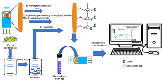
2. Materials and Methods

2.1. Lectin Extraction and Evaluation

All solutions were prepared in deionised water with resistance not less than 18 MΩ cm, purified by a Milli-Q system (Millipore, Bedford, MA, USA).
The HA was provided by the Biological Research Center of the Autonomous University of Hidalgo State. HA leaves were dried at room temperature and then pulverized in a mortar. The dried material (10.0 g) was stirred for 60 min in 150 mM NaCl (50.0 mL) (J.T. Baker, Phillipsburg, NJ, USA). The mixture was left for 16 h at 4 °C, and then filtered through gauze. The liquid phase was centrifuged (15 min, 4000 rpm) and stored at −20 °C [34]. Protein content was estimated by the Bradford method using bovine serum albumin (Sigma-Aldrich; St. Louis, MO, USA) as standard [35]. Protein molecular weight was determined by polyacrylamide gel electrophoresis (SDS-PAGE) (11% resolving gel and 4% stacking gel), using Coomassie Brilliant Blue G-250 for protein staining [36].
Lectin presence in HA extracts was confirmed by the evaluation of hemagglutinating activity of the aqueous extract. HA extract (50.0 μL) was mixed with human blood type A, B, and O (50.0 μL each), and the mixture was incubated at 37 °C for 30 min. Agglutination was observed under a microscope at 40× magnification. Negative controls used a 150.0 mM NaCl solution in place of HA extract. Inhibition of hemagglutinating activity was evaluated with the following carbohydrates: N-acetyl-D-glucosamine, N-acetylneuraminic acid, L-fucose, D-fructose, D-galactose, D-glucose, D-maltose, and D-mannose (all Sigma-Aldrich). Inhibition tests were performed following the reported procedure, adding 0.5 M carbohydrate solution (50.0 μL) for both tests. Negative controls used a 150.0 mM NaCl solution in place of carbohydrate [37].

2.2. Biosensor Construction

The biosensor was constructed on the surface of a low-temperature curing, screen-printed gold electrode (DropSens 220BT, 4 mm diameter, Asturias, Spain). Gold was used as a working electrode, and silver was used as both the auxiliary and pseudo-reference electrodes. A 25.0 mM solution (10.0 μL) of 16-mercaptohexadecanoic acid (MHDA; Sigma Aldrich, St. Louis, MO, USA) in anhydrous absolute ethanol (J.T. Baker) was deposited on the electrode surface. The droplet was dried for 24 h at 25 °C, then the electrode was rinsed twice with ethanol, and left to dry at 25 °C. The surface was then activated with 20.0 mM N-(3-dimethylaminopropyl)-N-ethylcarbodiimide (EDC; Sigma Aldrich) and 5.0 mM N-hydroxysulfosuccinimide (NHS; Sigma Aldrich) solution (10.0 μL each) for 1 h at 25 °C. Then, the biosensor was rinsed with phosphate buffer solution (PBS) at pH 7.4, containing: 137.0 mM NaCl, 3.0 mM KCl, 10.0 mM Na2HPO4 and 2.0 mM KH2PO4, with 0.5 mM of CaCl2, ZnCl2 and MgCl2. Once the electrode surface was activated, the HA extract (40.0 μL) was applied and allowed to stand for 1.5 h. The electrode was then immersed in a 20.0 mM ethanolamine (Sigma Aldrich) solution (prepared in deionised water) for 30 min, in order to block the still-active positions. The biosensor was rinsed and stored in PBS at 4 °C prior to use [38].
Micrographs of the biosensor surface were obtained by scanning electron microscopy (SEM; Carl Zeiss, model EVO-50, Oberkochen, Germany). The biosensor was evaluated after analyzing a sample containing 105 CFU mL−1 of Salmonella spp.

2.3. Impedimetric Measurements

All impedimetric measurements were carried out in a Metrohm Autolab galvanostat potentiostat electrochemical system (model PGSTATAT302N, Amsterdam, The Netherlands), controlled through Nova 2.0 software.
Cyclic voltammetry was used to select the direct current potential used for impedance measurements. Figure 1 shows the cyclic voltamograms of Fe(CN)64−/Fe(CN)63− redox couple at different modified electrode surfaces. A reversible Nersitan charge transfer was observed at Au bare surface at 350 mV (vs. Ag) whereas an absence of current peaks for modified surfaces was obtained due to the formation of a coating, indicating an increase in electron transfer resistance.
Impedance measurements were performed at the formal potential of the Fe(CN)64−/Fe(CN)63− redox pair. A direct current potential of 350 mV and an alternating current potential of 5 mV were applied. Impedance was determined by electrochemical impedance spectroscopy (EIS), at 25 frequencies from 0.010 Hz to 100 kHz. A volume of 40.0 µL of either the standard or sample was placed on the biosensor for 15 min at 25 °C, then the electrode was rinsed with PBS solution, and the impedance measurement was performed with 40 µL of the redox probe (5.0 mM potassium hexacyanoferrate (III) +5.0 mM potassium hexacyanoferrate (II) trihydrate) [38]. The impedance spectra were plotted in the form of Nyquist plots.
The biosensor was tested employing carbohydrates, which inhibited agglutination, at concentrations between 1 × 10⁻⁷–1 × 10⁻⁴ M. Listeria monocytogenes and Salmonella spp. were provided by the Food Microbiology laboratory at Autonomous University of Hidalgo State. Bacteria were grown in tryptic soy broth (Bioxon Becton Dickinson, Mexico), and incubated at 37 °C for 24 h. Decimal dilutions were made in 0.85% saline solution to obtain a concentration of 105 CFU mL−1 of each microorganism, to be analysed by EIS.
To evaluate the proposed biosensor in real samples for comparison with the traditional microbiological culture method, 10 samples of hen eggs were obtained from different commercial establishments in Pachuca, Hidalgo, Mexico. These were kept in polyethylene bags and stored at room temperature from their purchase until the analysis. An egg inoculated with 107 CFU mL−1 of Salmonella spp. acted as the positive control, and an egg washed with ethanol acted as the blank/negative control. A single egg was placed in lactose broth (LB; Bioxon) (225 mL) for 60 min at room temperature, then the egg was removed, and the broth was incubated for 24 h at 37 °C for pre-enrichment. For selective enrichment, 1.0 mL of the broth solution was placed in 10.0 mL of Rappaport-Vassiliadis broth (RVB; Bioxon) for 24 h at 37 °C. Two aliquots were taken, the first (40.0 µL) for analysis employing the biosensor, and the second for inoculation in Salmonella-Shigella (Bioxon) and brilliant green (Bioxon) for 24 h at 37 °C. Both Salmonella-Shigella and brilliant green agars are selective media for the growth of Salmonella spp. [39,40] (Figure 2).

3. Results and Discussion

3.1. Lectin Evaluation

The presence of lectin in the HA extract was determined by analysing the agglutination of the erythrocytes [41,42,43]. Three types of human blood showed agglutination in the presence of HA extract. Agglutination was observed as accumulations of the erythrocytes, which was not shown in their respective negative controls. The agglutination inhibition test for free carbohydrates is based on the affinity of lectin for these molecules [43,44,45,46]. The results showed that the carbohydrates that inhibited agglutination were N-acetyl-D-glucosamine, N-acetylneuraminic acid and D-mannose, indicating that the lectin presented active sites that allowed it to interact with these carbohydrates.
The total protein content in the HA extract was 131.7 mg L−1. Analysis by sodium dodecyl sulfate-polyacrylamide gel electrophoresis allowed us to identify the amount of proteins contained in the HA extract, as well as the molecular weight of the protein, when compared to a protein standard of known molecular weight. After staining polyacrylamide gel with Coomassie Brilliant Blue G-250, a single band near 27.4 kDa was observed, indicating that the HA extract could be used without any additional purification process.
The extracted lectin has a similar carbohydrate affinity compared to other lectins. For example, Cratylia mollis lectin (31 kDa) showed an affinity for D-glucose and D-mannose [47]. It was used in the development of an impedimetric biosensor based on self-assembled hybrid cysteine-gold nanoparticles for the analysis of S. enterica [48]. Also, a Triticum vulgaris lectin (36 kDa), with an affinity for glycoconjugates with N-acetylglucosamine or N-acetylneuraminic acid residues, was used for magnetic separation of S. enterica [49].

3.2. Biosensor Impedimetric Evaluation

Impedimetric biosensors are based on the immobilization of a biological recognition element related to the analyte. When recognition occurs on the electrode, changes in the interfacial electron transfer impedance occur. The interfacial changes generated by the recognition processes are detected by EIS [50], and are proportional to the concentration of the analyte [50,51].
The formation of self-assembled monolayers (SAMs) is one of the most-used methods for the immobilization of antibodies in immunoassays. The immobilization of the recognition agent on a surface requires the following characteristics: (1) the recognition element must be oriented with minimum steric impediment to effectively interact with the analyte, and (2) the recognition agent should not lose its ability to interact with the analyte. The main methods of immobilization are the formation of imines with glutaraldehyde and the formation of amides with the addition of EDC/NHS [38,52]. The amide formation offers a high conversion efficiency, mild reaction conditions, and excellent biocompatibility, with little influence on the bioactivity of the target molecules and much cleaner products than other reagents, such as glutaraldehyde [38,53].
In this work, the biosensor was evaluated for measuring the interaction between lectin and carbohydrates that inhibited agglutination in blood. Solutions of N-acetyl-D-glucosamine, N-acetylneuraminic acid, and D-mannose (1 × 10⁻⁷–1 × 10⁻⁴ M), were analysed, and an increase in impedance with the concentration of carbohydrates was observed (Figure 3A), while no significant changes were observed during analysis of D-galactose (Figure 3B). The lectin extracted from HA presented an affinity for N-acetyl-D-glucosamine, N-acetylneuraminic acid and D-mannose. These carbohydrates are contained in the cell wall of some Salmonella spp. [48,54,55] and could be exploited when designing a device for microbiological analysis of Salmonella spp.
The biosensor was applied for the analysis of independent solutions of a Gram-positive (L. monocytogenes) and a Gram-negative (Salmonella spp.) bacteria (each 105 CFU mL−1). The analysis of L. monocytogenes did not reveal any change in impedance (Figure 4A). Although a positive response was expected in Listeria spp., it being a Gram-positive bacterium with a cell wall containing high levels of peptidoglycan, a linear polymer made up of N-acetyl-D-glucosamine and N-acetylmuramic acid that interacts with amino acids [56]. However, since peptidoglycan is not the terminal carbohydrate, there is no interaction with the recognition agent from the HA lectin. An increase in impedance was observed when Salmonella spp. was analysed (Figure 4B). This impedance increase can be attributed to the lectin—Salmonella spp. interaction; the bacterium contains an external membrane made up of lipopolysaccharides containing different terminal carbohydrates, including D-mannose as the terminal carbohydrate [30].
The interactions that occur in the recognition between a lectin and carbohydrates are hydrogen bonding, metal coordination, van der Waals forces and hydrophobic interactions [26]. In microbial biosensors, the interaction occurs between microorganisms (lipopolysaccharides in the cell wall) and the recognition element (lectin) [57]. Figure 5 shows the recognition scheme for Salmonella spp. the presence of a coating (SAMs) on the surface of the electrode changes both the interfacial capacitance and the speed of electron transfer, causing an increase in impedance. This increase is because the bacteria, bound to the surface through interaction with lectin, act as a barrier for the transfer of electrons between the redox probe (Fe(CN)64−/Fe(CN)63−) and the biosensor.
Based on the results of agglutination inhibition and the change in the impedance of the device, it is probable that the lectin contained in HA extract interacts with D-mannose contained in the lipopolysaccharide of Salmonella spp.
Figure 6A shows an SEM micrograph of gold electrodes cured at low temperature, where an irregular surface with small agglomerations acting as single microelectrodes can be observed. The micrograph of the biosensor employed for analysis of 105 CFU mL−1 Salmonella spp. (Figure 6B) shows a layer formation on the electrode surface, and the presence of Salmonella spp. bacteria. This can be attributed to the lectin–carbohydrate interaction.

3.3. Sample Analysis

The biosensor was applied for analysis of hen eggs. The proposed methodology for the EIS analysis involves pre-enrichment, which restores the damaged bacterial cells to a stable physiological condition, then a selective enrichment used to increase the population of Salmonella spp. by inhibiting the growth of other organisms present in the sample, subsequently reducing interferences from other bacteria [6,8,21]. The analysis for Salmonella spp. was constructed using the Nyquist plots, showing that, in a concentration range of 103–107 CFU mL−1, there is an increase in impedance with respect to the logarithm of the concentration of Salmonella spp. (Figure 6). The biosensor resistance (ΔR) vs. logarithm of the concentration of Salmonella spp. were linear in the same interval (Figure 7), with a limit of detection of 5 CFU mL−1 and a limit of quantification of 15 CFU mL−1 (R2 = 0.9902). The reproducibility was evaluated analysing with three independent biosensors samples containing 0, 105 and 106 CFU mL−1, the relative standard deviation obtained from the analysis was 3.45, 1.69 and 0.90%, respectively. The values obtained were adequate for a biosensor.
Lectins are selective recognition elements for the detection of pathogens, in addition, the collection and extraction processes were not complicated. Lectins were investigated for the detection of some pathogens due to their ability to bonding selectively to the surfaces of viruses and bacteria [58]. Table 1 shows some lectins applied to analyse pathogens, Concanavalin is the most studied lectin and it was used for the detection of Escherichia coli. Currently, the development of electrochemical biosensors based on different lectins for the detection of different pathogens important to human health is an interesting area in biosensing [59].
On the other hand, the biosensor based on lectin obtained from Hechtia argentea is competitive compared to other lectins used for bacterial analysis (Table 1). Furthermore, the LOD obtained is comparable with other biosensors based on specific antigens. Yang et al. [65] developed a biosensor based on a self-assembled gold nanoparticle monolayer and grafted ethylenediamine on a vitreous carbon electrode, immobilizing specific monoclonal antibodies to Salmonella spp. in gold nanoparticles, achieving a limit of detection of 100 CFU mL−1.
The proposed biosensor was used to analyse 10 hen eggs, including negative and positive control samples. For all test samples and the negative control sample, a change in impedance in the analysis was not observed using the biosensor. The positive control showed the expected change in impedance. The microbiological analysis was congruent, and presumptive colonies of Salmonella spp. were not observed. The results obtained are preliminary, but there must be test more samples, including other analytical matrices, in order to evaluate if the biosensor is robust.

4. Conclusions

Hechtia argentea contains a lectin with a molecular weight near 27.4 kDa that is selective towards N-acetyl-D-glucosamine, N-acetylneuraminic acid, and D-mannose, allowing recognition of Salmonella spp. The biosensor achieved a limit of detection of 5 CFU mL−1 and a linear range of detection of 15–2.57 × 10⁷ CFU mL−1. The biosensor was applied successfully to the analysis of hen egg samples, and the results obtained were consistent with those obtained from the official analysis methodology. The proposed technique presents advantages over the official methodology for the detection of Salmonella spp., such as higher simplicity and shorter analysis time.

Author Contributions

Conceptualization, I.S.-O., C.T.H.-L. and J.A.R.; methodology J.L.-T.; formal analysis, E.M.S. and J.M.M.; investigation, J.L.-T.; writing—original draft preparation, J.L.-T.; writing—review and editing, J.M.M. and J.A.R. All authors have read and agreed to the published version of the manuscript.

Funding

This research received no external funding.

Conflicts of Interest

The authors declare no conflict of interest.

References

  1. World Health Organization. WHO Food Safety. April 2020. Available online: https://www.who.int/news-room/fact-sheets/detail/food-safety (accessed on 8 May 2020).
  2. Park, S.; Szonyi, B.; Gautam, R.; Nightingale, K.; Anciso, J.; Ivanek, R. Risk factors for microbial contamination in fruits and vegetables at the preharvest level: A systematic review. J. Food Prot. 2012, 75, 2055–2081. [Google Scholar] [CrossRef]
  3. Pui, C.F.; Wong, W.C.; Chai, L.C.; Nillian, E.; Ghazali, F.M.; Cheah, Y.K.; Radu, S. Simultaneous detection of Salmonella spp., Salmonella Typhi and Salmonella Typhimurium in sliced fruits using multiplex PCR. Food Control 2011, 22, 337–342. [Google Scholar] [CrossRef]
  4. Allerberger, F.; Liesegang, A.; Grif, K.; Khaschabi, D.; Prager, R.; Danzl, J.; Höck, F.; Ottl, J.; Dierich, M.P.; Berghold, C.; et al. Occurrence of Salmonella enterica serovar Dublin in Austria. Wien. Med. Wochenschr. 2002, 7, 65–70. [Google Scholar] [CrossRef] [PubMed]
  5. World Health Organization. WHO Salmonella (Non-Typhoidal). February 2018. Available online: https://www.who.int/news-room/fact-sheets/detail/salmonella-(non-typhoidal) (accessed on 8 May 2020).
  6. Musaj, A.; Llugaxhiu, D.; Hyseni, B. Detection of Salmonella in Eggs. J. Int. Environ. App. Sci. 2018, 13, 1–7. [Google Scholar]
  7. Odumeru, J.A.; León-Velarde, C.G. Salmonella detection methods for food and food ingredients. In Salmonella—A Dangerous Foodborne Pathogen; Mahmoud, B., Ed.; InTechOpen: Rijeka, Croatia, 2012; pp. 373–392. [Google Scholar] [CrossRef] [Green Version]
  8. Zourob, M.; Elwary, S.; Turner, A.P. Principles of Bacterial Detection: Biosensors, Recognition Receptors and Microsystems; Springer: New York, NY, USA, 2008. [Google Scholar]
  9. Grimont, P.A.; Weill, F.X. Antigenic Formulae of the Salmonella Servovars, 9th ed.; WHO Collaborating Centre for Reference and Research on Salmonella: Paris, France, 2007; pp. 1–166. [Google Scholar]
  10. Shipp, C.R.; Rowe, B. A mechanised microtechnique for Salmonella serotyping. J. Clin. Pathol. 1980, 33, 595–597. [Google Scholar] [CrossRef] [PubMed] [Green Version]
  11. Waters, S.M.; Murphy, R.A.; Power, R.F. Characterisation of prototype Nurmi cultures using culture-based microbiological techniques and PCR-DGGE. Int. J. Food Microbiol. 2006, 110, 268–277. [Google Scholar] [CrossRef]
  12. Lynch, M.J.; Leon-Velarde, C.G.; McEwen, S.; Odumeru, J.A. Evaluation of an automated immunomagnetic separation method for the rapid detection of Salmonella species in poultry environmental samples. J. Microbiol. Meth. 2004, 58, 285–288. [Google Scholar] [CrossRef]
  13. Španová, A.; Rittich, B.; Karpíšková, R.; Čechová, L.; Škapova, D. PCR identification of Salmonella cells in food and stool samples after immunomagnetic separation. Bioseparation 2000, 9, 379–384. [Google Scholar] [CrossRef]
  14. Rijpens, N.; Herman, L.; Vereecken, F.; Jannes, G.; De Smedt, J.; De Zutter, L. Rapid detection of stressed Salmonella spp. in dairy and egg products using immunomagnetic separation and PCR. Int. J. Food Microbiol. 1999, 46, 37–44. [Google Scholar] [CrossRef]
  15. Kumar, R.; Surendran, P.K.; Thampuran, N. Evaluation of culture, ELISA and PCR assays for the detection of Salmonella in seafood. Lett. Appl. Microbiol. 2008, 46, 221–226. [Google Scholar] [CrossRef]
  16. Lee, K.M.; Runyon, M.; Herrman, T.J.; Phillips, R.; Hsieh, J. Review of Salmonella detection and identification methods: Aspects of rapid emergency response and food safety. Food Control 2015, 47, 264–276. [Google Scholar] [CrossRef]
  17. Mirhosseini, S.A.; Fooladi, A.A.I.; Amani, J.; Sedighian, H. Production of recombinant flagellin to develop ELISA-based detection of Salmonella enteritidis. Braz. J. Microbiol. 2017, 48, 774–781. [Google Scholar] [CrossRef] [PubMed]
  18. Nicholas, R.A.; Cullen, G.A. Development and application of an ELISA for detecting antibodies to Salmonella enteritidis in chicken flocks. Veter. Rec. 1991, 128, 74. [Google Scholar] [CrossRef] [PubMed]
  19. Malorny, B.; Paccassoni, E.; Fach, P.; Bunge, C.; Martin, A.; Helmuth, R. Diagnostic real-time PCR for detection of Salmonella in food. App. Environ. Microbiol. 2004, 70, 7046–7052. [Google Scholar] [CrossRef] [PubMed] [Green Version]
  20. Moosavy, M.H.; Esmaeili, S.; Amiri, F.B.; Mostafavi, E.; Salehi, T.Z. Detection of Salmonella spp. in commercial eggs in Iran. Iran. J. Microbiol. 2015, 7, 50. [Google Scholar] [PubMed]
  21. Juneja, V.K.; Cherry, J.P.; Tunick, M. Advances in Microbial Food Safety; Symposium Series; American Chemical Society: Washington, DC, USA, 2006; pp. 14–27. [Google Scholar]
  22. Saleem, M. Biosensors a promising future in measurements. In Proceedings of the 1st International Conference on Sensing for Industry, Control, Communications, & Security Technologies (ICSICCST 2013), Karachi City, Pakistan, 24–26 June 2013; Volume 51. [Google Scholar] [CrossRef]
  23. Lei, Y.; Chen, W.; Mulchandani, A. Microbial biosensors. Anal. Chim. Acta 2006, 568, 200–210. [Google Scholar] [CrossRef] [PubMed]
  24. Thévenot, D.R.; Toth, K.; Durst, R.A.; Wilson, G.S. Electrochemical biosensors: Recommended definitions and classification. Biosens. Bioelectron. 2001, 16, 121–131. [Google Scholar] [CrossRef]
  25. Silva, N.F.; Magalhães, J.M.; Freire, C.; Delerue-Matos, C. Electrochemical biosensors for Salmonella: State of the art and challenges in food safety assessment. Biosens. Bioelectron. 2018, 99, 667–682. [Google Scholar] [CrossRef] [Green Version]
  26. Zeng, X.; Andrade, C.A.; Oliveira, M.D.; Sun, X.L. Carbohydrate–protein interactions and their biosensing applications. Anal. Bioanal. Chem. 2012, 402, 3161–3176. [Google Scholar] [CrossRef]
  27. Preston, A.; Mandrell, R.E.; Gibson, B.W.; Apicella, M.A. The lipooligosaccharides of pathogenic gram-negative bacteria. Crit. Rev. Microbiol. 1996, 22, 139–180. [Google Scholar] [CrossRef]
  28. Helander, I.M.; Mamat, U.; Rietschel, E.T. Lipopolysaccharides. eLS 2001. [Google Scholar] [CrossRef]
  29. Lam, J.S.; Graham, L.L.; Lightfoot, J.; Dasgupta, T.; Beveridge, T.J. Ultrastructural examination of the lipopolysaccharides of Pseudomonas aeruginosa strains and their isogenic rough mutants by freeze-substitution. J. Bacteriol. 1992, 174, 7159–7167. [Google Scholar] [CrossRef] [PubMed] [Green Version]
  30. Devyatyarova-Johnson, M.; Rees, I.H.; Robertson, B.D.; Turner, M.W.; Klein, N.J.; Jack, D.L. The Lipopolysaccharide Structures of Salmonella enterica Serovar Typhimurium and Neisseria gonorrhoeae Determine the Attachment of Human Mannose-Binding Lectin to Intact Organisms. Infect. Immun. 2000, 68, 3894–3899. [Google Scholar] [CrossRef] [PubMed] [Green Version]
  31. Van Damme, E.J.M.; Peumans, W.J.; Pusztai, A.; Bardocz, S. Handbook of Plant lectins: Properties and Biomedical Applications; John Wiley & Sons: Chichester, UK, 1998. [Google Scholar]
  32. Yagi, F.; Hidaka, M.; Minami, Y.; Tadera, K. A lectin from leaves of Neoregelia flandria recognizes D-glucose, D-mannose and N-acetyl D-glucosamine, differing from the mannose-specific lectins of other monocotyledonous plants. Plant Cell Physiol. 1996, 37, 1007–1012. [Google Scholar] [CrossRef] [Green Version]
  33. Ramírez-Morillo, I.M.; Hornung-Leoni, C.T.; González Ledesma, M.; Romero-Soler, K.J. “The Old White Lady of Kew Gardens”, Hechtia argentea (Bromeliaceae: Hechtioideae), found her homeland in Mexico. Taxon 2020. [Google Scholar] [CrossRef]
  34. Santana, G.M.S.; Albuquerque, L.P.; Simões, D.A.; Coelho, L.C.B.B.; Paiva, P.M.G.; Gusmão, N.B. Isolation of lectin from Opuntia ficus-indica cladodes. Acta Hortic. 2009, 811, 281–286. [Google Scholar] [CrossRef]
  35. Kruger, N.J. The Bradford method for protein quantitation. In The Protein Protocols Handbook, 3rd ed.; Walker, J.M., Ed.; Humana Press: Totowa, NJ, USA, 2009; pp. 17–24. [Google Scholar]
  36. Laemmli, U.K. Cleavage of structural proteins during the assembly of the head of bacteriophage T4. Nature 1970, 227, 680–685. [Google Scholar] [CrossRef]
  37. Moreira, R.D.A.; Perrone, J.C. Purification and partial characterization of a lectin from Phaseolus vulgaris. Plant Physiol. 1977, 59, 783–787. [Google Scholar] [CrossRef] [Green Version]
  38. Silva, M.L.S.; Gutiérrez, E.; Rodríguez, J.A.; Gomes, C.; David, L. Construction and validation of a Sambucus nigra biosensor for cancer-associated STn antigen. Biosens. Bioelectron. 2014, 57, 254–261. [Google Scholar] [CrossRef]
  39. Secretaria de Salud. NORMA Oficial Mexicana NOM-210-SSA1-2014, Products and Services. Microbiological Test Methods. Determination of Indicator Microorganisms. Determination of Pathogenic Microorganisms for Official Mexican Standard; Diario Oficial de la Federacion: Distrito Federal, Mexico, 2015; pp. 1–96. [Google Scholar]
  40. ISO. ISO 6579-1:2017. Microbiology of the Food Chain—Horizontal Method for the Detection, Enumeration and Serotyping of Salmonella—Part 1: Detection of Salmonella spp., 1st ed.; International Organization for Standardization: Geneva, Switzerland, 2017. [Google Scholar]
  41. Kennedy, J.F.; Palva, P.M.G.; Corella, M.T.S.; Cavalcanti, M.S.M.; Coelho, L.C.B.B. Lectins, versatile proteins of recognition: A review. Carbohydr. Polym. 1995, 26, 219–230. [Google Scholar] [CrossRef]
  42. Sharon, N.; Lis, H. History of lectins: From hemagglutinins to biological recognition molecules. Glycobiology 2004, 14, 53R–62R. [Google Scholar] [CrossRef] [PubMed] [Green Version]
  43. Van Damme, E.J.; Lannoo, N.; Peumans, W.J. Plant lectins. Adv. Bot. Res. 2008, 48, 107–209. [Google Scholar] [CrossRef]
  44. Boyd, W.C.; Shapleigh, E. Antigenic relations of blood group antigens as suggested by tests with lectins. J. Immun. 1954, 73, 226–231. [Google Scholar] [PubMed]
  45. Goldstein, I.J.; Hayes, C.E. The lectins: Carbohydrate-binding proteins of plants and animals. Adv. Carbohydr. Chem. Biochem. 1978, 35, 127–340. [Google Scholar] [CrossRef] [PubMed]
  46. Nilsson, C. Lectins: Analytical Technologies, 1st ed.; Elsevier: Amsterdam, The Netherlands, 2008. [Google Scholar] [CrossRef]
  47. Correia, M.T.; Coelho, L. C Purification of a glucose/mannose specific lectin, isoform 1, from seeds of Cratylia mollis Mart. (Camaratu bean). Appl. Biochem. Biotech. 1995, 55, 261–273. [Google Scholar] [CrossRef]
  48. Oliveira, M.D.; Andrade, C.A.; Correia, M.T.; Coelho, L.C.; Singh, P.R.; Zeng, X. Impedimetric biosensor based on self-assembled hybrid cystein-gold nanoparticles and CramoLL lectin for bacterial lipopolysaccharide recognition. J. Colloid Interface Sci. 2011, 362, 194–201. [Google Scholar] [CrossRef] [PubMed] [Green Version]
  49. Payne, M.J.; Campbell, S.; Kroll, R.G. Lectin-magnetic separation can enhance methods for the detection of Staphylococcus aureus, Salmonella enteritidis and Listeria monocytogenes. Food Microbiol. 1993, 10, 75–83. [Google Scholar] [CrossRef]
  50. Karunakaran, C.; Pandiaraj, M.; Santharaman, P. Immunosensors. In Biosensors and Bioelectronics, 1st ed.; Karunakaran, C., Bhargava, K., Benjamin, R., Eds.; Elsevier: Amsterdam, The Netherlands, 2015; pp. 205–245. [Google Scholar] [CrossRef]
  51. Yang, L.; Bashir, R. Electrical/electrochemical impedance for rapid detection of foodborne pathogenic bacteria. Biotech. Adv. 2008, 26, 135–150. [Google Scholar] [CrossRef]
  52. Putzbach, W.; Ronkainen, N.J. Immobilization techniques in the fabrication of nanomaterial-based electrochemical biosensors: A review. Sensors 2013, 13, 4811–4840. [Google Scholar] [CrossRef]
  53. Wang, C.; Yan, Q.; Liu, H.B.; Zhou, X.H.; Xiao, S.J. Different EDC/NHS activation mechanisms between PAA and PMAA brushes and the following amidation reactions. Langmuir 2011, 27, 12058–12068. [Google Scholar] [CrossRef]
  54. Serra, B.; Gamella, M.; Reviejo, A.J.; Pingarron, J.M. Lectin-modified piezoelectric biosensors for bacteria recognition and quantification. Anal. Bioanal. Chem. 2008, 391, 1853–1860. [Google Scholar] [CrossRef] [PubMed]
  55. Wang, Y.; Ye, Z.; Si, C.; Ying, Y. Monitoring of Escherichia coli O157: H7 in food samples using lectin based surface plasmon resonance biosensor. Food Chem. 2013, 136, 1303–1308. [Google Scholar] [CrossRef] [PubMed]
  56. Doyle, R.J.; Dziarski, R. The bacterial cell: Peptidoglycan. In Molecular Medical Microbiology; Sussman, M., Ed.; Academic Press: Cambridge, MA, USA, 2002; pp. 137–154. [Google Scholar] [CrossRef]
  57. Su, L.; Jia, W.; Hou, C.; Lei, Y. Microbial biosensors: A review. Biosens. Bioelectron. 2011, 26, 1788–1799. [Google Scholar] [CrossRef] [PubMed]
  58. Reina, J.J.; Díaz, I.; Nieto, P.M.; Campillo, N.E.; Paez, J.A.; Tabarani, G.; Fieschi, F.; Rojo, J. Docking, synthesis, and NMR studies of mannosyl trisaccharide ligands for DC-SIGN lectin. Org. Biomol. Chem. 2008, 6, 2743–2754. [Google Scholar] [CrossRef] [PubMed] [Green Version]
  59. Cesewski, E.; Johnson, B.N. Electrochemical biosensors for pathogen detection. Biosens. Bioelectron. 2020, 159, 112214. [Google Scholar] [CrossRef] [PubMed]
  60. Yang, H.; Zhou, H.; Hao, H.; Gong, Q.; Nie, K. Detection of Escherichia coli with a label-free impedimetric biosensor based on lectin functionalized mixed self-assembled monolayer. Sens. Actuators B Chem. 2016, 229, 297–304. [Google Scholar] [CrossRef]
  61. Ma, F.; Rehman, A.; Liu, H.; Zhang, J.; Zhu, S.; Zeng, X. Glycosylation of quinone-fused polythiophene for reagentless and label-free detection of E. coli. Anal. Chem. 2015, 87, 1560–1568. [Google Scholar] [CrossRef]
  62. Xi, F.; Gao, J.; Wang, J.; Wang, Z. Discrimination and detection of bacteria with a label-free impedimetric biosensor based on self-assembled lectin monolayer. J. Electroanal. Chem. 2011, 656, 252–257. [Google Scholar] [CrossRef]
  63. Jantra, J.; Kanatharana, P.; Asawatreratanakul, P.; Hedstrom, M.; Mattiasson, B.; Thavarungkul, P. Real-time label-free affinity biosensors for enumeration of total bacteria based on immobilized concanavalin A. J. Environ. Sci. Health 2011, 46, 1450–1460. [Google Scholar] [CrossRef]
  64. Wan, Y.; Zhang, D.; Hou, B. Monitoring microbial populations of sulfate-reducing bacteria using an impedimetric immunosensor based on agglutination assay. Talanta 2009, 80, 218–223. [Google Scholar] [CrossRef]
  65. Yang, G.J.; Huang, J.L.; Meng, W.J.; Shen, M.; Jiao, X.A. A reusable capacitive immunosensor for detection of Salmonella spp. based on grafted ethylene diamine and self-assembled gold nanoparticle monolayers. Anal. Chim. Acta 2009, 647, 159–166. [Google Scholar] [CrossRef] [PubMed]
Figure 1. Cyclic voltammograms of the electrode surfaces at different stages of biosensor preparation, obtained in 40 µL of 5.0 mM Fe(CN)64−/Fe(CN)63− solution, scan rate 20 mV s−1.
Chemosensors 08 00115 g001
Figure 2. Analysis scheme for Salmonella spp. detection. The sample is placed for pre-enrichment in lactose broth (LB) and enrichment in Rapaport-Vassiliadis broth (RVB). (A) Analysis of presumptive colonies, employing the microbiology method, involving inoculation in solid media (Salmonella-Shigella and brilliant green agars). (B) Analysis employing the biosensor, using 40 µL of RVB.
Chemosensors 08 00115 g002
Figure 3. Nyquist plots of (A) D-mannose (B) D-galactose, both in a concentration of 10−7–10−4 M.
Chemosensors 08 00115 g003
Figure 4. Nyquist plots of (A) L. monocytogenes, (B) Salmonella spp. Both analyses were performed at a concentration of 105 CFU mL−1.
Chemosensors 08 00115 g004
Figure 5. Recognition scheme for Salmonella spp. with the biosensor.
Chemosensors 08 00115 g005
Figure 6. (A) SEM micrograph of the unmodified gold electrode surface (DropSens 220BT). (B) SEM micrograph of the surface of the gold electrode modified with Salmonella spp. (105 CFU mL−1).
Chemosensors 08 00115 g006
Figure 7. Nyquist plots of Salmonella spp. in the concentration range of: (a) 0, (b) 103, (c) 104, (d) 105, (e) 106 and (f) 107 CFU mL−1.
Chemosensors 08 00115 g007
Table 1. Lectin-based biosensors for bacteria analysis.
Lectin SourceRecognition CarbohydrateBacteriaDetection TechniqueLODRef
Arachis hypogaeaN-acetyl-D-galactoseamine and D-galactoseL. monocytogenesSurface plasmon resonance-[55]
Canavalia ensiformisD-glucose and D-mannoseE. coli DH5αEIS75 cell/mL[60]
E. coli W1485Square wave voltammetry25 cell/mL[61]
E. coliSurface plasmon resonance12 CFU/mL[62]
Desulforibrio caledoiensisEIS1.8 CFU/mL[63]
Cratylia mollisD-glucoseSerratia. marcescens, E. coli, S. enterica and K. pneumoniaeEIS-[48]
Ricinus communisN-acetyl-D-galactosamine and D-galactoseE. coli DH5α Enterobacter cloacae, and Bacillus subtilisEIS-[64]
Triticum vulgarisN-acetylneuraminic acidE. coli O157: H7Surface plasmon resonance3 × 103 CFU/mL[55]
Hechtia argenteaN-acetyl-D-glucosamine, N-acetylneuraminic acid and D-mannoseSalmonella spp.EIS5 CFU/mLThis work
Publisher’s Note: MDPI stays neutral with regard to jurisdictional claims in published maps and institutional affiliations.

