

Approaches to Obtaining Water-Insoluble Fibrous Matrices from Regenerated Fibroin
 , ,
, ,  ,
,
Abstract
:1. Introduction
2. Materials and Methods
2.1. Chemicals
2.2. Preparation of Regenerated Fibroin
2.3. Preparation of Fibroin Solutions and Mixed Solutions of Fibroin and Chitosan
2.4. Kinetics of Change in Viscosity of SF Solutions
2.5. Measurement of Turbidity
2.6. Electrical Conductivity of Fibroin Solutions
2.7. Thermogravimetric Analysis
2.8. Solubility of SF Fibrous Matrices
2.9. Study of Gel Formation in Fibroin Solutions When Cross-Linked with Genipin
2.10. Preparation of Fibrous Matrices by Electrospinning
2.11. Modification of Fiber Matrices
2.12. Study of Fibroin Matrix Morphology Using Confocal Laser Scanning Microscopy
2.13. Study of the Structures of Fibrous Matrices by Confocal Microscopy
2.14. Cell Culture
2.15. Cell Cultivation in Fibrous Matrices
2.16. Study of Cell Proliferation in Fibrous Matrices
2.17. Morphology of Cells after Three Days of Cultivation in Fibrous Matrices
3. Results
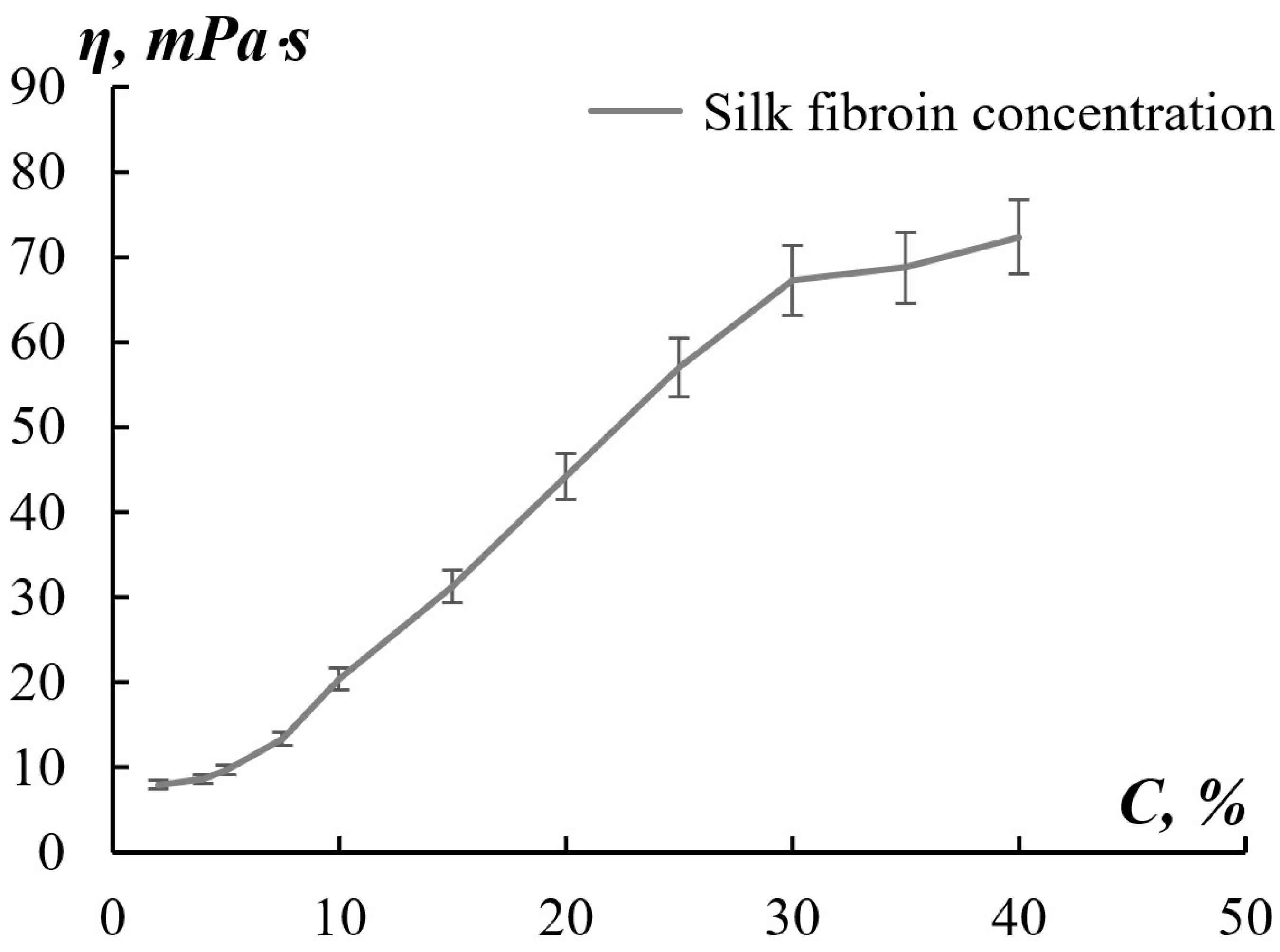
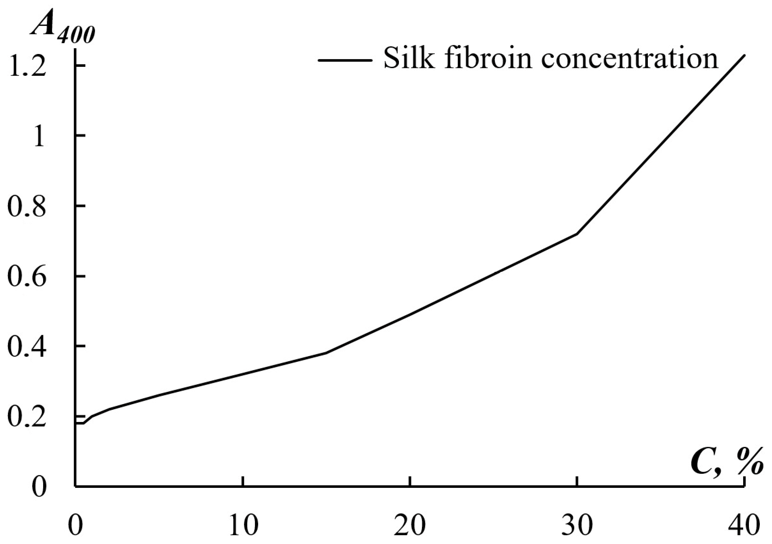
3.1. Effect of Silk Fibroin Solution Concentration on Fibrous Matrices from Regenerated Fibroin
3.2. Hydrophobization of Electrospun SF Fibers with An Ethanol Solution
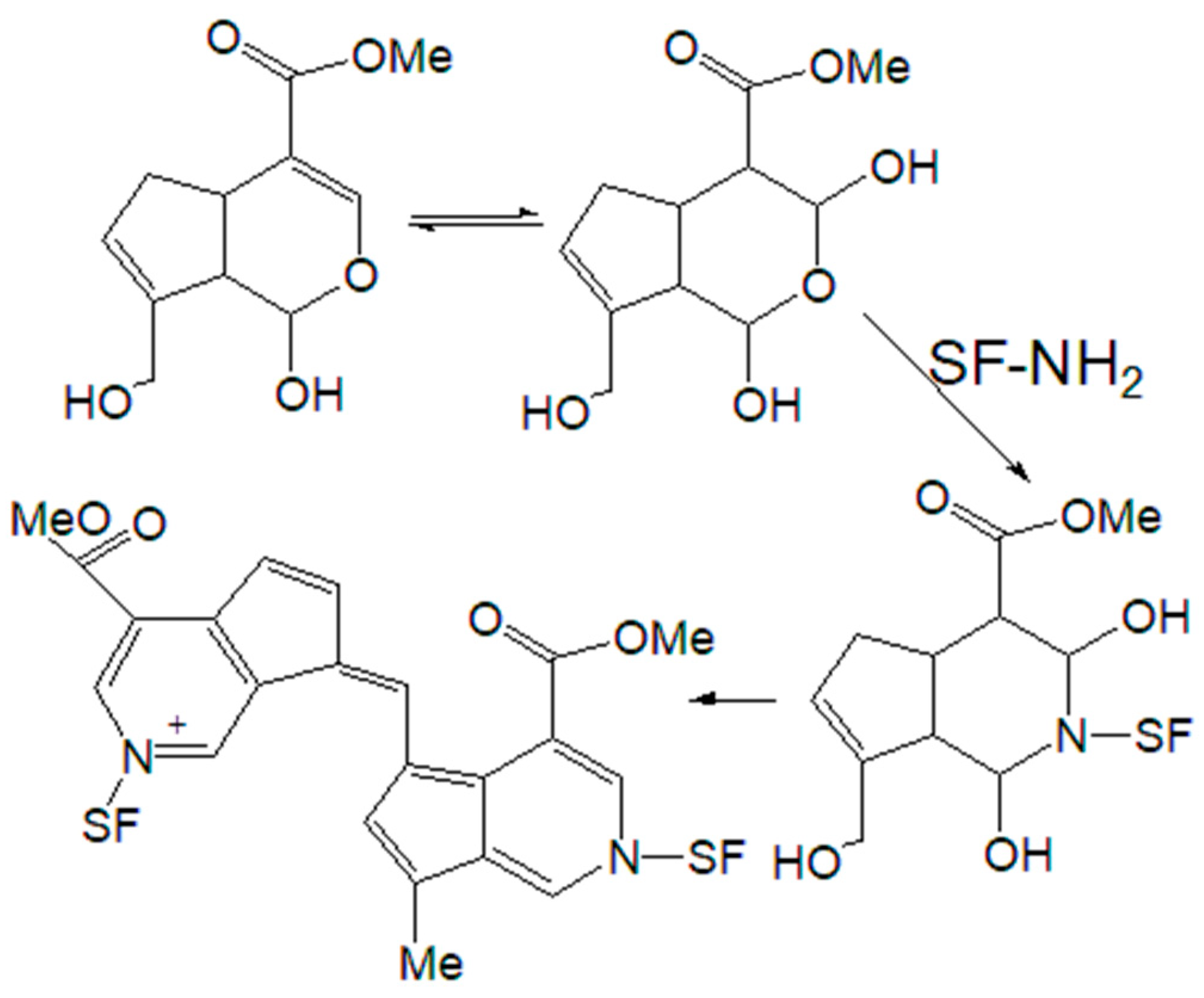
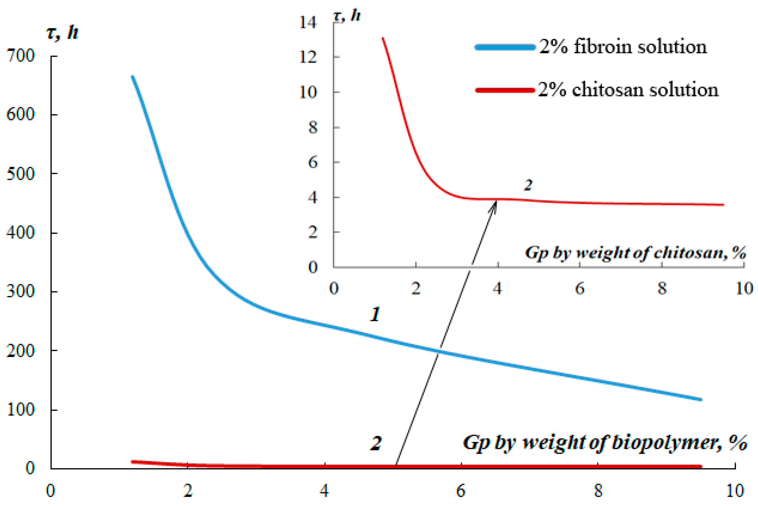
3.3. Use of a Genipin Cross-Linking Reagent for Preparation of Water-Insoluble Fibrous Matrices from Regenerated Fibroin
4. Conclusions
Author Contributions
Funding
Institutional Review Board Statement
Informed Consent Statement
Data Availability Statement
Acknowledgments
Conflicts of Interest
References
- Altman, G.H.; Diaz, F.; Jakuba, C.; Calabro, T.; Horan, R.L.; Chen, J.; Lu, H.; Richmond, J.; Kaplan, D.L. Silk-based biomaterials. Biomaterials 2003, 24, 401–416. [Google Scholar] [CrossRef]
- Zheng, Y.; Wang, L.; Zhao, L.; Wang, D.; Xu, H.; Wang, K.; Han, W. A flexible humidity sensor based on natural biocompatible silk fibroin films. Adv. Mater. Technol. 2021, 6, 2001053. [Google Scholar] [CrossRef]
- Zheng, H.; Zuo, B. Functional silk fibroin hydrogels: Preparation, properties and applications. J. Mater. Chem. B 2021, 9, 1238–1258. [Google Scholar] [CrossRef] [PubMed]
- Long, S.; Xiao, Y.; Zhang, X. Progress in preparation of silk fibroin microspheres for biomedical applications. Pharm. Nanotechnol. 2020, 8, 358–371. [Google Scholar] [CrossRef]
- Farokhi, M.; Mottaghitalab, F.; Fatahi, Y.; Saeb, M.R.; Zarrintaj, P.; Kundu, S.C.; Khademhosseini, A. Silk fibroin scaffolds for common cartilage injuries: Possibilities for future clinical applications. Eur. Polym. J. 2019, 115, 251–267. [Google Scholar] [CrossRef]
- Panda, P.K.; Sadeghi, K.; Seo, J. Recent advances in poly (vinyl alcohol)/natural polymer based films for food packaging applications: A review. Food Packag. Shelf Life 2022, 33, 100904. [Google Scholar] [CrossRef]
- Li, D.; Wang, Y.; Xia, Y. Electrospinning nanofibers as uniaxially aligned arrays and layer-by-layer stacked films. Adv. Mater. 2004, 16, 361–366. [Google Scholar] [CrossRef]
- Wang, Q.; Ma, J.; Chen, S.; Wu, S. Designing an innovative electrospinning strategy to generate PHBV nanofiber scaffolds with a radially oriented fibrous pattern. Nanomaterials 2023, 13, 1150. [Google Scholar] [CrossRef] [PubMed]
- Thanh, N.H.; Olekhnovich, R.; Sitnikova, V.; Kremleva, A.; Snetkov, P.; Uspenskaya, M. PHB/PEG Nanofiber Mat Obtained by Electrospinning and Their Performances. Technologies 2023, 11, 48. [Google Scholar] [CrossRef]
- Yang, N.; Qi, P.; Ren, J.; Yu, H.; Liu, S.-X.; Li, J.; Chen, W.; Kaplan, D.L.; Ling, S. Polyvinyl alcohol/silk fibroin/borax hydrogel ionotronics: A highly stretchable, self-healable, and biocompatible sensing platform. ACS Appl. Mater. Interfaces 2019, 11, 23632–23638. [Google Scholar] [CrossRef]
- Shi, W.; Que, Y.; Zhang, X.; Bian, L.; Yu, X.; Tang, X.; Yang, G.; Dai, Y.; Bi, S.; Lv, D.; et al. Functional tissue-engineered bone-like graft made of a fibrin scaffold and TG2 gene-modified EMSCs for bone defect repair. NPG Asia Mater. 2021, 13, 28. [Google Scholar] [CrossRef]
- Yang, W.; Xu, H.; Lan, Y.; Zhu, Q.; Liu, Y.; Huang, S.; Shi, S.; Hancharou, A.; Tang, B.; Guo, R. Preparation and characterisation of a novel silk fibroin/hyaluronic acid/sodium alginate scaffold for skin repair. Int. J. Biol. Macromol. 2019, 130, 58–67. [Google Scholar] [CrossRef] [PubMed]
- Grabska-Zielińska, S.; Sionkowska, A.; Reczyńska, K.; Pamuła, E. Physico-chemical characterization and biological tests of collagen/silk fibroin/chitosan scaffolds cross-linked by dialdehyde starch. Polymers 2020, 12, 372. [Google Scholar] [CrossRef] [PubMed]
- Roseti, L.; Parisi, V.; Petretta, M.; Cavallo, C.; Desando, G.; Bartolotti, I.; Grigolo, B. Scaffolds for bone tissue engineering: State of the art and new perspectives. Mater. Sci. Eng. C 2017, 78, 1246–1262. [Google Scholar] [CrossRef]
- Kasoju, N.; Bora, U. Silk fibroin in tissue engineering. Adv. Healthc. Mater. 2012, 1, 393–412. [Google Scholar] [CrossRef]
- Rinaudo, M. Chitin and chitosan: Properties and applications. Prog. Polym. Sci. 2006, 31, 603–632. [Google Scholar] [CrossRef]
- Wang, J.; Zhuang, S. Chitosan-based materials: Preparation, modification and application. J. Clean. Prod. 2022, 355, 131825. [Google Scholar] [CrossRef]
- Sazhnev, N.A.; Drozdova, M.G.; Rodionov, I.A.; Kil’deeva, N.R.; Balabanova, T.V.; Markvicheva, E.A.; Lozinsky, V.I. Preparation of chitosan cryostructurates with controlled porous morphology and their use as 3D-scaffolds for the cultivation of animal cells. Appl. Biochem. Microbiol. 2018, 54, 459–467. [Google Scholar] [CrossRef]
- Kildeeva, N.; Chalykh, A.; Belokon, M.; Petrova, T.; Matveev, V.; Svidchenko, E.; Surin, N.; Sazhnev, N. Influence of genipin crosslinking on the properties of chitosan-based films. Polymers 2020, 12, 1086. [Google Scholar] [CrossRef]
- Wang, H.Y.; Zhang, Y.Q. Processing silk hydrogel and its applications in biomedical materials. Biotechnol. Prog. 2015, 31, 630–640. [Google Scholar] [CrossRef]
- Koh, L.-D.; Cheng, Y.; Teng, C.-P.; Khin, Y.-W.; Loh, X.-J.; Tee, S.-Y.; Low, M.; Ye, E.; Yu, H.-D.; Zhang, Y.-W.; et al. Structures, mechanical properties and applications of silk fibroin materials. Prog. Polym. Sci. 2015, 46, 86–110. [Google Scholar] [CrossRef]
- Asakura, T.; Ohgo, K.; Komatsu, K.; Kanenari, M.; Okuyama, K. Refinement of repeated β-turn structure for silk I conformation of Bombyx mori silk fibroin using 13C solid-state NMR and X-ray diffraction methods. Macromolecules 2005, 38, 7397–7403. [Google Scholar] [CrossRef]
- Asakura, T. Structure of silk I (Bombyx mori silk fibroin before spinning)-type II β-turn, not α-helix. Molecules 2021, 26, 3706. [Google Scholar] [CrossRef] [PubMed]
- Yamada, H.; Nakao, H.; Takasu, Y.; Tsubouchi, K. Preparation of undegraded native molecular fibroin solution from silkworm cocoons. Mater. Sci. Eng. C 2001, 14, 41–46. [Google Scholar] [CrossRef]
- Farokhi, M.; Mottaghitalab, F.; Reis, R.L.; Ramakrishna, S.; Kundu, S.C. Functionalized silk fibroin nanofibers as drug carriers: Advantages and challenges. J. Control. Release 2020, 321, 324–347. [Google Scholar] [CrossRef]
- Chen, K.; Li, Y.; Li, Y.; Pan, W.; Tan, G. Silk fibroin combined with electrospinning as a promising strategy for tissue regeneration. Macromol. Biosci. 2023, 23, 2200380. [Google Scholar] [CrossRef]
- Yerra, A.; Dadala, M.M. Silk fibroin electrospun nanofiber blends with antibiotics and polyvinyl alcohol for burn wound healing. J. Appl. Polym. Sci. 2022, 139, 51930. [Google Scholar] [CrossRef]
- Sashina, E.S.; Sashina, E.S.; Bochek, A.M.; Bochek, A.M.; Novoselov, N.P.; Novoselov, N.P.; Kirichenko, D.A. Structure and solubility of natural silk fibroin. Russ. J. Appl. Chem. 2006, 79, 869–876. [Google Scholar] [CrossRef]
- Kiadeh, S.G.H.; Rahaiee, S. Extraction of silk fibroin protein from Bombyx mori cocoon by an optimized solvent system. In Proceedings of the 3rd International Conference on Modern Technologies in Sciences, Amol, Iran, 10 May 2023. [Google Scholar]
- Cheng, G.; Wang, X.; Wu, M.; Wu, S.; Cheng, L.; Zhang, X.; Dai, F. Insignificant Difference in Biocompatibility of Regenerated Silk Fibroin Prepared with Ternary Reagent Compared with Regenerated Silk Fibroin Prepared with Lithium Bromide. Polymers 2022, 14, 3903. [Google Scholar] [CrossRef]
- Li, G.; Sun, S. Silk fibroin-based biomaterials for tissue engineering applications. Molecules 2022, 27, 2757. [Google Scholar] [CrossRef]
- Cheng, G.; Wang, X.; Wu, M.; Wu, S.; Cheng, L.; Zhang, X.; Dai, F. Electrospinning and rheology of regenerated Bombyx mori silk fibroin aqueous solutions: The effects of pH and concentration. Polymer 2008, 49, 2880–2885. [Google Scholar]
- Singh, B.K.; Dutta, P.K. Chitin, chitosan, and silk fibroin electrospun nanofibrous scaffolds: A prospective approach for regenerative medicine. In Chitin and Chitosan for Regenerative Medicine; Springer: Berlin/Heidelberg, Germany, 2016; pp. 151–189. [Google Scholar]
- Kishimoto, Y.; Morikawa, H.; Yamanaka, S.; Tamada, Y. Electrospinning of silk fibroin from all aqueous solution at low concentration. Mater. Sci. Eng. C 2017, 73, 498–506. [Google Scholar] [CrossRef]
- Elango, J.; Lijnev, A.; Zamora-Ledezma, C.; Alexis, F.; Wu, W.; Marín, J.M.G.; de Val, J.E.M.S. The Relationship of Rheological Properties and the Performance of Silk Fibroin Hydrogels in Tissue Engineering Application. Process Biochem. 2022, 125, 198–211. [Google Scholar] [CrossRef]
- Tsukada, M.; Freddi, G.; Monti, P.; Bertoluzza, A.; Kasai, N. Structure and molecular conformation of tussah silk fibroin films: Effect of methanol. J. Polym. Sci. Part B Polym. Phys. 1995, 33, 1995–2001. [Google Scholar] [CrossRef]
- Shu, T.; Cui, J.; Lv, Z.; Cao, L.; Ren, J.; Ling, S. Moderate conformational transition promotes the formation of a self-reinforced highly oriented silk fibroin network structure. Soft Matter 2021, 17, 9576–9586. [Google Scholar] [CrossRef] [PubMed]
- Chen, X.; Shao, Z.; Knight, D.P.; Vollrath, F. Conformation transition kinetics of Bombyx mori silk protein. Proteins Struct. Funct. Bioinform. 2007, 68, 223–231. [Google Scholar] [CrossRef]
- Zuo, B.; Liu, L.; Wu, Z. Effect on properties of regenerated silk fibroin fiber coagulated with aqueous methanol/ethanol. J. Appl. Polym. Sci. 2007, 106, 53–59. [Google Scholar] [CrossRef]
- Nogueira, G.M.; Rodas, A.C.; Leite, C.A.; Giles, C.; Higa, O.Z.; Polakiewicz, B.; Beppu, M.M. Preparation and characterization of ethanol-treated silk fibroin dense membranes for biomaterials application using waste silk fibers as raw material. Bioresour. Technol. 2010, 101, 8446–8451. [Google Scholar] [CrossRef]
- Mo, C.; Wu, P.; Chen, X.; Shao, Z. The effect of water on the conformation transition of Bombyx mori silk fibroin. Vib. Spectrosc. 2009, 51, 105–109. [Google Scholar] [CrossRef]
- Li, M.; Tao, W.; Kuga, S.; Nishiyama, Y. Controlling molecular conformation of regenerated wild silk fibroin by aqueous ethanol treatment. Polym. Adv. Technol. 2003, 14, 694–698. [Google Scholar] [CrossRef]
- Tacias-Pascacio, V.G.; García-Parra, E.; Vela-Gutiérrez, G.; Virgen-Ortiz, J.J.; Berenguer-Murcia, Á.; Alcántara, A.R.; Fernandez-Lafuente, R. Genipin as an emergent tool in the design of biocatalysts: Mechanism of reaction and applications. Catalysts 2019, 9, 1035. [Google Scholar] [CrossRef]
- Yu, Y.; Xu, S.; Li, S.; Pan, H. Genipin-cross-linked hydrogels based on biomaterials for drug delivery: A review. Biomater. Sci. 2021, 9, 1583–1597. [Google Scholar] [CrossRef]
- Pizzolitto, C.; Cok, M.; Asaro, F.; Scognamiglio, F.; Marsich, E.; Lopez, F.; Donati, I.; Sacco, P. On the mechanism of genipin binding to primary amines in lactose-modified chitosan at neutral pH. Int. J. Mol. Sci. 2020, 21, 6831. [Google Scholar] [CrossRef] [PubMed]
- Bucciarelli, A.; Janigro, V.; Yang, Y.; Fredi, G.; Pegoretti, A.; Motta, A.; Maniglio, D. A genipin crosslinked silk fibroin monolith by compression molding with recovering mechanical properties in physiological conditions. Cell Rep. Phys. Sci. 2021, 2, 100605. [Google Scholar] [CrossRef]
- Zhou, C.-Z.; Confalonieri, F.; Jacquet, M.; Perasso, R.; Li, Z.-G.; Janin, J. Silk fibroin: Structural implications of a remarkable amino acid sequence. Proteins Struct. Funct. Bioinform. 2001, 44, 119–122. [Google Scholar] [CrossRef] [PubMed]


| Fibroin Solution Concentration, % | pH | Conductivity ϰ, mS/cm | Dynamic Viscosity η, mPa⋅s |
|---|---|---|---|
| 10 | 6.80 | 3.6 | 20.4 |
| 20 | 6.90 | 6.4 | 44.2 |
| 30 | 6.93 | 9.8 | 67.2 |
| Fibroin Solution Concentration, % w/w | Electrospinning Voltage E, kV | Characteristics of the Electrospinning Process | AFM Image | Fiber Diameter, μm |
|---|---|---|---|---|
| 10 | 22.2–25.8 | Stable |  | 0.61 ± 0.22 |
| 20 | 23.0–26.4 | Stable |  | 2.80 ± 0.20 |
| 30 | 24.1–28.0 | Unstable | 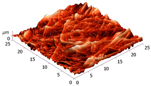 | 2.20 ± 0.80 |
| Concentration,% | Fibroin/Chitosan Ratio, g/g | pH | Conductivity ϰ, mS/cm | Dynamic Viscosity η, mPa⋅s | |
|---|---|---|---|---|---|
| Fibroin | Chitosan | ||||
| 20 | - | 1:0 | 6.90 | 6.4 | 44.13 |
| 10 | 2 | 5:1 | 4.84 | 27.3 | 62.21 |
| Fibrous Matrix Composition | Morphology of the Source Electrospun Fibrous Matrices | Morphology of the Fibrous Matrices Washed with PBS (pH 7.4) | Morphology of the Fibrous Matrices after 3 Days of Cultivation of hTERT-MSCs |
|---|---|---|---|
| 100% fibroin | 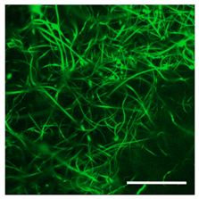 |  |  |
| Fibroin/Chitosan, 5:1 | 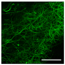 | 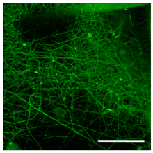 | 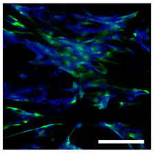 |
Disclaimer/Publisher’s Note: The statements, opinions and data contained in all publications are solely those of the individual author(s) and contributor(s) and not of MDPI and/or the editor(s). MDPI and/or the editor(s) disclaim responsibility for any injury to people or property resulting from any ideas, methods, instructions or products referred to in the content. |
© 2023 by the authors. Licensee MDPI, Basel, Switzerland. This article is an open access article distributed under the terms and conditions of the Creative Commons Attribution (CC BY) license (https://creativecommons.org/licenses/by/4.0/).
Share and Cite
Kildeeva, N.; Sazhnev, N.; Drozdova, M.; Zakharova, V.; Svidchenko, E.; Surin, N.; Markvicheva, E. Approaches to Obtaining Water-Insoluble Fibrous Matrices from Regenerated Fibroin. Technologies 2023, 11, 146. https://doi.org/10.3390/technologies11050146
Kildeeva N, Sazhnev N, Drozdova M, Zakharova V, Svidchenko E, Surin N, Markvicheva E. Approaches to Obtaining Water-Insoluble Fibrous Matrices from Regenerated Fibroin. Technologies. 2023; 11(5):146. https://doi.org/10.3390/technologies11050146
Chicago/Turabian StyleKildeeva, Nataliya, Nikita Sazhnev, Maria Drozdova, Vasilina Zakharova, Evgeniya Svidchenko, Nikolay Surin, and Elena Markvicheva. 2023. "Approaches to Obtaining Water-Insoluble Fibrous Matrices from Regenerated Fibroin" Technologies 11, no. 5: 146. https://doi.org/10.3390/technologies11050146