Article

H2O2 Induces Association of RCA with the Thylakoid Membrane to Enhance Resistance of Oryza meyeriana to Xanthomonas oryzae pv. oryzae

1
College of Plant Protection, Shenyang Agricultural University, Shenyang 210095, China
2
State Key Laboratory for Quality and Safety of Agro-products, Institute of Virology and Biotechnology, Zhejiang Academy of Agricultural Science, Hangzhou 310021, China
3
College of Plant Protection, Yunnan Agricultural University, Kunming 650000, China
4
Institute of Plant Virology, Ningbo University, Ningbo 315211, China
*
Author to whom correspondence should be addressed.
These authors contributed equally to this work.
Plants 2019, 8(9), 351; https://doi.org/10.3390/plants8090351
Submission received: 13 August 2019 / Revised: 4 September 2019 / Accepted: 10 September 2019 / Published: 16 September 2019
(This article belongs to the Section Plant Physiology and Metabolism)

Abstract

:
Oryza meyeriana is a wild species of rice with high resistance to Xanthomonas oryzae pv. oryzae (Xoo), but the detailed resistance mechanism is unclear. Ribulose-1, 5-bisphosphate carboxylase/oxygenase (Rubisco) activase (RCA) is an important enzyme that regulates photosynthesis by activating Rubisco. We have previously reported that Xoo infection induced the relocation of RCA from the chloroplast stroma to the thylakoid membrane in O. meyeriana, but the underlying regulating mechanism and physiological significance of this association remains unknown. In this study, “H2O2 burst” with rapid and large increase in the amount of H2O2 was found to be induced by Xoo invasion in the leaves of O. meyeriana. 3, 3-diaminobenzidine (DAB) and oxidative 2, 7-Dichlorodi-hydrofluorescein diacetate (H2DCFDA) staining experiments both showed that H2O2 was generated in the chloroplast of O. meyeriana, and that this H2O2 generation as well as Xoo resistance of the wild rice were dramatically dependent on light. H2O2, methyl viologen with light, and the xanthine-xanthine oxidase system all induced RCA to associate with the thylakoid membrane in vitro, which showed that H2O2 could induce the relocation of RCA. In vitro experiments also showed that H2O2 induced changes in both the RCA and thylakoid membrane that were required for them to associate and that this association only occurred in O. meyeriana and not in the susceptible cultivated rice. These results suggest that the association of RCA with the thylakoid membrane helps to protect the thylakoid membrane against oxidative damage from H2O2. Therefore, in addition to its universal function of activating Rubisco, RCA appears to play a novel role in the resistance of O. meyeriana to Xoo.

1. Introduction

Rice (Oryza sativa L.) is a primary staple cereal and a source of food for more than half of the world’s population, but it is susceptible to many pathogens that can dramatically decrease yields. Rice bacterial blight is caused by Xanthomonas oryzae pv. oryzae (Xoo) and it is one of the most destructive diseases in all the major rice growing countries, typically reducing yields by 20% to 30%, but by as much as 50% in years when the disease is prevalent [1,2,3,4]. Chemical control of this disease is temporarily effective, but it often causes environmental pollution, and the most effective method of control is generally recognized to be through the use of resistant cultivars. Nevertheless, it is difficult for plant breeders to find new resistance genes from within the currently narrow genetic base of rice cultivars. Although they have low grain yields, wild species of the genus Oryza have adapted through natural selection to survive in harsh environments, and therefore often possess many useful traits that are not present in cultivated rice, including high disease resistance. Many wild species are highly resistant to bacterial blight, including O. longistaminata [5], O. minuta [6], O. rufipogon [7], and O. officinalis [8]. The resistance mechanisms of these wild rice species to Xoo have been studied in detail, and their resistance traits have been introduced into cultivated rice in breeding programs to give sustained improvement of rice productivity [5,6,7,8]. However, such resistance is often overcome by rapidly evolving Xoo populations and it is recognized that a better use of the genetic diversity of wild rice provides the best way to broaden the genetic base and adaptability of rice cultivars.
O. meyeriana is one of the wild relatives of rice growing in tropical and subtropical areas of South and Southeast Asia, and has long been known to possess a high resistance to Xoo pathogens: in 1981 and 1982 Peng et al. had reported that the O. meyeriana from Yunnan province, China, had high Xoo resistance [9,10]. Zhang et al. evaluated the Xoo resistance of 871 accessions of 13 wild rice species and found that the O. meyeriana accessions ranked as the most highly resistant [11]. Our previous studies also confirmed the high resistance to Xoo pathogens of this wild rice species [3,4,12,13,14,15]. However, there has been no research regarding the underlying mechanisms of this resistance, and this has limited its use in breeding programmes.
Pathogen invasion often leads to distinct inhibition of plant photosynthesis. In tomato, Berger et al. showed that infection by Pseudomonas syringae or Botrytis cinerea suppressed photosynthetic gene expression and downregulated the photosynthetic rate [16]. In Arabidopsis, Bonfig et al. found that invasion by Pseudomonas syringae decreased the chlorophyll fluorescence parameters (maximum PSII quantum yield, effective PSII quantum yield, and nonphotochemical quenching), which indicate a decline in the photosynthetic reaction [17]. We also found that Xoo inoculation decreased the photosynthetic rate in rice [4]. The effect of pathogen invasion on photosynthesis and the response of the plant photosynthetic apparatus to this invasion are extremely complex, due to the complicated structure and functional mechanism of photosystems [16,17,18]. On the one hand, the pathogen attempts to manipulate the photosynthetic metabolism of the plant for its own advantage. On the other hand, the plant has to reorganize carbon fluxes to provide resources to fight the pathogens. The ways in which these reactions interact with one another needs more research.
In green plants, Ribulose-1,5-bisphosphate carboxylase/oxygenase (Rubisco) catalyses the carboxylation of ribulose-1, 5-bisphosphate and CO2 to form two 3-phosphoglyceric acid molecules, and this is a dominant rate-controlling step in photosynthetic CO2 assimilation. Rubisco activase (RCA) is a key enzyme in the regulation of photosynthesis, and its primary role is to activate Rubisco for maintaining its catalytic competency. RCA encircles the Rubisco molecule and the two proteins are chemically cross-linked [19,20,21]. Through protein-protein interactions and ATP hydrolysis, RCA can remove sugar-phosphate inhibitors from Rubisco catalytic sites to allow for the binding of Mg2+ and ribulose 1,5-bisphosphate [20,22]. Numerous studies have shown that RCA is sensitive to inactivation that arises from various biotic [23,24] and abiotic [25,26,27] stresses. However, to our knowledge, no data are available at present concerning the response of RCA to Xoo invasion in the wild rice O. meyeriana.
We have reported that the inoculation of O. meyeriana with Xoo induced the relocation of RCA from the chloroplast stroma to associate with the thylakoid membrane [14]. This association simultaneously led to a decline in the initial activity and the activation status of Rubisco in the wild rice. However, the underlying regulatory mechanism and physiological significance of this binding to the thylakoid membrane is not known. In this study, we found that Xoo infection induced an “H2O2 burst” with a rapid and transient increase in the amount of H2O2 in the leaves of O. meyeriana. Experiments, both in situ and in vitro, revealed that a large amount of H2O2 was generated in the chloroplast and that this then induced the redistribution of RCA from the chloroplast stroma to the thylakoid membrane. In vitro experiments showed that H2O2 induced the changes in both RCA and the thylakoid membrane that are required for their association, and that this effect only occurred in the wild rice and not in cultivated rice. Based on these results, we suggest that the association of RCA with the thylakoid membrane helps to protect the thylakoid membrane against oxidative damage by H2O2. Thus, RCA has a novel role in the resistance of O. meyeriana to Xoo pathogens, in addition to its universal function of activating Rubisco.

2. Results

2.1. Resistance Analysis

The resistance of O. sativa cv. Dalixiang and O. meyeriana to Xoo was compared by measuring lesion length on leaves three weeks after inoculation with Xoo or H2O (as control). There was a highly significant difference in the resistance between the two hosts (P < 0.001): the Xoo lesions on Dalixiang were approximately 16.6 cm long, while those on O. meyeriana were only about 0.62 cm (Figure 1). These results are in agreement with the earlier observations that O. meyeriana has a high level of resistance to Xoo [3,11,12].

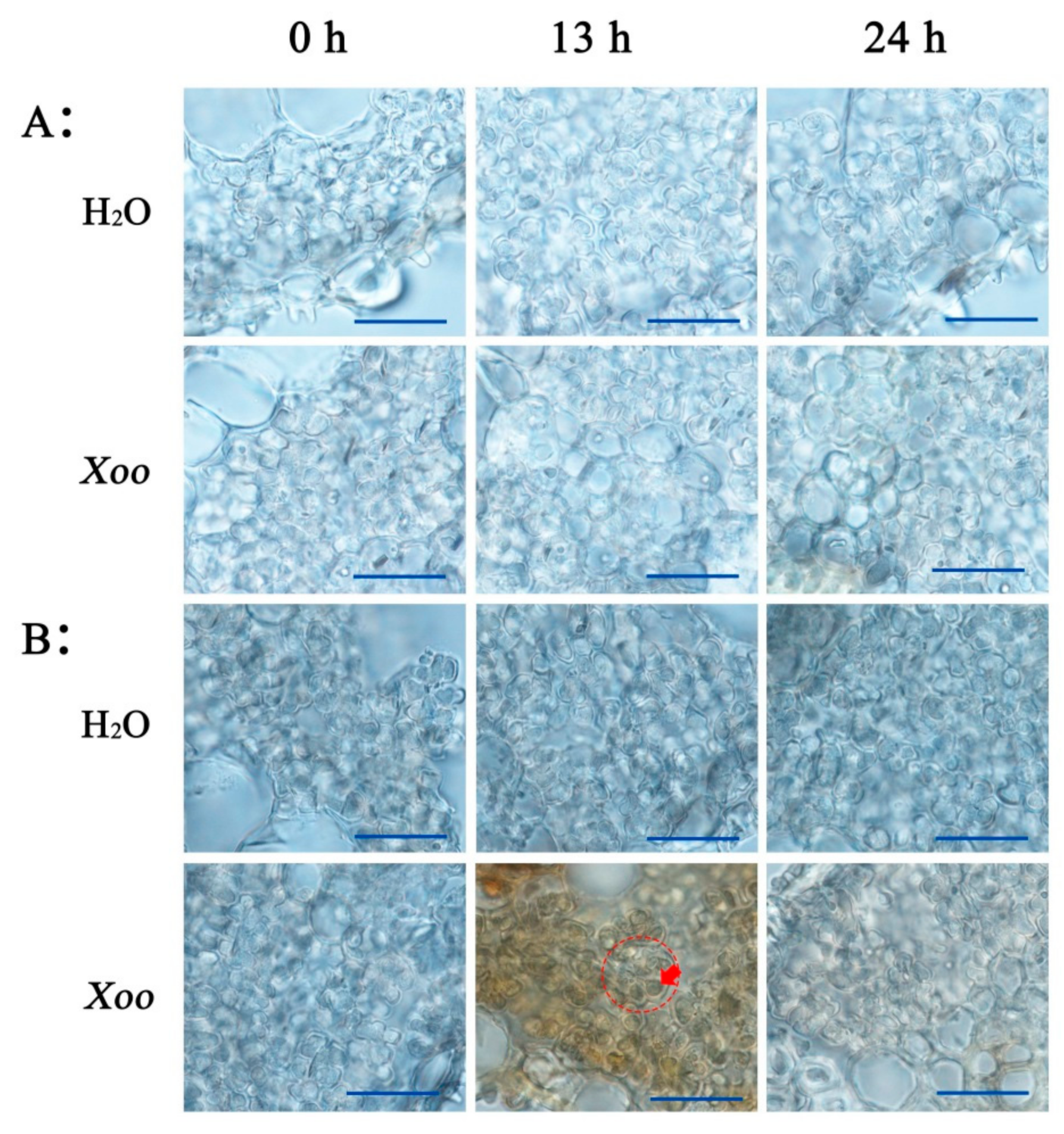
2.2. Effect of Xoo Inoculation on RCA Distribution

We first confirmed the earlier findings that Xoo infection induced the redistribution of RCA protein from the chloroplast stroma to the thylakoid membrane [14]. Intact chloroplasts were isolated from both O. meyeriana and the cultivar Dalixiang and the distribution of RCA between the chloroplast stroma and thylakoid membrane fractions were compared while using western blotting analysis. As shown in Figure 2, after mock inoculation with H2O, most RCA was found in the stroma fraction with only small amounts in the thylakoid membrane fraction of both plants. In cv. Dalixiang, there were no significant changes in either content or distribution of RCA after Xoo inoculation. However, in O. meyeriana RCA was sequestered to the thylakoid membrane from the stroma during the period from approximately 11 to 16 h after Xoo inoculation (Figure 2). The amounts of RCA in the soluble stroma decreased during this period and they did not recover to the original levels until approximately 17 h (Figure 2).
RCA activates Rubisco in the chloroplast stroma to maintain its catalytic competency, so the decline in the amount of RCA in chloroplast stroma decreased the initial activity and activation state of Rubisco in O. meyeriana not in O. sativa cv. Dalixiang [14]. The chlorophyll fluorescence parameters of Photosystem II (PSII) effective photochemical efficiency (ΦPSII) and PSII photochemical quenching (qP) reflect the part of PSII-absorbed light energy that was used for CO2 assimilation, and both of these parameters were notably decreased in O. meyeriana as the feedback of decline in CO2 assimilation due to the decreased Rubisco activation state (Supplementary Table S1). Plants have developed a photo-protective mechanism, which is called non-photochemical quenching (qN), to safely dissipate the excessive light energy as heat from PSII to protect the photosynthetic apparatus. In this study, in O. meyeriana inoculated with Xoo, qN was remarkably increased in parallel with the decrease in ΦPSII and qP (Supplementary Table S1). Fv/Fm, PSII maximum photochemical efficiency, often reflects whether or not the photosynthetic apparatus has suffered irreversible damage. There was no significant change in Fv/Fm in O. meyeriana (Supplementary Table S1), which indicated that the photosystem of the wild rice was not destroyed by the excessive light energy that was caused by Xoo infection due to the effective working of the protective mechanism. There were no significant differences in Fv/Fm, qP and qN in Dalixiang in the period shortly after Xoo inoculation (Supplementary Table S1) when compared to the wild rice, but all of the three chlorophyll fluorescence parameters were dramatically decreased three weeks after inoculation (Supplementary Table S1), which indicated that the photosystem of Dalixiang was severely damaged. These results are in agreement with the difference in resistance between O. meyeriana and Dalixiang shown by the lesion lengths in Figure 1.
There was no significant decline in the amount of RCA in the thylakoid membrane fraction of infected O. meyeriana following incubation in the HEPES-KOH buffer, and RCA was hardly detected in the buffer until the incubation lasted for 24 h (Figure 3A). The thylakoid membrane was also washed with 2 M NaCl or 2 M NaBr, or treated for 30 min. with 1% Triton X-100. Washing with 2 M NaBr has been shown to remove extrinsic thylakoid proteins [28]. However, none of these treatments released noticeable amounts of RCA (Figure 3B). However, limited proteolysis with thermolysin did decrease the amount of RCA in the thylakoid membrane (Figure 3B), indicating that thermolysin digested RCA thorough its protease activity. All of these results suggest that, in the wild rice, Xoo infection induces a tight association of RCA with the thylakoid membrane.

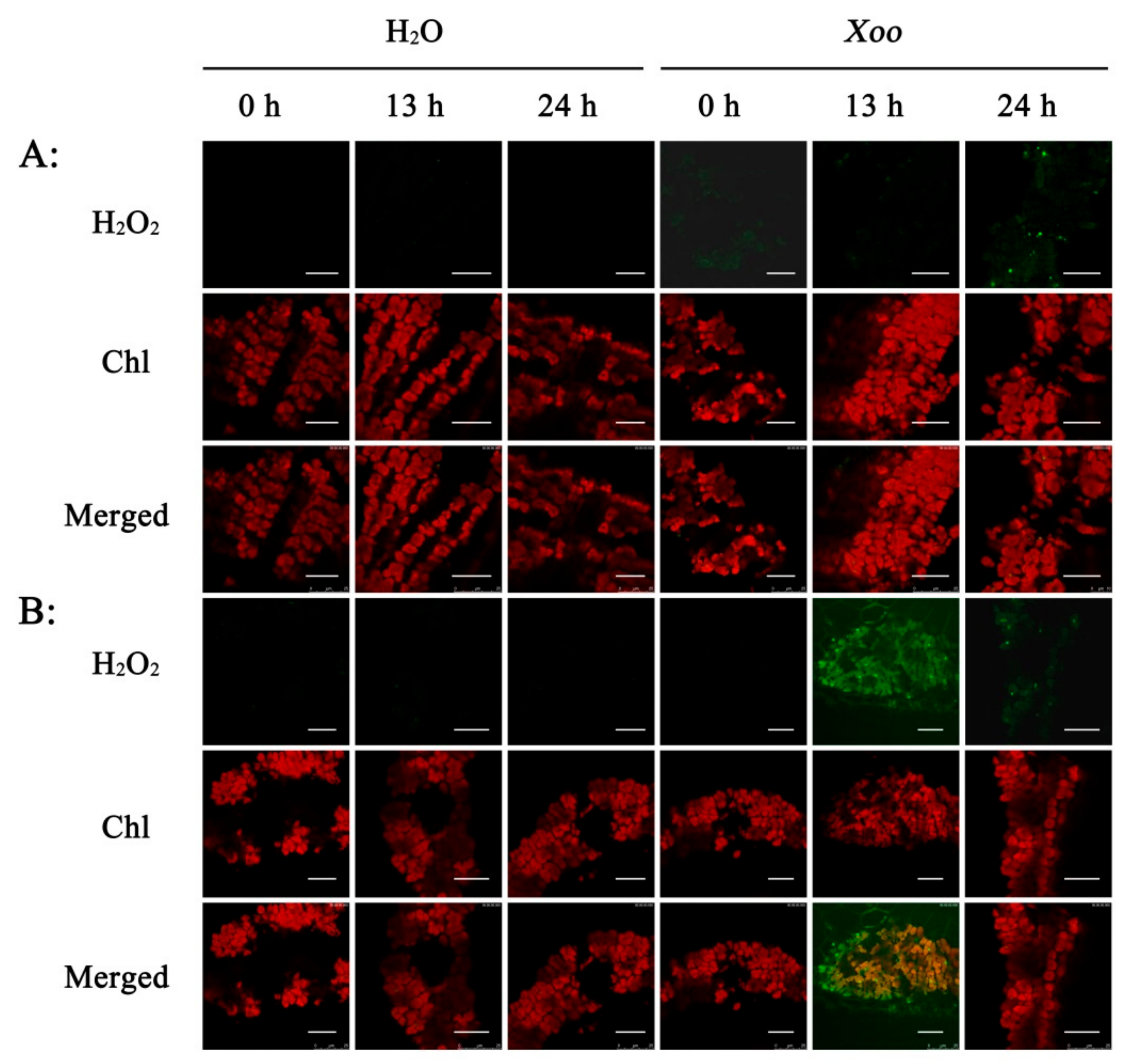
2.3. Effect of Xoo Inoculation on H2O2 Generation

H2O2 is a central signaling molecule that induces plant responses to pathogens. The H2O2 content of leaves was measured at different times after inoculation with Xoo to study the effect of Xoo inoculation on H2O2 generation. In leaves of cv. Dalixiang, H2O2 content increased slightly during the period 0-16 h after Xoo inoculation, and then remained stable (Figure 2). In O. meyeriana leaves inoculated with Xoo, there was a rapid increase in H2O2 content after approximately 10 h, peaking at about 13 h, and then declining rapidly until 16 h, after which the decrease was slight (Figure 2). During this period, the H2O2 content in the wild rice was much greater than in the cultivated rice; for example, at 13 h after Xoo inoculation, the H2O2 content was 12.70 µM g−1 FW in O. meyeriana, but only 4.49 µM g−1 FW in Dalixiang (Figure 2).
It is noteworthy that the period of rapid increase in H2O2 content was remarkably co-incident with that for the association of RCA with the thylakoid membrane (Figure 2), which strongly suggests some relationship between the rapidly increasing H2O2 content and the relocation of RCA within the chloroplasts of O. meyeriana.

2.4. Subcellular Localization of H2O2

We next investigated the subcellular localization of Xoo-induced H2O2 to determine whether it might be in contact with RCA. Two different methods were used.
After 3, 3-diaminobenzidine (DAB) staining, the mesophyll cells of O. meyeriana were stained brightly brown 13 h after Xoo inoculation as compared to the mock (water-inoculated) controls (Figure 4B), which indicates that Xoo infection induced a large H2O2 accumulation in the leaves of the resistant wild rice. This accumulation was mostly in the chloroplasts (Figure 4B). However, H2O2 accumulation was significantly less at 24 h after inoculation (Figure 4B). In contrast, there was little H2O2 accumulation in the leaves of Dalixiang at either 13 or 24 h after inoculation (Figure 4A). Therefore, these results are consistent with those from H2O2 examination of leaf extracts, as described above (Figure 2). In the water-inoculated control leaves, the color from DAB staining was slightly deeper in O. meyeriana than in Dalixiang, perhaps because the basal H2O2 content of the wild rice was slightly higher than that of the cultivated rice.
After oxidative 2, 7-Dichlorodi-hydrofluorescein diacetate (H2DCFDA) staining, there was no fluorescence of mesophyll cells of the cultivated rice that had been inoculated with either water or Xoo (Figure 5A). Green fluorescence was also hardly found in the wild rice that was inoculated with water (Figure 5B). However, strong fluorescence was detected in the wild rice at 13 h after Xoo inoculation, although this was much weaker by 24 h (Figure 5B). This fluorescence also appeared to overlap with the auto-fluorescence of the chloroplasts (Figure 5B). These results all show that the H2O2 induced by Xoo-infection mainly accumulates in the chloroplast, which is in agreement with the results from DAB staining.

2.5. Light-Dependent H2O2 Generation and Resistance in Wild Rice

We next studied the effects of shading on H2O2 concentrations and Xoo resistance (Figure 6A) to further investigate the relationship between chloroplast H2O2 and Xoo resistance in the wild rice. Although Xoo inoculation induced a large accumulation of H2O2 in chloroplasts under light conditions, there was little accumulation without light (Figure 6C), which indicated that H2O2 accumulation in the chloroplasts was strongly light dependent. In the presence of light, the wild rice was also highly resistant to Xoo and lesions were very short, but in the absence of light, the plants became much more susceptible (Figure 6B). These results suggest that, in the wild rice, H2O2 in the chloroplasts stimulates the resistance to Xoo.

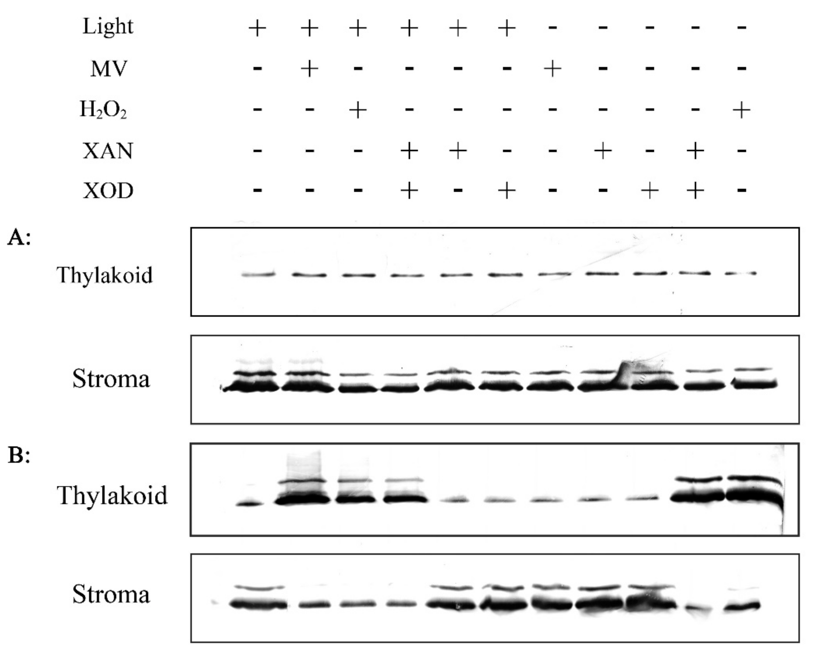
2.6. Effect of H2O2 on RCA Redistribution in Vitro

Different methods were then used to generate H2O2 in vitro and to test whether this H2O2 could induce the redistribution of RCA from the soluble stroma to the thylakoid membrane. The first method used methyl viologen (MV), which, in the light, can transfer electrons from Photosynthetic system I (PSI) to molecular H2O to produce H2O2 in plant chloroplasts [29,30]. The results showed that MV induced the moving of RCA from the stroma to the thylakoid membrane in the resistant wild rice, but not in the sensitive cultivated rice, and that this effect was light dependent (Figure 7). H2O2 induced RCA association with thylakoid membrane with or without light in O. meyeriana but not in Dalixiang (Figure 7). Xanthine (XAN)-xanthine oxidase (XOD) is a kind of oxidative system that can produce H2O2 in vitro [31,32,33]. When used alone, neither XAN nor XOD induced the association of RCA with the thylakoid membrane in either rice species, but when used together, RCA became bound to the thylakoid membrane in O. meyeriana, but not in Dalixiang, whether treated with light or not (Figure 7). All of these results show that H2O2 induces the redistribution of RCA to the thylakoid membrane from the soluble stroma in the resistant rice O. meyeriana.

2.7. In Vitro Investigation of the Association between RCA and Thylakoid Membrane

In vitro experiments were then performed to investigate factors that might affect the association of RCA with the thylakoid membrane in O. meyeriana. In experiments where H2O2 was used for different lengths of time, RCA began to redistribute from the stroma to thylakoid membrane after approximately 10 min. of H2O2 treatment. The amounts of RCA in the thylakoid membrane increased greatly after approximately 30 min. treatment, and then seemingly reached a plateau that was not exceeded by longer treatments (Figure 8A).
Next, the stroma and thylakoid membrane fractions were separately treated with H2O2 for 30 min. before mixing and incubation. RCA redistribution occurred within 5 min. of mixing (Figure 8B), which suggested that the 30 min. pre-treatment might have changed the RCA structure ready for association with the thylakoid membrane. As expected, a large amount of RCA was detected in the thylakoid membrane (Figure 8B). When the stroma fraction was pre-treated with H2O2 for 30 min. and it was then added to an untreated thylakoid membrane fraction, there was no redistribution of RCA after 5 min. (Figure 8B). Large amounts of RCA were detected in the thylakoid membrane fraction after 45 min. since the thylakoid membrane was exposed to H2O2 after mixing (Figure 8B). Pre-treatment of the thylakoid membrane fraction before mixing it with an untreated stroma fraction similarly resulted in little redistribution of RCA after 5 min. (Figure 8B). These results show that changes in the thylakoid membrane and in RCA take place to facilitate their H2O2-induced association.
In further experiments, the thylakoid membrane fraction that was isolated from O. meyeriana and the stroma fraction isolated from Dalixiang were separately treated with H2O2 before mixing and incubation. The reciprocal experiment was also done using the thylakoid membrane fraction from Dalixiang and the stroma fraction from O. meyeriana. In neither case did RCA redistribute to the thylakoid membrane (Figure 8C), showing that the redistribution effect was specific to both the RCA and the thylakoid membrane of the resistant wild rice and not to the susceptible cultivated rice.

3. Discussion

3.1. High Resistance of O. meyeriana to Xoo

Wild species of rice have adapted through natural selection to survive in harsh environments and they possess many useful traits that are not present in cultivated rice. For example, O. nivara has high resistance to the rice brown planthopper [34], O. officinalis is highly resistant to the rice small brown planthopper [35], and O. longistaminata and O. rufipogon are reported to have resistance to Xoo [5,7]. O. meyeriana is one of the wild relatives of rice growing in tropical and subtropical areas of South and Southeast Asia. In China, it is distributed in the Yunnan and Hainan provinces. This wild rice has been shown to possess a high resistance to Xoo pathogens [11] and this was further confirmed here (Figure 1). We have previously introduced this trait into a rice cultivar while using asymmetric somatic hybridization [3,12], but studies of the mechanism of this resistance are needed if this is to be widely used for genetic improvement of rice.

3.2. H2O2 Generation in Chloroplasts of O. meyeriana Infected by Xoo

H2O2 is recognized to be an important signaling molecule that triggers various downstream resistance reactions in the response of plants to pathogen infection [36,37,38,39]. In many plant resistance reactions, one of the earliest cellular responses following successful pathogen recognition is a large and transient production of reactive oxygen species, mainly H2O2, a phenomenon that is often called an “oxidative burst” [38,39,40,41,42]. In the present study, the inoculation of O. meyeriana with the bacterial pathogen Xoo also triggered an “oxidative burst” (Figure 2).
Plasma membrane-bound NADPH/NADH oxidase or cell wall-localized peroxidase are known to be the major sources of H2O2 during the “oxidative burst” [42,43]. NADPH/NADH oxidase, known as the respiratory burst oxidase, catalyzes the production of superoxide by one-electron reduction of molecular oxygen while using NADPH as an electron donor, and then other enzymes, such as superoxide dismutase catalyze superoxide, to its more stable dismutation product H2O2 [44]. The peroxidase catalyzes the oxidoreduction of various substrates to generate H2O2 [44,45]. In the “oxidative burst”, NADPH/NADH oxidase is localized in the plant plasma membrane and peroxidase is localized in the cell wall, and H2O2 generation is initiated in apoplasts and the extracellular spaces of plant tissues.
In addition to NADPH/NADH oxidase and peroxidase, the electron transport chain in the chloroplast is another important source of H2O2 in plants. However, it is not known whether and how H2O2 generation in chloroplasts is involved in plant resistance to pathogens. In this study, the results from both DAB staining and H2DCFDA fluorescence imaging showed that H2O2 was largely and transiently generated in the chloroplasts of the resistant wild rice, but not in the susceptible cultivated rice, when it was inoculated with Xoo (Figure 4 and Figure 5). Additionally, both Xoo resistance and H2O2 generation in O. meyeriana were distinctly light-dependent (Figure 6). Therefore, these results provide evidence that H2O2 from chloroplasts triggered the high Xoo resistance of O. meyeriana. Liu et al. and Caplan et al. have also demonstrated that chloroplasts are one of the major sources of the “oxidative burst” mediating the resistance of tobacco plants to tobacco mosaic virus [40,41]. Thus, the “oxidative burst” might be initiated as a defense response in plant chloroplasts as well as the apoplast.
Light is the important energy source that plants use to regulate their growth and development via various energy-dependent reactions in addition to the reaction of photosynthesis. In chloroplasts, light energy beyond what is used for photosynthesis can be used to generate reactive oxygen species, mainly H2O2, by a series of complicated oxidoreductive systems, and the regulation of the redox state in chloroplasts has been widely proven to play an important role in plant response to different stresses, such as cold [46], drought [47], salt [48,49], and waterlogging [50]. The “oxidative burst” is a process that is not only requires energy, but is also fine-regulated in the redox state. Therefore, it is entirely plausible that the H2O2 used by O. meyeriana in Xoo resistance is generated in the chloroplast.

3.3. Chloroplast H2O2 Induced the Association of RCA with Thylakoid Membrane Following Infection of O. meyeriana with Xoo

RCA is an important enzyme regulating photosynthesis. An increasing number of studies have been made regarding the structure and function of this enzyme since it was discovered based on an rca mutant of Arabidopsis in the 1980s. However, there are only a few reports on the cellular localization of RCA. Anderson and Carol found that labeling for RCA occurred in the chloroplast of pea plants [51]. Hong et al. also demonstrated that the RCA protein was distributed in the chloroplast of bundle sheath and mesophyll cells of the C4 plant Amaranthus tricolor [52]. Immuno-gold localization by Jin et al. showed that RCA was mainly detected in the chloroplast stroma and to a smaller extent in the thylakoid membrane in rice [53]. Interestingly, Chen et al. had demonstrated that the amount of RCA in the thylakoid membrane increased when the pH gradient across the thylakoid membrane and ATP in the chloroplast decreased in cultivated rice [54]. It has also been reported that moderate heat treatment (42 ℃) induced the redistribution of RCA from the stroma to the thylakoid membrane in spinach and tobacco [55,56]. We also reported that Xoo inoculation induced the association of RCA with the thylakoid membrane in the wild rice O. meyeriana [14] Figure 2. However, the mechanism that underlies the redistribution of RCA within the chloroplasts of the wild rice still remained unstudied.
A significant finding of the present study was that the period when Xoo infection triggered the “H2O2 burst” precisely coincided with the movement of RCA from the stroma to the thylakoid membrane (Figure 2), which suggested a causal link. In vitro experiments showed that H2O2 induced the redistribution of RCA in the wild rice but not in the cultivated rice, and that the effect could be reproduced if H2O2 was generated by two other methods, using MV-light or XAN-XOD. Additionally, the newly-generated H2O2 triggered by Xoo was clearly localized in chloroplasts (Figure 4 and Figure 5), and the resistance of O. meyeriana to Xoo and the H2O2 accumulation were both dependent on light. Taken together, we therefore propose that the chloroplast-generated “H2O2 burst” triggered by Xoo infection induces the association of RCA with the thylakoid membrane.
H2O2 is a small oxidative molecule that can cause significant modifications in the protein structure in the redox state and that is known to trigger a series of signaling transduction steps in response to various biotic or abiotic stresses in plants [18,38,39,40,42,47]. It seemed that H2O2 might induce a structural change in RCA to make it suitable for binding to the thylakoid membrane. Further experiments revealed that some H2O2-induced changes in the thylakoid membrane were also required for this association (Figure 8). Moreover, this H2O2-induced association was specific to the RCA and thylakoid membranes of O. meyeriana, rather than to those of the cultivated rice (Figure 8). It therefore appears that many changes in both RCA and the thylakoid membrane are required for them to associate with each other. Further study is needed to understand the detailed mechanism.

3.4. Potential Novel Role of RCA in the Xoo Resistance of O. meyeriana

It has been well documented that the primary role of RCA is to activate Rubisco to maintain its catalytic competency, and it is therefore expected that RCA would be localized in the chloroplast stroma coupled with Rubisco. Any RCA bound to the thylakoid membrane would not be involved in activating Rubisco, because the membrane is not the place where carboxylation takes place. Therefore, it seems likely that RCA has an additional role in the thylakoid membrane. Rokka et al. and Yang et al. found an increase in the amount of RCA that was bound to the thylakoid membrane under moderate heat treatment (42 ℃) and suggested that this was part of a system that helped to protect the thylakoid-associated translation machinery against heat inactivation [55,56]. However, Chen et al. proposed that the association of RCA with the thylakoid membrane was involved in the RCA activation of Rubisco due to its dependence on ATP and the pH gradient across the thylakoid membrane [54]. In the present study, the association of RCA with the thylakoid membrane was not found in the cultivated rice, but only in the wild rice, which suggested a resistance role rather than a general physiological one.
Reactive oxygen species damage plant cells because of their strong oxidative toxicity to membranes, proteins, pigments, and other essential macro-molecules, and such damage has been widely reported in plants under different biotic or abiotic stresses [47,48,57,58,59]. Therefore, H2O2 generated in the “oxidative burst” in response to pathogens could be double-edged: on the one hand it acts as a signaling molecule triggering the response reaction, but on the other hand it damages plant cells as an unstable oxidative molecule. The thylakoid membrane is crucial for the photosynthetic reaction as the site of various proteins and pigments that are involved in photosynthetic electron transport and photophosphorylation. The rapid binding of RCA to the thylakoid membrane might help to protect the thylakoid membrane system against the oxidative damage that might otherwise occur from the locally-produced H2O2 burst.
RCA is related to an AAA+ family of proteins, a class of chaperone-like ATPases associated with a variety of cellular activities, including the assembly, operation, or disassembly of protein complexes [60]. Proteins in the AAA+ family are also often covalently linked to other protein domains that mediate localization to cellular membranes. Therefore, RCA bound to the membrane could play a role as a chaperone in protecting the thylakoid-associated proteins against H2O2 damage. This protection might be helpful in explaining the high resistance of O. meyeriana to Xoo infection (Figure 1). On the basis of the results in this study, we therefore suggest that RCA may be involved in the resistance of O. meyeriana to Xoo in addition to its universal function in activating Rubisco.
O. meyeriana is highly resistant to Xoo, perhaps as the result of many mechanisms, rather than a single one. For example, we have found that the xylem secondary cell-wall thickening is involved in this resistance [4]. Here, we report that RCA is one of the factors that contribute to the resistance of O. meyeriana. Additionally, we have transferred the trait of this resistance of O. meyeriana into cultivated rice while using asymmetric somatic hybridization. Using the hybrid progeny as a resistant parent, we constructed a segregating population and mapped three major quantitative trait loci (QTLs) for Xoo resistance on chromosomes 1, 3, and 5 [3]. It seems that the three major QTLs are not associated with the RCA gene, because they are at different loci of different chromosomes, indicating that the RCA gene from O. meyeriana was not transferred into the cultivated rice through asymmetric somatic hybridization. However, the RCA gene of the wild rice might interact with the major QTLs to make a contribution to the high resistance, so it is necessary that these QTLs are isolated and characterized by map-based cloning and that additional genetic analysis is conducted to reveal the relationship between the RCA gene and the three major QTLs for resistance.

4. Materials and Methods

4.1. Plant Materials

O. meyeriana, a wild species of rice with high Xoo resistance, was from the Zhejiang Academy of Agricultural Sciences collection. It was originally provided by Professor Xin-hua Wei, China National Rice Research Institute and its accession No. was ABB658. O. sativa ssp. japonica (cv. Dalixiang), a rice cultivar that is susceptible to rice Xoo pathogens, was from the Zhejiang Academy of Agricultural Sciences collection. The rice plants were grown at 28/25 °C (day/night; 16 h/8 h), with photon flux density 600–800 µM m−2 s−1, and relative humidity 60–80%.

4.2. Pathogen Inoculation and Resistance Identification

In this study, Xoo strain PXO99 was used to inoculate the rice leaves. The leaf-clipping method was used to inoculate Xoo pathogens, as previously described [61,62]. Briefly, The Xoo bacterial inoculum was prepared from a 48 h culture on potato semisynthetic agar (PSA) [62] and its density was adjusted to 109 CFU/mL. Approximately 40 days after transplanting (booting stage), a pair of scissors was dipped into the inoculum and used to clip about 1 cm from the tip of the fully expanded leaves. At least, 16 leaves from four plants of each of the wild and cultivated rice were inoculated with the bacteria. Bacterial lesion length was measured for resistance evaluation after three weeks of inoculation.

4.3. Measurements of Chlorophyll Fluorescence

Chlorophyll fluorescence was measured with an integrating chlorophyll fluorometer (LI-6400 leaf chamber fluorometer, LI-COR, USA). The rice leaves were kept in dark for 1 h before the minimum fluorescence yield in the dark (Fo) was recorded under a measuring light (0.1 µmol m−2 s−1). The maximum fluorescence yield in the dark (Fm) was then measured with a 600 ms saturating flash (8000 µmol m−2 s−1). Subsequently, an actinic light (1000 µmol m−2 s−1) was applied for 30 min. to obtain the stable fluorescence yield during actinic light (Fs), which was followed by a measurement of the maximum fluorescence yield druing actinic illumination (Fm) with another saturating flash. A far-red light was then applied to determine the minimum fluorescence yield (Fo). Different chlorophyll fluorescence parameters were defined, as follows:
Fv/Fm = (Fm − Fo)/Fm;
ΦPSII = (Fm’ − Fs)/Fm’;
qP = (Fm’ − Fs)/(Fm’ − Fo);
qN = [(Fm − Fm)(Fo − Fo)]/(Fm − Fo).

4.4. Isolation and Sub-Fractionation of Chloroplasts

Intact chloroplasts were isolated according to Cline et al. and Gunasinghe and Berger with some modifications [63,64]. In the procedure finally adopted, leaves (20 g) were homogenized in 200 mL of grinding buffer containing 330 mM sorbitol, 50 mM Hepes-KOH (pH 7.5), 2 mM EDTA, 1 mM MgCl2, 1 mM MnCl2, 0.5% BSA, and 4 mM ascorbic acid until the tissue was smaller than 1 mm2. The homogenate was filtered through eight layers of cheesecloth, and centrifuged for 1 min. at 4000 rpm in a Sorvall GSA rotor at 4 ℃. The pellet was suspended in 8 mL of grinding buffer and then overlayered onto a 40% to 80% Percoll gradient containing 0.6 mM glutathione, gradients of 0.7% to 2.7% (w/v) of polyethyleneglycol 3350 and 0.25% to 0.92% (w/v) of both BSA and Ficoll 400, and the same ingredients as in the grinding buffer. The gradient tubes were centrifuged for 8 min. at 7500 rpm in the Sorvall HB-4 rotor. Two green-colored bands were found in the tube after centrifugation. The lower green band of intact chloroplasts was collected and diluted with about 25 mL of grinding buffer, and then was centrifuged for 5 min. at 3500 rpm in the HB-4 rotor. The pellet was suspended and centrifuged again in 25 mL of HSM suspension medium containing 50 mM Hepes-KOH (pH 8.0), 330 mM sorbitol, and 42 mM methionine. The pellet was washed again with HSM and drained at 4 ℃. The resulting pellet of intact plastids was resuspended in about 3 mL HSM and the final concentration was adjusted to 3.0 mg Chl/mL. The intactness of the chloroplasts was examined by observation under an inverted microscope (ECLIPSE Ts2-FL, Nikon, Japan). If there were ruptured chloroplasts in the HSM buffer, the buffer would be re-extracted by repeating the procedure until the intact chloroplasts remained.
Purified intact chloroplasts were ruptured in 1 mM PMSF and the suspension was placed on ice for 45–60 min. then at room temperature for 30 min. The suspension of broken chloroplasts was centrifuged for 10 min. at 14000 g. The sediment was dissolved in loading buffer as the membrane fraction for SDS-PAGE analysis. An equal volume of precooled 20% TCA was added to the supernatant and the mixture was incubated for at least 30 min. on ice, followed by centrifugation for 20 min. at 14000 g. The stroma proteins were washed twice with 80% acetone and drained at 4 ℃, and were then dissolved in loading buffer for SDS-PAGE analysis.

4.5. SDS-PAGE and Immunological Analyses

Two fractions of thylakoid membrane and stroma of chloroplasts were separated by differential centrifugation following an osmotic shock to intact chloroplasts. SDS-PAGE with 12% (w/v) acrylamide gels was used to separate the proteins in the fractions and the samples were loaded on the total protein basis (5 µg protein). Antibodies that were raised against RCA were used to detect the RCA proteins in western blotting after the polypeptides had been electrophoretically transferred to a polyvinylidene fluoride (PVDF) membrane (Bio-Rad, Hercules, USA). RCA antibodies were produced while using prokaryotically expressed products as antigens of RCA genes that were derived from O. meyeriana and the cultivar rice, respectively. After the proteins were transferred to the PVDF membrane, the secondary anti-mouse IgG antibody conjugated with alkaline phosphatase (Sigma) and the nitroblue tetrazolium (NBT)/5-bromo-4-chloro-3-indolyl phosphate (BCIP) system were used to detect and visualize the immunoblotting signals.

4.6. Determination of H2O2 in Leaf extracts

The content of H2O2 in leaves was measured by examining the absorbance of the titanium-peroxide complex at 415 nm, according to a previous method [65]. One g fresh leaves was homogenized in an ice bath with 10 mL 0.1% TCA. After the homogenate was centrifuged at 10,000 g for 15 min., one mL of the supernatant was added to 1 mL of 10 mM potassium phosphate buffer with 2 mL of 1M KI and pH 7.0, and the absorbance of the supernatant was then measured at 415 nm. The absorbance was quantified while using a standard curve generated from known concentrations of H2O2.

4.7. Subcellular Localization of H2O2

Two different approaches were used on rice leaf frozen sections to visualize the subcellular localization of H2O2 in leaves. DAB staining was performed, as previously described [66,67]. Briefly, DAB (Sigma) was dissolved in 10 mM Tris-acetate (pH 5.0) at a concentration of 1 mg mL−1. Rice leaf sections were incubated in DBA solution for 3 h at room temperature and then were washed three times with distilled water. Subsequently, the sections were observed and photographed with an inverted microscope (ECLIPSE Ts2-FL, Nikon, Japan) at 200× magnification. A second method with H2DCFDA fluorescence imaging was conducted while using an H2O2 Assay Kit (Invitrogen), according to the manufacturer’s instruction. A 10 mM stock solution of H2DCFDA (Molecular Probes) was prepared by dissolving H2DCFDA in anhydrous dimethyl sulfoxide, and after a subsequent dilution step with distilled water, a 10 µmol working concentration was set. The rice leaf sections were incubated in the working solution for 30 min., and then washed three times with distilled water. Green fluorescence from oxidized H2DCFDA was monitored by confocal laser scanning microscopy (LSM510, Zeiss, Germany) while using the dye-specific excitation/emission wavelength of 488/529 nm. Background chloroplast autofluorescence emission was monitored with a 650 nm long-pass filter.

4.8. Salt Washing and Thermolysin Treatment of Isolated Thylakoids

The thylakoid membrane fractions with 2 mg chlorophyll mL−1 were washed with 2 M NaCl or 2 M NaBr in 10 mM HEPES-KOH with 5 mM MgCl2 (pH 7.8), and then incubated in 1% (v/v) Triton X-100 for 30 min. on ice. The thylakoid membrane fractions were then added to in 0.4 mg mL−1 of thermolysin (pH 7.8, Sigma, St. Louis, USA) with 50 mM Tricine, 100 mM sorbitol, 5 mM NaCl, 10 mM MgCl2, and 2 mM CaCl2 for about 8 min. on ice, after which EDTA (50 mM) was added to stop the digestion. The thylakoid membrane fractions were then collected by 10 min. centrifugation at full speed in an Eppendorf centrifuge.

4.9. Oxidative Treatment of Ruptured Chloroplasts

A suspension of ruptured chloroplasts was mixed with 10 mM of H2O2 (Sigma, St. Louis, USA), 20 µM MV (Sigma, St. Louis, USA), 10 mM XAN (Sigma, St. Louis, USA), or 10 mM XOD (Sigma, St. Louis, USA), and the mixture was then incubated for 30 min. at room temperature usually using a light photon flux density of about 600 µM m−2 s−1, but up to 1000 µM m−2 s−1 for the MV treatment to stimulate enough H2O2 production from the photosynthetic electron transport chain. The chloroplasts were then divided into thylakoid membrane and stroma fractions for RCA protein detection, as described above.

4.10. Shading Treatment of Wild Rice Leaves

To investigate the influence of light on the resistance of the wild rice to Xoo, the leaves were shaded with a 2–3 cm piece of aluminum foil for 1 wk under the light condition of 600 µM m−2 s−1 after inoculation. The foil was then removed and light was re-instated for 4 d. Subsequently, the lengths of lesions on inoculated leaves were measured for resistance evaluation, and the accumulation and subcellular localization of H2O2 was determined.

Supplementary Materials

The following are available online at https://www.mdpi.com/2223-7747/8/9/351/s1, Table S1: Effects of Xoo inoculation on Fv/Fm, ΦPSII, qP and qN in the leaves of O. meyeriana and O. sativa during a 336 h test period.

Author Contributions

Conceptualization, J.C. and Y.Y.; formal analysis, Y.Y.; investigation, Q.M., Y.Y., S.Y., W.L., J.Z., X.W., C.L.Y. and C.Q.Y.; data curation, Y.Y. and Q.M.; writing—original draft preparation, Y.Y.; writing—review and editing, J.C.; All authors have read the final manuscript and approved the submission.

Funding

This research was funded by grants from the National Key Research and Development Program of China (2016YFD0200804, 2016YFD0100601-15), the Key Program of Zhejiang Provincial Foundation for Natural Science (LZ16C130002), Zhejiang Provincial Key Research and Development Plan (2019C02006), Zhejiang Fundamental Public Welfare Research Program (LGN19C140008), and State Key Laboratory for Quality and Safety of Agro-products (2010DS700124-ZZ1907)

Acknowledgments

The authors thank M. J. Adams for critically reading the manuscript.

Conflicts of Interest

The authors declare no conflict of interest.

References

  1. Nino-Liu, D.O.; Ronald, P.C.; Bogdanove, A.J. Xanthomonas oryzae pathovars: Model pathogens of a model crop. Mol. Plant Pathol. 2006, 7, 303–324. [Google Scholar] [CrossRef] [PubMed]
  2. Zhang, Q. Genetics and Improvement of Resistance to Bacterial Blight in Rice, 1st ed.; Science Press: Beijing, China, 2007; pp. 1–4. [Google Scholar]
  3. Han, X.Y.; Yang, Y.; Wang, X.M.; Zhou, J.; Zhang, W.H.; Yu, C.L.; Cheng, C.; Cheng, Y.; Yan, C.Q.; Chen, J.P.; et al. Quantitative trait loci mapping for bacterial blight resistance in rice using bulked segregant analysis. Int. J. Mol. Sci. 2014, 15, 11847–11861. [Google Scholar] [CrossRef]
  4. Yang, Y.; Xie, L.; Yan, C.Q.; Wang, X.M.; Yu, C.L.; Cheng, X.Y.; Cheng, Y.; Chen, J.P. Xylem secondary cell-wall thickening involved in defense responses of Oryza meyeriana to Xanthomonas oryzae pv. oryzae. Acta Phytopathol. Sin. 2012, 5, 505–514. [Google Scholar]
  5. Song, W.Y.; Wang, G.L.; Chen, L.L.; Kim, H.S.; Pi, L.Y.; Holsten, T.; Gardner, J.A. Receptor kinase-like protein encoded by the rice disease resistance gene, Xa21. Science 1995, 270, 1804–1806. [Google Scholar] [CrossRef] [PubMed]
  6. Gu, K.; Yang, B.; Tian, D.; Wu, L.F.; Wang, D.J.; Sreekala, C.; Yang, F.; Chu, Z.Q.; Wang, G.L.; White, F.F.; et al. R gene expression induced by a type-III effector triggers disease resistance in rice. Nature 2005, 435, 1122–1125. [Google Scholar] [CrossRef] [PubMed]
  7. Wang, C.L.; Chen, L.T.; Zeng, C.Z.; Zhang, Q.Y.; Liu, P.Q.; Liu, Y.G.; Fan, Y.L.; Zhang, Q.; Zhao, K.J. Chromosome walking for fine mapping of Xa23 gene locus by using genomic libraries. Chinese J. Rice Sci. 2006, 20, 355–360. [Google Scholar]
  8. Tan, G.X.; Ren, X.; Weng, Q.M.; Shi, Z.Y.; Zhu, L.L.; He, G.C. Mapping of a new resistance gene to bacterial blight in rice line introgressed from Oryza officinalis. Acta Genet. Sin. 2004, 31, 724–729. [Google Scholar]
  9. Peng, S.Q.; Wei, Z.S.; Mao, C.X.; Luo, K.; Huang, H.Q.; Xiao, F.H. Identification of multiresistance of wild rice: Resource for high resistance to rice bacterial blight—Oryza meyeriana. Acta Agricul. Hunan. 1981, 5, 47–48. [Google Scholar]
  10. Peng, S.Q.; Wei, Z.S.; Mao, C.X.; Huang, H.Q.; Xiao, F.H.; Luo, K. Identification of multiresistance of O. meyeriana, O. officinalis and O. sativa f. spantanea growing in Yunan province. Acta Phytopathol. Sin. 1982, 12, 58–61. [Google Scholar]
  11. Zhang, Q.; Wang, C.; Shi, A. Evaluation of resistance to bacterial blight (Xanthomonas oryzae pv. oryzae) in wild rice species. Sci. Agricul. Sin. 1994, 27, 1–9. [Google Scholar]
  12. Yan, C.Q.; Qian, K.X.; Yan, Q.S.; Zhang, X.Q.; Xue, G.P.; Huangfu, W.G.; Wu, Y.F.; Zhao, Y.Z.; Xue, Z.Y.; Huang, J.; et al. Use of asymmetric somatic hybridization for transfer of the bacterial resistance trait from Oryza meyeriana L. to O. sativa L. ssp. japonica. Plant Cell Rep. 2004, 2, 569–575. [Google Scholar] [CrossRef] [PubMed]
  13. Yan, C.Q.; Qian, K.X.; Xue, G.P.; Wu, Z.C.; Chen, Y.L.; Yan, Q.S.; Zhang, X.Q.; Wu, P. Production of bacterial blight resistant lines from somatic hybridization between Oryza sativa L. and Oryza meyeriana L. J. Zhejiang Univ. Sci. 2004, 5, 1199–1205. [Google Scholar] [CrossRef] [PubMed]
  14. Yang, Y.; Yu, C.L.; Wang, X.M.; Yan, C.Q.; Cheng, Y.; Chen, J.P. Inoculation with Xanthomonas oryzae pv. oryzae induces thylakoid membrane association of Rubisco activase in Oryza meyeriana. J. Plant Physiol. 2011, 168, 1701–1704. [Google Scholar] [CrossRef] [PubMed]
  15. Chen, L.N.; Yang, Y.; Yan, C.Q.; Wang, X.M.; Yu, C.L.; Zhou, J.; Zhang, W.L.; Cheng, Y.; Cheng, X.Y.; Chen, J.P.; et al. Identification of quantitative trait loci for bacterial blight resistance derived from Oryza meyeriana and agronomic traits in recombinant inbred lines of Oryza sativa. J. Phytopathol. 2012, 160, 461–468. [Google Scholar] [CrossRef]
  16. Berger, S.; Papadopoulos, M.; Schreiber, U.; Kaiser, W.; Roitsch, T. Complex regulation of gene expression, photosynthesis and sugar levels by pathogen infection in tomato. Physiol. Plant. 2004, 122, 419–428. [Google Scholar] [CrossRef]
  17. Bonfig, K.B.; Schreiber, U.; Gabler, A.; Roitsch, T.; Berger, S. Infection with virulent and avirulent P. syringae strains differentially affects photosynthesis and sink metabolism in Arabidopsis leaves. Planta 2006, 225, 1–12. [Google Scholar] [CrossRef]
  18. Shapiguzov, A.; Vainonen, J.P.; Wrzaczek, M.; Kangasjarvi, J. ROS-talk-how the apoplast, the chloroplast, and the nucleus get the message through. Front. Plant Sci. 2012, 3, 292. [Google Scholar] [CrossRef]
  19. Yokota, A.; Tsujimoto, N. Characterization of ribulose-1,5-bisphosphate carboxylase/oxygenase carrying ribulose-1,5-bisphosphate at its regulatory sites and the mechanism of interaction of this form the enzyme with ribulose-1,5-bisphosphate carboxylase/oxygenase activase. Eur. J. Biochem. 1992, 204, 901–909. [Google Scholar] [CrossRef]
  20. Portis, A.R.J. Rubisco activase - Rubisco’s catalytic chaperone. Photosynth. Res. 2003, 75, 11–27. [Google Scholar] [CrossRef]
  21. Zhang, Z.L.; Komatsu, S. Molecular cloning and characterization of cDNAs encoding two isoforms of ribulose-1, 5-bisphosphate carboxylase/oxygenase activase in rice (Oryza sativa L.). J. Biochemy. 2000, 128, 383–389. [Google Scholar] [CrossRef]
  22. Parry, M.A.J.; Keys, A.J.; Madgwick, P.J.; Carmo-Silva, A.E.; Andralojc, P.J. Rubisco regulation: A role for inhibitors. J. Exp. Bot. 2008, 59, 1569–1580. [Google Scholar] [CrossRef]
  23. Giri, A.P.; Wünsche, H.; Mitra, S. Molecular interactions between the specialist herbivore Manduca sexta (lepidoptera, sphingidae) and its natural host Nicotiana attenuata. VII. changes in the plant’s proteome. Plant Physiol. 2006, 142, 1621–1641. [Google Scholar] [CrossRef]
  24. Nabity, P.D.; Zavala, J.A.; DeLucia, E.H. Indirect suppression of photosynthesis on individual leaves by arthropod herbivory. Ann. Bot. 2009, 103, 655–663. [Google Scholar] [CrossRef]
  25. Crafts-Brandner, S.J.; van de Loo, F.J.; Salvucci, M.E. The two forms of ribulose-1, 5-bisphosphate carboxylase/oxygenase activase differ in sensitivity to elevated temperature. Plant Physiol. 1997, 114, 439–444. [Google Scholar] [CrossRef]
  26. Sharkey, T.D.; Badger, M.R.; von Caemmerer, S.; Andrews, T.J. Increased heat sensitivity of photosynthesis in tobacco plant with reduced Rubisco activase. Photosynth. Res. 2001, 67, 147–156. [Google Scholar] [CrossRef]
  27. Saibo, N.J.M.; Lourenco, T.; Oliveira, M.M. Transcription factors and regulation of photosynthetic and related metabolism under environmental stresses. Ann. Bot. 2009, 103, 609–623. [Google Scholar] [CrossRef]
  28. Hurt, E.; Hauska, G. A cytochrome b6/f complex of five polypeptides with plastoquinol-plastocyanin-oxidoreductase activity from spinach chloroplasts. Eur. J. Biochem. 1981, 17, 591–595. [Google Scholar] [CrossRef]
  29. Li, D.Y.; Liu, H.J.; Zhang, H.J.; Wang, X.; Song, F.M. OsBIRH1, a DEAD-box RNA helicase with functions in modulating defence responses against pathogen infection and oxidative stress. J. Exp. Bot. 2008, 59, 2133–2146. [Google Scholar] [CrossRef]
  30. Xu, J.; Duan, X.; Yang, J.; Beeching, J.R.; Zhang, P. Enhanced reactive oxygen species scavenging by overproduction of superoxide dismutase and catalase delays postharvest physiological deterioration of cassava storage roots. Plant Physiol. 2013, 161, 1517–1528. [Google Scholar] [CrossRef]
  31. Iwata, S.; Fukaya, Y.; Nakazawa, K.; Okuda, T. Effects of tannins on the oxidative damage of mouse ocular lens. I. Using the oxidative damage model induced by the xanthine-xanthine oxidase system. J. Ocul. Pharmacol. 1987, 3, 227–238. [Google Scholar] [CrossRef]
  32. Li, B.; HU, B.; Liao, D.F. Protective effect of gypenosides on free radical damage of isolated guinea pig papillary muscles. Chin. Pharmacol. Bull. 1993, 9, 287–290. [Google Scholar]
  33. Hussein, G.; Goto, H.; Oda, S.; Iguchi, T.; Sankawa, U.; Matsumoto, K.; Watanabe, H. Antihypertensive potential and mechanism of action of astaxanthin: II. Vascular reactivity and hemorheology in spontaneously hypertensive rats. Biol. Pharm. Bull. 2005, 28, 967–971. [Google Scholar] [CrossRef]
  34. Liu, Y.Q.; Wu, H.; Chen, H.; Liu, Y.L.; He, J.; Kang, H.Y.; Sun, Z.G.; Pan, G.; Wang, Q.; Hu, J.L.; et al. A gene cluster encoding lectin receptor kinases confers broad-spectrum and durable insect resistance in rice. Nat. Biotech. 2015, 33, 301–307. [Google Scholar] [CrossRef]
  35. Dong, Y.; Fang, X.P.; Yang, Y.; Xue, G.P.; Chen, X.; Zhang, W.L.; Wang, X.M.; Yu, C.L.; Zhou, L.; Mei, Q.; et al. Comparative proteomic analysis of susceptible and resistant rice plants during early infestation by small brown planthopper. Front. Plant Sci. 2017, 8, 1744. [Google Scholar] [CrossRef]
  36. Tian, S.; Wang, X.; Li, P.; Wang, H.; Ji, H.; Xie, J.; Qiu, Q.; Shen, D.; Dong, H. Plant aquaporin AtPIP1;4 Links Apoplastic H2O2 induction to disease immunity pathways. Plant Physiol. 2016, 171, 1635–1650. [Google Scholar] [CrossRef]
  37. Jajic, I.; Sarna, T.; Strzalka, K. Senescence, stress, and reactive oxygen species. Plants 2015, 4, 393–411. [Google Scholar] [CrossRef]
  38. Lamb, C.; Dixon, R.A. The oxidative burst in plant disease resistance. Annu. Rev. Plant Physiol. Plant Mol. Biol. 1997, 48, 251–275. [Google Scholar] [CrossRef]
  39. Grant, M.; Brown, I.; Adams, S.; Knight, M.; Ainslie, A.; Mansfield, J. The RPM1 plant disease resistance gene facilitates a rapid and sustained incre Torres ase in cytosolic calcium that is necessary for the oxidative burst and hypersensitive cell death. Plant J. 2000, 23, 441–450. [Google Scholar] [CrossRef]
  40. Liu, Y.D.; Ren, D.T.; Pike, S.; Pallardy, S.; Gassmann, W.; Zhang, S.Q. Choroplast-generated reactive oxygen species are involved in hypersensitive response-like cell death mediated by a mitogen-activated protein kinase cascade. Plant J. 2007, 51, 941–954. [Google Scholar] [CrossRef]
  41. Caplan, J.L.; Kumar, A.S.; Park, E.; Padmanabhan, M.S.; Hoban, K.; Modla, S.; Czymmek, K.; Dinesh-Kumar, S.P. Chloroplat stromules function during innate immunity. Develop. Cell 2015, 34, 45–57. [Google Scholar] [CrossRef]
  42. Torres, M.A.; Jones, J.D.G.; Dangl, J.L. Reactive oxygen species signaling in response to pathogens. Plant Physiol. 2006, 141, 373–378. [Google Scholar] [CrossRef]
  43. Bolwell, G.P. Role of active oxygen species and NO in plant defense responses. Curr. Opin. Plant Biol. 1999, 2, 287–294. [Google Scholar] [CrossRef]
  44. Apel, K.; Hirt, H. Reactive oxygen species: Metabolism, oxidative stress, and signal transduction. Annu. Rev. Plant Biol. 2004, 55, 373–399. [Google Scholar] [CrossRef]
  45. Bindschedler, L.V. Peroxidase-dependent apoplastic oxidative burst in Arabidopsis required for pathogen resistance. Plant J. 2006, 47, 851–863. [Google Scholar] [CrossRef] [Green Version]
  46. Xia, X.J.; Wang, Y.J.; Zhou, Y.H.; Tao, Y.; Mao, W.H.; Shi, K.; Asami, T.; Chen, Z.; Yu, J.Q. Reactive oxygen species are involved in brassinosteroid-induced stress tolerance in cucumber. Plant Physiol. 2009, 150, 801–814. [Google Scholar] [CrossRef]
  47. Miao, Y.; Lv, D.; Wang, P.; Wang, X.C.; Chen, J.; Miao, C.; Song, C.P. An Arabidopsis glutathione peroxidase functions as both a redox transducer and a scavenger in abscisic acid and drought stress responses. Plant Cell 2006, 18, 2749–2766. [Google Scholar] [CrossRef]
  48. Yang, Y.; Yan, C.Q.; Cao, B.H.; Xu, H.X.; Chen, J.P.; Jiang, D.A. Some photosynthetic responses to salinity resistance are transferred into the somatic hybrid descendants from the wild soybean Glycine cyrtoloba ACC547. Physiol. Plant. 2007, 129, 658–669. [Google Scholar] [CrossRef]
  49. Parvin, K.; Hasanuzzaman, M.; Bhuyan, M.; Mohsin, S.M.; Fujita, M. Quercetin mediated salt tolerance in tomato through the enhancement of plant antioxidant defense and glyoxalase systems. Plants 2019, 8, 247. [Google Scholar] [CrossRef]
  50. Anee, T.I.; Nahar, K.; Rahman, A.; Mahmud, J.I.; Bhuiyan, T.F.; Alam, M.U.; Fujita, M.; Hasanuzzaman, M. Oxidative damage and antioxidant defense in Sesamum indicum after different waterlogging durations. Plants 2019, 8, 196. [Google Scholar] [CrossRef]
  51. Anderson, L.E.; Carol, A.A. Enzyme co-localization with Rubisco in pea leaf chloroplasts. Photosyn. Res. 2004, 82, 49–58. [Google Scholar] [CrossRef]
  52. Hong, J.; Jiang, D.A.; Weng, X.Y.; Wang, W.B.; Hu, D.W. Leaf anatomy, chloroplast ultrastructure, and cellular localisation of ribulose-1,5-bisphosphate carboxylase/oxygenase (RuBPCO) and RuBPCO activase in Amaranthus tricolor L. Photosyn. 2005, 43, 519–528. [Google Scholar] [CrossRef]
  53. Jin, S.H.; Hong, J.; Li, X.Q.; Jiang, D.A. Antisense inhibition of Rubisco activase increases Rubisco content and alters the proportion of Rubisco activase in stroma and thylakoids in chloroplasts of rice leaves. Ann. Bot. 2006, 97, 739–744. [Google Scholar] [CrossRef]
  54. Chen, J.; Wang, P.; Mi, H.; Chen, G.Y.; Xu, D.Q. Reversible association of ribulose-1, 5-bisphosphate carboxylase/oxygenase activase with the thylakoid membrane depends upon the ATP level and pH in rice without heat stress. J. Exp. Bot. 2010, 61, 2939–2950. [Google Scholar] [CrossRef] [Green Version]
  55. Rokka, A.; Zhang, L.; Aro, E.N. Rubisco activase: An enzyme with a temperature-dependent dual function? Plant J. 2001, 25, 463–471. [Google Scholar] [CrossRef]
  56. Yang, X.H.; Zheng, L.; Lu, C.M. Genetic engineering of the biosynthesis of glycinebetaine enhances photosynthesis against high temperature stress in transgenic tobacco plants. Plant Physiol. 2005, 138, 2299–2309. [Google Scholar] [CrossRef]
  57. Koh, E.; Carmieli, R.; Mor, A.; Fluhr, R. Singlet oxygen-induced membrane disruption and serpin-protease balance in vacuolar-driven cell death. Plant Physiol. 2016, 171, 1616–1625. [Google Scholar] [CrossRef]
  58. Dietz, K.; Turkan, I.; Krieger-Liszkay, A. redox-and reactive oxygen species-dependent signaling into and out of the photosynthesizing chloroplast. Plant Physiol. 2016, 171, 1541–1550. [Google Scholar] [CrossRef]
  59. Bhuyan, M.; Hasanuzzaman, M.; Mahmud, J.A.; Hossain, M.S.; Bhuiyan, T.F.; Fujita, M. Unraveling morphophysiological and biochemical responses of Triticum aestivum L. to exreme pH: Coordinated actions of antioxidant defense and glyoxalase systems. Plants 2019, 8, 24. [Google Scholar] [CrossRef]
  60. Neuwald, A.F.; Aravind, L.; Spouge, J.L.; Koonin, E.V. AAA+: A class of chaperone-like ATPases associated with the assembly, operation and disassembly of protein complexes. Genome Res. 1999, 9, 27–43. [Google Scholar]
  61. Kauffman, H.E.; Reddy, A.P.K.; Hsieh, S.P.Y.; Merca, S.D. An improved technique for evaluating resistance of rice varieties to Xanthomonas oryzae. Plant Dis. Rep. 1973, 57, 537–541. [Google Scholar]
  62. Sanchez, A.C.; Ilag, L.L.; Yang, D.; Brar, D.S.; Ausubel, F.; Khush, G.S.; Yano, M.; Sasaki, T.; Li, Z.; Huang, N. Genetic and physical mapping of Xa13, a recessive bacterial blight resistance gene in rice. Theor. Appl. Genet. 1999, 98, 1022–1028. [Google Scholar] [CrossRef]
  63. Cline, K.; Andrews, J.; Mersey, B.; Newcomb, E.H.; Keegstra, K. Separation and characterization of inner and outer envelope membranes of pea chloroplasts. Proc. Natl. Acad. Sci. USA 1981, 78, 3595–3599. [Google Scholar] [CrossRef] [Green Version]
  64. Gunasinghe, U.B.; Berger, P.H. Association of potato virus Y gene products with chloroplast in tobacco. Mol. Plant Microbe Interact. 1991, 4, 452–457. [Google Scholar] [CrossRef]
  65. Velikova, V.; Yordanov, I.; Edreva, A. Oxidative stress and some antioxidant system in acid rain-treated bean plants: Protective role of exogenous polyamines. Plant Sci. 2000, 151, 59–66. [Google Scholar] [CrossRef]
  66. Thordal- Christensen, H.; Zhang, Z.G.; Wei, Y.D.; Collinge, D.B. Subcellular localization of H2O2 in plants, H2O2 accumulation in papillae and hypersensitive response during the barley-powdery mildew interaction. Plant J. 1997, 11, 1187–1194. [Google Scholar] [CrossRef]
  67. Zhu, Y.; Du, B.; Qian, J.; Zou, B.; Hua, J. Disease resistance gene-induced growth inhibition is enhanced by rcd1 independent of defense activation in Arabidopsis. Plant Physiol. 2013, 161, 2005–2013. [Google Scholar] [CrossRef]
Figure 1. The high resistance of Oryza meyeriana to Xanthomonas oryzae pv. oryzae (Xoo). Rice was inoculated by the leaf-clipping method and at least 16 leaves from four plants were inoculated. Lesion length was measured three weeks after inoculation. (A) O. sativa ssp. japonica cv. Dalixiang. Left is plant inoculated with water (control) and right is plant inoculated with Xoo. White scale bar = 3 cm. (B) O. meyeriana. Left is plant inoculated with water and right is plant inoculated with Xoo. (C) Leaves inoculated with water and Xoo. Left is Dalixiang and right is O. meyeriana. Red scale bar = 3 cm. (D) Lesion length caused by Xoo infection. Bars indicate the standard error.
Plants 08 00351 g001
Figure 2. Effect of Xoo inoculation on H2O2 content in the leaves of Dalixiang and O. meyeriana during a 26 h test period (upper panel) and rubisco activase (RCA) content in the thylakoid membrane fraction and soluble stroma fraction of chloroplasts in Dalixiang and O. meyeriana (lower panel). (A): Dalixiang, (B): O. meyeriana. Intact chloroplasts were isolated from rice leaves at different times after Xoo inoculation. The protein samples were loaded on the basis of 5 µg total protein and western blot analysis was performed with an antibody raised against RCA.
Plants 08 00351 g002
Figure 3. Stability of the association of RCA with the thylakoid membrane. (A) Thylakoids were isolated from leaves of O. meyeriana inoculated with Xoo and then incubated in HEPES-KOH buffer solution to release RCA from the thylakoid membrane for 0, 1/6, 1/2, 1, 2, 3, 6, 12, 24 hours. (B) Isolated thylakoid membrane washed with 2 M NaCl or 2 M NaBr, or treated for 30 min. with 1% Triton X-100.
Plants 08 00351 g003
Figure 4. Subcellular localization of H2O2 detected by After 3, 3-diaminobenzidine (DAB) staining in Dalixiang and O. meyeriana 0 h, 13 h, and 24 h after Xoo inoculation. Rice leaf sections were incubated in 3, 3-diaminobenzidine (DAB) solution and then examined in an inverted microscope. (A): Dalixiang, (B): O. meyeriana. Red circle indicates a mesophyll cell and the arrow indicates a chloroplast. Scale bars = 20 µm.
Plants 08 00351 g004
Figure 5. Subcellular localization of H2O2 detected by H2DCFDA fluorescence imaging in Dalixiang and O. meyeriana 0 h, 13 h, and 24 h after Xoo inoculation. Rice leaf sections were incubated in 2, 7-Dichlorodi-hydrofluorescein diacetate (H2DCFDA) solution and then examined by confocal laser scanning microscopy with excitation/emission wavelength of 488/529 nm. Chloroplast autofluorescence emission was 650 nm. (A): Dalixiang, (B): O. meyeriana. Chl: chloroplast. Scale bars = 20 µm.
Plants 08 00351 g005
Figure 6. Resistance to Xoo and H2O2 accumulation in chloroplasts were dependent on light in O. meyeriana. (A) Shading treatment with aluminum foil to the leaves of O. meyeriana. (B) Effect of light on the lesion length caused by Xoo infection. (C) Effect of light on the subcellular localization of H2O2. Chl: chloroplast. Scale bars = 20 µm.
Plants 08 00351 g006
Figure 7. H2O2 induced association of RCA with the thylakoid membrane. Chloroplasts were isolated from leaves of O. meyeriana and then ruptured by osmotic shock. The suspension of ruptured chloroplasts was treated with H2O2, methyl viologen (MV) or the xanthine (XAN)-xanthine oxidase (XOD) system, before the chloroplasts were sub-fractionated into soluble stroma and thylakoid membrane fractions followed by separation of RCA proteins while using SDS-PAGE. (A): Dalixiang, (B): O. meyeriana.
Plants 08 00351 g007
Figure 8. In vitro experiments on RCA association with the thylakoid membrane. (A) Effect of different treatment times of exposure to H2O2 on the association of RCA with the thylakoid membrane of O. meyeriana. The suspension of ruptured chloroplasts was treated with H2O2 during the 240 min. chase phase, and the chloroplasts were sub-fractionated into soluble stroma and thylakoid membrane fractions followed by separation of RCA proteins using SDS-PAGE. (B) Effect of pre-treatment with H2O2 on the stroma or thylakoid membrane fractions of O. meyeriana. Intact chloroplasts isolated from leaves were ruptured into soluble stroma fraction and thylakoid membrane fraction, and each of the fractions was treated with H2O2 for 30 min. before mixing and incubation. At 5 min. or 45 min. after mixing, samples were re-fractionated and the distribution of RCA protein was tested by SDS-PAGE. (C) Effect of pre-treatment with H2O2 as in B but using mixtures of the stroma fraction from susceptible O. sativa with the thylakoid membrane fraction of O. meyeriana or vice versa.
Plants 08 00351 g008

